Faculdade de Engenharia da Universidade do Porto
An endotelial cell-‐delivery system
for therapeutic angiogenesis
Joana Bianchi Marques Bettencourt Gesta
Dissertação realizada no âmbito do Mestrado Integrado em Bioengenharia
Major Engenharia Biomédica
Orientador: Dr. Cristina Barrias Co-‐orientador: Dr. Sílvia Bidarra
20 de Setembro de 2013
© Joana Gesta 2013
Abstract
Neovascularization is a crucial step towards recovery of injured tissues. Since endothelial cells (ECs) are primary angiogenic cells, their delivery has been prominently studied as a pro-‐angiogenic strategy. Yet, up to now, clinical trials of ECs transplantation have not resulted in consistent benefits. The outcome might presumably be improved using biomaterial-‐based vehicles to protect cells from the harsh in vivo environment, enhancing their survival and engraftment. These carriers might also provide ECs with instructive signals to assist and promote their 3D organization, enhancing functional integration.
Alginate is an injectable polymer, widely used for cell entrapment, which is biologically inert but can be modified in order to stimulate specific cellular responses, namely through changes in its viscoelastic properties and/or grafting of bioactive peptides. In particular, previous studies showed that 3D culture in soft RGD-‐alginate hydrogels promote the self-‐assembly of entrapped cells, including ECs and mesenchymal stem cells (MSC), and the deposition of an endogenous fibronectin-‐rich extracellular matrix (ECM) by MSCs.
Therefore, the main aim of this work was to create an ideal microenvironment for EC entrapment using an integrative approach, combining this optimized hydrogel matrix with the use of co-‐entrapped MSCs as mural cells.
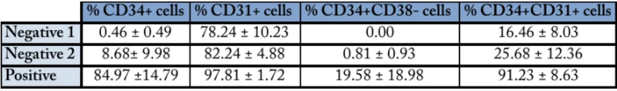
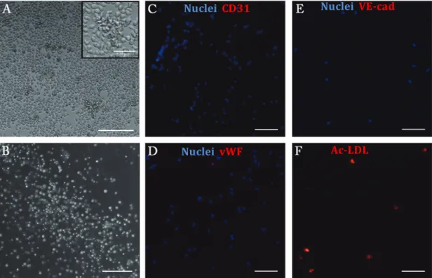
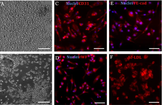
A variety of cell populations have been investigated in clinical revascularization trials. Here, the initial plan was to test two different cell types: human umbilical vein endothelial cells (HUVECs), a well-‐established model of mature ECs, and endothelial progenitor cells (EPCs) derived from human umbilical cord blood (UCB), which appear to have superior angiogenic properties than fully differentiated ECs such as HUVECs. UCB is a promising source of EPCs for therapeutic applications, as cells can be obtained through a non-‐ invasive procedure, support long-‐term storage without losing biological properties, and have low immunogenicity, which makes them an interesting candidate for allogeneic transplantation. So, the first step was to isolate and characterize CD34+ cells from UCB, and promote their differentiation into EPCs, which was performed using a previously published protocol. The isolated CD34+ cells were able to form different hematopoietic colonies, in a standard methylcellulose assay, which confirmed their multipotency. The evaluation of phenotypic expression before and after a differentiation period of 21 days showed that differentiated CD34+ cells expressed some EC-‐lineage markers like CD31, VE-‐ cadherin, vWF and uptake of Ac-‐LDL and presented cluster formation with surrounding
spindle-‐shaped cells. Also, these cells were not able to form tube-‐like structures in Matrigel at any time-‐point. Overall, CD34+ -‐derived cells phenotype resembled the so-‐ called early EPCs.
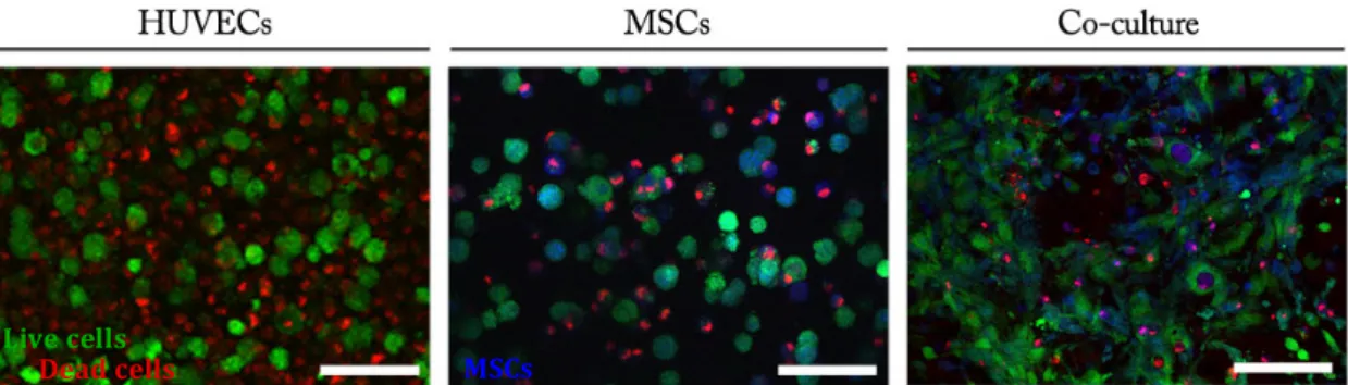
Subsequently, soft RGD-‐alginate hydrogels were used as matrices for the 3D culture of ECs. HUVECs were cultured alone or in combination with MSCs, whose pro-‐angiogenic effects and pericyte-‐like roles have been widely reported. Cells viability and functionality were increased in co-‐cultured constructs, where the formation of multicellular structures, including EC cord-‐like structures, and deposition of endogenous ECM were also stimulated. When placed in a tissue mimic (Matrigel), co-‐cultures also promoted higher outward migration and cell sprouting. 3D cultures of CD34+ cells in monoculture or co-‐ cultured with MSC were also established. However, only freshly isolated CD34+ cells were tested in a preliminary study, not only because it was important to assess how these cells behaved in soft-‐RGD alginate matrices, but also because the differentiation process was too lengthy to be implemented in 3D cultures in due time. Although CD34+ cells did not perform well in 3D monocultures, interesting results were obtained when these were co-‐ cultured with MSCs, and both cell types seemed to exert some influence over each other. Finally, a preliminary characterization of the in vivo performance of cell-‐laden soft RGD-‐ alginate hydrogels was carried out using the chorioallantic membrane (CAM) assay. Matrices were implanted immediately after preparation or following a pre-‐culture time of 5 days, and their angiogenic potential was evaluated. Although no significant differences were found between the different types of cultures (mono-‐ vs. co-‐cultures), the pre-‐ cultured matrices seemed to result in an increased stimulation of new vessels formation, suggesting that it might be advantageous to implant more mature cellular-‐ECM structures.
Quando se pretende regenerar tecidos lesionados, a estimulação da neovascularização é um passo crucial. Tendo em conta que as principais células envolvidas no processo de angiogénese são células endoteliais (ECs), a transplantação das mesmas é uma das estratégias mais estudadas hoje em dia. Contudo, até hoje, os ensaios clínicos que envolveram o transplante de ECs não conseguiram assegurar os seus benefícios. Uma das maneiras de melhorar os resultados obtidos poderá ser, então, a utilização de biomateriais como veículos que protejam as células do ambiente hostil in vivo e permitam melhorar a sua sobrevivência e integração. Para além disso, estes veículos poderão ser modificados com sinais que orientem e suportem a sua organização em 3D, melhorando a integração do sistema.
O alginato é um polímero injetável natural muito usado para encapsular células. Este polímero é biologicamente inerte, podendo ser modificado de forma a estimular respostas celulares específicas e desejáveis através de ajustes nas suas capacidades viscoelásticas e/ou por adição de péptidos bioativos às suas cadeias. Mais especificamente, estudos recentes com células endoteliais e mesenquimais estaminais (MSCs) mostraram que culturas 3D em hidrogeis suaves de RGD-‐alginato promovem a reorganização das células e estimulam a deposição de uma matriz extracelular (ECM) endógena, rica em fibronectina, por MSCs.
Assim sendo, o principal objectivo deste trabalho seria o de criar o microambiente ideal para a encapsulação de ECs, combinando os conhecimentos relativos à matriz de hidrogel optimizada com o uso de MSCs como células murais.
Hoje em dia, já foram testados vários tipos de células em ensaios clínicos com vista a revascularização. Neste estudo, o plano inicial era o de testar dois tipos de células humanas: células endoteliais da veia do cordão umbilical (HUVECs) e células progenitoras endoteliais (EPCs) derivadas do sangue do cordão umbilical (UCB), sendo que a primeira população é correntemente usada como modelo de ECs maduras e a segunda, apesar de parecer possuir mais propriedades angiogénicas, ainda não se encontrar bem definida. O facto de as EPCs poderem ser isoladas de uma fonte rica em células progenitoras como UCB é uma grande vantagem pois permite a obtenção de células não imunogénicas por meios não invasivos. Para além disso, as EPCs podem ser armazenadas por longos períodos de tempo sem perder propriedades biológicas. Todos estes factores fazem destas células as candidatas ideais para transplantes alogénicos. Desta forma, o primeiro passo deste trabalho foi isolar, caracterizar as células CD34+ presentes no UCB e promover a sua
diferenciação em EPCs segundo um protocolo previamente publicado. A análise do fenótipo exibido antes e depois de um período de diferenciação de 21 dias demonstrou que as células diferenciadas a partir de células CD34+ exprimiram marcadores característicos de linhagens endoteliais como CD31, VE-‐caderina, vWF, incorporaram Ac-‐ LDL e formaram agregados de células rodeados de células fusiformes. Para além disso, as células CD34+ formaram colónias hematopoiéticas, e nem estas nem as células diferenciadas conseguiram formar estruturas tubulares em Matrigel. De uma maneira geral, as células derivadas de células CD34+ apresentaram um fenótipo semelhante ao das EPCs precoces.
De seguida, hidrogeis moles de RGD-‐alginato foram usados como matrizes para a cultura 3D de ECs. HUVECs foram postas em cultura sozinhas ou em co-‐cultura com MSCs, cujos efeitos pro-‐angiogénicos e capacidade de atuar como pericitos foram vastamente reportados. A viabilidade e funcionalidade das células em co-‐cultura foram aumentados, da mesma maneira que foram estimuladas a formação de estruturas multicelulares tubulares, formadas por ECs, e deposição de ECM endógena. Quando estas matrizes foram colocadas em Matrigel, a co-‐cultura também promoveu maior migração para o exterior. Células CD34+ foram também testadas em culturas 3D, sozinhas ou em co-‐cultura com MSCs. No entanto, só foram utilizadas células isoladas no momento, por ser importante analisar o seu comportamento nas matrizes utilizadas, mas também por o seu processo de diferenciação ser demasiado demorado para ser implementado em culturas 3D a tempo.
Apesar de o comportamento das células CD34+ não ter sido satisfatório em monocultura 3D, foram obtidos resultados interessantes quando em co-‐cultura com MSCs, e ambos os tipos celulares pareceram exercer algum tipo de influência um sobre o outro.
Por fim, foi feita uma caracterização preliminar da performance dos hidrogeis moles de RGD-‐alginato com células embebidas in vivo, utilizando o ensaio na membrana corio-‐ alântica de um embrião de galinha. As matrizes foram implantadas imediatamente após preparação ou após um período de pré-‐cultura de 5 dias, e o seu potencial de angiogénese foi avaliado. Apesar de não se terem detectado diferenças significativas entre os tipos de cultura (mono-‐ e co-‐cultura), as matrizes previamente preparadas pareceram aumentar a estimulação da formação de novos vasos sanguíneos, indicando que talvez possa ser vantajosa a implantação de matrizes contendo estruturas de ECM mais maduras.
Acknowledgements
First of all, I’d like to thank Prof. Inês Gonçalves for helping me find a project of my interest and making the connections with the needed people. Likewise, I’d like to thank Prof. Mário Barbosa for giving me the opportunity to work at INEB.
Prof. Cristina, thank you for letting me in so “out of the blue” and trusting in my capabilities. Also, thank you for all the support, the insights and the help and, of course, the good humour that always made me feel welcomed.
Prof. Sílvia, thank you for all your help and your availability to answer my “doubts”. Most of all, thank you for your patience!
David, thank you for introducing me around. Thank you for giving up your time to accompany me and sharing your knowledge.
Raquel Gonçalves, thank you for all your help with the flow citometry and methilcelulose, and for sharing your antibodies with me.
Raquel Maia, thank you for your help every time I needed, and of course for letting me use your alginate!
Filipa, thank you for the pleasant conversations outside, and for letting me drag you to the confocal microscope every time I could.
I’d also like to thank to Prof. Isabel Amaral and Estrela Neto, for kindly letting me borrow their antibodies, and Sara for helping me with the Live/Dead.
Last but not least, I’d like to thank all of the Biomatrix and INEB members (Daniela, Ana Luísa, Estrela, Filipa Lourenço, Ana Rita Ferreira, Tiago) for being so prompt to help and always having an encouraging (or funny!) thing to say.
Finally, I’d like to thank my family and friends for the support and comprehension, and especially to my mom, who always believes in me even when I don’t.
Index:
Resumo ... I Abstract ... III Acknowledgements ... I Index: ... V List of figures: ... VII List of tables: ... VIII List of abbreviations: ... IXIntroduction ... 11
1. DINAMICS OF NEOVASCULARISATION ... 13
2. NEOVASCULARISATION AS A TISSUE REGENERATION STRATEGY ... 14
3. ENDOTHELIAL CELL DELIVERY TOWARDS NEOVASCULARISATION STIMULATION .... 16
3.1. MATURE ENDOTHELIAL CELLS FOR NEOVASCULARISATION ... 16
3.2. ENDOTHELIAL PROGENITOR CELLS ... 17
3.3. CO-‐CULTURE: THE SUPPORTING ROLE OF MESENCHYMAL STEM CELLS ... 19
3.4. CELL DELIVERY VEHICLES ... 19
4. PRELIMINARY IN VIVO ASSESSMENT OF PROANGIOGENIC PROPERTIES ... 20
Material And Methods ... 23
1. ISOLATION AND CHARACTERIZATION OF CD34+ CELLS ... 25
1.1. ISOLATION OF MNCS FROM UCB ... 25
1.2. ISOLATION OF CD34+ CELLS FROM MNCS ... 25
1.3. FLUORESCENCE-‐ACTIVATED CELL SORTING ANALYSIS ... 26
1.4. METHYLCELLULOSE ASSAY FOR COLONY CHARACTERIZATION ... 26
1.5. DIFFERENTIATION OF CD34+ CELLS INTO ECS ... 26
1.6. DETECTION OF EXPRESSION OF EC MARKERS ... 27
1.7. ASSESSMENT OF PHENOTYPE EXPRESSION ... 27
1.8. IN VITRO TUBE FORMATION ON MATRIGEL PLATE ... 27
2. IN VITRO STUDIES WITH 3D CULTURES ... 28
2.1. CELL CULTURE CONDITIONS ... 28
2.2. CELL INCORPORATION WITHIN RGD-‐GRAFTED ALGINATE HYDROGEL MATRICES ... 28
2.3. CHARACTERIZATION OF CELL CULTURES WITHIN ALGINATE DISKS ... 29
2.4. METABOLIC ACTIVITY AND CELL VIABILITY ... 29
2.5. CELL MORPHOLOGY AND FIBRONECTIN EXPRESSION ... 30
2.6. IN VITRO MIGRATION ASSAY OF 3D CULTURE ... 30
3. IN VIVO STUDIES WITH 3D CULTURES ... 31
4. STATISTICAL ANALYSIS ... 31
Isolation and characterization of CD34+ Cells ... 33
AIM ... 35
RESULTS ... 35
1. ISOLATION OF CD34+ CELLS ... 35
2. CHARACTERIZATION OF CD34+ CELLS ... 36
DISCUSSION ... 39
In vitro studies with 3D cultures ... 43
AIM ... 45
RESULTS: ... 45
1. HUVECS AND MSCS 3D CULTURES ... 45
1.1 METABOLIC ACTIVITY AND VIABILITY ... 45
1.2. CELL REARRANGEMENT AND MATRIX FORMATION IN HUVECs/MSCs 3D CULTURES .... 46
1.3. OUTWARD CELL MIGRATION ... 49
2. CD34+ CELLS AND MSCS 3D CULTURES ... 51
2.1. METABOLIC ACTIVITY AND VIABILITY ... 51
2.2. CELL REARRANGEMENT AND MATRIX FORMATION IN CD34+ CELLS/MSCs 3D CULTURES ... 52
DISCUSSION: ... 53
1. HUVECs/MSCs 3D CONSTRUCTS: ... 53
2. CD34+ CELLS/MSCS 3D CULTURE ... 55
In vivo studies with 3D cultures ... 57
AIM: ... 59
RESULTS: ... 59
5.3. DISCUSSION: ... 61
Main conclusions and future prospects ... 63
References ... 67
Supplementary Data ... 73
List of figures:
Figure 1 – Overview of the neovascularization processes and the main steps it involves. 18 Figure 2 – Different approaches for cell-‐based neovascularisation enhancement in bioengineered
tissues. 19
Figure 3 – Putative circulating EPCs kinetics. 22
Figure 4 – Common methods of EPCs isolation and culture. 23
Figure 5 – Representative time-‐dependent diagram of the in vivo CAM assay. 25
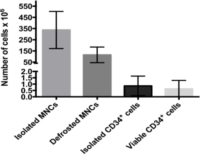
Figure 6 – Efficiency of CD34+ cells isolation. 39
Figure 7 – FACS analysis scatter images. 41
Figure 8 – CD34+ cells morphology and phenotypic expression after 5 days in culture. 42
Figure 9 – CD34+-‐derived cells morphology and phenotypic expression after 21 days in culture. 43
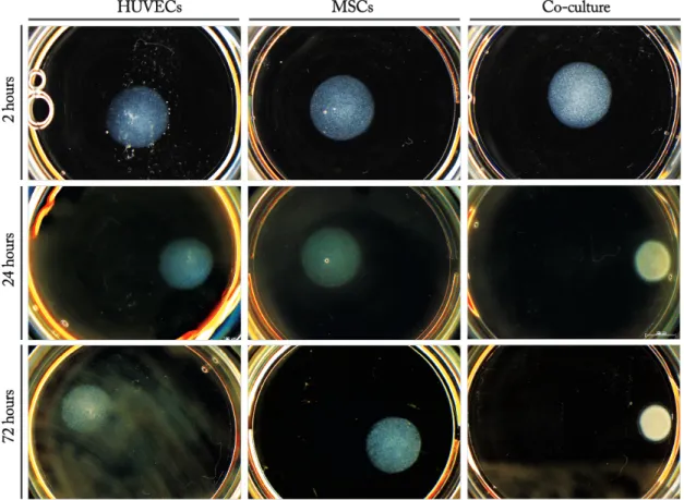
Figure 10 – Metabolic activity of HUVECs/MSCs 3D cultures throughout a 3-‐day culturing period. 50 Figure 11 – Live/Dead Assay – 24 hours after cell embedment. 50 Figure 12 – General appearance of alginate hydrogel disks 2, 24 and 72 hours after cell
embedment. 51
Figure 13 – Morphology and spatial organization of HUVECs and MSCs in 3D co-‐culture. 51 Figure 14 – Extracellular matrix and multicellular networks in HUVECs/MSCs 3D culture. 52 Figure 15 –Decomposed confocal fluorescent microscopy image of 3D HUVECs/MSCs culture
after 72 hours. 52
Figure 16 – Representative phase-‐contrast micrographs of cell-‐laden disks embedded in Matrigel. 53 Figure 17 – Metabolic activity of cell-‐seeded alginate disks before and after culture in EGM. 54 Figure 18 – Metabolic activity of CD34+ cells/MSCs 3D cultures throughout a 3-‐day culturing
period. 55
Figure 19 – Live/Dead Assay – 24 and 72 hours after cell embedment. 56 Figure 20 – Cell functionality, morphology and extracellular matrix production in CD34+
cells/MSCs 3D culture. 57
Figure 21 – Effect of different cell cultures and pre-‐incubation of alginate hydrogel disks on blood
vessel density in CAM assay without VEGF. 64
Figure 22 – Photograph of representative CAM of 13 day-‐old chick embryo (Day 3 after
implantation). 64
Figure S-‐1 – Location of the ring formed by MNCs. 79
Figure S-‐2 – Representation of a 3D culture migration assay. 79 Figure S-‐3 – Number of cells isolated from each donor's blood. 79
Figure S-‐4 – Flow cytometry scatter images of cells marked with PI. 80
Figure S-‐5 – Flow cytometry scatter images of isotype. 81
Figure S-‐6 – Analysis of protein expression in HUVECs using immunohistochemistry. 82 Figure S-‐7 – Morphology of CD34+-‐derived cells, 21 days after plating. 82 Figure S-‐8 – HUVECs stained with CellTracker Green and MSCs stained with CellTracker Blue. 83
List of tables:
Table I – FACS analysis of CD34, CD31 and CD38 expression. 40
Table S-‐I – FACS analysis of CD34, CD31 and CD38 expression in each sample. 80
List of abbreviations:
BM Bone Marrow
BFU-‐E Burst-‐forming Unit -‐ Erithroyd CAC Circulating Angiogenic Cell
CCFM Recovery Cell Culture Freezing Medium CD Cluster of Differentiation
CF Colony-‐forming CFU Colony-‐forming Unit
CLSM Confocal Laser Scanning Microscope DMSO Dimethyl Sulfoxide
EBM Endothelial Basal Medium ECFC Endothelial Colony Forming Cell ECM Extracellular Matrix
ECGS Endothelial Cell Growth Supplement ECs Endothelial Cells
EGF Epidermal Growth Factor EGM Endothelial Growth Medium EOC Endothelial Outgrowth Cells EPCs Endothelial Progenitor Cells EXP Experiment
FBS Fetal Bovine Serum FGF Fibroblast Growth Factor FN Fibronectin
GA Gentamicin Amphotericin GDL Glucone Delta-‐Lactone
GEMM Granulocyte, Eritrocyte, Monocyte, Megakaryocyte GM Granulocyte, Macrophage
HMW High Molecular Weight HSC Hematopoietic Stem Cells
HUVECs Human Umbilical Vein Endothelial Cells IFM Inverted Fluorescence Microscope IGF Insulin Growth Factor
LMW Low Molecular Weight MC Mural Cells
MMP Matrix Metalloproteinase MNCs Mononuclear Cells
MSCs Human Mesenchymal Stem Cells PFA Paraformaldehyde
PI Propidium Iodide
PBS Phosphate Buffered Saline Pen/Strep Penicillin/Streptomycin RGD Arginylglycylaspartic acid RT Room Temperature SD Supplementary Data TBS Tris-‐Buffered Saline UCB Umbilical Cord Blood
VE-‐cad Vascular Endothelial Cadherin VEGF Vascular Endothelial Growth Factor vWF Von Willebrand Factor
Introduction
13
1. DINAMICS OF NEOVASCULARISATION
Angiogenesis is the physiological process that leads to formation of new blood vessels from pre-‐existing vasculature in post-‐embryonic development. Blood vessels provide adequate oxygenation, nutrient delivery and removal of waste products in surrounding cells, as well as signaling molecules that might be involved in communication between organs. The importance of vascularization is based on the fact that diffusion between blood vessels and the surrounding cells is limited to a distance of up to 150-‐300 µm [1].
When new blood vessels are formed, it can happen either from the longitudinal splitting of an existing vessel – intussusceptive angiogenesis – or from the outgrowth of a new branch from preexisting blood vessels – sprouting angiogenesis. Yet, both of these processes occur via proliferation and migration of endothelial cells (ECs), which can be influenced by interaction with their extracellular matrix (ECM) [2] and involves the release of matrix metalloproteinases (MMP) by ECs to degrade the ECM [3]. Communication between ECs, between ECs and other cells and between cells and ECM is vital throughout the entire process.
Vasculogenesis is other type of neovascularization that happens more frequently in embryonic development (although reports have been made of its occurrence in adult life [4]), and consists in the de novo formation of blood vessels from angioblasts: endothelial progenitor cells form blood islands that fuse and sprout, forming a primary plexus that later expands via angiogenesis and vasculogenesis [5]. In this case, endothelial progenitor cells (EPCs) are mobilized to sites of neovascularization and differentiate into ECs in situ – see Figure 1.
Mature endothelial vessels are formed by an endothelial layer that is stabilized by mural cells (which depend on the size of tube: pericytes in cappilaries and vascular smooth muscle cells in more complex vessels) and a basement membrane that embeds them [6].
14
2. NEOVASCULARISATION AS A TISSUE REGENERATION STRATEGY
The main goal of tissue engineering strategies is to repair damaged, injured or missing body tissues in a way that its functions maintain assured. Engineered tissues of a clinically relevant size and complexity must have their own vasculature or easily develop it after implantation, allowing rapid and stable perfusion so that the area and its surrounding tissue is repopulated, preventing cell death and tissue necrosis. Many approaches have been devised in order to improve angiogenesis and vasculogenesis in bioengineered tissues for later implantation, taking into consideration the knowledge on phisiological mechanisms of neovascularisation.
Implants can present neovascularization that results from either the invasion of host blood vessels and/or neovascularization in vivo, or from the prevascularization in vitro or in vivo before implantation [6]. Figure 2 depicts the main cell-‐based approaches to promote neovascularization of a bioengineered tissue. Also, Novosel et al. published a review that delves into these subjects [1]. Prevascularization is of great importance in
VASCULOGENESIS ANGIOGENESIS
Angioblast
Hematopoiesis
Primary vascular plexus
Neovascularization VEGF/FGF 1. Proteolysis 2. Migration Proliferation 3. Maturation Differentiation Pericyte/SMC Endothelial Cell Endothelial progenitor cell
15
thick constructs, since it accelerates functional anastomosis, through connection with the host existing vasculature upon implantation. Prevascularization can be stimulated by cell seeding and neovascularization stimulation in vitro (Figure 7-‐A) [7], or by implantation of unseeded scaffolds into a host body (Figure 7-‐B). Host blood vessels penetrate the scaffold, building a perfusable vascular network, and these scaffolds are then explanted and reimplanted into the ischemic target site [1].
Anyhow, neovascularization in vitro or in vivo, can be achieved by seeding relevant cell types in the target area. Endothelial cells (ECs) compose the inner lining of blood vessels, and secrete several paracrine factors (such as growth factors) that are known to be involved in the stimulation of angiogenesis – hence, these are the most comon “single-‐cell-‐ type” cultures used for angiogenesis stimulation assays [8]. However, it is known that ECs and their progenitors (EPCs) are not the only cells involved in neovascularization and that more cell types are found in mature blood vessels, leading to the use of more than one cell type in these assays (Figure 7-‐E).
16
The ECM that surrounds blood vessels consists mostly of hydrated proteins and proteoglycans, yet is responsible for mechanical and biochemical stimuli that regulate cell behavior [9]. The influence of grafted growth factors (Figure 7-‐C) and adhesion peptides (Figure 7-‐D) that stimulate scaffold-‐cells interaction on angiogenesis is a major subject currently under study, in order to fully understand and ultimately mimic the normal biological processes occurring inside the body during neovascularization [6].
3. ENDOTHELIAL CELL DELIVERY TOWARDS NEOVASCULARISATION
STIMULATION
3.1. MATURE ENDOTHELIAL CELLS FOR NEOVASCULARISATION
For neovascularisation in cell delivery therapies, ECs are one of the primary types of cells to be seeded. Mature endothelial cells have limited regenerative capacity, since they are fully differentiated, and their phenotype is slightly different in every source. Still, they can be isolated from many parts of the human body, such as the umbilical vein (HUVECs), dermal microvasculature (HDMECs) and vasculature in general (HVECs), and are easy to identify. Mature endothelial cells preferentially express some genes and molecular markers: expression of CD31, CD34, von Willebrand factor (vWF) and dil-‐acetylated low density lipoprotein (Ac-‐LDL) uptake are the most common markers when distinguishing ECs from other cells in culture using flow cytometry or immunohistochemistry. Other relevant markers used to discriminate mature ECs during differentiation are E-‐selectins, VE-‐cadherin (vascular endothelial cadherin) and N-‐cadherin: these molecules are involved in cell adhesion between ECs or between ECs and other cells present in angiogenesis like pericytes, fibroblasts and smooth muscle cells (SMCs) [10]. Previous studies from our group where HUVECs were used have shown that these cells can proliferate, reorganize into cellular networks and even migrate when they were encapsulated into alginate [11].
A comon disadvantage of mature endothelial cells is that, in order to prevent an immune reaction, the patient’s own cells would have to be collected from a blood vessel, a process that can cause morbidity at the donor site, and they also present low proliferation rates and low availability. As a result of these impairments, the scientific community’s attention has been turning to endothelial progenitor cells (EPCs), a heterogeneous minor subpopulation of blood mononuclear cells (MNCs) that play a significant role in postnatal vasculogenesis [1]. EPCs are believed to be mobilized to damaged tissues in case an emergent vascular regenerative process is happening, and represent an advantageous cell type for cell delivery therapies, since blood can be collected from the patient and these
17
cells can be isolated and expanded in vitro [12]; this way, the implant will stimulate tissue regeneration without causing an immune response. They also present aditional advantages, as detailed below.
3.2. ENDOTHELIAL PROGENITOR CELLS
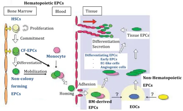
Since Asahara et al. [13] originally reported the isolation of EPCs, efforts have been made to characterize each type of cells comprised under the term “EPCs”. However, there is not a standardized isolation and culture protocol being used yet. Asahara’s group claims that there are two main types of EPCs, acording to their origin: hematopoietic lineage EPCs and nonhematopoietic lineage EPCs [14]. “Hematopoietic EPCs” represent a heterogeneous subpopulation of hematopoietic stem cells (HSCs) with provasculogenic characteristics. These EPCs can be isolated from bone marrow (BM) or blood and include colony-‐forming EPCs (CF-‐EPCs), non-‐colony forming “differentianting” EPCs or even adherent circulating angiogenic cells (CACs), among others. “Nonhematopoietic EPCs” are adhesive angiogenic and vasculogenic cells that present mature EC-‐like phenotypes or differentiate into it., yet do not form hematopoietic colonies in methylcelulose. Nonhematopoietic EPCs can be isolated from blood or tissue samples; however, these cells primary origin is still unknown. Endothelial outgrowth cells (EOC) are the main member of this group of EPCs. Figure 3 describes the general EPC dynamics: although all of the EPCs seem to be responsive to stimulation that causes them to migrate to damaged tissues, the role they play in tissue regeneration is different.
Bone Marrow Blood Tissue
Tissue EPCs Differentiation Secretion Differentiating EPCs: -‐ Early EPCs -‐ EC-‐like cells -‐ Angiogenic cells Proliferation Commitment HSCs CF-‐EPCs Differentiation Non-‐Hematopoietic EPCs Non-‐colony forming EPCs Adhesion BM-‐derived EPCs EOCs Hematopoietic EPCs
Figure 3 – Putative circulating EPCs dynamics. Adapted from Asahara, T. et al. Mobilization
Monocyte
18
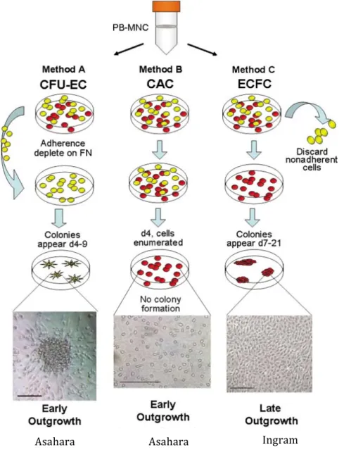
On the other hand, Prater’s group has categorized EPCs regarding their phenotype: early EPCs and late EPCs [15]. Accordingly to this classification, CF-‐EPCs and CACs constitute early EPCs, since they appear in culture after 4-‐9 days. Early EPCs have angiogenic and vasculogenic potential, are capable of vascular integration and express EC markers, but do not form tube-‐like structures or present a cobblestone-‐like morphology when in culture [14]-‐[16]. Endothelial colony forming cells (ECFCs) are classified as late EPCs, since they take about 7 to 21 days to be detected in culture [17]. These EPCs form tube-‐like structures in culture and present a phenotype that is similar to mature endothelial lineage [15], [18]. Figure 4 presents common methods of EPC culture, their morphology and the main groups that characterized each type of cells.
Figure 4 – Common methods of EPCs isolation and culture. Method A: CFU-‐ECs are obtained after a culture
period of 5-‐days, where non-‐adherent MNCs differentiate into adherent EPC colonies. Scale bar = 100 μm. Method B: Circulating angiogenic cells appear after 4-‐7 days in culture, and typically do not form CAC cultures. Scale bar = 200 μm. Method C: ECFCs derive from adherent MNCs and are detected after 7 to 21 days in culture. These cells usually display a cobblestone-‐like appearance. Scale bar = 400 μm. Yellow: non-‐adherent cells; Red:
adherent cells.
Adapted from Prater et al.
Asahara
19
Recently, several groups have been focusing in CD34+-‐selected populations, isolated from MNCs, as an EPC-‐enriched fraction. Even though this marker is also expressed on hematopoietic stem and progenitor cells, this fraction has been yielding positive results regarding neovascularisation stimulation and angiogenesis potential [19], [20]. However, CD34+ cells still form a heterogeneous population [21], and their differentiation and phenotype varies according to their culturing method [22].
CD34+-‐derived EPCs have the advantage of being isolated through non-‐invasive means [23] and having great expansion potential. However, not only their expansion and maintenance is more successful when they are co-‐cultured with mesenchymal stem or progenitor cells (MSCs) [24], their viability and angiogenic behavior has already been proven to be increased when they are co-‐cultured with many types of cells, like MSCs [25], CD34-‐ cells [26] and even CD34+-‐derived ECs [20].
3.3. CO-‐CULTURE: THE SUPPORTING ROLE OF MESENCHYMAL STEM CELLS
So far, bone marrow-‐derived and cord blood-‐derived MSCs were proven to be able to act as pericytes (perivascular cells), providing paracrine signals that stimulate ECs to form tubular structures, and promote and stabilize newly forming structures in vitro and in vivo [23], [27]. The stimulation MSC-‐derived perivascular cells confer to the growing blood vessels is attained by secretion of pro-‐angiogenic cytokines and regulation of cell-‐ cell adherens junctions, which leads to regulation of the vessels permeability and perfusion and makes them less susceptible to regression. Also, our group has already shown that MSCs can self-‐assemble and produce ECM within RGD-‐grafted alginate [28], as well as promote multicellular networks formation by HUVECs when encapsulated in alginate microspheres [11]. Another advantage of MSCs is that their isolation does not yield donor site morbidity, allowing us to use the patient’s own cells and prevent an immune response.
3.4. CELL DELIVERY VEHICLES
Many research groups have tried to mimic the ECM in vitro, not only using naturally derived biomaterials but also synthetic biomaterials with some type of modification; however, there are many factors and limitations that have to be controlled [9]. In order to mimic vascularisation in engineered tissues and/or deliver vascular cells, a suitable extracellular environment should also be developed. Therefore, scaffolds should ideally
20
promote cell survival, allow cellular reorganization, and allow cell-‐driven remodeling processes.
Hydrogels can be formed from several natural and synthetic materials, mostly under mild conditions. Also, they are easily modified and highly permeable to oxygen and water-‐ soluble molecules. From the range of possibilities within hydrogels, natural hydrogels have the advantage of innately exhibiting some of the properties that characterize soft tissues [29].
Alginate is one of the most widely used hydrogels: it is a polysaccharide, derived from brown algae, which can crosslink in situ in the presence of divalent cations (e.g. Ca2+), with low toxicity, and therefore can be injected into the target site, what makes it a minimally invasive therapy. Thanks to the alginate’s versatility, its non-‐fouling characteristics and non-‐adhesiveness to cells can be overcome by covalently modifying it with cell-‐adhesion peptides (namely, containing the arginine-‐glycine-‐aspartic acid aminoacid sequence – RDG). This strategy has already been studied in previous works within our group [11], [30], [31]. Besides that, it is also possible to adjust its mechanical properties through molecular weight distribution and partial oxidation, and their degradation rate can be regulated using crosslinking peptides that are susceptible to cleavage by MMPs [32]. Regarding injectable pro-‐angiogenic therapies, these hydrogels have been most commonly tested as growth-‐factor delivery (usually VEGF) vehicles [33], [34]. In order to validate these hydrogels as endothelial cell celivery vehicles and their pro-‐angiogenic properties, in vitro and in vivo studies have to be performed.
4. PRELIMINARY IN VIVO ASSESSMENT OF PROANGIOGENIC PROPERTIES
In vitro assays have the advantage of being easy to interpret and involving well-‐ controlled conditions, what facilitates the assessment of angiogenic effects [35]. However, in vivo studies have to be performed in order to analyze the host’s response and the pro-‐ angiogenic properties of endothelial cell delivery vehicles.
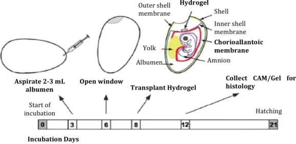
The simplest and most extensively used in vivo assay is performed in the chorioallantoic membrane (CAM) of a chick embryo. The CAM is an extra-‐embryonic membrane with an extensive vascular network that grows rapidly and lines the inner shell membrane, being so thin that becomes almost transparent and planar. To implant scaffolds onto the CAM, a window is opened in the shell, exposing the CAM and allowing the placement of the scaffold (Figure 4). Afterwards, this window can be closed with transparent tape or a glass slide to prevent dehydration [36], and the grafts can be recovered after an appropriate length of incubation time. Therefore, this assay provides an
21
easy way to directly assess and quantify the formation of blood vessels using stereo-‐ microscopy; also, the embryo’s inflamatory reaction to the implant and morphology of the newly formed vessels can be assessed by immunocytochemistry. Finally, fertilized specific pathogen-‐free (SPF) chicken eggs are relatively cheap and easy to obtain, which makes the CAM assay a lot more appealing [37].
For all of the above reasons, the CAM assay is the best stepping stone between in vitro 3D studies and more detailed in vivo studies with a mammalian model.
Figure 5 – Representative time-‐dependent diagram of the in vivo CAM assay. The transplantation and
collection of the hydrogel can be made at different times, as long as one does not get too close to the hatching date. (LIU, XI et al.)
Transplant Hydrogel Open window
Aspirate 2-‐3 mL albumen
Collect CAM/Gel for histology Start of incubation Hatching Amnion Chorioallantoic membrane Inner shell membrane Hydrogel Shell Outer shell membrane Incubation Days Yolk Albumen
Material And Methods
25
1. ISOLATION AND CHARACTERIZATION OF CD34
+CELLS
1.1. ISOLATION OF MNCS FROM UCB
Umbilical cord blood samples were collected during labor at Hospital S. João. All of the donors signed an informed consent form that is in compliance with the Portuguese legislation and the ethical committee of the referred hospital approved the collection. After collection, the samples were stored and transported in 250 mL sterile bags that contained 35 mL of CPDA-‐1 (Citrate, Phosphate, Dextrose and Adenine) anti-‐coagulant solution. Mononuclear cells were isolated from blood using Ficoll (Histopaque-‐1077 Hybri Max; Sigma-‐Aldrich, St. Louis, USA) density gradient separation (see Figure S-‐1, in Supplementary Data – SD). MNCs rings were pipetted onto 50 mL Falcon tubes, washed with twice their volume of IMDM (Iscoves Modified Dulbecco’s Medium; Invitrogen, Carlsbad, USA) and afterwards resuspended in CCFM (Recovery Cell Culture Freezing Medium; Invitrogen) at a density of about 108 cells/mL. Samples were frozen and stored at -‐80ºC.
1.2. ISOLATION OF CD34+ CELLS FROM MNCS
MNCs were defrosted and the freezing medium was neutralized in 10% v/v FBS-‐ enriched (Fetal Bovine Serum; Invitrogen) IMDM. Cells were resuspended in MACS buffer (PBS; 0.5% w/v BSA, Sigma-‐Aldrich; 2mM EDTA, VWR, Pennsylvania, USA) at a density of 1 x 10⁸ cells/300 μL and marked using a CD34 MicroBead Kit (Myltenyi Biotec, Bergisch Gladbach, Germany). Succinctly, MNCs were incubated for 30 min at 4ºC with magnetic CD34 microbeads (microbeads conjugated to monoclonal mouse anti-‐human CD34) and FcR blocking reagent (human IgG) to prevent non-‐specific binding, using 100 μL of each solution for every 108 cells. After being washed with MACS buffer, the labeled cells were positively selected for CD34 expression using the mini-‐MACS immunomagnetic separation system (Myltenyi Biotec). The suspension was filtered through a 30-‐mm nylon mesh and loaded onto a column within a magnetic field. CD34+ cells (bound to the CD34 microbeads) were eluted after the column was removed from the magnet. The resulting cell solution was loaded onto a new column and the purification step was repeated. The final cell solution was submitted to several characterization assays: assessment of expression of EC markers using fluorescence-‐activated cell sorting (FACS), detection and characterization of colony formation in methylcellulose and in vitro cell culturing towards ECs differentiation.
26
1.3. FLUORESCENCE-‐ACTIVATED CELL SORTING ANALYSIS
CD34+ cells suspension and suspensions of CD34-‐ cells (which resulted from negative selection in the first and second columns) were aliquoted (1.25-‐2.0 x 10⁵ cells per condition), centrifuged and resuspended in FACS Buffer (PBS; 0.5% w/v BSA; 0.01% w/v Azide, Sigma-‐Aldrich). Half of each suspension was incubated for 30 minutes at 4ºC with isotype controls (Mouse IgG1 FITC and Mouse IgG1 R-‐PE; Caltag Medsystems, Buckingham, UK) or antigen-‐specific mouse anti-‐human antibodies: CD31-‐APC (Myltenyi Biotec), CD34-‐FITC and CD38-‐PE (both from Caltag Medsystems). In the first assays, dead cells were marked with PI (Propidium Iodide Staining Solution; BD Biosciences, USA, www.bdbiosciences.com) before washing with FACS buffer. However, after identifying this population in the FACS results, cell suspensions were just washed and fixed with 1% w/v paraformaldehyde (PFA; Merck Millipore, Darmstadt, Germany). Markers expression analysis was carried out by three-‐color flow cytometry on a FACSCalibur flow cytometer (BD Biosciences). Data analysis was made using FlowJo software. At least three experiments were tested for each sample.
1.4. METHYLCELLULOSE ASSAY FOR COLONY CHARACTERIZATION
CD34+ cells were resuspended in complete endothelial growth medium (EGM) and 50 ng/mL of VEGF – endothelial basal medium (EBM™-‐2; Lonza, Gaithersburg, Maryland, USA); SingleQuots® growth factors (EGM™-‐2 SingleQuots; Lonza): Hydrocortisone, hFGF-‐ B, VEGF, IGF-‐1, Ascorbic acid, hEGF, GA-‐1000 and Heparin; 20% v/v FBS (Invitrogen); and 1% v/v Penicillin/Streptomycin (Pen/Strep; PAA, New Jersey, USA). 1 x 10⁴ cells were aliquoted from the cell suspension, mixed with methylcellulose-‐based semi-‐solid culture medium (MethoCult®H4230; StemCell Technologies Inc., London, UK) and distributed among 3 wells of a 4-‐well plate. The 4th well of each plate was filled with PBS to prevent dehydration and the plates were incubated for 14 days at 37ºC in a humidified atmosphere with 5% v/v CO2 in air. Three types of colonies were identified and counted after the incubation period: CFU-‐GM; BFU-‐E, CFU-‐GEMM.
1.5. DIFFERENTIATION OF CD34+ CELLS INTO ECS
Isolated CD34+ cells were resuspended in EGM with 50 ng/mL of vascular endothelial growth factor (VEGF; Sigma-‐Aldrich) and plated onto 1% w/v gelatin-‐coated 48-‐well plates, at a density of approximately 1 x 10⁵ cells/well. Starting on the 5th day of culture, every 2 days half of the medium was replaced and fresh VEGF was added. The cells were incubated for 21 days at 37ºC in a humidified atmosphere with 5% v/v CO2 in air. The