tology Centers, were invited by e-mail in November 2017 to collaborate in a multicentric project which main objective was to identify and characterize OPK’s cases ever diagnosed in these Centers. Cases were iden-tified by asking the physicians of each Center if they had ever diagnosed this condition in any patient. Six cases were reported, from 3 Rheumatology Centers, the earliest diagnosed 19 years ago. Data were collected through retrospective analysis of clinical processes. Four of the six OPK patients (66.7%) were men, with a median age at diagnosis of 29 years (IQR 9.5, mini-mum 22, maximini-mum 42). Table I summarizes all these case reports.
cAsE 1
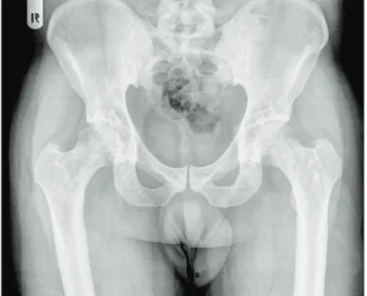
A previously healthy 42-year-old Caucasian woman de-veloped complaints of mechanical knee pain and bi-lateral gluteal pain. She performed a pelvic conven-tional radiography which revealed multiple dense rounded lesions scattered in the iliac bones and ischio-pubic branches. A computed tomography (CT) scan was requested to exclude a tumor, and it confirmed the presence of irregularly contoured osteocondensing fo-cal lesions, some of them showing a spiculate appear-ance strongly suspicious of a secondary lesion. Blood tests, including phospho-calcium metabolism, did not show relevant changes and serum tumor markers were negative. Mammography and thoraco-abdominopelvic CT scan were also normal. Technetium-99m (99mTc) whole body bone scintigraphy did not show any fixa-tion zone thus supporting the diagnosis of OPK. cAsE 2
A 22-year-old Caucasian male, soldier, smoker (20 cigarettes a day for 4 years), with no known personal or family history, suffered trauma to the left wrist. He per-formed radiographs (Figure 1) which excluded fracture and revealed the presence of small, multiple symmetri-cally distributed, oval and rounded lesions of increased and well-defined density located at the distal radii and 1. Rheumatology, Instituto Português de Reumatologia
2. Rheumatology, Centro Hospitalar São João 3. Rheumatology, Hospital Garcia de Orta
Osteopoikilosis: case series from
Portuguese Rheumatology centers
Madeira N1, Ganhão S2, Ferreira RM2, Duarte AC3, Freitas R3, Cunha-Miranda L1
ACTA REUMATOL PORT. 2019;44:78-83
AbstrAct
Osteopoikilosis (OPK) is a rare, hereditary, usually asymptomatic disease characterized by the presence of multiple, well-defined sclerotic lesions distributed in periarticular locations, frequently diagnosed as an inci -dental finding. Differential diagnosis with osteoblastic metastases is fundamental. This article reports six ca -ses of OPK diagnosed in Portuguese Rheumatology Centers.
Keywords: Case series; Osteopoikilosis.
INtrODUctION
Osteopoikilosis (OPK), osteopathy condensans dissemina
te or “spotted bone disease” is an uncommon osteo
-sclerotic dysplasia1. It is a benign and usually asympto
-matic disease of unknown cause. Inheritance is auto-somal dominant; although cases with no family histo-ry have been reported. Radiologically it is characterized by the presence of multiple, well-defined, oval or rounded sclerotic lesions distributed in peri-articular locations, epiphysis and metaphysis of long bones. It is not uncommon for its diagnosis to be obtained during the investigation carried out after a conventional ra-diography requested in the context of another
con-comitant clinical situation, such as a trauma1–3. Herein
we report 6 cases of OPK, identified in Portuguese Rheumatology Centers, describing clinical and radio-graphic features.
cAsE sErIEs
Rheuma-tA b LE I. O st EO P O IK IL O sI s cA sE s D IA G N O sE D IN P O r tU G U Es E r H EU M At O LO G Y c EN tE r s A g e at K n o w n R el ev an t d ia g n o si s fa m il y R ea so n cl in ic al D er m at o -Im ag in g (y ea rs h is to ry fo r th e A rt ic u la r ex am in at io n K n o w n b o n e lo g ic al L ab o ra to ry ex am s C as e G en d er C o m o rb id it ie s o ld ) o f O P K fi rs t X -r ay sy m p to m s fi n d in g s in v o lv em en t in v o lv em en t ch an g es p er fo rm ed 1 F em al e N o n e 4 2 N o B il at er al B il at er al N o P B N o N o , C R , C T o f g lu te al g lu te al in cl u d in g P B , p ai n p ai n se ru m m am m o g ra p h y, tu m o r T A P C T , W h o le m ar k er s b o d y b o n e sc in ti g ra p h y 2 M al e S m o k in g 2 2 N o L ef t w ri st V er y N o H , R , U , C B , N o N o C R , C T o f tr au m a sp o ra d ic M C , P g es o f h an d s, W h o le ar th ra lg ia h an d s an d f ee t, b o d y b o n e o f fe et a n d P A R , P B , F em ., sc in ti g ra p h y w ri st s F ib u l. , T , M T , T ar sa l b o n es 3 M al e N o n e 2 6 N o M in o r N o N o H , R , U , C B , M C N o N o C R tr au m a o f P g es o f h an d s le ft f o re ar m an d f ee t, P B , F em ., F ib u l. , T , M T , T ar sa l b o n es 4 M al e N o n e 2 9 Ye s F am il y N o N o H an d s (M C , N o N o C R (s ib li n g h is to ry o f P ge s) , P B , K , F ee t of c as e 3) O P K 5 F em al e JI A , to ta l 3 2 N o In fl am . In fl am . N o n e o th er S h , H ip s, K N o N o n e o th er C R , W h o le b o d y b il at er al h ip p o ly ar th ra lg ia p o ly ar th -th an t h o se th an t h o se b o n e re p la ce m en t, an d p ro lo n g ed ra lg ia as si g n ed as si g n ed t o sc in ti g ra p h y ty p e 2 D M , M S i n t h e to J IA JI A A H T , C R F co n te x t o f JI A 6 M al e R en al l it h ia si s 2 9 N A N S a rt h ra lg ia N S a rt h ra l-N o n e S h , H ip s, K N o N A ( L T F ) C R (L T F ) o f S h , h ip s, K g ia o f S h , h ip s, K O P K : o st eo p o ik il o si s; J IA : ju v en il e id io p at h ic a rt h ri ti s; C T : co m p u te d t o m o g ra p h y ; H : h u m er i, U : u ln ae , C B : ca rp al b o n es ; P A R : p er i-ac et ab u la r re g io n ; P B : p el v ic b o n es ; S h : sh o u ld er s; P g es : p h al an g es ; N A : n o t ap p li ca b le ; C R : co n v en ti o n al r ad io g ra p h y ; D M : d ia b et es m el li tu s; T A P : th o ra co -a b d o m in o p el v ic ; C R F : ch ro n ic r en al f ai lu re ; L T F : lo st t o f o ll o w -u p ; M S : m o rn in g st if fn es s; A H T : ar te ri al h y p er te n si o n ; In fl am .: I n fl am m at o ry ; R : ra d ii ; M C : m et ac ar p al s; M T : m et at ar sa ls ; T : ti b ia e; F em : fe m u r; K : k n ee ; N S : n o n -s p ec if ic .
ulnae, carpal bones and peri-articular regions of the metacarpals and phallanges. The patient was referred to a Rheumatology appointment in the following month, in which he reported episodes of very sporadic feet and wrists pain in the past. Clinical examination and labo-ratory tests were unremarkable. Additional conven-tional radiographs were requested and similar lesions were found in peri-acetabular region, ischium and ilio-pubic branches, proximal epiphysis of humeri and fe-mura, distal epiphysis of fibulae and ti biae, tarsal bones, peri-articular areas of the metatarsal bones and inter-phalangeal regions of the feet, bilate rally; no changes were found in skull and chest conventional radiographs. 99mTc whole body bone scintigraphy exclu -ded the existence of focal osteoblastic lesions or suspi-cious alterations of metabo lic bone disease. CT scans of the hands (Figure 1) demonstrated micronodular ima -ges of increased density, dispersed by all the bone struc-tures, compatible with OPK.
cAsE 3
A 26-year-old Caucasian male, with no relevant per-sonal or family history, was observed at the emergen-cy department due to a minor trauma of his left fore-arm. At that time, he performed a conventional radio-graphy of the left forearm and hand which revealed several sclerotic oval and rounded lesions of approxi-mately 2 to 4 mm in diameter in the periarticular areas of the carpal bones, metacarpophalangeal and interphalangeal regions, distal radii and ulnae. He was then referred for a rheumatologic evaluation, where he did not present any symptom or relevant finding on clinical examination. To confirm the diagnosis, addi-tional convenaddi-tional radiographs were requested, which showed similar lesions, symmetrically distributed in the pelvic bones (Figure 2), metaphysis and epiphysis of femura, fibulae, tibiae, carpal and tarsal bones, metatarsals and phalanges of feet, humeri, radii and ulnae. Blood tests requested were also normal, ex-cluding other etiologies and supporting the diagnosis of OPK.
cAsE 4
A 29yearold previously healthy Caucasian male wi -thout any relevant finding at clinical evaluation,
per-formed multiple conventional radiographsafter his
brother was diagnosed with OPK (case 3). They re-vealed similar lesions on hands, pelvic bones, knees and feet (Figure 3). These findings were not present on their mother’s conventional radiographs. Their father refused the investigation. Also, it was not possible to establish if anyone else in the family had this condition. cAsE 5
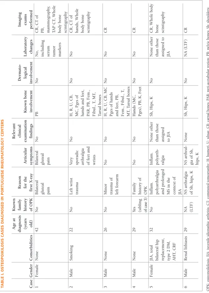
A 32-year-old Caucasian female, diagnosed with rheumatoid factor negative polyarticular juvenile idio -pathic arthritis (JIA) since she was 8 years old, with a FIGUrE 1.Hands AP radiography and computed tomography (case 2)
past history of bilateral total hip replacement, arterial hypertension, type 2 diabetes mellitus and chronic re-nal failure, referred complaints of inflammatory polyar thralgia and prolonged morning stiffness at a Rheumatology follow-up visit. Multiple conventional radiographswere requested which revealed accidental findings of OPK lesions on shoulders (Figure 4), hips and knees. 99mTc whole-body bone scintigraphy did not reveal any image suggestive of metastases and showed mild hyperfixation in these locations. Blood tests of phospho-calcium metabolism were normal. She denied a family history of rheumatic diseases. cAsE 6
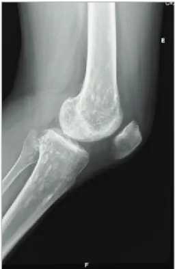
A 29-year-old Caucasian male attended his assistant physician because of non-specific arthralgia of shoulders, hips and knees, without any associated sym -ptoms. He reported a history of renal lithiasis and de-nied any relevant family history. Clinical examination was normal, without arthritis or limited range of mo-tion. Conventional radiographs showed multiple small, well defined, circular or ovoid sclerotic areas on the shoulders, hips and knees (Figure 5). Unfortu-nately, the patient missed the following appointments and did not perform the requested blood tests with
phospho-calcium metabolism.
DIscUssION
The first description of OPK dates from 1915, by
Al-bers-Schönberg1. It is a rare bone dysplasia, with an
estimated incidence of 1 in 50 0004. It is inherited in
an autosomal dominant pattern, with variable pene-FIGUrE 3.Feet AP radiography (case 4)
FIGUrE 4.Right shoulder AP radiography (case 5)
trance, and characterized by an abnormality in the endo chondral bone maturation process that develops during childhood and persists throughout life, causing focal deposits of compact lamellar bone within
can-cellous bone. Sporadic cases have also been repor ted2,5.
According to the published literature, men and
women are equally affected6. However, in our natio nal
case series, male sex was twice as frequent, although it is important to notice that we are only describing six patients. Diagnosis was established at a relatively young age and regarding personal history, there was no common previous pathology in these patients. As
very often described1–4,7, we too found that in half of
the cases, diagnosis was an incidental finding, either because the patient had some trauma (case 2 and 3) or because of probably unrelated pain (case 5). Two pa-tients referred joint pain (case 1 and 6).
According to the published literature, clinical manifes tations, if present, are usually mild. Mild ar-ticular pain with or without joint effusion have been
reported in 15 to 20% of the cases7. In our series, only
two patients were totally asymptomatic. As for the others, it is difficult to clinically demonstrate that the symptoms could be directly attributed to the OPK le-sions, since they were very unspecific and each one had multiple lesions in multiple places but not all were symptomatic. Physical abnormalities were only found in the patient with JIA, given her concomitant rheumatic disease. The coexistence of other rheuma -tic diseases such as rheumatoid arthritis, systemic lu-pus erythematosus, spondyloarthritis (including pso-riatic arthritis), systemic sclerosis, family Mediter-ranean fever or fibromyalgia were previously described
in published report cases2,6.
Interestingly, none of these patients had dermato-logic disorders. However, it has been reported that in 25% of the cases, OPK can be associated with several dermatologic manifestations, such as dermatofibrosis lenticularis disseminata (Buschke-Ollendorff syn-drome), characterized by multiple papular fibromas on the back, arms, and thighs, or a predisposition to keloid formation, scleroderma-like lesions or discoid
lupus erythematosus6,7. According to some authors,
OPK has been associated with other conditions such as dacryocystitis, heart or renal malformations and
en-docrine disorders2. As comorbidities, we highlight the
history of renal lithiasis in case 6 and type 2 diabetes mellitus and chronic kidney failure in case 5.
Radiologically, OPK lesions appear as sclerotic, nu-merous, well defined, homogenous, circular or ovoid,
varying in size from a few millimeters to several
cen-timeters6. They are symmetrically distributed in
peri-articular locations, more frequently in the epiphysis and metaphysis of long tubular bones, carpal and tarsal bones, metacarpals, metatarsals, phalanges of hands
and feet and pelvic bones7,8. The involvement of the
skull, ribs and clavicles is rare8. Our case series is
con-sistent with the published literature, since the most in-volved bones were carpal bones (3/6), metacarpals (3/6), humeri (3/6), femura (3/6), knees (3/6), ischio-pubic branches (3/6), radii (2/6), ulnae (2/6), metatarsals (2/6), tarsals (2/6), phalanges of hands and feet (2/6) and iliac bones (2/6).
Despite being a benign entity, differential diagnosis with other bone pathologies is essential, especially with osteoblastic metastases, mastocytosis, melorheostosis
and tuberous sclerosis3,7. Unlike OPK lesions,
os-teoblastic metastases are usually asymmetric, highly variable in size, associated with bone destruction and located in vertebral bodies, ribs and diaphysis of long
bones2,9. In mastocytosis and tuberous sclerosis,
scle-rotic lesions are more often asymmetric, less defined and have less preference for peri-articular
localiza-tion2,7. 99mTc bone scintigraphy with radiotracer
up-take is suggestive of metastases or systemic diseases such as mastocytosis and tuberous sclerosis, while a negative bone scan hints at benign bone disease. Bone scintigraphy can also be useful to exclude the diffe -rential diagnosis of melorheostosis, a rare sclerosing bone dysplasia, characterized by cortical and medullary hyperostosis with typical “dripping candle
wax” appearance, usually involving the long bones7,11.
Although bone scintigraphy is usually normal in OPK, it must be borne in mind that cases with mild hyper-fixation had been previously reported in the published literature, as described in case 5, reflecting active os-seous remodelling similar to what had been observed
in bone islands10. If doubts remain in the differential
diagno sis, a bone biopsy may be considered to exclude other processes.
In half of the cases described the diagnosis was established by the presence of typical radiographic fin -dings together with the absence of laboratory abnor-malities, and for one of these patients, also supported by the family history. For the remaining three patients, 99mTc whole body scintigraphy was requested and one exclude metastases but showed mild hyperfixa-tion at the sites where radiographic lesions were iden-tified, probably due to active osseous remodeling. CT scans were also requested in two of these patients to
cOrrEsPONDENcE tO
Nathalie Madeira
IPR - Rua da Beneficência, 7 1050 - 034 Lisboa
E-mail: nathalie.almeida.madeira@gmail.com
rEFErENcEs
1. Inci MF, Vurdem UE, Gumus H, Inci R. Case report of a patient with osteopoikilosis. Rheumatol Int 2012; 32: 2829–2832. 2. Woyciechowsky TG, Monticielo MR, Keiserman B, Monticielo
OA. Osteopoikilosis: what does the rheumatologist must know about it? Clin Rheumatol 2012; 31: 745–748.
3. Carvalho ACP, Santos Beze R, Picinini SE. Osteopoiquilose -Apresentação de um caso e revisão da literatura. Radiol Bras 2002; 35: 191–192.
4. Carpintero P, Abad JA, Serrano P, Serrano JA, Rodriguez P, Cas-tro L. Clinical features of ten cases of osteopoikilosis. Clin Rheu-matol 2004; 23: 505–508.
5. Cravo AR, Villacreses C, Canas da Silva J. Osteopoiquilose: dois casos clínicos. Acta Reum Port 2006; 31: 255–260. 6. Serdaroglu M, Capkin E, Uçuncu F, Tosun M. Case report of a
patient with osteopoikilosis. Rheumatol Int 2007; 27: 683–686. 7. Negi RS, Manchanda KL, Sanga S, Chand S, Goswami G.
Os-teopoikilosis - Spotted bone disease. Med J Armed Forces In-dia 2013; 69: 196–198.
8. Czerniak B. Sclerosing Bone Lesions. In: Saunders, editor Dorf-man and Czerniak’s Bone Tumors Second. Philadelphia: Else-vier; 2016: 1317–1345.
9. Ogbonnaya A, Middleton B, Cady T, Ho C. Osteopoikilosis. Lancet 2014; 383: e4.
10. Tsai S-Y, Wang S-Y, Shiau Y-C, Wu Y-W. Benign incidental fin-dings of osteopoikilosis on Tc-99m MDP bone SPECT/CT: A case report and literature review. Medicine (Baltimore) 2016; 95: e3868.
11. Bullough PG. Bone-forming tumors and tumor-like conditions. In: Mosby, editor Orthopaedic Pathology Fifth Edit. Maryland Heights: Elsevier; 2010: 361–398.
confirm conventional radiographs findings. For one case only (case 1) some of the lesions had a spiculate appearance that raised the suspicion of a secondary le-sion. Complementary evaluation with mammography and thoraco-abdominopelvic CT excluded a primary malignancy. For this particular case, a normal scintigra -phy confirmed the diagnosis.
The majority of patients are asymptomatic and do not require treatment. For symptomatic patients, treat-ment relies essentially on pain relief by using non--steroidal anti-inflammatory drugs, acetaminophen and/or weak opioids. Previously published case reports have reported occasional complications or coexisting pathological conditions such as premyelopathic syn-drome due to spinal canal stenosis, hip fracture and tumors (osteosarcoma, chondrosarcoma and giant cell tumor) in OPK patients, suggesting that a regular clini
-cal follow-up of these patients is needed4.
Neverthe-less, the association with these diseases has not yet
been confirmed3, and none of these conditions was
seen in our patients.
In conclusion, after exclusion of common differen-tial diagnosis, particularly osteoblastic metastases, diagno sis of OPK should be considered in the presen -ce of the typical, well-defined radiographic findings, in an asymptomatic patient or with mild joint symptoms and without laboratory tests abnormalities.