CLINICAL SCIENCE
Earthquake-related pelvic crush fracture vs.
non-earthquake fracture on digital radiography and
MDCT: a comparative study
Tian-wu Chen,I,IIZhi-gang Yang,II,IIIZhi-hui Dong,IISi-shi Tang,IIZhi-gang Chu,IIHeng ShaoII
IDepartment of Radiology, West China Hospital of Sichuan University, 37#Guo Xue Xiang, Chengdu, Sichuan 610041, China.IISichuan Province Key
Laboratory of Medical Imaging, and Department of Radiology, Affiliated Hospital of North Sichuan Medical College, 63#Wen Hua Lu, Nanchong, Sichuan 637000, China.IIIState Key Laboratory of Biotherapy, West China Hospital of Sichuan University, 37#Guo Xue Xiang, Chengdu, Sichuan 610041, China.
OBJECTIVE:To determine the features of earthquake-related pelvic crush fractures versus non-earthquake fractures with digital radiography and multidetector row computed tomography.
METHODS:One hundred and sixty-seven survivors with pelvic crush fractures in the 2008 Sichuan earthquake were entered in our study as the earthquake-related group (139 underwent digital radiography, 28 underwent multidetector row computed tomography); 70 victims with non-earthquake pelvic fractures were enrolled into this study as the non-earthquake group (54 underwent digital radiography, 16 underwent multidetector row computed tomography). Data were reviewed retrospectively between groups, focusing on anatomic distributions, status of pelvic bone fractures, numbers of pelvic bones involved, and classification of pelvic ring fractures according to the Tile classification system.
RESULTS:Pelvic fractures occurred more frequently in the pubis in the earthquake-related group than in the non-earthquake group (135/167, 81% vs. 48/70, 69%). In addition, comminuted fractures were more common in the earthquake-related group than in the non-earthquake group (55/167, 33% vs. 10/70, 14%). Multiple fractures were less common in the earthquake-related group than in the non-earthquake group (81/167, 49% vs. 46/70, 66%). Regarding the classification of pelvic ring fractures, Type C predominantly composed of subtype C3 occurred more frequently (64/167, 38% vs. 12/70, 17%), and Type A was less common in the earthquake-related group than in the non-earthquake group (31/167, 19% vs. 23/70, 32%). All differences were statistically significant (p,0.05). No difference was found in Type B fractures between the groups (72/167, 43% vs. 35/70, 50%).
CONCLUSION:Earthquake-related pelvic crush fractures can be characterized by a high incidence of pelvic fractures occurring in the pubis, comminuted fractures, and Type C fractures predominantly composed by subtype C3, despite a low incidence of multiple fractures.
KEYWORDS: Earthquake; Pelvis; Fracture; Digital radiography; Multidetector row computed tomography.
Chen TW, Yang ZG, Dong ZH, Tang SS, Chu ZG, Shao H. Earthquake-related pelvic crush fracture vs. non-earthquake fracture on digital radiography and MDCT: a comparative study. Clinics. 2011;66(4):629-634.
Received for publication onDecember 24, 2010;First review completed onJanuary 17, 2011;Accepted for publication onJanuary 17, 2011 E-mail: [email protected]
Tel.: 86-28-85423817
INTRODUCTION
During the past 20 years, natural disasters have claimed more than three million lives worldwide and affected at least 800 million people.1 In comparison with other natural
disasters, such as floods, volcanic eruptions and droughts, earthquakes are much more harmful in terms of loss of life.2-3
For example, in the 2008 Sichuan earthquake, 374,643 people were injured, 69,227 were killed, and 17,923 were missing.4-7
In earthquake settings, pelvic crush fractures are one of the most common traumas due to high-force impacts.5,8-9As
demonstrated by Chen et al., 5 the profile of pelvic crush fractures may largely include multiple fractures, predomi-nantly occurring in the bilateral pubis, composed of Type C3 followed by Types B3 and B2, according to the Tile classification system for pelvic ring fractures. This observa-tion suggests that earthquake-related pelvic crush fractures might be different from non-earthquake fractures.
To evaluate the differences between earthquake-related pelvic crush fractures and non-earthquake fractures, radio-graphy and computed tomoradio-graphy (CT) are considered standard procedures. However, until now, only two reports regarding radiography and computed tomography used with earthquake-related pelvic fractures have been Copyrightß2011CLINICS– This is an Open Access article distributed under
published.5,10The limitations of published reports include a
lack of comparisons between earthquake-related pelvic crush fractures and non-earthquake fractures to confirm the features of earthquake-related pelvic fractures. To the best of our knowledge, there are no relevant reports to illustrate the features of earthquake-related pelvic crush fractures that are different from non-earthquake fractures. Based on the published reports of Chen et al.,5we aimed to retrospectively investigate the differences between earth-quake-related pelvic crush fractures and non-earthquake fractures using radiography and multidetector row CT (MDCT). We also sought to confirm the features of earth-quake-related fractures to obtain a better understanding and to promote effective treatment planning of victims with earthquake-related pelvic crush fractures in the future.
MATERIALS AND METHODS
Participants
The present study was approved by our institution’s human research committee, and informed consent was waived because of the retrospective nature of this study. There were two groups in our study: an earthquake-related group and a non-earthquake-related group. Patients were entered in this study in the earthquake-related group according to the following criteria: (1) the pelvic fracture was initially confirmed by DR or CT, and (2) the mechanism of the fracture was a crush injury caused by building collapse or projectile objects in the 2008 Sichuan earthquake. Patients were excluded from the earthquake-related group if the mechanism was not a crush injury, but rather a jump or accidental fall from a building. Patients with pelvic fractures were enrolled into the non-earthquake group if the fracture resulted from trauma other than an earthquake-related injury.
At 2:28 pm local time on May 12, 2008, an earthquake of magnitude 8.0 on the Richter scale occurred in the mountainous region of Sichuan in China. The epicenter was in Wenchuan County in Sichuan Province, China. In view of the massive morbidity arising from this earthquake, an undamaged key university hospital equipped with 4300 beds in the earthquake-affected area, but 92 kilometers away from the epicenter, received and treated a total of 2728 victims with earthquake-related injuries over a period of 15 days after the earthquake. Among these victims, 167 consecutive victims (65 men and 102 women; mean age, 42.87 years; age range, 6-103 years) with pelvic crush fractures resulting from the Sichuan earthquake presented in the emergency department of this university hospital met the inclusion criteria and were entered into the present study as the earthquake-related group. In this group, 13 victims with pelvic fractures had crush injuries in one or more other anatomic regions, including the thorax in 11 patients, the abdomen in 6, the spine in 3, the extremities in 3, and the cranial region in 1. They initially underwent pelvic CT scans along with thoracic, abdominal or cranial CT scans to explore the pelvic fractures, as well as traumas in the pleural space, peritoneum or skull.
To detect pelvic fractures in a great number of injured patients in a timely manner in this earthquake setting for emergency medical treatment, 11 digital radiography (DR) scanners and 5 CT scanners in this key university hospital were utilized to image the fractures as quickly as possible. Because it would be faster and easier to utilize DR rather
than CT scanners, 139 patients with pelvic fractures who did not have severe traumas suggested by clinical data under-went DR scans in this major earthquake situation. Because CT scanning can improve the assessment reliability of both pelvic fractures and hemorrhage, 28 patients with pelvic fractures in combination with possible pelvic hemorrhage or severe traumas as suggested by clinical data underwent MDCT scans. Among the 28 patients receiving CT scans, pelvic hemorrhage was found in 9. The mean time from the crush fracture to imaging was 5.4 days, with a range of 6 hours to 14 days. Few critically ill patients survived until they could be conveyed to hospitals to receive effective treatment, although some of the survivors received anti-biotics to prevent infection in the disaster areas.
For reduction and fixation of pelvic fractures or for controlling pelvic hemorrhage, 136 victims, including the 28 severely traumatized patients, underwent pelvic surgery, and the remaining with incomplete fractures had conserva-tive treatment. In the cohort, 166 patients recovered due to appropriate treatment, and 1 died of fatal crush injuries.
Between May 1 and June 6, 2009, 70 consecutive victims (51 men and 19 women; mean age, 38.84 years; age range, 2-88 years) with non-earthquake pelvic fractures presenting in the above-mentioned university hospital that met the inclusion criteria were enrolled into this study as the non-earthquake group. According to the etiology, 29, 22, 14, and 5 patients had pelvic fractures due to traffic accidents, falls, crush and collision injuries, and work-related accidents, respectively. In this group, 42 victims had pelvic crush fractures in combination with injuries in one or more other anatomic systems, including the abdomen in 5 patients, the thorax in 5, the spine in 6, the extremities in 30, and the craniocerebral region in 3. In this cohort, one DR scanner and CT scanner were used specifically for imaging the fractures. Fifty-four victims underwent DR scans, and the remaining victims with severe traumas suggested by clinical data underwent CT scans. The mean time from pelvic fracture to imaging was 4.3 days, ranging from 1 hour to 13 days. According to the image findings and clinical data, 47 victims underwent open reduction and internal fixation, and the remaining underwent external fixation. In this cohort, 69 victims recovered, and 1 died of fatal injury.
Digital Radiography
In the earthquake-related and non-earthquake-related groups, 139 and 54 victims, respectively, were imaged in a standard anteroposterior (AP) view using a digital radio-graphic system (Digital Diagnost, Philips Healthcare, Hamburg, Germany). To detect the pelvic fractures in a timely way in an emergency setting, particularly with a great number of injured patients due to the earthquake, all victims in both groups had only an AP pelvic radiograph (Figure 1) without inlet/outlet views. In the earthquake-related and non-earthquake groups, a total of 139 and 54 radiographic examinations, comprising a total of 139 and 54 radiographs, were obtained, respectively. The scanning parameters used for the scanners were as follows: 70 kV, 24.9 mAs, an active imaging area of 43643 cm, a theoretical spatial resolution of 3.5 line pairs per millimeter, and a matrix of 300163001 pixels.
Computed Tomography
with a Somatom Sensation 16-row MDCT (Siemens Medical Systems, Forchheim, Germany), a Brilliance 16-row MDCT (Philips Healthcare, Eindhoven, the Netherlands), or a Somatom Sensation 4-row MDCT (Siemens Medical Systems, Forchheim, Germany). After the Sichuan earth-quake, this analysis was performed without intravenous contrast material as soon as possible after presentation due to the suspicion of acute renal failure. In the non-earthquake group, 16 victims with suggested severe traumas underwent similar non-enhanced emergency scans with the previously mentioned Somatom Sensation 16-row MDCT. The scanning parameters used for the 16-row MDCT scanners were as follows: 120 kV, 200 mAs, 0.5-s gantry rotation time, a pitch of 0.85, a collimation of 12 mm, 1-mm reconstructed section thickness, 380-mm field of view, and a matrix of 5126512 mm. The scanning parameters used for the 4-row MDCT were similar to those used for the 16-row MDCT scanners, except for a pitch of 1.5 and a collimation of 10 mm.
Image Data Analysis
Image data for the groups were transferred to a picture-archiving communication system (Syngo–Imaging, Siemens Solution Systems, Forchheim, Germany) and were retro-spectively reviewed by an associate radiological professor (the first author, with 12 years of experience in radiology) and one experienced radiologist (the third author, with 11 years of experience in radiology), focusing on the anatomic distribu-tions, the status of the pelvic bone fractures, the numbers of pelvic bones involved, and classification of the pelvic ring fracture by consensus to maintain accuracy in the analysis.
Based on the status of the pelvic bone fractures, the cases were classified as comminuted or non-comminuted frac-tures, including transverse, oblique, linear and spiral fractures. According to the numbers of pelvic bones involved, the cases were considered as single or multiple fractures when the numbers were 1 or more than 1, respectively. Regarding the classification of pelvic ring fractures, the Tile classification system was used in this study because the Tile system is more useful as compared to the Young-Burgess classification system for specialists in pelvic and acetabular surgery.11According to the increasing
severity of pelvic ring fractures described by Tile 12, the
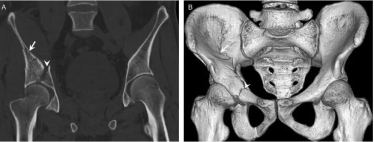
cases were classified according to the pelvic ring stability (Table 1). In addition, the CT data were used to perform multiplanar reconstruction and generate volume-rendering images (Figure 2) with a slab of 7-10 mm to depict pelvic fractures in detail.
Statistical Analysis
Statistical analysis was performed with the SPSS statistical package (version 13.0 for Windows, SPSS Inc., Chicago, IL, USA). To investigate the features of earthquake-related pelvic crush fractures as compared to non-earthquake fractures, we compared the pelvic bones predominantly involved in the earthquake-related group, the incidence of comminuted fractures and multiple fractures and the classification of pelvic ring fractures between groups by Fisher’s exact tests. A two-tailed p-value of less than 0.05
was considered significant.
RESULTS
General Anatomic Distributions of Pelvic Bones Involved
In the two groups, the fracture occurred in one or more of the pelvic bones including the pubis, ilium, ischium and sacrum. Regarding the pelvic bones involved in the earth-quake-related group, a total of 80.84% (135/167) and 47.31% (79/167) of patients had a pelvic fracture in the pubis and in one or more of the other pelvic bones, respectively. In the non-earthquake group, fractures of the pubis and of one or more of the other pelvic bones occurred in a total of 68.57% (48/70) and 75.71% (53/70) of patients, respectively. Detailed results of the pelvic bones involved in the pelvic fractures are listed in Table 2. Interestingly, fractures of the pubis occurred more frequently in the earthquake-related group than in the non-earthquake group (p= 0.044), whereas
fractures in one or more of the other pelvic bones were less common in the earthquake-related group than in the non-earthquake group (p,0.001).
Status of Pelvic Bone Fractures
Pelvic bone fractures appeared as both comminuted and non-comminuted fractures. In the earthquake-related group, pelvic comminuted fractures occurred in 32.93% (55/167) of patients. In the non-earthquake group, 14.29% (10/70) of patients had comminuted fractures. Therefore, comminuted fractures occurred more frequently in the earthquake-related group than in the non-earthquake group (p= 0.004).
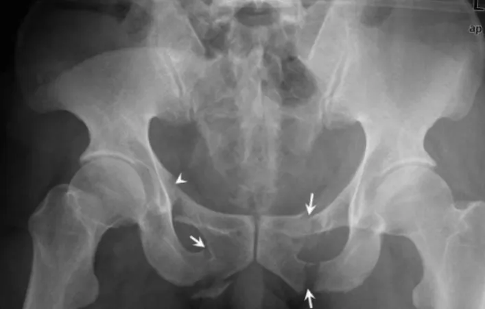
Regarding the pelvic bones involved in the earthquake-related group, comminuted fractures occurred in the pubis in 22.75% of patients (38 of 167) and in any other pelvic bone in Figure 1 -The digital radiograph of fractured pelvis of a
45-year-old man depicts fractures in left inferior and superior pubic ramus, in right inferior pubic ramus (white arrows), and in superior pubic ramus involving acetabulum (white arrowhead); and the pelvic ring fracture is classified as Type C.
Table 1 -Tile Classification of pelvic ring fractures.
Type A: Pelvic ring stable
A1: fractures not involving the ring (i.e. avulsions, iliac wing or crest fractures)
A2: stable minimally displaced fractures of the pelvic ring Type B: Pelvic ring rotationally unstable, vertically stable
B1: open book
B2: lateral compression, ipsilateral
B3: lateral compression, contralateral or bucket handle-type injury Type C: Pelvic ring rotationally and vertically unstable
C1: unilateral C2: bilateral
not more than 10.78% (18 of 167). Based on the pelvic bones involved in the two groups, patients with comminuted fractures are listed in Table 3. The comminuted fractures occurred more frequently in the pubis in the earthquake-related group than in the non-earthquake group (p= 0.01).
Number of Pelvic Bone Fractures
According to the number of bones involved, pelvic fractures appeared as single and multiple fractures. In the earthquake-related group, single and multiple fractures occurred in 51.5% (86/167) and 48.5% (81/167) of patients, respectively. In the non-earthquake group, however, 34.29% (24/70) and 65.71% (46/70) of patients had single and multiple fractures, respectively. Thus, single fractures were observed more often, whereas multiple fractures were less common in the earthquake-related group than in the non-earthquake group (p= 0.022).
Regarding the bones involved, single fractures occurred in the pubis in 66.28% of patients (57 of 86) and in the other pelvic bones in a total of 33.72% (29 of 86) in the earthquake-related group, while the fracture occurred in the pubis in 75% of patients (18 of 24) and in the other pelvic bones in a total of 25% (6 of 24) in the non-earthquake group. According to the pelvic bones involved, detailed results of single fractures are shown in Table 4. Single fractures occurred more frequently in the pubis in the earthquake-related group than in the non-earthquake group (p,0.001). According to the bones involved, multiple fractures occurred in the pubis in 96.3% of patients (78 of 81) and in
the other pelvic bones in a total of 60.49% (49 of 81) in the earthquake-related group. Fractures occurred in the pubis in 91.3% of patients (42 of 46) and in the other pelvic bones in a total of 76.07% (35 of 46) in the non-earthquake group. Based on the pelvic bones involved, detailed results of multiple fractures are listed in Table 4. There were no significant differences in multiple fractures occurring in the pubis between the groups (p= 0.457).
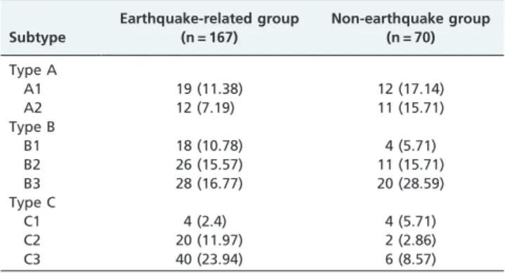
Classification of Pelvic Ring Fractures
According to the Tile classification system for pelvic ring fractures, Type A, Type B and Type C, respectively, occurred in 18.56% (31/167), 43.11% (72/167) and 38.32% (64/167) of patients in the earthquake-related group, whereas the patients of the non-earthquake group had 32.86% (23/70), 50% (35/70) and 14.16% (12/70). Based on each subtype in these groups, the patients with pelvic ring fractures are listed in Table 5. Interestingly, Type C pelvic ring fractures occurred more frequently in the earthquake-related group than in the non-earthquake group (p= 0.001), whereas Type A pelvic ring
fractures were less common in the earthquake-related group than in the non-earthquake group (p= 0.026). No significant
differences were observed for Type B pelvic ring fractures between the groups (p= 0.391).
DISCUSSION
Pelvic fractures represent a serious, potentially life-threatening injury in patients with major trauma.13 The most common mechanism of injury resulting in pelvic Figure 2-The CT coronal reconstruction (a) and 3D reconstruction (b) images of a 51-year-old man illustrate fractures in right ilium (white arrow) involving acetabular roof and columns (white arrowhead); and the pelvic ring fracture is classified as Type C.
Table 2-Patients with pelvic fractures according to each pelvic bone involved.
Anatomic distribution
Earthquake-related group (n = 167)
Non-earthquake group (n = 70)
Pubis 135 (80.84) 48 (68.57)
Ilium 40 (23.95) 24 (34.29)
Ischium 10 (5.99) 9 (12.86)
Sacrum 23 (13.77) 19 (27.14)
Acetabular bone 25 (14.97) 17 (24.29)
Note.-Numbers in parentheses are percentages of the patients.
Table 3-Patients with pelvic comminuted fractures according to each pelvic bone involved.
Anatomic distribution
Earthquake-related group (n = 167)
Non-earthquake group (n = 70)
Pubis 38 (22.75) 10 (14.29)
Ilium 17 (10.18) 4 (5.71)
Ischium 6 (3.59) 3 (4.29)
Sacrum 10 (5.99) 8 (11.13)
Acetabular bone 18 (10.78) 3 (4.29)
fracture is road traffic collisions, followed by pedestrian collisions and motorcyclist collisions.14-17 Due to the mechanism of crush injury resulting from high-force impacts in an earthquake, earthquake-related pelvic crush fractures might be different from non-earthquake fractures. To illustrate the features of earthquake-related pelvic crush fractures that are different from non-earthquake fractures, we performed this comparative study with digital radio-graphy and MDCT. We chose consecutive patients with non-earthquake pelvic fractures presenting in the same university hospital during a similar period one year after a major earthquake as the non-earthquake group to avoid the confusing influence of the earthquake situation.
As shown in our study, earthquake-related pelvic crush fractures occurred more frequently in the pubis in earthquake situations than in non-earthquake settings. The high frequency of earthquake-related pubic crush fractures might be due to the fact that the pubis is the weakest part of the pelvis.5,18-19As presumed by Dong et al.,7shaking and obstacles might cause victims to fall down in a prone posture and become trapped, which may result in persistent compression striking one side of the pelvis and a counteracting force exerted on the other side, eventually leading to a high frequency of pubic fractures because of the weak nature of the pubis.
In view of the severity of pelvic bone fractures, comminuted fractures may be a good indicator because these require more treatment. In this study, we found that pelvic comminuted fractures occurred more frequently in earthquake situations than in non-earthquake settings. Furthermore, according to the pelvic bone involved, comminuted fractures occurred more frequently in the pubis in the earthquake situation, which may be due to both the weak nature of the pubis and high-force impacts.18
In addition, the number of pelvic bones involved may be another indicator for the severity of pelvic fractures in earthquake situations. As demonstrated in our study, single fractures predominantly occurring in the pubis were observed more often, and multiple fractures were less common in the earthquake than in the non-earthquake group. This low frequency of multiple fractures may be explained by their potentially life-threatening nature in contrast to single fractures. We could presume that some patients with multiple fractures in the major earthquake died before being rescued and thus could not be entered into our study, resulting in this low frequency.
Clinically, it is extremely important to accurately assess the instability pattern of pelvic ring fractures for effective treatment. According to the classification of pelvic ring fractures, Type C occurred more frequently in the earth-quake setting, Type A occurred more frequently in the non-earthquake setting, and Type B occurred with equal incidence in both the earthquake-related and non-earth-quake settings. As for the subtype of pelvic ring crush fractures, Type C was predominantly composed of the most severe subtype (Type C3), which suggests that both rotational and vertical instability of the pelvic ring might be more common in an earthquake.
There was an inevitable limitation in our study. For example, some critically ill patients with severe pelvic crush fractures died before rescue in this earthquake and could therefore not be entered into our study. The use of a population that survived long enough to reach the hospital might lead to a selection bias. Despite this limitation, our comparisons of earthquake-related pelvic crush fractures with non-earthquake fractures confirm the characteristic features of earthquake-related fractures in survivors, which may be helpful in better understanding pelvic crush fractures in the future.
CONCLUSIONS
Earthquake-related pelvic crush fractures can be char-acterized by a high incidence of pelvic fractures occurring in the pubis, comminuted fractures occurring predominantly in the pubis, and Type C pelvic ring fractures predomi-nantly composed of subtype C3, despite a low incidence of multiple fractures. We further expect our results to be helpful in providing a better understanding and treatment planning for survivors with similar earthquake-related injuries in the future.
ACKNOWLEDGEMENT
This study was supported by the Science Foundation for Distinguished Young Scholars of Sichuan Province in China (Grant No. 2010JQ0039).
REFERENCES
1. Peek-Asa C, Kraus JF, Bourque LB, Vimalachandra D, Yu J, Abrams J. Fatal and hospitalized injuries resulting from the 1994 Northridge earthquake. Int J Epidemiol. 1998;27:459-65.
2. Reitherman R. How to prepare a hospital for an earthquake. J Emerg Med. 1986;4:119-31.
Table 4 -Patients with pelvic single and multiple fractures according to each pelvic bone involved.
Earthquake-related group (n = 167)
Non-earthquake group (n = 70)
Single fractures
Pubis 57 (34.13) 6 (8.57)
Ilium 15 (8.98) 7 (10)
Ischium 2 (1.2) 0
Sacrum 5 (2.99) 2 (2.86)
Acetabular bone 8 (4.79) 9 (12.86)
Multiple fractures
Pubis 78 (46.71) 42 (60)
Ilium 25 (14.97) 17 (24.29)
Ischium 8 (4.79) 9 (12.86)
Sacrum 18 (10.78) 17 (24.29)
Acetabular bone 17 (10.18) 8 (11.43)
Note.-Numbers in parentheses are percentages of the patients.
Table 5 -Patients with pelvic ring fractures according to each subtype.
Subtype
Earthquake-related group (n = 167)
Non-earthquake group (n = 70)
Type A
A1 19 (11.38) 12 (17.14)
A2 12 (7.19) 11 (15.71)
Type B
B1 18 (10.78) 4 (5.71)
B2 26 (15.57) 11 (15.71)
B3 28 (16.77) 20 (28.59)
Type C
C1 4 (2.4) 4 (5.71)
C2 20 (11.97) 2 (2.86)
C3 40 (23.94) 6 (8.57)
3. Mahoney LE, Reutershan TP. Catastrophic disasters and the design of disaster medical care systems. Ann Emerg Med. 1987;16: 1085-91, doi: 10. 1016/S0196-0644(87)80764-3.
4. Chen TW, Yang ZG, Wang QL, Dong ZH, Yu JQ, Zhuang ZP, et al. Crush extremity fractures associated with the 2008 Sichuan earthquake: anatomic sites, numbers and statuses evaluated with digital radiography and multidetector computed tomography. Skeletal Radiol. 2009;38:1089-97, doi: 10.1007/s00256-009-0743-5.
5. Chen TW, Yang ZG, Dong ZH, Chu ZG, Yao J, Wang QL. Pelvic crush fractures in survivors of the Sichuan earthquake evaluated by digital radiography and multidetector computed tomography. Skeletal Radiol. 2010;39:1117-22, doi: 10.1007/s00256-010-0912-6.
6. Dong ZH, Yang ZG, Chen TW, Feng YC, Wang QL, Chu ZG. Spinal injuries in the Sichuan earthquake. N Engl J Med. 2009;361: 636-7. 7. Dong ZH, Yang ZG, Chen TW, Feng YC, Chu ZG, Yu JQ, et al. Crush
thoracic trauma in the massive Sichuan earthquake: evaluation with multidetector CT of 215 cases. Radiology. 2010;254:285-91, doi: 10.1148/ radiol.09090685.
8. Yang C, Wang HY, Zhong HJ, Zhou L, Jiang DM, Du DY, et al. The epidemiological analyses of trauma patients in Chongqing teaching hospitals following the Wenchuan earthquake. Injury. 2009;40: 488-92, doi: 10.1016/j.injury.2009.01.102.
9. Sheng ZY. Medical support in the Tangshan earthquake: a review of the management of mass casualties and certain major injuries. J Trauma. 1987;27:1130-5.
10. Hussain P. Earthquake radiography. Radiogr Today. 1990;56: 22-5.
11. Koo H, Leveridge M, Thompson C, Zdero R, Bhandari M, Kreder HJ, et al. Interobserver reliability of the Young-burgess and Tile classification systems for fractures of the pelvic ring. J Orthop Trauma. 2008;22:379-84. 12. Tile M. Acute pelvic fractures: I. causation and classification. J Am Acad
Orthop Surg. 1996;4:143-51.
13. Lee C, Porter K. The prehospital management of pelvic fractures. Emerg Med J. 2007;24:130-3, doi: 10.1136/emj.2006.041384.
14. Poole GV, Ward EF, Muakkassa FF, Hsu HS, Griswold JA, Rhodes RS. Pelvic fracture from major blunt trauma. Outcome is determined by associated injuries. Ann Surg. 1991;213:532-8; discussion 538-9., doi: 10. 1097/00000658-199106000-00002
15. Gustavo Parreira J, Coimbra R, Rasslan S, Oliveira A, Fregoneze M, Mercadante M. The role of associated fractures on outcome of blunt trauma patients sustaining pelvic fractures. Injury. 2000;31: 677-82, doi: 10.1016/S0020-1383(00)00074-7.
16. Dalal SA, Burgess AR, Siegel JH, Young JW, Brumback RJ, Poka A, et al. Pelvic fracture in multiple trauma: classification by mechanism is key to pattern of organ injury, resuscitative requirements, and outcome. J Trauma. 1989;29:981-1000.
17. Moffatt CA, Mitter EL, Martinez R. Pelvic fractures crash vehicle indicators. Accid Anal Prev. 1990;22:561-9, doi: 10.1016/0001-4575(90)90028-J. 18. Durkin A, Sagi HC, Durham R, Flint L. Contemporary management of
pelvic fractures. Am J Surg. 2006;192:211-23.