1. Serviço de Medicina Interna, Centro Hospitalar Baixo Vouga, EPE, Aveiro
2. Serviço de Reumatologia, Centro Hospitalar Baixo Vouga, EPE, Aveiro
The most common mali gnan cies noted are lung, breast and gastrointestinal cancers9. The occurrence of lym
-phoma is a rare event, with less than 50 cases reported, although a study found an increased incidence of this lymphomas associated with the disease10. Many
subty-pes of NHL were described but to our knowledge the-re has been no the-report of MCL.
cAse report
A 76-year-old female was admitted to the hematology clinic due to recent unintended weight loss of 6 kilo-grams and occasional night sudoresis. She had a 4-year history of multiple adenopathies, which had recently become painful and with progressive enlargement, and occasional gastro-intestinal symptoms namely abdo-minal pain, diarrhea and rectorrhagia in the past 6 years. She also reported a progressive, symmetrical and mild swelling of hands and feet in the past year. Her medical history was positive for hypertension, dyslipi-demia and gastritis. At examination, she presented mul-tiple adenopathies – submandibular, cervical, supra-clavicular, axillary, epitrochlear and inguinal, the grea-ter on the left axilla, with 10 cm. No palpable masses or any organomegally were noticed on abdominal eva-luation and rectal examination showed no masses or blood. The laboratory tests revealed a normal hemo-gram; erythrocyte sedimentation rate of 64 mm at first hour; normal liver and renal function; lactic dehydro-genase 208 U/L (normal < 190 U/L); C-reactive protein 5.61 mg/dL; protein electrophoresis with elevation of alpha 2 and gamma globulins; β2 microglobulin 6740 ng/mL (normal range 1100-2300 ng/mL). Colonosco-py demonstrated multiple polyps, particularly distal to the splenic angle of the colon; ileocecal valve was en-gorged, with a pseudopolypoid aspect, seeming affec-ted by an expansive process. Histopathological exami-nation showed a lymphoproliferative disease with small/medium-sized lymphoid cells, with irregular and hyperchromatic nucleus, without nucleolous and scar-ce cytoplasm. Immunohistochemical study of the po-lyps and ilecocecal valve revealed positivity to CD20,
Mantle cell lymphoma and systemic sclerosis
Marto G1, Aguiar R2, Barcelos A2
AbstrAct
Systemic sclerosis (SSc) is a rare disease with an in-creased incidence of cancer, but the ocorrence of Non--Hodgkin lymphoma (NHL) is a very uncommon event. We report a case of a 76yearold female addmi ted to the hematology clinic with longterm adenopa -thies and ocasional gastro-intestinal symptomatology. Progressive symmetrical swelling of hands was also no-ticed. Colonoscopy revealed multiple polyps and histopathology was consistent with MantleCell Lym -phoma (MCL)-NHL. R-CHOP (rituximab, cyclophos-phamide, doxorubicine, vincristine and prednisone) regimen was promptly iniated with complete respon-se. Persistent swelling of both hands was observed, with thickening of the skin of both hands with proximal ex-tension until the forearm. Biopsy confirmed the diag-nosis of scleroderma. Symptomatic and rehabilitation treatment was initiated with mild improvement of symptoms. To our knowledge this is the first case of MCL associated with SSc.
Keywords: Mantle cell lymphoma; Systemic sclerosis;
Non-Hodgkin lymphoma.
IntroductIon
Autoimmune-inflamatory systemic diseases, including rheumatoid arthirits, systemic lupus erythematosus, Sjogren’s syndrome, dermatomyositis and SSc have an increased incidence of solid tumors, but also lympho-proliferative disorders1. SSc is a chronic multisystemic
autoimmune disorder characterized by fibrosis and vas-cular abnormalities on both skin and internal organs. The increased risk of ma lignan cy in patients with SSc has already been described2, but the cause is not fully
understood. Immunossupressive agents3-5,
paraneo-plastic phenomena6,7and chronic B-cell stimulation8
were linked to the biopa thogenesis of cancer in SSc.
lective dysphagia. She denied dyspnoea, cough, digi-tal ulcers or any other symptoms. Physical examination revealed tight, shiny and atrophic skin over the face and fingers and face telangiectasias. There was no cal-cinosis. Nifedipine was added and physiotherapy was started. The patient didn’t tolerate nifedipine due to dizziness and headache, but the hand pain relieved with physiotherapy. Chest high resolution CT didn’t revealed pulmonary fibrosis. Lung function tests (DLCO, spirometry, vital capacity) were inconclusive due to lack of collaboration. Transthoracic echocar-diography revealed mild left ventricular hypertrophy, preserved systolic function, impared relaxing diastolic pattern and an estimated pulmonary systolic arterial pressure of 41 mmHg. The patient refused any further investigation. After three years of follow-up, the patient is stable, with sustained remission of the lym -phoma and no new symptoms related to systemic scle-rosis.
dIscussIon
MCL was known like as an indolent variant of mature B-cell NHL11, but it often behaves as an aggresive form,
requiring treatment at presentation. MCL was pre-viously referred to as mantle zone lymphoma, centro-cytic lymphoma, intermediate lymphocentro-cytic lympho-ma or lymphocytic lympholympho-ma of intermediate diffe-rentiation12. Incidence increases with age, with a
me-dian age of 68 years at diagnosis. Approximately 75 percent of affected individuals are male and IT is more common in caucasians than in afro-americans13. The
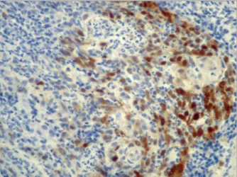
blc2, CD43, CD79a and D1 cyclin, consistent with the diagnosis of a MCL-NHL of the colon (Figures 1 and 2). Upper gastrointestinal endoscopy showed erosive bulbitis. Toracoabdominopelvic computed tomogra -phy (CT) demonstrated multiple adenopathies: para-tracheal, bilateral axilar, celiac trunk, para-aortic and intercavoaortic. The biopsy of an axillary lymph node was consistent with the same diagnosis, with a Ki67 of 50%. The flux cytometry of bone marrow showed no monoclonality of B-lymphocytes, with a normal im-munologic phenotype. The diagnosis of a IIIB-staged Mipi 7.5 (highrisk) MCLNHL was therefore establi -shed and the patient started a R-CHOP regimen (rituxi mab, cyclophosphamide, doxorubicine, vin-cristine and prednisone). After 6 cycles, she achieved a completed response. NHL-related symptoms dra-matically improved, but bilateral hand swelling was still present. The patient was referred to the dermato-logy clinic. Physical examination revealed thickening of the skin in both hands with proximal extension up to the forearm, without skin ulcers. Histopathological analysis of a biopsy of the skin confirmed the diagno-sis of scleroderma. The autoimmunity study showed a strongly positive test for antinuclear antibodies (ANA) (>1/1260 IU with a centromeric pattern) and anti-cen-tromere antibodies (>1/640 IU) with negative anti-scl 70 antibodies. The patient was started on pentoxifyl-line and referred to the rheumatology clinic. In a more exhaustive inquiring, the patient confirmed she had Raynaud phenomena triggered by cold exposition on her hands and feet with 2 years duration. The sclero-dactily was quite painful and caused functional limi-tation. Moreover, she also had recent onset of
non-se-FIGure 1.H & E (400x): Biopsy of colon showing diffuse proliferation of small/medium-sized lymphoid cells
FIGure 2.H & CyclinD1 (400x): Biopsy of colon showing intense reactivity for CyclinD1
sequent development of adenocarcinoma of the eso -phagus19,20. There is no clear explanations at the
mo-ment for the frequency of lymphomas and hematolo-gical malignancies in patients with SSc. It has been suggested that it may be incidental or secondary to an emergence of a malignant clone from a pool of poly-clonal chronically stimulated B-cells21, deficiency of T
and NK cells21-23, or common genetic background that
predisposes to both disorders, like the HLA-DR5 ha-plotype24.
Recently, a systematic review of all cases of NHL re-ported in association with SSc was published by Vettori et al25. The T-cells NHL were angioimmunoblastic and
diffuse large cell subtypes. Subtypes of NHL were all of the B-cell type and included chronic lymphocytic
leu-kemia10, mucosa-associated lymphoid tissue-type
(MALT)26,27, Helicobacter pylori associated MALT28,
small lymphocytic follicular10, diffuse large cell29,8,
Pin-kus variant of skin and muscle30and intermediate
lymphocytic31. More recently, was described a case
re-port of a small lymphocytic lymphoma in a patient with CREST syndrome was described32.
Raynaud’s phenomenon and scleroderma-like syn-drome may occasionally be a paraneoplastic rheuma-tic disease33. While several studies34have demonstrated
an increased frequency of lung, esophageal and breast cancer in patients with scleroderma, there are only a few observations of scleroderma-like syndrome prece-ding the manifestation of cancer. Very little is known about potential mechanisms underlying this connec-tion35.
Several mechanisms could explain the relationship between malignancy and scleroderma. For example: multiple chemotherapeutic agents have been implica-ted as potential causes of scleroderma-like disease or se-vere Rayhnaud’s phenomena36, radiation therapy may
cause localized scleroderma in patients without a prior history of connective tissue disease37.
In the other hand, lymphoproliferative diseases are one of the most common group of disorders associated with autoimmune disturbances38. The presence of ANA
in patients with NHL isn’t an uncommon phenomena since in the past, a significant incidence of ANA was de-monstrated before any treatment in NHL patients. Spe-cifically, ANA directed against mitotic-associated or mi-totic apparatus proteins have been found in the sera of patients with MCL39. Neverthless, its significance or
prognostic importance isn’t established.
In this particular case, the patient had a close tem-poral relationship between cancer diagnosis and syste-mic sclerosis clinical onset, which raises the possibili-ty that the presence of one disease may be implicated majority of patients present with advanced stage
di-sease. The primary presentation is often lymphadeno-pathy (75%), with the remaining presenting initially with extranodal disease14. MCL can involve any region
of the gastrointestinal tract, with a colic involvement of 90 percent of cases, usually with polyps found in co-lon and small bowel15, as we found in our case.
Histo-lologicaly, the pattern may be diffuse, nodular, mantle zone, or a combination of the three and tumor cells are usually small to medium-sized B lymphocytes with ir-regular nuclei, tipically CD5+ and CD23-, with nuclear staining cyclin D1 present in more than 90 percent of cases. The majority of MCL cases are believed to deri-ve from naïderi-ve pre-germinal centre B cells of the mantle zone, and a minority from marginal zone or peri -pheral blood memory B cells. All patients with MCL demonstrate increased cell division and replication. Overexpression of the cyclin D1 gene is present in most cases16, while a minority is thought to overexpress
other cell cycle mediatores, like cyclin D2 and D3. Most patients will require treatment of MCL at the moment of diagno sis, but there is a small subset of patients with a more indolent course who won’t. Chemoimmuno -therapy remains the main treatment modality. R-CHOP and R-CVP (cyclophosphamide, vincristine, predniso-ne plus rituximab) are the preferred treatments. Inten-sive chemoimmunotherapy R-Hyper-CVAD (rituximab plus hyperfractionated cyclophosphamide, vincristine, doxorubicin and dexamethasone) and off-label BR (bendamustine and rituximab) are alternative treatments, occasionally employed. The course and progno -sis of MCL are variable, with median overall survival between three to four years. MIPI (Mantle Cell Lym -phoma International Prognostic Index), IPI (International Prognostic Index) and FLIPI (Follicular Lym -phoma International Prognostic Index) are examples of prognosis indexes that were developed in the last years, with evidence showing that FLIPI gathers better prognostic information17. Recently, Ki-67 staining, a
cellular marker of proliferation was associated with a worse prognosis18.
The prevalence of malignant neoplasia in SSc is es-timated between 3.6% to 10.7% and identified risk fac-tors for malignancy are old age, female gender and dif-fuse skin involvement2. Malignancy in SSc may
coinci-de with the diagnosis or may be a consequence of treat-ment for it or another condition.
Lung and breast cancer remain the most frequently reported types of malignancy, followed by gastrointes-tinal cancers2. There is a close relationship between
scleroderma-related abnormalities of esophageal moti-lity, the development of Barret metaplasia and the
sub-in the subsequent development of the second or may influence the behaviour of the second patology.
To our knowledge, this is the first case report of MCL associated with SSc. Older age and female gender were known risk factors present in our case. Our patient didn’t seek medical care until severe symptomatology related to lymphoma developed. We postulate that a humoral or cell mediated immune process initiated by the lymphoma was responsible for the development of SSc in our patient.
In conclusion, patients with SSc probably need clo-se vigilance for symptoms suggestive of malignancy. At the same time, the clinicians should further investiga-te concomitant symptoms not relainvestiga-ted with LNH (such as Raynaud phenomena) and/or their treatment, parti-cularly if persistent after complete remission of the di-sease.
correspondence to Gonçalo Marto
Rua dos Arcos, nº 10 2660-130 Santo Antão do Tojal Portugal
E-mail: [email protected] reFerences
1. Szekanecz É, Szamosi S, Horváth Á, et al. Malignancies asso-ciated with systemic sclerosis. Autoimmu Rev 2012; 11(12): 852-855.
2. Wooten M. Systemic sclerosis and malignancy: A review of the literature. South Med J 2008; 101(1):59-62.
3. Askling J et al. Anti-tumour necrosis factor therapy in rheuma-toid arthritis and risk of malignant lymphomas: Relative risks and time trends in the Swedish Biologics Register. Ann Rheum Dis 2009; 68(5): 648-653.
4. Asten P, Barrett J, Symmons D. Risk of developing certain ma-lignancies is related to duration of immunosuppressive drug ex-posure in patients with rheumatic diseases. J Rheumatol 1999; 26 (8): 1705-1714.
5. Bernatsky S, Clarke AE, Suissa S. Hematologic malignant neo-plasms after drug exposure in rheumatoid arthritis. Arch Intern Med 2008; 168(4): 378-381.
6. Shah AA, Rosen A. Cancer and systemic sclerosis: Novel insights into pathogenesis and clinical implications. Curr Opin Rheu-matol 2011; 23 (6):530-535.
7. Racanelli V, Prete M, Minoia C, Favoino E, Perosa F. Rheumatic disorders as paraneoplastic syndromes. Autoimmunity Reviews 2008; 7 (5): 352-358.
8. Kojima M et al. Malignant lymphoma in patients with systemic rheumatic disease (rheumatoid arthritis, systemic lupus eryt-hematosus, systemic sclerosis, and dermatomyositis): A clini-copathologic study of 24 Japanese cases. Int J Surg Pathol; 2006; 14 (1):43-48.
9. Derk CT, Rasheed M, Artlett CM, Jimenez SA. A cohort study of cancer incidence in systemic sclerosis. J Rheumatol 2006; 33 (6):1113-1116.
10. Szekanecz E, Szamosi S, Gergely L, Keszthelyi P, Szekanecz Z, Szucs G. Incidence of lymphoma in systemic sclerosis: a
re-trospective analysis of 218 Hungarian patients with systemic sclerosis. Clin Rheumatol 2008; 27 (9):1163-1166.
11. Harris NL et al. A revised European-American classification of lymphoid neoplasms: A proposal from the International Lymp-homa Study Group. Blood 1994; 84 (5):1361-1392.
12. Weisenburger DD, Kim H, Rappaport H. Mantle-zone lympho-ma: A follicular variant of intermediate lymphocytic lymphoma. Cancer 1982; 49 (7):1429-1438.
13. Zhou Y et al. Incidence trends of mantle cell lymphoma in the United States between 1992 and 2004. Cancer 2008; 113 (4):791-798.
14. Argatoff LH, Connors JM, Klasa RJ, Horsman DE, Gascoyne RD. Mantle cell lymphoma: A clinicopathologic study of 80 cases. Blood 1997; 89 (6):2067-2078.
15. Ruskone-Fourmestraux A et al. Multiple lymphomatous poly-posis of the gastrointestinal tract: Prospective clinicopathologic study of 31 cases. Gastroenterology 1997; 112 (1):7-16. 16. Rimokh R, Berger F, Delsol G, et al. Detection of the
chromoso-mal translocation t(11;14) by polymerase chain reaction in man-tle cell lymphomas. Blood 1994; 83 (7):1871-1875.
17. Salaverria I, Zettl A, Beà S, et al. Specific secondary genetic al-terations in mantle cell lymphoma provide prognostic infor-mation independent of the gene expression-based proliferation signature. J Clin Oncol 2007; 25 (10):1216-1222.
18. Andersen NS, Jensen MK, De Nully Brown P, Geisler CH A. Da-nish population-based analysis of 105 mantle cell lymphoma patients: Incidences, clinical features, response, survival and prognostic factors. Eur J Cancer 2002; 38 (3):401-408. 19. Halpert RD, Laufer I, Thompson JJ, Feczko PJ.
Adenocarcino-ma of the esophagus in patients with scleroderAdenocarcino-ma. Am J Roent-genol 1983; 140 (5):927-930
20. Katzka DA, Reynolds JC, Saul SH, et al. Barrett’s metaplasia and adenocarcinoma of the esophagus in scleroderma. Am J Med 1987; 82 (1):46-52.
21. Frizzera G. Immunosuppression, autoimmunity, and lympho-proliferative disorders. Hum Pathol 1994; 25 (7):627-629. 22. Sato S, Fujimoto M, Hasegawa M, Takehara K. Altered blood B
lymphocyte Homeostasis in systemic sclerosis - Expanded nai-ve B cells and diminished but activated memory B cells. Arth-ritis Rheum 2004; 50 (6):1918-1927. doi:10.1002/art.20274 23. Horikawa M, Hasegawa M, Komura K, Hayakawa I, Yanaba K,
Matsushita T, Takehara K, Sato S. Abnormal natural killer cell function in systemic sclerosis: Altered cytokine production and defective killing activity. J Invest Dermatol 2005; 125 (4):731--737.
24. Lee P, Alderdice C, Wilkinson S, Keystone EC, Urowitz MB, Gladman DD. Malignancy in progressive systemic sclerosis-as-sociation with breast carcinoma. J Rheumatol 1983; 10 (4): 665--666.
25. Vettori S, Staibano S, Mascolo M, Ilardi G, Valentini G. Non--Hodgkin’s lymphoma in systemic sclerosis: case and literature review. Clin Rheumatol 2010; 29 (1):1-6.
26. Prochorec-Sobieszek M, Mielnik P, Wagner T, Chwalinska-Sa-dowska H. Mucosa-associated lymphoid tissue lymphoma (MALT) of salivary glands and scleroderma: a case report. Clin Rheumatol 2004; 23 (4):348-350.
27. Miller M, Loffe V, Ruffin WK, Giri PGS. Primary MALT lym -phoma of the dura in a patient with active scleroderma. Clin Adv Hematol Oncol 2004; 2 (12):815-819.
28. Arnaud L, Chryssostalis A, Terris B, Pavy S, Chaussade S, Kahan A, Allanore Y. Systemic sclerosis and gastric MALT lym
-phoma. Joint Bone Spine 2006; 73 (1):105-108.
29. Suzuki A, Ohoike H, Kitagawa Y, Matsuoka Y, Irimajiri S. Pro-gressive Systemic-Sclerosis Complicated with Primary Cerebral Malgnant-Lymphoma. Intern Med 1994; 33 (9):557-559. 30. Parma M, Sita G, Guffanti A, Bianchi F, Colombi M, Baldini L.
Multilobated B cell lymphoma (Pinkus variant) with isolated muscular localization in a patient with systemic sclerosis: A case report. Ann Hematol 1996; 73 (1):43-45.
31. Baldini L, Guffanti A, Ferrari A, Castagnone D, Mascagni B, Fracchiolla N, Colombi M, Maiolo AT. Uncommon Clinical Pre-sentation of a Lymphocytic Lymphoma of Intermediate Diffe-rentiation in a Patient with Systemic Sclerosis. Br J Haematol 1994; 86 (3):657-658.
32. William BM, Harbert T, Ganti AK, Bierman PJ (2011) Small lymphocytic lymphoma in a patient with CREST syndrome. He-matol Oncol Stem Cell Ther 2011; 4 (3):132-135.
33. Chakravarty E, Genovese MC. Rheumatic syndromes associated with malignancy. Curr Opin Rheumatol 2003; 15:35–43.
34. Abu-Shakra M, Guillemin F, Lee P. Cancer in systemic sclerosis. Arthritis Rheum 1993; 36: 460–4.
35. Ami A. Shah, Antony Rosen, Laura Hummers, Fredrick Wigley, and Livia Casciola-Rosen. Close Temporal Relationship Between Onset of Cancer and Scleroderma in Patients With RNA Poly-merase I/III Antibodies. Arthritis Rheum 2010; 62, (9): 2787–2795.
36. Clowse ME, Wigley FM. Digital necrosis related to carboplatin and gemcitabine therapy in systemic sclerosis. J Rheumatol 2003; 30:1341–1343.
37. Colver GB, Rodger A, Mortimer PS, Savin JA, Neill SM, Hunter JA. Post-irradiation morphoea. Br J Dermatol 1989;120:831–5. 38. Kaden BR et al, Immune thrombocytopenia in
lymhoprolifera-tive disease. Blood, 1979; 53: 545-551.
39. Guyomard S et al, Prevalence and pattern of antinuclear au-toantibodies in 347 patients with non-Hodkin’s lymphoma. Br J Haematol 2003; 123, 90-99.