i
Evolution of genotypes of Group A
streptococci from colonization and
infection
© Renato Pires
ISBN:978-989-20-2602-2
iii
To Professor Ilda Santos-Sanches, Head of the Laboratory at Centro de Recursos Microbiológicos (CREM), Faculdade de Ciências e Tecnologia (FCT), Universidade Nova de Lisboa (UNL), my supervisor, for having accepted me in her laboratory and encouraged me to start the Doctoral Thesis work. I would also like to thank her for having always believed in my working capacity, for encouraging me to go further and for all the criticism.
To Professor Josefina Liñares, for having accepted me in the Laboratory of Resistance to Antibiotics at Microbiology Department, Hospital Universitari de Bellvitge, Barcelona, where I performed part of the experimental work presented in this Doctoral Thesis. It is also with great satisfaction that I thank her for the discussion and sharing of scientific ideas during my stay in Barcelona.
To Dr. Carmen Ardanuy, for the supervision of my work in Barcelona. For her creativity, inspiration, and brilliant ideas and for the contagious enthusiasm that she always demonstrated for my work, making me believe in it.
To Professor Isabel Spencer-Martins, who passed away during my PhD studies, for the trust in my Doctoral Project and help in all the logistic process.
To Professor Isabel Sá-Nogueira, for continuing to believe in my Project and the help in its conclusion.
To my friend and colleague, Dora Rolo, who taught me some of the laboratory skills that I have today. I would like to thank her unconditional support, patience and encouragement in the most stressful moments, and also the accommodation during my first stay in Barcelona.
To José Gonçalo-Marques (Hospital de Santa Maria, Lisbon), for his criticism during the production of abstracts for submission to scientific meetings.
To Professor Birgitta Henriques-Normark, Christina Johansson, Gunnar Möllerberg and Ingrid Andersson (Swedish Institute for Infectious Disease Control, Solna) for T serotyping of part of the isolates included in this Doctoral Thesis.
iv
Faculdade de Ciências, Universidade de Lisboa (FC/UL) for guidance with BioNumerics software.
To Ana Morais, António Brito-Avô, Teresa Ramos, Clotilde Gameiro, Filomena Andrade, Ana Lopes, Joana Queiroga, Inês Dias, Fátima Vaz and Luísa Romeiro (Centro de Saúde de Oeiras, Oeiras), Patrícia Broeiro, Paula Correia and Carla Pereira (Centro de Saúde do Lumiar, Lisbon), and Dr. Rosario Mato (Instituto de Tecnologia Química e Biológica, Universidade Nova de Lisboa) who participated in the collection of samples from healthy carriers.
To Luís Lito and Maria José Salgado (Hospital de Santa Maria, Lisbon), Isabel Peres and Rosa Maria Barros (Hospital D. Estefânia, Lisbon), Carlos Cardoso and Graça Trigueiro (Laboratório Joaquim Chaves, Miraflores), and Maria da Conceição Faria (Centro Hospitalar da Covilhã, Covilhã), who provided the infection isolates included in this Doctoral Thesis.
To Gabriela Ribeiro, Dora Rolo, Leonor Gama-Norton, Ana Margarida Sousa, Cláudia Marques, Rita Cabral, Patrícia Diogo, Sónia Custódio, Alexandra Nunes, Luís Sobreira, Sónia Cândido, Débora Tavares, Inês Faustino, Maria João Santos, Vera Oliveira, Seila Espiniella, Montserrat Alegre, Meritxell Cubero, Carolina Alves, Filipe Esteves, Lara Lino and Paula Fernandes, for all the technical assistance at the laboratory.
To Márcia Rato, Pedro Arede, Ana Portelinha and Joana Ministro for all the good moments shared outside the laboratory.
To Fundação para a Ciência e a Tecnologia (FCT) for the financial support during this work (SFRH/BD/32374/2006).
To all my friends, who supported me during all these years, sharing the good and bad moments with me.
v
Streptococcus pyogenes é uma bactéria patogénica do Homem associada a uma
variedade de infecções e doença, desde a pouco grave mas altamente prevalente faringite até a infecções extremamente severas, tais como a fasceíte necrosante e o síndroma do choque tóxico estreptocócico.
Este trabalho teve como objectivo o estudo de aspectos importantes e não explorados da epidemiologia, transmissão e evolução de S. pyogenes causadores de colonização e doença.
A amostra em estudo incluiu 1629 isolados de S. pyogenes associados a colonização e
infecções. Destes 1629 isolados, 1026 foram recolhidos de 10578 exsudados da orofaringe de populações assintomáticas (crianças e adultos) durante 2000-2007 e 603 isolados foram provenientes de pacientes diagnosticados com infecções: 487 com amigdalite/faringite em 2000-2006, 72 com infecções de pele/tecidos moles em 1999-2005 e 44 com infecções invasivas em 1999-2005.
Este estudo revelou que os portadores saudáveis foram colonizados por uma população muito heterogénea de S. pyogenes. A taxa de colonização média foi maior entre as crianças em
idade pré-escolar (0-6 anos) do que entre crianças com idade escolar (7-16 anos) e a frequência de colonização foi superior durante os períodos de inverno, o que sugere que o contacto entre crianças nas creches poderá favorecer o aumento de colonização por S. pyogenes em crianças
saudáveis do pré-escolar.
Foi ainda observada uma elevada diversidade de estirpes de S. pyogenes associada a
colonização de longo prazo e detectada co-colonização por várias estirpes de S. pyogenes.
Neste trabalho foi reportada, em portadores, a persistência de longo prazo da linhagem
emm28/ST52, com reduzida resistência à bacitracina, a qual é prevalente na Europa.
Adicionalmente, foi reportada pela primeira vez, a elevada resistência à bacitracina associada à linhagem emm74/ST120, a qual não era conhecida por incluir isolados resistentes à bacitracina.
Neste estudo foram reportadas, também pela primeira vez, inversões temporais de fenótipos de resistência a macrólidos em isolados de colonização, reforçando a importância da vigilância de portadores, pois estes podem ser indicadores do conjunto de isolados presentes na comunidade e que podem causar infecções. A elevada prevalência (>20%) dos genes de virulência speC, prtF1 e ssa foi causada provavelmente por disseminação clonal (speC) ou por
eventos de transferência génica horizontal (prtF1 e ssa).
Neste trabalho foi observado que a taxa de susceptibilidade reduzida à ciprofloxacina foi ligeiramente inferior em isolados de colonização (4.3%) do que em isolados clínicos (6.0%). À excepção de um isolado, nos restantes foram identificadas mutações em parC-QRDR,
vi
doença estreptocócica, tais como amigdalite/faringite, infecções de pele/tecidos moles ou doença invasiva, do que em colonização. Este estudo, em particular, contribuiu para um maior conhecimento dos factores de virulência de S. pyogenes que circulamneste país.
De entre os 1629 isolados, foram descobertos novos genótipos, tais como os subtipos
emm 6.63, 28.9, 53.10, st4040.0 e stMrp6.0, assim como as sequências tipo ST380, ST397,
ST398, ST401, ST402, ST427, ST428, ST429, ST430, ST431 e ST581.
Em conclusão, os resultados desta dissertação ampliam o nosso conhecimento sobre o estado de portador de S. pyogenes, a susceptibilidade a agentes antimicrobianos, epidemiologia
molecular e virulência de isolados de S. pyogenes provenientes de colonização orofaríngica e
infecções sintomáticas.
Palavras-chave: Streptococcus pyogenes, colonização, infecção, resistência antimicrobiana,
vii
Streptococcus pyogenes is one pathogenic bacterium of humans and is associated with a
wide variety of infections and disease states, ranging from uncomplicated but highly prevalent pharyngitis to extremely severe infections, such as necrotizing fasciitis and streptococcal toxic shock syndrome.
This work aimed to study important and unexplored aspects of the epidemiology, transmission and evolution of S. pyogenes causing colonization and a wide range of diseases.
Our sample included 1,629 nonduplicated S. pyogenes isolates associated with
colonization and infections. Out of the 1,629 isolates, 1,026 were recovered from 10,578 throat swabs of asymptomatic populations (children and adults) during 2000-2007 and 603 isolates were from patients diagnosed with clinical infections: 487 with tonsillitis/pharyngitis in 2000-2006, 72 with skin/soft tissue infections in 2005, and 44 with invasive diseases in 1999-2005.
This study demonstrated that a very heterogeneous population of S. pyogenes colonized
healthy carriers. The mean carrier rate was higher among pre-school children (0-6 years) than among school-aged children (7-16 years) and colonization frequency was higher during Winter periods, which suggests that the crowding of children in day-care centers may possibly increase the carrier rate in healthy pre-school children.
Moreover, it was also found that a high diversity of S. pyogenes strains was associated
with long-term colonization, and it was detected co-colonization of the oropharynx by multiple
S. pyogenes strains.
In this work, it was reported the long term persistence among carriers of the low-level bacitracin-resistant emm28/ST52 lineage, which is prevalent in Europe. It was also reported for
the first time a high-level bacitracin-resistant isolate of the emm74/ST120 lineage, which was
not previously known to include bacitracin-resistant isolates.
In this study were also reported for the first time temporal inversions of macrolide resistance phenotypes among colonization isolates, reinforcing the importance of surveillance of carriers, as they may be indicators of the pool of isolates circulating in the community that may cause infections. The high prevalence (>20%) of virulence genes speC, prtF1 and ssa was
probably caused either by clonal dissemination (speC), or to horizontal gene transfer events
(prtF1 and ssa).
In this work it was observed that ciprofloxacin-nonsusceptibility rate was slightly lower among colonization isolates (4.3%) than among the clinical isolates (6.0%). All but one ciprofloxacin-nonsusceptible isolates had parC-QRDR mutations generating the aminoacid
viii
It was observed a higher frequency of virulence genes among isolates from disease, such as tonsillitis/pharyngitis, skin/soft tissue infections or invasive disease, than among colonization. In particular, this study contributed to a better knowledge of S. pyogenes virulence
factors that circulate in this country.
Among the 1,629 isolates, novel genotypes were discovered, such as emm-subtypes
6.63, 28.9, 53.10, st4040.0 and stMrp6.0, as well as sequence types ST380, ST397, ST398, ST401, ST402, ST427, ST428, ST429, ST430, ST431 and ST581.
In conclusion, the results from this dissertation extend our knowledge about the carrier state, the susceptibility to antimicrobial agents, molecular epidemiology and virulence of S.
pyogenes isolates from oropharyngeal colonization and symptomatic infections.
ix
The studies presented in this Doctoral Thesis focused on the antimicrobial susceptibility patterns, molecular epidemiology and virulence profiling of both colonizing- and infection-derived Streptococcus pyogenes isolates.
The order of presentation of each chapter in the present Doctoral Thesis does not necessarily reflect a chronological order, since some of the works described below were done simultaneously and the results obtained during one particular work would influence the progress of the other and vice-versa.
Chapter I consists of a general introduction and briefly describes the current state of the art of S. pyogenes species. Special attention was given to the general characteristics of the species, the
carrier state and symptomatic infections, the most common virulence factors reported in this species, the resistance that antimicrobials confer to, the molecular tools used in S. pyogenes
epidemiological studies, the S. pyogenes genome and vaccine candidates.
Chapter II reports important features of S. pyogenes oropharyngeal colonization, such as the
clonal structure of sporadic and persistent strains, the long-term colonization status and the occurrence of co-colonization of the oropharynx by multiple strains.
Chapter III describes the molecular epidemiology, macrolide susceptibility and virulence
profiles of S. pyogenes from healthy carriers.
Chapter IV presents the description of ciprofloxacin-nonsusceptible S. pyogenes isolates
recovered from colonized and infected children, as well as the characterization of the associated clones and resistance mechanisms.
Chapter V describes the characterization of bacitracin-resistant S. pyogenes collected from
oropharyngeal carriers and patients with diagnosed infections bythe assessment of the putative variability of genetic backgrounds and of virulence genotypes of the isolates.
Chapter VI reports the screening for the presence of virulence genes in isolates from
oropharyngeal colonization and symptomatic infections and the study of gene expression in selected isolates.
Chapter VII presents the major findings of this Thesis, which are highlighted and discussed.
xi 2YT – 2X Yeast-tryptone
A – Alanine
ABC – ATP-binding cassette AP1 – Ancillary protein 1 AP2 – Ancillary protein 2
Arp – Immunoglobulin A receptor protein ATP – Adenosine triphosphate
BHI – Brain-heart infusion BP – Backbone protein bp – Base pair
C – Colonization
CDC – Centers for Disease Control and Prevention cDNA – Complementary DNA
CFU – Colony-forming units CIP – Ciprofloxacin
CLSI – Clinical and Laboratory Standards Institute
cMLSB– Constitutive resistance to macrolides, lincosamides and streptogramins B CO2– Carbon dioxide
CSO – Centro de Saúde de Oeiras D – Aspartic acid
DCC – Day-care center DEPC – Diethylpyrocarbonate
DID – Defined daily doses/1,000 inhabitants/day DNA – Deoxyribonucleic acid
DNase – Deoxyribonuclease
dNTP – Deoxynucleotide triphosphate DTT – Dithiothreitol
E – Glutamic acid
EDTA – Ethylenediamine tetraacetic acid EL – Expression level
erm– Erythromycin ribosome methylation
ESAC – European Surveillance of Antimicrobial Consumption FCT – Fibronectin-binding, collagen-binding, T antigen FISH – Fluorescent in situ hybridization
xii
gtr – Glutamine transporter protein
GyrA – DNA gyrase subunit A GyrB – DNA gyrase subunit B IFN-γ – Gamma interferon IL-1 – Interleukin-1
iMLSB– Inducive resistance to macrolides, lincosamides and streptogramins B L – Leucine
LVX – Levofloxacin Mb – Megabase pair
mef – Macrolide efflux
MgCl2– Magnesium chloride
MHC – Major histocompatibility complex MIC – Minimum inhibitory concentration
MLSB– Co-resistance to macrolides, lincosamides and streptogramins B MLST – Multilocus sequence typing
murI – Glutamate racemase
mutS – DNA mismatch repair protein
MXF – Moxifloxacin N – Asparagine
NaCl – Sodium chloride NK/ND – Not known/not done NOR – Norfloxacin
NT – T nontypeable O2– Oxygen
OC – Oropharyngeal colonization OD – Optical density
P – Proline
PANDAS – Pediatric autoimmune neuropsychiatric disorders associated with streptococcal infections
ParC – Topoisomerase IV subunit C ParE – Topoisomerase IV subunit E PBP – Penicillin-binding protein PBS – Phosphate buffer
xiii PYR – L-pyrrolidonil--naphthylamide
QRDR – Quinolone resistance-determining region
recP – Transketolase
RFLP – Restriction fragment length polymorphism RNA – Ribonucleic acid
RNase – Ribonuclease rRNA – Ribosomal RNA
RT-PCR – Reverse transcriptase PCR S – Serine
SAg – Superantigen
SfbI – Streptococcal fibronectin-binding protein I SlaA – Streptococcal phospholipase A2
SME-Z – Streptococcal mitogenic exotoxin Z SOF – Serum opacity factor
Spd1 – Streptococcal phage DNase SPE – Streptococcal pyrogenic exotoxin SSA – Streptococcal superantigen S/STI – Skin/soft tissue infections ST – Sequence type
STSS – Streptococcal toxic shock syndrome TBE – Tris-borate EDTA buffer
TCR – T-cell receptor TE – Tris-EDTA buffer T/P – Tonsillitis/pharyngitis
Tris – 2-Amino-2-hydroxymethyl-propane-1,3-diol UP – Undecaprenol monophosphate
UPGMA – Unweighted pair-group method with arithmetic mean UPP – Undecaprenol pyrophosphate
xpt– Xanthine phosphoribosyl transferase
Y – Tyrosine
xv
ACKNOWLEDGEMENTS iii
RESUMO v
ABSTRACT vii
THESIS OUTLINE ix
ABBREVIATIONS xi
TABLE OF CONTENTS xv
FIGURE INDEX xix
TABLE INDEX xxi
CHAPTER I – GENERAL INTRODUCTION 1
1. Streptococcus pyogenes or Group A Streptococcus (GAS) 3
1.1. General features 3
1.2. S. pyogenes carrier state: meaning and clinical relevance 4
1.3. S. pyogenes symptomatic infections 5
1.4. Virulence factors 6
1.4.1. M protein and M-like proteins 6
1.4.2. Superantigens 7
1.4.3. Streptococcal phospholipase A2 (SlaA) and streptococcal phage DNase (Spd1) 8 1.4.4. Protein F1 (PrtF1) and other fibronectin-binding proteins 9
1.4.5. Pili 9
1.5. Antimicrobial therapy 9
1.5.1. Macrolides, lincosamides and streptogramins 11 1.5.1.1. Structure and mechanisms of action 11
1.5.1.2. Resistance mechanisms 11
1.5.2. Fluoroquinolones 13
1.5.2.1. Structure 13
1.5.2.2. Use of fluoroquinolones 13
1.5.2.3. Mechanisms of action 14
1.5.2.4. Resistance mechanisms 14
1.5.3. Tetracycline 15
1.6. Epidemiology of S. pyogenes 16
1.7. S. pyogenes genome 18
1.8. Vaccine candidates 19
xvi
LONGITUDINAL OROPHARYNGEAL ASYMPTOMATIC COLONIZATION AND OF
MULTICOLONIZATION BY DIFFERENT STRAINS OF STREPTOCOCCUS
PYOGENES 21
Abstract 23
Keywords 24
Introduction 25
Materials and methods 26
Results 29
Discussion 36
Acknowledgements 39
CHAPTER III – DESCRIPTION OF MACROLIDE-RESISTANT AND POTENTIAL
VIRULENT CLONES OF STREPTOCOCCUS PYOGENES CAUSING ASYMPTOMATIC
COLONIZATION DURING 2000-2006 IN LISBON AREA 41
Abstract 43
Keywords 43
Introduction 44
Materials and methods 45
Results 47
Discussion 53
Acknowledgements 56
CHAPTER IV – EMERGENCE OF CIPROFLOXACIN-NONSUSCEPTIBLE
STREPTOCOCCUS PYOGENES FROM HEALTHY CHILDREN AND PEDIATRIC
PATIENTS IN PORTUGAL 57
Abstract 59
Keywords 59
Text 60
Acknowledgements 66
CHAPTER V – RESISTANCE TO BACITRACIN IN STREPTOCOCCUS PYOGENES FROM OROPHARYNGEAL COLONIZATION AND NONINVASIVE INFECTIONS IN
PORTUGAL WAS CAUSED BY TWO CLONES OF DISTINCT VIRULENCE
GENOTYPES 67
xvii
Materials and methods 71
Results and discussion 74
Conclusion 78
Acknowledgements 79
CHAPTER VI – NON-RANDOM DISTRIBUTION OF VIRULENCE GENES IN GROUP
A STREPTOCOCCUS (GAS) FROM COLONIZATION AND INFECTION 81
Abstract 83
Keywords 83
Introduction 84
Materials and methods 85
Results 90
Discussion 96
Acknowledgements 98
CHAPTER VII – CONCLUDING REMARKS 99
Insights into the carrier state 101
Bacitracin resistance in S. pyogenes from colonization and disease 102
Resistance to macrolides among healthy children 102 Ciprofloxacin nonsusceptibility in healthy children and pediatric patients 103 Virulence factors in S. pyogenes from colonization and disease 104
xix beta-hemolysis (This study)
Figure 1.2. The basic outer cell antigenic structure of S. pyogenes (adapted from Steer et al.,
2007)
Figure 1.3. Diagrammatic representation of the M protein molecule on the cell surface of
Group A streptococci (Adapted from Bisno et al., 2003)
Figure 1.4. Examples of macrolides, lincosamides and streptogramins chemical structures:
A- Erythromycin (14-membered macrolide); B- Azithromycin (15-membered macrolide); C- Josamycin (16-membered macrolide); D- Clindamycin (lincosamide); E- Streptogramin B
Figure 1.5. Molecular structures of quinolones: A- Nalidixic acid (first generation
quinolone); B- Ciprofloxacin (second generation quinolone); C- Gatifloxacin (third generation quinolone); D- Moxifloxacin (fourth generation quinolone)
Figure 2.1. Frequency of S. pyogenes oropharyngeal colonization by age group of the
surveyed individuals
Figure 2.2. Frequency of S. pyogenes oropharyngeal colonization by school period
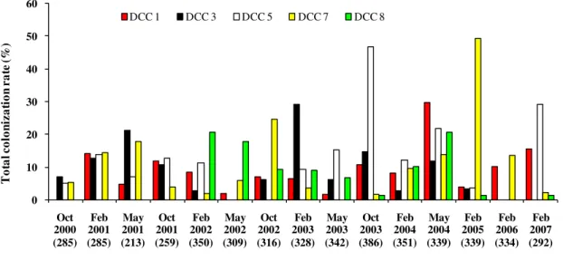
Figure 2.3. Variation of colonization rates among DCCs that participated along the study
period (2000-2007)
Figure 2.4. Variation of colonization rates among schools
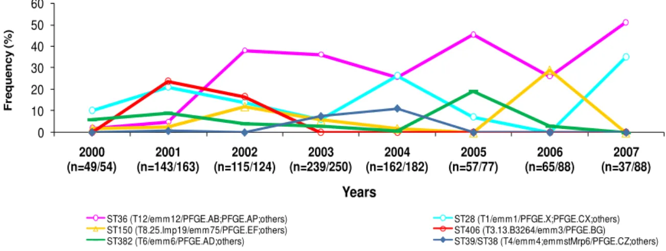
Figure 2.5. Resistance to macrolides and clindamycin along the study period (2000-2007) Figure 2.6. Evolution of frequencies of the six major clonal lineages from oropharyngeal
colonization
Figure 2.7. Evolution of frequencies of the ten minor clonal lineages from oropharyngeal
colonization
Figure 2.8. Distribution of PFGE types and associated emm types among isolates from
recurrent carriers
Figure 3.1. Temporal evolution of macrolide resistance frequency and phenotypes among S. pyogenes colonization isolates in children from Lisbon area (2000-2006)
Figure 3.2. Dendrogram of the PFGE profiles of macrolide-resistant S. pyogenes from
asymptomatic oropharyngeal colonization (2000-2006) in children
Figure 4.1. Diagram representing the methodologies used for the selection and
characterization of ciprofloxacin-nonsusceptible S. pyogenes
Figure 5.1. Annual distribution of PFGE patterns of the 45 bacitracin-resistant S. pyogenes
isolates
Figure 5.2. Properties of the bacitracin-resistant S. pyogenes from asymptomatic
xx
Figure 6.3.A. Representative dendrogram of virulence genes searched for among 208 GAS isolates
Figure 6.3.B. Distribution by clinical origin of the 208 GAS isolates of clusters A-O Figure 6.4. Growth curves in 2YT and BHI media
xxi
Table 1.1. List of sequenced S. pyogenes strains
Table 2.1. Characteristics of S. pyogenes isolates recovered from individuals included in the
multicolonization study
Table 3.1. Antimicrobial consumption among children carriers of macrolideresistant and
-susceptible S. pyogenes isolates at sampling period, 30 days and 2-6 months before
sampling, during 2000-2006 in Lisbon area, Portugal
Table 3.2. Distribution of virulence genes among emm types of macrolide-resistant S.
pyogenes from oropharyngeal colonization in children (2000-2006)
Table 3.3. Characteristics of macrolide-resistant S. pyogenes isolates from oropharyngeal
colonization (2000-2006) in children and distribution of clonal lineages by year of study
Table 4.1. MICs to ciprofloxacin and 12 other antimicrobial agents and susceptibility rates
among 66 ciprofloxacin-nonsusceptible S. pyogenes isolates collected from different origins
in Portugal (1999-2006)
Table 4.2. Genotypes, phenotypes and origins of the 66 ciprofloxacin-nonsusceptible S. pyogenes isolates collected in Portugal (1999-2006)
Table 6.1. Primers and associated controls used in PCR reactions
Table 6.2. Primers used in PCR reactions
xxii
3
1. Streptococcus pyogenes or Group A Streptococcus (GAS)
1.1.General features
In this section we describe the general characteristics of the species, an overview of the identification methods used for differentiation of S. pyogenes, the niche of this pathogen and its
host.
S. pyogenes is also known as beta-hemolytic Group A Streptococcus or Lancefield’s
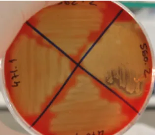
Group A strep (GAS) (Facklam, 2002). It is a facultative anaerobic microorganism, catalase and oxidase negative, and its metabolism is fermentative. Individual cells have spherical shape and no motion. Colonies produce streptolysin-O, lysing completely the erythrocytes and forming large zones of beta-hemolysis (two or four times the diameter of the colony), when cultured on blood agar plate after 18 to 24 hours of incubation at 37ºC (see Figure 1.1) (Facklam, 2002). The cell wall of S. pyogenes contains an antigenic polysaccharide composed of
N-acetilglucosamine linked to a rhamnose polimer backbone, whose serologic reactivity allows identifying primarily this species as Lancefield Group A (Cunningham, 2000). The true incidence of non-S. pyogenes GAS strains found in human infections, such as S. dysgalactiae
subsp. equisimilis and S. anginosus group, is unknown, but from the information available at the
Centers for Disease Control and Prevention (CDC) Streptococcus laboratory, these strains are
not common (Facklam, 2002). Presumptive identification can also be made by bacitracin
susceptibility or by the ability to enzymatically hydrolyse PYR (L-pyrrolidonil- -naphthylamide). S. pyogenes strains are the only beta-hemolytic streptococci that are positive in
both of these tests (Facklam, 2002). Other techniques have been used for S. pyogenes
identification, including 16S rRNAsequencing and assessment of phenotypic characteristics by biochemical tests (Facklam, 2002), as well as fluorescent in situ hybridization (FISH)
(Tajbakhsh et al., 2011). Usually, S. pyogenes is a free-living organism; however, its ecological
niche appears to be quite narrow, being limited to upper-respiratory tract mucosa, as well as skin. The only known natural reservoir of this pathogen is the human (Bessen and Hollingshead, 2000).
As it was referred to above, susceptibility to bacitracin remains to be used as a criterion for presumptive identification of S. pyogenes. However, resistance to this antimicrobial agent in
S. pyogenes was previously described (Facklam and Washington II, 1991), raising questions
concerning the reliability of this criterion. Resistance to bacitracin in S. pyogenes has been also
4
Figure 1.1. Streptococcus pyogenes strains grown on a blood agar plate with observation of
beta-hemolysis (This study).
1.2. S. pyogenes carrier state: meaning and clinical relevance
Although it not be considered normal flora, S. pyogenes can colonize oropharyngeal
respiratory tract without manifestation of clinical infection symptoms by the host (Cunningham, 2000). Colonization is also considered an infection; however the association between the microorganism and the host is commensal (Berkovitch et al., 2002). The S. pyogenes carrier is
an asymptomatic individual with a positive oropharyngeal swab culture and without serological response, or with a positive culture after completing the appropriate treatment with antimicrobial agents (Martin et al., 2004). Pichichero and Casey (2003) showed that 7% of 140
children were S. pyogenes carriers after 10 to 21 days of treatment with an antimicrobial from
macrolide class. These studies indicated that, although the disappearance (or attenuation) of the symptoms, the microorganism was not eradicated after the treatment with several antimicrobial classes (Martin et al., 2004). Nguyen et al. (1997) demonstrated that asymptomatic individuals
previously exposed to treatments for streptococcal infection can be colonized by new S.
pyogenes strains (new clones). S. pyogenes can be spontaneously eradicated from asymptomatic
individuals without antimicrobial therapy, suggesting that active immunization can eliminate bacteria from oropharyngeal flora. The re-emergence can arise from family contacts that coexist with sick individuals.
The S. pyogenes prevalence in the throat is known to be more common in school-aged
children (5-21%) (Gunnarsson et al., 1997). The carrier state has an important role in the
dissemination of this bacterium via aerosol, especially among children at schools, day-care centers and at home. Since carriers could be the source of infection, the study of the prevalence of healthy S. pyogenes carriers and the molecular epidemiology of the isolates may improve
5
control measures (Kim, 2000; Durmaz et al., 2003; Fazeli et al., 2003). The genotypic
comparison of S. pyogenes strains from asymptomatic carriers and from individuals with disease
symptoms is also crucial for the understanding of global epidemiology of this pathogenic agent and from factors (from the host or from the bacterium) that control the development and severity of the disease (Efstratiou, 2000; Kim, 2000; Hoe et al., 2002; Blandino et al., 2011). An
increase of epidemicity in an evolutive perspective can result from the acquisition of virulence or of antibiotic resistance genes by some strains, which could have selective advantage on
others. On the other hand, the knowledge of hosts’ population is important because susceptibility to disease can vary among individuals.
1.3. S. pyogenes symptomatic infections
S. pyogenes is considered to be the most pathogenic bacterium in the genus
Streptococcus. The reason why this microorganism is a major public health concern is because
is one of the most versatile and common human pathogens, causing a wide spectrum of diseases, ranging from mild infections to life-threatening systemic diseases (Facklam, 2002). Infections typically begin in the throat (examples are tonsillitis/pharyngitis or scarlet fever) or skin (impetigo). S. pyogenes may also cause disease in the form of nonsuppurative sequelae.
These complications follow a small percentage of infections and include rheumatic fever, acute poststreptococcal glomerulonephritis and PANDAS (from “pediatric autoimmune neuropsychiatric disorders associated with streptococcal infections”). Rheumatic fever is
characterized by inflammation of the joints and/or heart following an episode of streptococcal pharyngitis. Acute glomerulonephritis, which is inflammation of the renal glomerulus, can follow streptococcal pharyngitis or skin infection (Efstratiou, 2000). PANDAS is characterized by the presentation of exacerbated neuropsychiatric symptoms following oropharyngeal infections caused by S. pyogenes. Among the invasive diseases caused by this pathogen,
erysipelas and cellulitis are characterized by multiplication and lateral spread of S. pyogenes in
deep layers of the skin. S. pyogenes invasion and multiplication in the fascia can lead to
necrotizing fasciitis, a potentially life-threatening condition requiring surgical treatment. It can also lead to streptococcal toxic shock syndrome (STSS) (Cunningham, 2000). Although invasive diseases cause high morbidity and mortality (19-44%), its frequency of occurrence in Europe is very low (3/100,000) (Lamagni et al., 2008). According to estimates, about 600
6
1.4. Virulence factors
The wide variety of disease syndromes caused by S. pyogenes is probably in part a
reflection of the virulence gene products that are either secreted into the environment or localized on cell surface (Cunningham, 2000; Efstratiou, 2000). S. pyogenes utilises its many
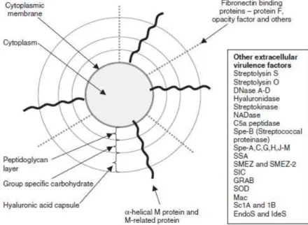
cell surface structures to adhere, internalise, and move across epithelia (Cunningham, 2000). A carbohydrate capsule composed of hyaluronic acid surrounds the bacterium, protecting it from phagocytosis by neutrophils. In addition, the capsule and several factors embedded in the cell wall, including M protein, lipoteichoic acid, and protein F (PrtF1/SfbI) facilitate attachment to various host cells (Figure 1.2.).
Figure 1.2. The basic outer cell antigenic structure of S. pyogenes (adapted from Steer et al.,
2007).
1.4.1. M protein and M-like proteins
The major virulence factor associated with S. pyogenes is the M-protein antigen. This
7
1.4.2. Superantigens
The superantigen (SAg) family can induce massive secretion of inflammatory cytokines, such as gamma interferon (IFN-γ), interleukin-1 (IL-1), and tumor necrosis factor-α.
These molecules are stimulated by large numbers of T cells by the SAg cross-linking major histocompability complex (MHC) class II antigens and T-cell receptors (TCR) (Cunningham, 2000). Overproduction of these cytokines can lead to hypotension, fever, tissue damage, organ failure, and shock (Kotb, 1995). The discovery of novel SAg has accelerated with the completion of several genome sequencing projects and the total number now stands at 11: SPE-A, SPE-C, SPE-G, SPE-H, SPE-I, SPE-J, SPE-K, SPE-L, SPE-M, SSSPE-A, and the highly polymorphic SME-Z (Sriskandan et al., 2007; Fraser and Proft, 2008).
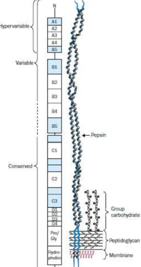
Figure 1.3. Diagrammatic representation of the M protein molecule on the cell surface of Group
A streptococci (Adapted from Bisno et al., 2003).
In the later 1990s, four novel SAggenes were identified, by mining the S. pyogenes M1
genomic database, at the University of Oklahoma. The genes for speG, speH, speI, and speJ
were cloned and expressed in Escherichia coli, and the purified recombinant proteins showed
the typical SAg features (Proft et al., 1999; Proft et al., 2001). The S. pyogenes genome project
8
2001; Proft et al., 2003). One it is named speK, which is a pseudogene with an incomplete open
reading frame (ORF) (Ferretti et al., 2001). The other two are speL and speM genes and they are
located on a mobile element (ΦspeL/M) that enables gene transfer between individual isolates
and between streptococci from different Lancefield groups (Proft et al., 2003; Commons et al.,
2008).
SPE-A and SPE-C have been epidemiologically associated with the development of severe invasive diseases like STSS, or nonsuppurative sequelae such as rheumatic fever (McCormick and Schlievert, 2000). However, the speA gene was detected with comparable
incidence among throat isolates and in invasive cases; in contrast, speC incidence was
surprisingly higher among throat of paediatric populations than in invasive isolates (Creti et al.,
2005).
Most SAg-encoding genes are associated with bacteriophages, except for speG, speJ
and smeZ, which are believed to be chromosomally encoded (Commons et al., 2008). The gene
distribution of superantigens has been used as an epidemiologic tool to explore genomic heterogeneity and the possible correlation between SAg gene content and clinical manifestation (Rivera et al., 2006; Chang et al., 2011).
The chromosomally encoded superantigens speG and smeZ have been described as the
most prevalent, ranging from 84% to 100% of infection isolates (Proft et al., 2000, 2003; Rivera
et al., 2006; Commons et al., 2008). Isolates in which these superantigens were not detected
were found to be restricted to certain emm types, e.g. speG was absent from emm4 isolates,
suggesting that these isolates may contain an allele with mutations in the primer-binding sites (Commons et al., 2008). The use of PCR with internal primers or DNA-DNA hybridization
experiments could confirm the presence or absence of speG among isolates of emm4 type.
Streptococcal superantigen (SSA) is a toxin that has high similarity in sequence to SPE-A and is also phage-encoded. The toxin is usually produced by isolates of streptococci of the M protein capsular type M3 associated with STSS and by other M types of streptococci, but rarely by M1 isolates (McCormick and Schlievert, 2000; Commons et al., 2008).
1.4.3. Streptococcal phospholipase A2 (SlaA) and streptococcal phage DNase 1 (Spd1)
The slaA and spd1 genes are virulence factors also associated to phages: slaA is present in prophage Φ6180.2, being contiguous of speK gene; spd1 is contiguous of speC gene in prophage Φ6180.1 (Green et al., 2005b). The slaA gene encodes for an enzyme that hydrolyzes
ester bonds of phospholipids (Nagiec et al., 2004). The extracellular proteins encoded by spd1
9
1.4.4. Protein F1 (PrtF1) and other fibronectin-binding proteins
The fibronectin-binding proteins attach bacteria to the extracellular matrix, which acts as a bridge between streptococci and host cells. There are at least 11 fibronectin-binding proteins in S. pyogenes, including PrtF1, protein F2 (PrtF2), serum opacity factor (SOF), FbaA,
and several M proteins (Nobbs et al., 2009).
PrtF1, also known as SfbI (streptococcal fibronectin binding protein I), facilitates adherence to respiratory epithelial cells (Hanski and Caparon, 1992; Bisno et al., 2003).
Although M protein is known to mediate S. pyogenes adherence to skin keratinocytes,
PrtF1/SfbI plays a major role in S. pyogenes adherence to cutaneous Langerhans cells (Okada et
al., 1994; Bisno et al., 2003). Expression of PrtF1 is enhanced in an O2-rich environment while that of M protein is greater at higher partial pressures of CO2 (Caparon et al., 1992; Bisno et al., 2003). Thus, it was postulated that the organism displays PrtF1 on its surface when it wishes to adhere to the cutaneous surface but expresses M protein in deeper tissues where it is more likely to encounter phagocytic cells (Bisno et al., 2003).
1.4.5. Pili
Pili are structures that extend 1 to 3 µm from the bacterial cell surface (Nobbs et al.,
2009). They are heteropolymeric structures consisting of a backbone protein (BP) and either 1 or 2 ancillary proteins (AP1 and AP2) covalently assembled and linked to the cell wall by a series of sortase-mediated transpeptidase reactions (Falugi et al., 2008). In S. pyogenes, there
are nine pilus islands so far reported that are inserted exclusively into a single highly variable
genetic locus known as the fibronectin-binding, collagen-binding, T antigen (FCT) region (Falugi et al., 2008) that forms part of the Lancefield T-serotyping system (Nobbs et al., 2009).
Pili play an important role in bacterial interaction with the human host. Streptococcal pili have
recently been associated with the capacity to adhere to human epithelial cells and form biofilm,
a process believed to be important in pathogenesis. For this reason, and because of their expression on the bacterial surface, pili have attracted interest as potential components of vaccines (Falugi et al., 2008).
1.5. Antimicrobial therapy
Diverse classes of antimicrobials are used for the treatment of infections caused by S.
pyogenes, such as streptococcal pharyngitis. Despite this microorganism is the most common
10
pharyngitis must be done when is guaranteed that pharyngitis has bacterial origin, usually attributed to S. pyogenes, especially because the signs and symptoms of bacterial and viral
pharyngitis are frequently overlapped (Bisno et al., 2002).
The evaluation of antimicrobial susceptibility of all pathogenic microorganisms for infection treatment, S. pyogenes in particular, is crucial so that the therapy is appropriate,
especially because it has been observed that antibiotics’ consumption can be associated with the
emergence of resistance in several countries (Bingen et al., 2002; Bergman et al., 2004). The β-lactamic antibiotics are the most diversified and most frequently used antimicrobial class (Fluit et al., 2001). This class includes penicillins (benzylpenicillin,
amoxicillin, ampicillin, methicillin), cephalosporins (ceflacor, cefotaxime) and carbapenems (biapenem, ertapenem). These antibiotics act at penicillin-binding proteins (PBPs), which are involved in the bacterial cell wall synthesis. Bacterial resistance, in the most part of the cases, is
due to the presence of β-lactamases(Fluit et al., 2001), however in Streptococcus pneumoniae
and in other streptococci, the emergence of resistance to β-lactamic antibiotics among natural populations is associated to the genesis of mosaic genes (Ferretti et al., 2001), when segments
of PBPs-encoding genes from susceptible strains are replaced by homologous blocks from resistant strains. These transfers are mediated by natural transformation with exogenous DNA and can cross the species boundary (Ferretti et al., 2001). However, in the case of S. pyogenes, it
was revealed the existence of two genes that encode for PBPs, both with low affinity and without homology comparing with other streptococcal corresponding genes (Ferretti et al., 2001). By this way, acquisition of β-lactams resistance by homologous recombination with genetic material of other streptococcal species is unlikely. Besides that, and taking into account that there is no evidence that S. pyogenes is naturally competent for transformation, it is possible that β-lactams resistance in this bacteria has to appear de novo (Ferretti et al., 2001). At the
moment, it was not found any S. pyogenes strain that was resistant in vitro to β-lactams
antibiotics, namely to penicillin (Ferretti et al., 2001; Bingen et al., 2004; Pires et al., 2005),
possibly due to the reasons referred to above and suggested by Ferretti and collaborators (2001). Other possible explanations for this remarkable state of continued susceptibility to penicillin are
that β-lactamases may not be expressed or may be toxic to the organism and/or that low-affinity PBPs either are not expressed or render organisms nonviable (Horn et al., 1998). In the last 50
years, penicillin is the most recommended antibiotic for the treatment of infections originated by S. pyogenes, due to its proven effectiveness, safety, short activity spectrum and low cost
(Bisno et al., 2002). However, penicillin treatment can fail in some cases, possibly due to the production of β-lactamases by other microorganisms from oral flora; so, a good choice for the empirical treatment of S. pyogenes will be the use of combination amoxicillin/clavulanic acid
(Gerber et al., 1999; Cunningham, 2000). In patients who are allergic to penicillins, macrolides
11
clindamycin) seem to be adequate choices for the treatment of streptococcal infections (Bisno et
al., 2002).
1.5.1. Macrolides, lincosamides and streptogramins
1.5.1.1. Structure and mechanisms of action
The macrolides are a group of antibiotics that have a large lactone ring structure. These may be 14- (like erythromycin, see Fig. 1.4A, and clarithromycin), 15- (like azithromycin, see Fig. 1.4B) or 16-membered (like josamycin, see Fig. 1.4C) rings. These are relatively nontoxic antibiotics, most active against Gram-positive bacteria (Roberts et al., 1999). Macrolides inhibit
protein synthesis by stimulating dissociation of the peptidyl-tRNA molecule from the ribosomes during elongation (Weisblum, 1995). This results in chain termination and a reversible stoppage of protein synthesis (Roberts et al., 1999). The lincosamide antibiotic lincomycin and its
semi-synthetic derivative clindamycin (see Fig. 1.4D) have a similar mode of action.
The streptogramins fall into two groups, A and B. Streptogramin belonging to Group A has a large nonpeptide ring, which is polyunsaturated. Streptogramins related to streptogramin B are cyclic peptides (see Fig. 1.4E). They differ in their modes of action although both inhibit bacterial protein synthesis. Group A streptogramins distort the ribosome to prevent binding of the tRNA; Group B streptogramins are thought to block translocation of the growing peptide.
1.5.1.2. Resistance mechanisms
Resistance to macrolides in S. pyogenes can be mediated by three different mechanisms:
target site modification, active efflux and rRNA 23S or ribosomal proteins mutations. The modification of the target site is based in dimethylation N6 of an adenine residue of rRNA 23S through the action of an enzyme family encoded by erm(“erythromycin ribosome methylation”)
genes class. The methylation seems to induce a conformational alteration among 50S subunit of the ribosome, leading to a reduced affinity and to co-resistance to macrolides, lincosamides and streptogramin B (MLSB antibiotics), whose binding sites probably overlap. The MLSB resistance can be expressed constitutively (cMLSB phenotype) or can be induced (iMLSB phenotype) and it is mediated by two classes of methylase-encoding genes, the erm(B) and
erm(A) [which includes the erm(TR) subclass)]. The induction is stimulated by 14- and
12
Figure 1.4. Examples of macrolides, lincosamides and streptogramins chemical structures: A-
Erythromycin (14-membered macrolide); B- Azithromycin (15-membered macrolide); C- Josamycin (16-membered macrolide); D- Clindamycin (lincosamide); E- Streptogramin B. Adapted from http://www.bmb.leeds.ac.uk/mbiology/ug/ugteach/icu8/antibiotics/protein.html, http://www.antibioticslist.com/azithromycin.htm, http://www.antibioticslist.com/josamycin.htm and http://www.antibioticslist.com/clindamycin.htm.
The active efflux of antibiotic is mediated by hydrophobic membrane proteins, encoded by mef(A) (“macrolide efflux”) resistance gene. A small portion of strains carry different mef
subclasses, such as mef(E) and mef(I) (Del Grosso et al., 2011).These proteins use the proton
energy in order to pump out the antibiotic, maintaining its intracellular concentration low and consequently, the ribosome free of its activity. The mechanism occurs without antibiotic and its target modification and became the strains resistant to 14- and 15-membered macrolides, but susceptible to 16-membered macrolides, to lincosamides and to streptogramin B (Roberts et al,
1999). This resistance phenotype is named M phenotype.
The third resistance mechanism was described for S. pyogenes by Malbruny et al.
(2002), and it was previously observed among Streptococcus pneumoniae as associated to a
13
to erythromycin, clindamycin and streptogramin B (M phenotype). Mutations in the gene that encodes this protein had been previously detected in S. pyogenes by Bingen et al. (2002).It was
also described by Jalava et al. (2004) one S. pyogenes strain that was resistant to macrolides,
lincosamides and streptogramin B, but did not present any macrolide resistance gene, but had a mutation in rRNA 23S in a different location as that described by Malbruny et al. (2002). This
strain presented the cMLSB phenotype, similarly with strains that constitutively expressed the
erm(B) genes.
1.5.2. Fluoroquinolones
1.5.2.1. Structure
The fluoroquinolones are synthetic antibiotics that belong to quinolones family. They are modified molecules that contain one or more fluorine atoms as well as other chemical alterations (Ball, 2000) (see Figure 1.5). Differences in the in vitro activity of the quinolones
primarily form the basis of their classification in four generations, and these differences are based on antibacterial activity and potency against pneumococci and anaerobic organisms (Andriole, 2005). The first generation quinolones (nalidixic acid, oxolinic acid, cinoxacin, piramidic acid, pipemidic acid, and flumequine) exhibit an excellent activity against aerobic and Gram-negative bacteria. The second generation quinolones include norfloxacin, ciprofloxacin, ofloxacin, levofloxacin, enoxacin, flexacin, lomefloxacin, pefloxacin, and rufloxacin (Andriole, 2005). They were introduced when norfloxacin was synthesized by adding a fluorine atom in C-6 carbon and a cyclic piperazine diamine in C-7 carbon (Andriole, 2005). These changes added antimicrobial activity against aerobic Gram-positive bacteria and improved activity against Gram-negative bacteria, when compared with first generation quinolones. The third generation fluoroquinolones, such as grepafloxacin, gatifloxacin, sparfloxacin, temafloxacin, tosufloxacin, or pazufloxacin, are very efficient against Gram-positive bacteria, in particular against pneumococci, and they also had good activity against anaerobic bacteria. The fourth generation fluoroquinolones (trovafloxacin, clinafloxacin, sitafloxacin, moxifloxacin, and gemifloxacin) had potent activity against anaerobic bacteria and increased activity against pneumococci (Andriole, 2005).
1.5.2.2. Use of fluoroquinolones
14
tract infections (Andriole, 2005). These antibiotics are not recommended for the treatment of infections among children because of high risk of tendon damage (Gendrel et al., 2003).
Figure 1.5. Molecular structures of quinolones: A- Nalidixic acid (first generation quinolone);
B- Ciprofloxacin (second generation quinolone); C- Gatifloxacin (third generation quinolone); D- Moxifloxacin (fourth generation quinolone).
Adapted from http://www.bmb.leeds.ac.uk/mbiology/ug/ugteach/icu8/antibiotics/dna.html, http://www.chemicalbook.com/ProductChemicalPropertiesCB8227559_EN.htm and http://www.antibioticslist.com/moxifloxacin.html.
1.5.2.3. Mechanisms of action
The bactericidal power of fluoroquinolones is originated by inhibition of DNA replication and transcription, targeting two cellular enzymes: DNA gyrase and topoisomerase
IV. The DNA gyrase, that originates DNA’s negative supercoiling, is constituted by two GyrA
and two GyrB subunits, which are encoded by gyrA and gyrB genes, respectively, the preferable
target of fluoroquinolones in Gram-negative bacteria. Topoisomerase IV separates DNA chains during replication and cell division, and is constituted by ParC and ParE subunits, which are encoded by parC and parE genes, respectively, the preferable target among Gram-positive
bacteria (Perichon et al., 1997; Alonso et al., 2002; Jacoby, 2005).
1.5.2.4. Resistance mechanisms
Fluoroquinolone resistance is mainly caused by point mutations in the target-encoding genes (gyrA, gyrB, parC and parE). Mutations tend to cluster in a defined region of these genes
called the quinolone resistance-determining region (QRDR). Fluoroquinolone resistance appears to occur stepwise, with moderate levels of resistance arising from a single mutation in the primary target of the drug (topoisomerase IV, parC gene) (Alonso et al., 2005; Orscheln et al.,
15
additional mutations in the secondary target (DNA gyrase, gyrA gene) (Orscheln et al., 2005).
The mutations occur most frequently in codons S79 for ParC and S81 for GyrA (Yan et al.,
2000; Richter et al., 2003). Recently, mutations in codons D83 for ParC (Pires et al., 2010) and
E85 for GyrA (Arai et al., 2011) were described. Fluoroquinolone efflux by a
reserpine-sensitive pump is also a common resistance mechanism to fluoroquinolones in Streptococcus
pneumoniae (Brenwald et al., 1998) and viridans group streptococci (Ferrándiz et al., 1999),
although this mechanism still remains to be demonstrated for S. pyogenes.
1.5.3. Tetracycline
Tetracycline is not recommended for therapy of upper respiratory tract infections originated by S. pyogenes. However, the selective pressure derived from its use in the treatment
of other human and animal infections can have contributed for the worldwide emergence of resistance to this antibiotic among isolates of S. pyogenes (de Melo et al., 2003). The transfer of
genes from animals to humans through food is a possible explanation for the emergence of resistance (Nielsen et al., 2004). Tetracycline resistance levels among enterococci are high in
both humans and their feeding animals. As the horizontal transfer of tetracycline resistance genes from enterococci to streptococci has been demonstrated (Nielsen et al., 2004), this
transfer mediated by transposons is a real possibility in the oral cavity. In the transfer of tetracycline resistance genes from enterococci, the oral flora can also be used as a transition state because both tet(M) and tet(O) genes have been found in several bacterial species from
oral flora (Nielsen et al., 2004). The existence of S. pyogenes strains resistant to tetracycline
contributes to the importance of including this antibiotic in antibiotyping studies.
Tetracycline is included in a group of antiobiotics with wide spectrum of activity and low toxicity. These antibiotics penetrate the bacterial cells by passive diffusion and bind to the 30S subunit of the ribosome, inhibiting the protein synthesis by blocking the connection between the aminoacil-tRNA and the ribosome A place. Several tetracycline resistance genes are described for S. pyogenes, such as tet(K), tet(L), tet(O) and tet(T), being prevalent the tet(M)
gene (Shlaes, 2006). Some of these genes are often associated to conjugative transposons, which in part can explain their wide distribution among bacterial species, as well as their association with other antibiotic resistance genes, particularly the association between tet(M) and erm(B)
genes (Fluit et al., 2001).There are three known tetracycline resistance mechanisms: enzymatic
inactivation of the antibiotic, antibiotic efflux by protons antiporte [tet(K) and tet(L)], and
protection of the ribosomes by the production of one protein which interacts with the ribosome allowing the protein synthesis autonomy in the presence of the antibiotic [tet(M) and tet(O)]
16
1.6. Epidemiology of S. pyogenes
In this topic the most common methods used in epidemiological studies of S. pyogenes
will be presented: M-typing, T-typing, emm-typing, sof-typing, multilocus sequence typing
(MLST) and pulsed-field gel electrophoresis (PFGE). It is important to refer that most studies on the epidemiology of S. pyogenes are based on the analysis of antimicrobial resistance
patterns (Cocuzza et al., 1997; Descheemaeker et al., 2000; Cresti et al., 2002), T-typing (Beall
et al., 1997; De Azavedo et al., 1999; Melo-Cristino et al., 1999; Bingen et al., 2000, Pires et al., 2005), and emm-typing (Tyrrell et al., 2010; Bahnan et al., 2011; Chen et al., 2011),
although resistance frequencies and phenotypes vary geographically.
M-typing - In 1928, Rebecca Lancefield published a method for serotyping S. pyogenes
based on its M protein (Facklam, 2002). M type-specific antisera are used in a precipitin reaction against S. pyogenes acid extracts. Currently, there are approximately 90 validated and
specific M types. There are some problems associated with M serotyping such as limited availability of M typing sera, the production of M sera is laborious, expensive, and only available to a few centers, difficulty in interpretation, newly encountered M types overtime and an increased frequency of nontypeable isolates (Neal et al., 2007). In the United States and
Europe, it was found a high prevalence of M1 and M3 serotypes among invasive disease (Efstratiou, 2000). Type M28 strains also are common causes of invasive infections, as well as pharyngitis in many countries (Green et al., 2005b).
T-typing - In 1946, Rebecca Lancefield described the serologic classification of S.
pyogenes isolates based on their surface T antigen. Typing of the T protein uses polyvalent
pooled and monovalent antisera in a slide agglutination test. In S. pyogenes, type-specific
T-protein antigens are basic markers for typing and can be divided into approximately 30 different T-types. Like M serotyping, T-serotyping have the same associated problems and a careful interpretation by experienced staff is needed. A high number of nontypeable isolates have been reported (Neal et al., 2007). The T-protein serotyping and its comparison with emm types
provide additional information for strain identification (Johnson et al., 2006).
emm-typing - In recent years, an alternative system called emm-typing has been
developed, which uses the sequence of the hypervariable region of the gene that encodes the M-protein (Beall et al.,1996). The method and a public database are of public access at The
Centers for Disease Control and Prevention (CDC), Streptococcus Laboratory
(http://www.cdc.gov/ncidod/biotech/strep/strepindex.htm). The definition of an emm-type sequence is based upon the identity of 180 bases at the 5’ terminal end of the hypervariable
portion of the emm gene (see Fig. 1.3). Determination of the emm gene sequence has become a
17
The emm-types are further divided into subtypes that are explicitly based on minor sequence
variation within the type specific hypervariable region of the gene. The website curator maintains the S. pyogenesemm sequence database with of all validated M and emm types, as
well as provisional types and subtypes yet to be officially designated. In most cases the emm
type reflects the M-protein serologic type (Facklam, 2002). Some restricted emm-types, such as
emm1 and emm3, were more representative in invasive strains (Creti et al., 2005), whereas emm12 was more common among asymptomatic carriers (Blandino et al., 2011).
sof sequence typing - S. pyogenes sof (serum opacity factor) is approximately a 1000
residue cell-surface-bound apoproteinase named for its property of rendering various opaque sera. sof is a virulence factor since it has fibronectin-binding activity that resides in a relatively
short C-proximal domain. sof gene detection and sequencing are based upon PCR and sequence
analysis of a variable-length 450-650 bp PCR fragment using methods and primers decribed in Beall et al., 2000. T-pattern and sof information about a S. pyogenes strain, especially when
combined with knowledge of M or emm-type, provides an important link to information from
studies published over many decades, when the serological methods were primary available tools (Johnson et al., 2006).
Multilocus sequence typing (MLST) is a method based on nucleotide sequence of internal fragments of seven housekeeping genes (highly conserved genes) that are assumed to be neutral in their genetic variation (www.mlst.net). This allelic profile (a sequence of 7 numbers –
one number for each allele and in a specific order: gki, gtr, murI, mutS, recP, xpt and yqiL)
defines the sequence type (ST). The STs provide unambiguous results that are easily portable and a central database (http://spyogenes.mlst.net/) allows for comparison of results obtained in different laboratories (Enright et al., 2001; Doktor et al., 2005). Until September 15, 2011, a
total of 624 STs were published at the international database. Also, it is considered to be the method of choice for global epidemiological studies and to evaluate the bacterial population structure and evolution by identifying lineages and clonal complexes.
Pulsed-field gel electrophoresis (PFGE) has been the gold standard method used to assess strain similarity in epidemiologic studies (Tenover et al., 1995; Cocuzza et al., 1997;
Haukness et al., 2002). However, it is a method particularly useful for short-term
epidemiological studies or outbreak situations and not for global epidemiological studies. Several authors also consider that PFGE, like other methods based on DNA-band analysis, is not reproducible among different laboratories and band-patterns are difficult to interpret.
Although the advantages of sequence-based methods, S. pyogenes virulence has been
related to the presence of phages and to horizontal transfer. This highlights the notion documented by several authors that PFGE may be more discriminatory than sequence-based methods (Bahnan et al., 2011), since phage insertions can alter band positions in an agarose gel
18
1.7. S. pyogenes genome
To date, the genome sequences of 15 strains of S. pyogenes have been determined,
including strains that were associated with various clinical conditions and representatives of the following ten serotypes: M1, M2, M3, M4, M5, M6, M12, M18, M28 and M49; the genomes of two separate strain isolates have been determined for serotypes M1, M3, M12 and M49. The genome is a circular chromosome between 1,3 and 1,9 Mb (Table 1.1). The genomes share 1.7 Mb of closely related genetic material (Beres et al., 2002; Nakagawa et al., 2003; McShan et
al., 2008), and phages, phage-like elements, and insertion sequences are the major sources of
variation between the genomes (Smoot et al., 2002). Genetic variation is essential to survival for
all organisms (McShan et al., 2008). These sequenced strains have genes encoding a novel array
of prophage virulence factors, cell-surface proteins, and other molecules likely to contribute to host-pathogen interactions. Genetic relationship between strains causing invasive disease episodes to strains of the same serotype recovered from asymptomatic carriers was not examined yet. Importantly, it is not known whether strains cultured from asymptomatic carriers differ in virulence compared to invasive isolates (Beres et al., 2006).
Table 1.1. List of sequenced S. pyogenes strains.
Strain Size (bp) M type No. of prophages
Accession no. Reference
SF370 1,852,441 1 4 AE004092 Ferretti et al., 2001
MGAS5005 1,838,554 1 3 CP000017 Scott et al., 2008
MGAS10270 1,928,252 2 5 CP000260 Beres et al., 2006
MGAS315 1,900,521 3 6 AE14074 Beres et al., 2002
SSI-1 1,894,275 3 6 BA000034 Nakagawa et al., 2003
MGAS10750 1,937,111 4 4 CP000262 Beres et al., 2006
Manfredo 1,841,271 5 5 AM295007 Holden et al., 2007
MGAS10394 1,899,877 6 8 CP000003 Banks et al., 2004
MGAS2096 1,860,355 12 2 CP000261 Beres et al., 2006
MGAS9429 1,836,467 12 3 CP000259 Beres et al., 2006
MGAS8232 1,895,017 18 5 AE009949 Smoot et al., 2002
MGAS6180 1,897,573 28 4 CP000056 Green et al., 2005b
NZ131 1,815,783 49 3 CP000829 McShan et al., 2008
19
1.8. Vaccine candidates
Because of the high colonization rates (for example 32%, Martin et al., 2004) and
increased frequency in some countries of severe infections rates caused by this pathogen, and concerning that penicillin-resistant strains may emerge, there is a strong incentive to develop a safe and effective vaccine against S. pyogenes. One of the benefits of a successful vaccination
scheme would be the reduction of streptococcal colonization in general, thus reducing the total number of these pathogens in the population. Since the main reservoir for S. pyogenes for most
streptococci-related illness is the human oropharynx, reducing the carriage could have a profound impact on the dissemination of streptococci in the environment and thus a significant reduction in streptococcal disease in general.
The M protein has been a prime vaccine candidate to prevent Group A streptococcal infections since Lancefield showed clearly that M protein-specific human and animal antibodies have the capacity to opsonize streptococci in preparation for phagocytic clearance. A type-specific vaccine necessary to protect against a streptococcal infection would require a multivalent antigen corresponding to stable immunodeterminants on serotypes that together account for the majority of the isolates prevalent within the population at a given time (Fischetti, 2000). Attempts to develop an M protein-based vaccine have been hindered by the fact that some M proteins elicit both protective antibodies and antibodies that cross-react with human tissues. New molecular techniques have allowed the previous obstacles to be largely overcome. A 26-valent vaccine has successfully completed a phase I/II clinical trial involving adults and was proposed, suggesting that it could have significant impact on the overall burden of streptococcal disease (McNeil et al., 2005).
Although promising, these multivalent M protein vaccines will not provide immunity against infections caused by strains of all M serotypes because of the variable nature of this protein. As consequence of these considerations, eight non-M protein vaccines has been in development, such as Group-A carbohydrate, C5a peptidase (ScpA), cysteine protease (SpeB), binding proteins like fibronectin, opacity factor, lipoproteins, Spes (superantigens) and streptococcal pili (Guilherme et al., 2009). However, no vaccines containing these antigens have
reached clinical trials (Steer et al., 2009).
1.9. Objectives of the research
This Thesis was designed to study important and unexplored aspects of the epidemiology, transmission and evolution of Streptococcus pyogenes causing colonization and a
C
C
h
h
a
a
p
p
t
t
e
e
r
r
I
I
I
I
AN EIGHT-YEAR SURVEILLANCE STUDY (2000-2007) OF OROPHARYNGEAL
COLONIZATION IN PORTUGAL AND CHARACTERISTICS OF LONGITUDINAL
OROPHARYNGEAL ASYMPTOMATIC COLONIZATION AND OF
MULTICOLONIZATION BY DIFFERENT STRAINS OF STREPTOCOCCUS PYOGENES