RESUMO: Frutos de maracujá (Passiflora edulis) foram encontra-dos com sintomas de podridão no campo, na cidade de Tanhaçu, Bahia. Após isolamento do patógeno associado a essas podridões, o presente trabalho teve como objetivo realizar a caracterização de um isolado de Ceratocystis de maracujá para melhor compreender esse patossistema. A caracterização molecular foi realizada com base no sequenciamento da região ITS-5.8S rDNA. Realizou-se a caracteri-zação patogênica em mudas e frutos de maracujá. A colonicaracteri-zação dos frutos de maracujá foi acompanhada com técnicas de microscopia de varredura (MEV). A análise do DNA do isolado de Ceratocystis mostrou que este pertence à espécie Ceratocystis fimbriata. As mudas de maracujá inoculadas apresentaram lesão 30 dias após a inocula-ção, e não foram observadas murcha nem morte das plantas inocu-ladas. Nos frutos não foram constatadas diferenças nas lesões causa-das por esse fungo entre os cultivares, com médias de lesões de 1,0 a 2,2 cm de diâmetro, aos 7 e 11 dias, respectivamente. Nas análises realizadas em MEV, foram observadas a germinação dos esporos e a penetração do fungo nos frutos no período entre 2 e 6 horas após a inoculação. Doze e 24 horas após a inoculação foi visualizada a colonização do fruto, tanto externa como internamente, e 48 horas após a inoculação se notou o início da degradação da parede externa da casca do fruto. Noventa horas após a inoculação, observou-se a formação de novos peritécios, tanto na parte interna como na parte externa do fruto. Este estudo vem complementar as informações relacionadas à interação desse fungo com frutos de maracujazeiro. PALAVRAS-CHAVE: podridão; espécies; microscopia. ABSTRACT: Passion fruits (Passiflora edulis) were found with
symptoms of rot in the field, in the city of Tanhaçu, Bahia. After isolating the pathogen associated with this rot, in the present study we aimed to characterize the Ceratocystis isolate from passion fruit for better understanding this pathosystem. Molecular characterization was done based on the region ITS-5.8S rDNA. Pathogenic characterization was carried out for seedlings and fruits of passionflower. Passion fruit colonization was monitored by means of scanning electron microscopy techniques (SEM). DNA analysis of the Ceratocystis isolate from passionflower pointed out that this species belongs to Ceratocystis fimbriata. The inoculated passionflower seedlings showed injury at 30 days post-inoculation, but no inoculated plant showed wilt or died. Considering fruits, no differences were found for lesions caused by this fungus among cultivars, and lesions had average diameters of 1.0 and 2.2 cm at 7 and 11 days, respectively. The analysis using SEM indicated fungus spore germination and penetration in the fruit between 2 and 6 hours post-inoculation. At 12 and 24 hours post-inoculation, fruit colonization was noted both externally and internally, while fruit wall degradation started at 48 hours post-inoculation. At 90 hours post-inoculation, formation of new perithecia was observed inside and outside the fruit. This study complements the available information about the interaction of this fungus with passion fruit.
KEYWORDS: rot; species; microscopy.
Characterization of
Ceratocystis fimbriata
from passion fruits
Caracterização de
Ceratocystis fimbriata
de frutos de maracujazeiro
Ana Carolina Firmino1*, Ivan Herman Fischer2, Gabriel Leonardi Antonio1,
Quelmo Silva De Novaes3, Hugo José Tozze Júnior1, Edson Luis Furtado4
1Faculdade de Ciências Agrárias e Tecnológicas (FCAT), Universidade Estadual Paulista “Júlio de Mesquita Filho” (UNESP) – Dracena (SP), Brazil. 2Agência Paulista de Tecnologia dos Agronegócios (APTA) – Bauru (SP), Brazil.
3Departamento de Fitotecnia e Zootecnia, Universidade Estadual do Sudoeste da Bahia (UESB) – Vitória da Conquista (BA), Brazil. 4Departamento de Proteção Plantas, Faculdade de Ciências Agronômicas (FCA), UNESP – Botucatu (SP), Brazil.
*Corresponding author: anacarfir@gmail.com
INTRODUCTION
The genus Ceratocystis covers several fungal species distrib-uted among different places of the world. In Brazil, there are reports of few species belonging to this genus, including
Ceratocystis cacaofunesta, Ceratocystis paradoxa and the most important species Ceratocystis fimbriata. Considering woody plants, C. fimbriata is a pathogen typical of xylem and its marked symptoms are dark radial striae from the medulla to the outer part of the xylem (Ferreira; Milane, 2002; Baker; Harrington, 2004). Generally, a plant infected with such a pathogen presents symptoms like leaf wilting and, conse-quently, drought. Cultures of C. fimbriata give off a smell of a ripe fruit. These volatile substances play an important role in the epidemiology of this disease since they attract the vector insect.
In Brazil, up to the middle of the 1990s, C. fimbriata was only considered a problem for crops of mango (Mangifera indica L.). Currently, this fungus has been shown harmful to other cultures like cacao (Theobroma cacao) (Bezerra, 1997), fig (Ficus carica) (Valarini; Tokeshi, 1980), teak (Tectona grandis) (Firmino et al., 2012b), atemoya (hybrid of Annona cherimola and Annona squamosa) (Firmino et al., 2012a), and, recently, yellow passion fruit (Passiflora edulis
f. flavicarpa) (Firmino et al., 2013). In this last case, the fungus causes rot to the fruits without causing any damage to the plant.
According to Harrington et al. (2011), the genus
Ceratocystis can be divided into four distinct clades, or groups: Latin America, North America, Asia and Africa. Within the Latin American clade, Baker et al. (2003) studied different isolates of cacao, Herrania sp., sweet potato, Platanus sp., coffee, mango, Annona sp., eucalyptus and Gmelina sp. They verified, based on a pathogenicity test, that there is a specialization level within this host clade. Thus, Baker et al. (2003) hypothesized that local populations of C. fim-briata are host-specialized. Based on these studies, it was suggested that the evolution and divergence of species of
Ceratocystis may have been conducted by host specializa-tion, since there is little morphological differences between species of these fungus (Ferreira, 2009). Analysis on elec-tronic scanning microscope, that were aimed to monitor the colonization of five Ceratocystis isolates from different hosts (eucalyptus, cocoa, mango, teak and atemoya) on the surface of eucalyptus plants to show that all isolates were capable of germinating, penetrating and developing in the vessel elements of eucalyptus plants within 6 hours, dem-onstrated that these fungal isolates, even from other hosts, are capable of developing in the xylem of eucalyptus plants (Firmino et al., 2015).
Report of Ceratocystis causing rot to passion fruits is some-thing new. So far, the reported occurrence of this disease has been limited to drought symptoms related to xylem invasion
by this fungus. Thus, the present study aimed to conduct molecular and pathogenic characterization of this fungus in plants and fruits of passionflower to improve the understand-ing of this pathosystem.
MATERIALS AND METHODS
Fruits of passionflower showing holes and rot were found in the field, in the city of Tanhaçu, Bahia, Brazil. These fruits had perithecia typical of Ceratocystis, which was isolated and deposited in the mycology collection located at the Laboratory of Forest Pathology, of the School of Agronomical Sciences of Universidade Estadual Paulista “Júlio de Mesquita Filho” (UNESP), in Botucatu, São Paulo, Brazil (Firmino et al., 2013). For its molecular characterization, the iso-late was recovered, removed from the oil and cultured on PDA medium (potato, dextrose and agar) for DNA extrac-tion. The DNA was extracted according to the method developed by Murray e Thompson (1980), with modi-fication. This DNA was used to amplify the region ITS-5.8S rDNA based on the protocol described by Johnson et al. (2005). The obtained DNA was sequenced and edited by using the software BioEdit Sequence Alignment Editor (1997–2005). After edition, that sequence was used to search for similar sequences by adopting the soft-ware Basic Local Alignment Search Tool (Blastn) of the National Center for Biotechnology Information (NCBI). The obtained sequences were aligned and processed with the software Mega 5.05, so that a phylogenetic tree could be built, using the method “Tamura-3-parameter” (Tamura, 1992). The distance matrix was constructed based on the Neighbor-Joining method. A bootstrap was applied with 10,000 replicates.
This Ceratocystis isolate from passionflower plants (one month of age) was subjected to pathogenic characterization by adopting two methods: inoculation with mycelial disks of this fungus, as described by Silveira et al. (2006), and deposit of a suspension of 108 cylindrical spores on the stem of the plant (Zauza et al., 2004). Evaluation occurred at 30 days post-inoculation. Seedlings were transversally sec-tioned in the stem to monitor the fungal invasion through the xylem. This fungal invasion into the xylem has as char-acteristic the vessel discoloration and darkening due to the collapse of tissues invaded by this fungus. Thus, the inva-sion could be measured from the inoculation site with the aid of a ruler. Five plants were employed for each inocula-tion method.
an experimental orchard located in São Manuel, São Paulo, Brazil, when approximately 75% of their shell was yellow, washed with neutral soap and dried at room temperature in the laboratory. Inoculation was done by depositing 40 µL of the spore suspension (105 conidia/mL) on the equato-rial region of the fruit, perforating 5 mm of it, through the drop, with a histological needle. Then, after 24 hours in a moist chamber, the fruits were kept for 11 days, at 25ºC, in biochemical oxygen demand (BOD). Rot severity was evaluated by perpendicularly measuring the lesion at 7 and 11 days. Six fruits were adopted per cultivar and the experi-ment was repeated once.
This characterization in fruits was complemented with colonization analyses by means of scanning electron micros-copy. In this case, besides the isolate from fruits of passion-flower, monitoring of the colonization of these fruits was done for other five Ceratocystis isolates from different host species: mango (ACF1), cacao (ACF15), atemoya (ACF24), eucalyptus (ACF38) and teak (ACF50). Thus, their capabil-ity to cause injury to passionflower could also be verified. For this, passion fruits were washed with neutral soap and sanitized with hypochlorite at 2% for 30 minutes. After sanitization, fruits were allowed to dry at room temperature for more than 12 hours. Once they were completely dried, fruits were placed on plastic trays, separating one tray for each studied isolate. On each tray, eight fruits were depos-ited. Each fruit received five drops of a spore suspension of the Ceratocystis isolate. No injuries were done to the fruit in this inoculation.
Following inoculation, the trays containing the fruits were kept in a moist chamber at 25ºC, in the dark. Disks were collected from the inoculation site in the fruit (around 5 mm diameter) at pre-determined intervals (2, 4, 6, 12, 24, 48, 72 and 96 hours) and fixed in “Karnovsky” solution (glutaraldehyde at 2.5%, paraformaldehyde at 2.0%, phos-phate buffer at 0.05M, pH 7.2) during a minimal period of 24 hours for preparation and analysis under a scanning electron microscope. Each inoculated fruit represented one collection time. For each collection, five disks were removed from the fruit.
Fragments from the inoculated fruits were transferred from “Karnovsky” fixative to 1.5 mL microtubes containing sodium phosphate buffer, 0.05 M, where they were kept for 10 minutes. Then, the samples were dehydrated in a series of increasing acetone concentrations (30, 50, 70 and 90% for 10 minutes each, and 100% for three times of 10 minutes). Following this step, they were taken to the critical point device to complete the drying. Once dehydration was completed, the samples were mounted on stubs and covered with gold. Sample preparation and observation were carried out at the Center for Electron Microscopy located at Escola Superior de Agricultura “Luiz de Queiroz” (ESALQ) of Universidade de São Paulo (USP).
RESULTS AND DISCUSSION
The studied isolate from passionflower was identified as
C. fimbriata, as shown in the phylogenetic tree (Fig. 1). It was close to the fungal isolates from Latin American euca-lyptus, but did not group to the isolates from Uruguay or Bahia, which shows that this isolate can be different from those that attack eucalyptus in Brazil. The isolates from Brazilian mango also kept very distant from the isolate from passionflower.
Only two plants inoculated with mycelial disk died due to collar necrotic lesion. The remaining inoculated plants, regardless of the method, did not show any symp-tom. Symptomatic plants had slight xylem darkening at the inoculation site, not exceeding 5 mm, which did not seem to result in damage to the plants. Wilt or drought symptoms were not observed in inoculated plants, but this may be due to the short evaluation period to which plants were subjected. There are stories of susceptibility to this fungus in eucalyptus plants which, even after 30 days of inoculation, did not show external symptoms of the dis-ease, but had significant lesions internally in the xylem (Zauza et al., 2004).
Considering the pathogenicity tests of fruits, there were no differences among cultivars, according to Tukey’s test (p<0.05), and lesions had average diameters of 1.0 and 2.2 cm at 7 and 11 days, respectively, evidencing the lack of resistance of the tested materials (Table 1).
As shown in Figures 2 to 4, only the isolate from passion-flower was capable of developing on the tested fruits. The inoc-ulated spores of isolates from mango, cacao, atemoya, euca-lyptus and teak showed no germination, even after 96 hours of inoculation in passion fruits (Fig. 3).
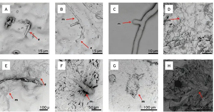
As regards fruit colonization by the isolate from pas-sionflower, spore germination and penetration in the fruit wall occurred between 2 and 6 hours post-inoculation. Differently from what was observed for xylem colonization, this isolate seems to form a structure similar to an appres-sorium, which helps it fix and penetrate the fruit wall with-out the aid of any injury. After 12 and 24 hours of inocu-lation, it was already possible to notice fruit colonization either externally or internally, in the inner part of the shell. At 48 hours post-inoculation, the outer part of the fruit shell started to undergo degradation due to the increased mycelium quantity in this region (Fig. 4). This fact became more frequent with time, and at 96 hours post-inoculation formation of new perithecia could already be seen, both in the inner and in the outer part of the fruit shell (Fig. 2). These new perithecia could be seen with the naked eye as small black dots.
exit of the new structures, both sexual and asexual (Fig. 2). These findings are of great importance since, as proven in the pathogenicity test, the isolate from passionflower is not capable of causing wilt to plants when inoculated in the xylem of this same species, differently from the tested
C1587 (C. cacaofunesta/Bahia/cacao tree)
C1548 (C. cacaofunesta/Costa Rica/cacao tree)
CF3 (C. cacaofunesta/Bahia/cacao tree)
CMW14809 (C. cacaofunesta/Ecuador/cacao tree)
C1004 (C. cacaofunesta/Ecuador/cacao tree)
CMW15051 (C. cacaofunesta/Costa Rica/cacao tree)
CMW14812 (C. fimbriata/Brazil/cacao tree) C1442 (C. fimbriata/Bahia/eucalyptus) JX477136 (C. fimbriata/Bahia/passion fruit) CMW7387 (C. fimbriata/Uruguay/eucalyptus) C1345 (C. fimbriata/Bahia/eucalyptus) CMW7383 (C. fimbriata/Uruguay/eucalyptus) CMW14802 (C. platani/United States) CMW23918 (C. platani/Greece) Ceratocystis papillata CMW5746 Ceratocystis papillata CMW8860
CMW27306 (C. mangicola/São Paulo/mango) CMW14797 (C. mangicola/São Paulo/mango) CMW4793 (C. fimbriata/Congo/eucalyptus) CMW5312 (C. fimbriata/Uganda/eucalyptus) CMW5761 (C. colombiana/coffee)
CMW5768 (C. colombiana/coffee)
CMW22579 (C. manginecans/Indonesia/mango) CMW21132 (C. manginecans/Indonesia/mango) CMW14789 (C. populicola/Poland)
CMW14819 (C. populicola/United States) CMW20935 (C. variospora/United States) CMW20936 (C. variospora/United States) CMW6579 (C. pirilliformis/Australia) CMW6569 (C. pirilliformis/Australia) CMw28920 (C. tyalla/Australia) Ceratocystis paradoxa (KF612475) Thielaviopsis basicola (BRIP40191)
99 50
53
97 21
41
97 98 75
61
35
94
99
47 99 34
73 99
74 99 63 44 21
39 43
99 88 50 91 68
Figure 1. Phylogenetic tree, based on the ITS-5.8S region, of the Ceratocystis isolate collected from passionflower plants.
Table 1. Severity of rot caused by Ceratocystis fimbriata in cultivars of yellow passion fruit in the post-harvest.
Cultivar
Lesion diameter (cm) 7 days
post-inoculation
11 days
post-inoculation
Sol de Cerrado 1.1 a1 2.4 a
Ouro Vermelho 1.1 a 2.4 a
Gigante Amarelo 0.9 a 2.2 a
Afruvec 0.9 a 2.2 a
FB 100 1.0 a 2.2 a
FB 200 1.0 a 2.0 a
CV (%) 17.4 18.6
1Followed by the same letter in the column do not differ according to Tukey’s test, at 5% significance level; CV = coefficient of variation.
p
100 µm
p m
15 µm
m
p
100 µm
p
m
15 µm 15 µm
m
m
10 µm
m
e
15 µm
e A
D
B
E
C
F
E: spore; m: mycelium; p: perithecium.
Figure 2. Details of the surface (A, B, C, D and E) and the inner part (F) of the passion fruit at 96 hours post-inoculation of
Ceratocystis isolate from passionflower.
A B C D E
Figure 3. Spores of isolates from cacao (A), mango (B), atemoya (C), eucalyptus (D) and teak (E) on the surface of passion fruits at
E: spore; m: mycelium; r: rostrum of the perithecium.
Figure 4. Isolate from passionflower at 2, 4, 6, 12, 24, 48, 72 and 96 hours (A, B, C, D, E, F, G and H, respectively) post-inoculation in fruits.
e
15 µm
e
m
100 µm
e e
m
m
15 µm ee15 µm
m
100 µm 50 µm
e e
m m
m
15 µm 10 µm
m
100 µm
m m
10 µm 100 µm
m m
r
100 µm 100 µm
r A
E
B
F
C
G
D
H
field, it seems to require specific temperature and humid-ity conditions, similar to those found in its place of origin.
CONCLUSIONS
The Ceratocystis isolate from passion fruit can germinate and pen-etrate in the fruit between 2 and 6 hours post-inoculation. At 12 and 24 hours post-inoculation, fruit colonization was noted both externally and internally. At 90 hours post-inoculation, there is for-mation of new perithecia. The Ceratocystis isolated from other plant species have not penetrated passion fruit.
ACKNOWLEDGEMENT
The authors thank the doctoral student Ana Karolina da Silva Ripardo and Professor Aloísio Costa Sampaio, from Faculdade de Ciências Agronômicas of Universidade Estadual Paulista “Júlio de Mesquita Filho” (FCA/UNESP), for providing the yellow passion fruits. We also thank the São Paulo Research Foundation (FAPESP) (Process No. 2011/05710-0) and the National Counsel of Technological and Scientific Development (CNPq), for financial support; the Electron Microscopy Research Support Nucleus of Escola Superior de Agricultura “Luiz de Queiroz” (ESALQ) of Universidade de São Paulo (USP), and Professor Doctor Francisco André Ossamu Tanaka.
REFERENCES
BAKER, C.J.; HARRINGTON, T.C. Ceratocystis fimbriata.In: BAKER,
C.J.; HARRINGTON, T.C. Crop Protection Compendium. Kew: CABI
Publishing, 2004. 14p.
BAKER, C.J.; HARRINGTON, T.C.; KRAUSS, U.; ALFENAS, A.C. Genetic variability and host specialization in the Latin American
clade of Ceratocystis fimbriata. Phytopathology, v.93, n.10,
p.1274-1284, 2003. DOI: 10.1094/PHYTO.2003.93.10.1274
BEZERRA, J.L. Ceratocystis fimbriata causing death of budded cocoa seedlings in Bahia, Brazil. Incoped Newsletter, v.1, p.6, 1997.
FERREIRA, F.A.; MILANE, D. Diagnose visual e controle de doenças
abióticas e bióticas do eucalipto no Brasil. Mogi Guaçu: International Paper, 2002. 104p.
FERREIRA, M.A. Estrutura genética de populações de Ceratocystis
fimbriata e padrão espaço-temporal da murcha-de-Ceratocystis. 2009. 107f. Tese (Doutorado em Fitopatologia) – Universidade Federal de Viçosa, Viçosa, 2009.
FIRMINO, A.C.; NOVAES, Q.S.; TOZZE JUNIOR, H.J.; ROCHA SOBRINHO, G.G.; SANTOS, A.; BEZERRA, J.L.; FURTADO, E.L.
First report of Ceratocystis fimbriata causing fruit-rot of Passiflora
edulis in Brazil. New Disease Reports, v.27, p.4, 2013. DOI: 10.5197/j.2044-0588.2013.027.004
FIRMINO, A.C.; TOZZE JUNIOR, H.J.; COSTA, P.N.; FURTADO, E.L. Ceratocystis fimbriata causando murcha em atemóia na região de Botucatu-SP. Summa Phytopathologica, v.38, n.2, p.171, 2012a. DOI: 10.1590/S0100-54052012000200016
FIRMINO, A.C.; TOZZE JUNIOR, H.J.; FURTADO, E.L. First
report of Ceratocystis fimbriata causing wilt in Tectona grandis in Brazil. New Disease Reports, v.25, 2012b. DOI: 10.5197/j.2044-0588.2012.025.024
HARRINGTON, T.C.; THORPE, D.J.; ALFENAS, A.C. Genetic variation and variation in aggressiveness to native and exotic
hosts among Brazilian populations of Ceratocystis fimbriata. Phytopathology, v.101, n.5, p.555-566, 2011. DOI: 10.1094/
PHYTO-08-10-0228
JOHNSON, J.A.; HARRINGTON, T.C.; ENGELBRECHT, C.J.B. Phylogeny and taxonomy of the North American clade of the Ceratocystis fimbriata complex. Mycologia, v.97, n.5, p.1067-1092, 2005.
MURRAY, M.G.; THOMPSON, W.F. Rapid isolation of high molecular weight
plant DNA. Nucleic Acid Research, v.8, n.19, p.4321-4325, 1980.
SILVEIRA, S.F.; HARRINGTON, T.C.; MUSSI-DIAS, V.; ENGELBRECHT, C.J.B.; ALFENAS, A.C.; SILVA, C.R. Annona squamosa, a new host
of Ceratocystis fimbriata. Fitopatologia Brasileira, Brasília, v.31, p.394-397, 2006.
TAMURA, K. Estimation of the number of nucleotide substitutions
when there are strong transition-transversion and G + C-content
biases. Molecular Biology and Evolution, v.9, n.4, p.678-687, 1992.
VALARINI, P.J.; TOKESHI, H. Ceratocystis fimbriata: agente causal
da seca da figueira e seu controle. Summa Phytopathologica, v.6, p.102-106, 1980.
ZAUZA, E.A.V.; ALFENAS, A.C.; HARRINGTON, T.C.; MIZUBUTI, E.S.; SILVAI, J.F. Resistance of Eucalyptus clones to Ceratocystis