MUSEU NACIONAL DE ARQUEOLOGIA IMPRENSA NACIONAL-CASA DA MOEDA
Ana Ávila de Melo
conselho eDitorial
Prof. Doutor Armando Coelho F. da Silva – Universidade do Porto Prof. Doutor João Luís Cardoso – Universidade Aberta
Prof. Doutor Jorge de Alarcão – Universidade de Coimbra Prof. Doutor Nuno Bicho – Universidade do Algarve
Prof. Doutora Rosa Varela Gomes – Universidade Nova de Lisboa Prof. Doutor Victor S. Gonçalves – Universidade de Lisboa
traDução para língua inglesa
Maria Florinda Costa
Design gráfico
Artlandia
pré-impressão e impressão
Imprensa Nacional-Casa da Moeda
tiragem ? Impresso em Setembro de 2011 Periodicidade anual ISSN 0870-094X Depósito legal n.º 3161/83
Solicita-se permuta – On prie l’échange – Exchange wanted – Tauschverkehr erwunscht – Sollicitiamo scambio
As opiniões expressas em texto e imagens são da exclusiva responsabilidade dos seus respectivos autores, salvo quando devidamente assinalado.
© Toda e qualquer reprodução de texto e imagem é interdita, sem a expressa autorização dos autores ou dos deten-tores dos direitos legais, nos termos da lei vigente, nomeadamente o DL 63/85 de 14 de Março, com as alterações subsequentes.
Museu Nacional de Arqueologia Praça do Império 1400-260 Lisboa Portugal Tel. 21 362 00 00; Fax 21 362 00 16 E-mail mnarq.oap@imc-ip.pt www.mnarqueologia-ipmuseus.pt
Remains from the galeria da
cisterna (Almonda karstic
system, torres Novas, Portugal)
and their Archeological context
eriktrinkaus*, sharae. bailey**, siMonJ. M. daVis***, JoãoZilhão**** 1 2 3 4
AbStRAct
The Galeria da Cisterna yielded an ensemble of human remains in Pleistocene remnant deposits radiocarbon–dated to the later part of the Magdalenian, in good agreement with their scarce stone tool and faunal content. The archeological context also includes a set of perforated shell beads, suggesting that the human remains entered the site as a result of burial practices. The dental remains of Cisterna 1 to 3 represent minimally a young child, a late juvenile, and an adolescent/young adult, and the three isolated manual and pedal remains (Cisterna 4 to 6) could belong to the same individuals. The remains are notable for their absence of developmental or degenerative lesions and the dimensions of the teeth, generally larger than those of most Late Upper Paleolithic Europeans and similar to those of earlier Upper Paleolithic Europeans.
Keywords: Upper Paleolithic – modern humans – teeth – stratigraphy – fauna
* Department of Anthropology, Campus Box 1114.Washington University, St. Louis MO 63130, USA E-mail: trinkaus@wustl.edu
** Department of Anthropology, New york University, 25 Waverly Place, New york Ny 10003, USA. E-mail: sbailey@nyu.edu
*** Zooarcheology Laboratory, Instituto de Gestão do Património Arquitectónico e Arqueológico, Rua Bica do Marquês 2, 1300–087 Lisboa, Portugal. E-mail: sdavis@igespar.pt
**** ICREA Research Professor, Universitat de Barcelona, Seminari d’Estudis i Recerques Prehistòriques, Departament de Prehistòria, Història Antiga i Arqueologia, Facultat de Geografia i Història, C/ Montalegre 6, 08001 Barcelona, Spain E-mail: joao.zilhao@ub.edu
RESuMo
Os depósitos plistocénicos residuais da Galeria da Cisterna forneceram um conjunto de restos humanos contidos em níveis que o radiocarbono data da parte final do Magdalenense, datação que se ajusta ao seu escasso conteúdo em fauna e indústria lítica. O respectivo contexto arqueológico inclui também conchas perfuradas, sugerindo que a presença de restos humanos estará relacionada com práticas funerárias. Os restos dentários dos indivíduos Cisterna 1 a Cisterna 3 representam, no mínimo, uma criança de pouca idade, um imaturo avançado e um adolescente ou adulto jovem, e é possível que os três restos isolados de mãos e pés (Cisterna 4 a 6) pertençam aos mesmos indivíduos. O conjunto destaca–se pela ausência de lesões degenerativas ou de desenvolvimento e pelas dimensões dos dentes, em geral superiores às da maioria dos europeus do Paleolítico Supe-rior final e semelhantes às dos europeus de momentos mais antigos do Paleolítico Superior.
Palavras–chave: Paleolítico Superior – homem moderno – dentes – estratigrafia – fauna
INtRoDuctIoN
Upper Paleolithic human remains are relatively rare from Iberia south of the Pyrenees (Ferembach & Roche, 1971; Aguirre et al., 1991; Zilhão, 1997; Trinkaus
et al., 2001; Arsuaga et al., 2001; Zilhão & Trinkaus, 2002), and it is therefore of
interest to describe in detail those remains which are known and have reliable stratigraphic contexts. With this in mind, we describe here a series of fragmentary human remains from the Galeria da Cisterna (also known as «Gruta da Nascente do Almonda»), Torres Novas, Portugal (39°30’18” N; 08°36’55” W) (Fig-ure 1). These dental and postcranial remains derive from a Magdalenian context (level 3) and provide additional data on these poorly known Upper Paleo-lithic populations of southwestern Iberia.
ARcHEoLogIcAL coNtEXt AND AgE
The Galeria da Cisterna is a fossil spring. The river now flows out of the karst ~5 m below, at the base of a ~75 m high rock face, part of the NE–SW fault escarpment that separates Portuguese Estrema-dura’s Central Limestone Massif from the Tertiary basin of the Tagus, of which the Almonda is a tribu-tary. Until 1989, and since its exposure by a landslide in the 1920s, the Galeria da Cisterna provided the only access to the interior of the karstic system, which
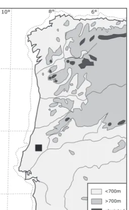
Fig. 1 – Paleogeography of western Iberia in Late Glacial times. The black square denotes the location of the Almonda karstic spring.
currently features some 12 km of mapped underground galleries. Over the last 20 years, several other collapsed entrances were found at different elevations, and some were reopened for archeological excavation of the sealed Lower, Middle or Upper Paleolithic sequences they contain (Zilhão et al., 1991, 1993; Zilhão & Mckinney, 1995; Zilhão, 1997; Marks et al., 1999, 2001, 2002; Chabai et al., 2000/2001; Trinkaus et al., 2003, 2007).
The length of the narrow, meandering Galeria da Cisterna is approximately 100 m, and its cross–section is in general less than 2×2 m (Figure 2). Archeologi-cal investigation of this gallery was carried out for the first time between 1937 and 1942 (Nogueira et al,. 1941; Paço et al., 1947; Guilaine & Ferreira, 1970). As described by Paço et al., the work consisted of the collection of surface material and the excavation of a small chamber located ~15 m from the entrance, and generated an assemblage of Iron Age, bronze Age and Early Neolithic pottery cur-rently kept at the Geological Museum, Lisbon.
In 1987, a small sediment pocket adjacent to Paço et al.’s excavations yielded three typical Solutrean points (Maurício, 1988). Following–up on these finds, two subsequent excavation seasons (1988 and 1989) revealed and explored other pockets of sediments accumulated in discontinuous depressions of the limestone bedrock of the Galeria da Cisterna that had not been touched by previous work: zones AMD1, AMD2 and AMD3 (Figures 2 & 3). The latter two featured Holocene deposits directly atop bedrock or lying on archeologically sterile river–accumu-lated Pleistocene sands. Zone AMD1, however, preserved remnant Upper Paleo-lithic deposits in two areas physically separated by an outcrop of the cave floor, described in detail elsewhere (Zilhão, 1997).
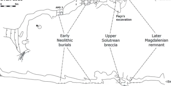
Fig. 2 – Topographical plan and profile of the Galeria da Cisterna. The areas preserving archeological deposits of different periods are indicated.
In grid unit M15, the AMD1 deposits consisted of a thin film of heavily indu-rated reddish sands (level β) coating the walls of a narrow fissure filled with loose, redeposited orange–brown sands (level α). The latter yielded a mix of Holocene and Solutrean material (including the three 1987 finds), while the level β breccia yielded fourteen lithic artifacts, including twelve Upper Solutrean projectile points of diverse typology. In grid units L–M/11–12, an area of <2 m² located some 10 m from the entrance, the AMD1 Pleistocene deposits were preserved at the bottom of a ~90 cm deep depression, under a Holocene «dark–earth» deposit, level 1, which yielded Copper and bronze Age finds (Figures 3 & 4). Against the West wall of the gallery, the immediately underlying Pleistocene sequence (levels 2–3) had been eroded away down to the indurated top of level 4, leaving only a remnant pre-served against the East wall. In squares L–M/11, levels 3 and 4 were separated by a thin black lens, very rich in organic material (sublevel 3a), and in squares L–M/12 most of level 3 was tainted black. This lens was interpreted as the result of a post– depositional process of lateral impregnation, occurring in the early Holocene and caused by the formation of stagnant pools in the depression left by the excavation of the erosional channel mentioned above. Geochemical analysis of the sediments provided results consistent with this interpretation (Cruz, 1993).
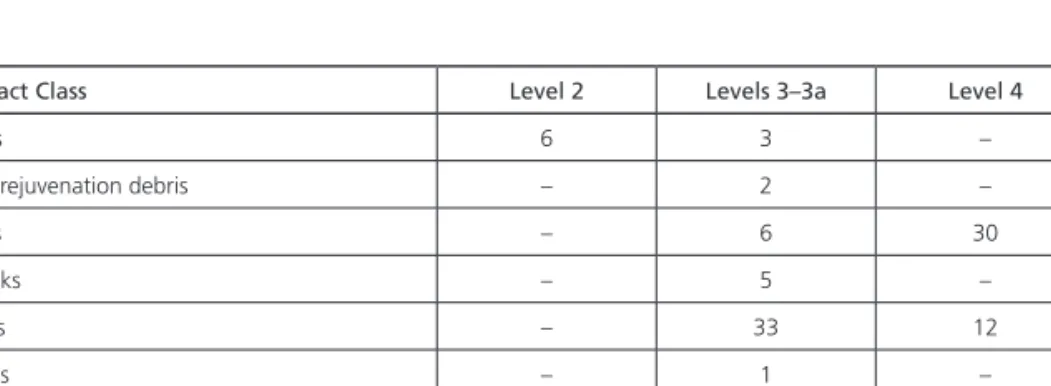
basal level 5 was a sterile fluviatile deposit, but the other levels yielded stone tools and faunal remains (Tables 1–3). The lithic material was generally undi-agnostic, and the sequence was initially assigned, tentatively, to the Last Glacial Maximum (level 4 to the Late Gravettian and level 3 to the Proto–Solutrean), on
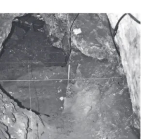
Fig. 3 – View over zone AMD1 towards the interior of the gallery, half way through the excavation process (before removal of the Pleistocene sediments in row 12 of the excavation grid), with the transversal stratigraphic profile exposed.
Fig. 4 – Pleistocene stratigraphy of zone AMD1: level 2, dense eboulis of mid -sized (5 -10 cm long) angular clasts packed in brecciated red sands, ~12 cm thick; level 3, non -brecciated red sands containing scattered angular clasts, ~12 cm thick; level 4, dense eboulis of mid--sized (5 -10 cm long) rounded clasts with a few larger blocks (20 -30 cm long), packed in variably indurated yellow -brown sands, ~10 cm thick; level 5, fluviatile yellow sands, grading into reddish -brown silt at contact with bedrock, containing scattered blocks and speleothem fragments, ~16 cm thick, the upper part forming a horizontal, highly -indurated, 5 -10 cm thick continuous plaque.
the basis of arguments relating to technology, raw–material procurement patterns, and the preservation of Solutrean material in the adjacent M15 breccia (Zilhão, 1997). Subsequent radiocarbon dating (Figure 5), however, proved that level 3 is later Magdalenian in age, which in turn suggests that level 4 is probably of Upper Magdalenian age, in good agreement with both the multiple dihedral burin found therein and the backed bladelet recovered at the interface between levels 3 and 4.
Fig. 5 – Climatic oscillations of the Tardiglacial as recorded in Greenland ice cores and AMS radiocarbon dates for level 3 of zone AMD1, Galeria da Cisterna. The shaded areas in the graph represent the 2 σ calibrated age ranges. Calibration data and paleoclimate curves generated by CalPal, 2007 Hulu version (Weninger and Jöris, 2004).
Artifact Class Level 2 Levels 3–3a Level 4
Cores 6 3 –
Core rejuvenation debris – 2 –
Chips – 6 30
Chunks – 5 –
Flakes – 33 12
Blades – 1 –
Retouched tools 2 2 (a) 2 (b)
Total 8 52 (c) 44 (d)
(a) one pointed blade and one backed bladelet (b) one denticulated blade and one multiple dihedral burin (c) 31% flint, 31% quartz, 36% quartzite, 2% basalt (d) 20% flint, 9% quartz, 71% quartzite
Stratigraphic unit
Latin name English name 4 4–3 3 2
Cervus Red deer – – 3 1
Capra Ibex 4 1 3 –
Sus Wild boar – – 1 –
Lepus Hare – 2 – –
Oryctolagus Rabbit – 3 10 1
Lynx Lynx +? – – –
Felis Wildcat 1 + 1 –
Vulpes Fox 3 4 – –
Chiroptera Bats – – Several 14
Aves indet Birds – – 2 –
Galliform – – 1
Turdus ?Thrush – 2 4 –
Corvus ?Crow 1 2 1 –
Amphibia – – 1 –
Table 2 – Numbers of bones recorded at Galeria da Cisterna. Recording procedure follows Davis (1992, 2002). «+» refers to cases where bones other than «Parts of the Skeleton Always Counted» (POSACS) were found and could be identified to genus.
Number Layer Bone Taxon Fus GL Bd Dd DLS SLC M1 length
L12–2 3a Phalanx III Cervus – – – – 472 – –
M12–4 2 Astragalus Cervus – 499 298 278 – – –
M11–177 4 Scapula Felis silvestris F – – – – 133 –
L11–1 3a Mandibula Lynx? – – – – – – ~100–110 M11–70 3a Tibia Oryctolagus F – 108 – – – – M12–97 3a Calcaneum Oryctolagus F 210 – – – – – M11–116 4 Tibia Oryctolagus F – 105 – – – – M11–22 4 Calcaneum Vulpes F 336 – – – – – M11–19 4 Calcaneum Vulpes F 323 – – – – –
Table 3 – Measurements (following Driesch, 1976), in tenths of a millimeter, of the Galeria da Cisterna mammal bones and teeth. «Fus» refers to the state of epiphysial fusion, where «F» is fused (i.e., adult).
This small Pleistocene remnant thus corresponds to a succession of Tar-diglacial deposits, with their erosion signaling a return to a pattern of regular (seasonal?) water flow in the gallery during the following, wetter–than–present Preboreal period. Subsequent to the excavation of the Galeria da Cisterna, cave– sheltering around the Almonda spring in such terminal Paleolithic times has been rather more extensively documented at the Lapa dos Coelhos, some 10 m higher
up (Gameiro & Almeida, 2004). The radiocarbon dates for Galeria da Cisterna place the formation of its level 3 in the later part of the Allerød and the earlier part of the Dryas III, ~13,000 cal bP. Level 2, therefore, must date to later Dryas III times, while the discontinuity cum cementation at the top of level 4 probably reflects the warm peak at the beginning of the bølling/Allerød interstadial (Fig-ure 5), implying an age >14,500 cal bP for the underlying deposits.
This chronostratigraphic reading of the evidence is consistent with the faunal evidence (Table 2). Three large herbivore taxa – red deer, a caprine (presumably the Iberian ibex), and wild boar were identified, as well as hare and rabbit. Remains of wildcat, fox, possibly lynx, bats, several birds (a galliform; Turdus pilaris, fieldfare;
T. cf merula, blackbird; Corvus cf frugilegus, rook; and C. monedula, jackdaw) and an
amphibian were also found. It is not possible to determine who was responsible for accumulating these taxa, although it seems probable that at least the bats and song-birds accumulated naturally or were predated by small carnivores, while the larger mammals were brought into the cave by people. None of the bones shows any signs of partial digestion. Among the carnivores, the damaged felid carnassial is tenta-tively identified as a very small lynx, although wildcat cannot be ruled out. With a crown length of 10 to 11 mm (Table 3), it is outside the range of measurements of a large collection of modern Felis silvestris from Europe, the Maghreb and the Near East. It is also outside the range for wildcats from the Upper Pleistocene levels of Caldeirão cave, but smaller than a small sample of present–day lynx carnassial teeth as well as the specimens from Caldeirão (Davis, 2002, Fig. 33, 35).
Cervus, Capra and Sus were among the more common taxa recovered in the
Mousterian, EUP, Solutrean and Magdalenian levels of Caldeirão, situated at a similar elevation and ~25 km to the NE as the crow flies. by comparison with this material, the Cisterna red deer astragalus is very small, although the cervid terminal phalanx with a DLS of 47.2 mm plots among the larger specimens of Cervus elaphus from Caldeirão (Davis, 2002). The absence of Equus in the Galeria da Cisterna sample may be due to the smallness of the sample, although at Caldeirão equids became scarcer with time and by the Magdalenian they comprised a mere 6% of the large herbivores. The fact that ibex is a major component of the fauna in both levels 3 and 4 implies for a lowland site such as Galeria da Cisterna a rather open immediate landscape, in agreement with regional environmental reconstructions for the colder phases of last glacial’s Dansgaard–Oeschger cycles (Zilhão, 1997).
StRAtIgRAPHIc PRoVENIENcE AND tAPHoNoMy
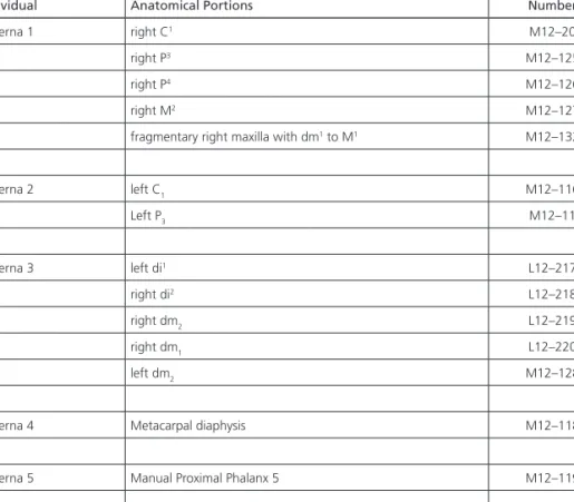
The human remains recovered in the Pleistocene deposits of the Galeria da Cisterna (Table 4) all come from level 3 (including sublevel 3a) of zone AMD1, the excavation of which was initiated and completed during the July 1988 field
season. The Cisterna 1 and Cisterna 3 groups described below include material collected in both level 3 and the 3a black lens. The fact that each of these groups is likely to represent a single individual provides additional support for the inter-pretation of that lens as a lateral variation without stratigraphic significance. The fact that Cisterna 6, found at the interface between levels 3 and 4, could belong to the same individual as Cisterna 2, whose left P3 is a piece–plotted item recovered at the base of the black lens, supports the inclusion of Cisterna 6 in the human bone assemblage from level 3.
Individual Anatomical Portions Number
Cisterna 1 right C1 M12–20
right P3 M12–125
right P4 M12–126
right M2 M12–127
fragmentary right maxilla with dm1 to M1 M12–132
Cisterna 2 left C1 M12–116 Left P3 M12–11 Cisterna 3 left di1 L12–217 right di2 L12–218 right dm2 L12–219 right dm1 L12–220 left dm2 M12–128
Cisterna 4 Metacarpal diaphysis M12–118
Cisterna 5 Manual Proximal Phalanx 5 M12–119
Cisterna 6 Pedal Proximal Phalanx M12–157
Table 4 – Human remains from levels 3–3a of the Galeria da Cisterna.
Assessing the taphonomy of the human bone requires consideration of the fact that the preserved remnant is but a small percentage of the Magdalenian deposits that once, prior to early Holocene erosion, must have filled the irregular rock bottom of the initial section of the Galeria da Cisterna. The deposits con-taining the human bone assemblage also yielded four shell beads – three pierced
Theodoxus fluviatilis and one pierced Hynia reticulata – making it reasonable to
speculate that ritual mortuary activity (burial?) occurred at the site during the later part of the Magdalenian.
MAtERIALS AND MEtHoDS
The Cisterna human remains are described using standard paleontological approaches and measurements (e.g., bräuer, 1988). The dental crown morphol-ogy is scored in part using the Arizona State University Dental Anthropolmorphol-ogy Sys-tem (ASUDAS) (Turner et al., 1991).
In order to assess the size and proportions of the remains, the dental and postcranial remains are compared, where possible, to European Upper Paleo-lithic and recent human remains. The fossil sample is divided into «Early Upper Paleolithic» (EUP) and «Late Upper Paleolithic» (LUP) samples, with the division between them being the last glacial maximum. The former Paleolithic sample is mostly Gravettian, whereas the latter is principally Magdalenian in associa-tion. Sample sizes vary depending upon both preservation and the current detail of publication of these comparative samples. The comparative data derive from primary paleontological descriptions or personal study of the original material. Since the remains from the Galeria da Cisterna are Magdalenian in age, they fall within the LUP time span. Recent European (or Euroamerican) data are included for the dental and phalangeal metrics; the permanent tooth metrics are from Twi-esselmann & brabant (1967), the deciduous dental metrics are for the Euroameri-can males from black (1978), the hand metrics derive from Musgrave (1970), and the pedal metrics are from Trinkaus & Hilton (1996). In the calculation of sample summary statistics, right and left sides (when available) were averaged to provide a value per individual prior to the computations.
tHE HuMAN REMAINS
The Galeria da Cisterna human remains are listed in Table 4. They consist principally of isolated teeth which, based on morphology and developmental age, derive from three individuals (Cisterna 1 to 3). In addition, there are three isolated postcranial elements, one of them immature, which are described here as separate specimens but might have derived from two or more of the dentally identified individuals.
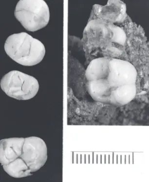
cisterna 1
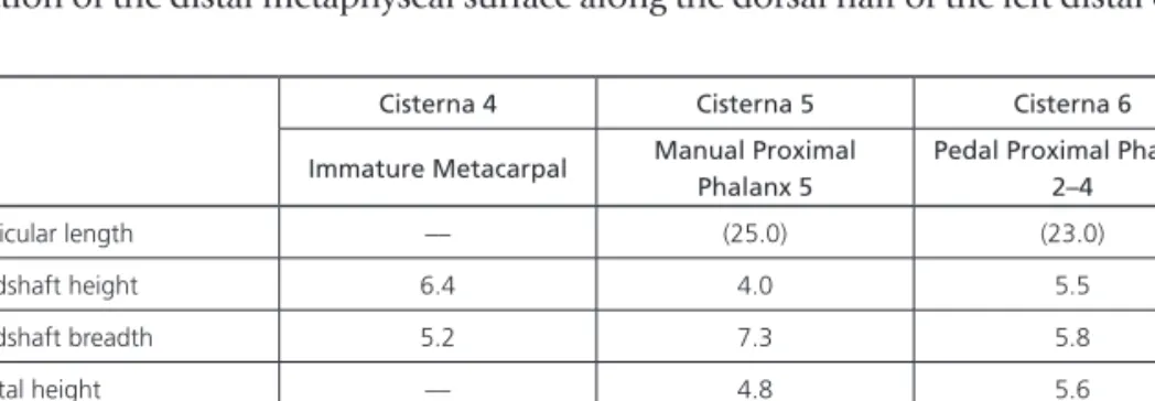
This specimen consists of a fragment of the right maxillary alveoli in matrix with the dm1, dm2 and M1 preserved, plus the isolated right C1, P3, P4 and M2
(Table 4; Figure 6).
The dm1 crown is substantially worn and has suffered postmortem damage
resulting in the loss of the mesiobuccal corner (about one–quarter of the tooth is missing). The exposed lingual root exhibits slight root resorption, whereas the buccal root is enclosed in bone and is not observable. The distal portion of the
dm2 is partially covered by matrix. Enamel
has chipped off distally, exposing the dentin horn of the hypocone.
The fully formed crown and partial (~4 mm) root of a right C1 are preserved. The
crown is complete and in good condition with only a small fracture that runs sagittally along the crown.
The fully formed crown and partial root (~5 mm) of a right P3 are preserved. The tooth
is in good condition, with only minor frac-tures running mesio–distally along the occlu-sal surface of the buccal cusp. A fully formed crown and partial root (~3 mm) of a right P4
are preserved. The crown is in good condition with a small fracture that runs mesiodistally along the buccal cusp.
The M1 crown is complete and well
pre-served. Only a small sagittal fracture runs along the hypocone. The lingual root is exposed distally but obscured by matrix otherwise. The incomplete crown and partial root (~2.7 mm) of a right M2 are preserved. Approximately one–quarter of
the distal crown is missing. Postmortem damaged has resulted in the loss of tooth enamel from the buccal tooth surface. The crown is fractured along the mesiobuc-cal fissure and along the mesial bucmesiobuc-cal cusp; its root was lost postmortem.
The occlusal morphology of the dm1 has been obliterated by extreme wear,
but some occlusal details remain on the dm2. It is a four–cusped tooth, and the
lingual surface is featureless (i.e., it lacks a Carabelli’s trait); no other morphology is observable due to damage and wear.
On the C1, the lingual surface exhibits weak shoveling (ASUDAS grade 2) and
a well–developed distal accessory ridge (ASUDAS grade 4). There is slight swelling of the gingival area but no lingual tubercle is present. Lingually, the tooth exhibits barely perceptible mesial and distal marginal ridges. In addition, short (1–2 mm) and shallow grooves radiate up from the cervix but are not pathological (i.e., enamel hypoplasia).
The buccal and lingual tooth surfaces of the P3 are featureless. Occlusally,
the crown exhibits small accessory mesial and distal cuspules but lacks accessory ridges on either the buccal or lingual cusp. In contrast, both the buccal and lin-gual cusps of the P4 exhibit mildly expressed accessory ridges mesially and distally
(MxPAR: burnett, 1998); they are more strongly expressed on the buccal cusp
Fig. 6 – Occlusal views of the Cisterna right maxillary teeth. Left: C1, P3, P4 and M2. Right: dm1 to M1. Scale in millimeters.
than on the lingual cusp. The buccal tooth surface of the P4 exhibits barely
percep-tible mesial and distal ridges, while the lingual surface is featureless.
The M1 is a four–cusped tooth. The protocone is the largest cusp (43.7 mm2)
followed by the paracone (28.7 mm2), with the remaining two cusps (metacone
and hypocone) equal in size (28.2 and 28.3 mm2 respectively) (cf., bailey, 2004).
The occlusal polygon area is large (38.8 mm2) suggesting that the cusp tips are
widely spaced. Finally, the cusp angles (bailey, 2004) are very close to the mean for a pooled Upper Paleolithic European sample, reflecting the squared outline of the tooth. Lingually, the protocone presents a medium sized Carabelli’s cusp (ASUDAS grade 3), while the buccal surface is featureless.
Occlusally, the tooth is morphologically simple. It lacks accessory cusps and presents only a weakly developed mesial marginal ridge. The M2 is a four–cusped
tooth that exhibits a much reduced hypocone (ASUDAS grade 2). The relative cusp areas are protocone > paracone > metacone > hypocone. Occlusally, the tooth is more complex than the M1, exhibiting two accessory ridges that emanate
from a mesial marginal ridge and run toward the central fossa. both buccal and lingual tooth surfaces are featureless.
The deciduous molars are quite worn. The dm1 exhibits heavy, basin–like
wear that has exposed the tooth’s small pulp chamber, and the dm2 is more
mod-erately worn, with more dentin exposed lingually than buccally. The M1 exhibits
only occlusal wear facets but no dentin exposure. The unerupted P3, P4 and M2 are
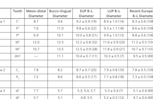
Tooth Mesio–distal Diameter Bucco–lingual Diameter EUP B–L Diameter LUP B–L Diameter Recent European B–L Diameter Cisterna 1 C1 8.7 9.6 9.2 ± 0.9 (19) 8.9 ± 1.0 (14) 8.3 ± 0.6 (108) P3 7.6 11.0 9.8 ± 0.6 (22) 9.3 ± 1.1 (18) 8.6 ± 0.6 (108) P4 6.9 10.1 10.0 ± 0.8 (21) 9.9 ± 1.0 (13) 8.8 ± 0.6 (106) M1 12.0 12.5 12.2 ± 0.8 (32) 11.6 ± 0.9 (20) 11.2 ± 0.5 (104) M2 10.7 13.5 12.5 ± 0.9 (28) 11.8 ± 0.9 (21) 10.7 ± 0.7 (102) dm2 –– 11.1 10.4 ± 0.7 (11) 10.3 ± 0.5 (7) 9.5 ± 0.5 (69) Cisterna 2 C1 7.8 8.2 8.7 ± 0.7 (20) 7.9 ± 0.8 (19) 7.8 ± 0.5 (109) P3 7.2 8.6 8.6 ± 0.5 (17) 7.7 ± 0.8 (18) 7.3 ± 0.5 (108) Cisterna 3 di1 7.7 5.7 5.3, 5.4, 5.7 5.3 ± 0.4 (7) 5.1 ± 0.4 (69) di2 5.7 5.1 4.8, 5.5 5.2 ± 0.5 (12) 4.7 ± 0.4 (69) dm1 9.0 7.7 7.8 7.2 ± 0.6 (13) 7.4 ± 0.5 (69) dm2 11.1 9.7 9.2 ± 0.7 (12) 9.0 ± 0.5 (19) 8.9 ± 0.4 (69) Table 5 – Dental metrics for Cisterna 1 to 3 dental crowns and comparative bucco–lingual diameters. Mean ± standard deviation. Measurements are in millimeters and sample sizes are in parentheses.
unworn. There is no distal interproximal wear facet on the M1, confirming that
the M2 was unerupted.
The presence of both deciduous molars with the first molar indicates a juve-nile age for Cisterna 1. The degree of development of the premolar roots (about one–half formed) narrows its age–at–death to between eight and ten years post-natal, although the degree of canine root development may suggest an age closer to eight years (Smith, 1991). This is supported by the presence of resorption on the dm1 lingual root, indicating that the dm1 is not far from exfoliation, and
hence the P3 would have been erupting within a year or two if the individual had
survived.
None of the tooth crowns exhibits macroscopic enamel hypoplasias or caries. The bucco–lingual diameters of the teeth (Table 5) are all relatively large, fall-ing at least 0.5 mm above the mean values for the LUP sample for all except the P4. They are above all of the recent human means and most similar to the mean
values for the EUP sample. They nonetheless remain within two standard devia-tions of the LUP means, although they are all more than two standard deviadevia-tions above the recent European values.
cisterna 2
The two teeth assigned to Cisterna 2 (Table 4; Figure 7) consist of mandibular left C1 and P3, both of which retain the complete crown and the fully formed root. both teeth are in good condition. The C1 crown has small fractures on the buccal and lingual surfaces that run sag-ittally from the crown tip to the root.
The lingual surface of the C1 exhib-its barely perceptible mesial and dis-tal marginal ridges (ASUDAS grade 1 shoveling) and is otherwise featureless. The single root exhibits shallow mesial and distal developmental grooves. The P3 presents a mesially oriented trans-verse ridge that connects the buccal and lingual cusps. This ridge, combined with the mesial and distal marginal ridges of the tooth, creates a small anterior fovea and a larger (but shallower) posterior fovea.
Fig. 7 – Views of the Cisterna 2 and 3 teeth. Above: Cisterna 2 left C1 and P3 in lingual view. Below from left to right: Cisterna 3 left dm2 in occlusal view, left di1 and right di2 in lingual view, and right dm
1 with mandibular fragments in occlusal view. Scale in millimeters.
The C1 cusp tip is slightly worn, but the wear does not expose the dentin below. The position of the mesial wear facet may indicate dental crowding and/ or that the canine was rotated lingually. The distal interproximal wear facet on this tooth and the mesial interproximal wear facet on the P3 match in size and shape. In addition the P3 exhibits small occlusal wear facets with no dentin exposure.
Metrically, these teeth are moderately large (Table 5). The C1 breadth falls between the EUP and the LUP means, and the P3 breadth is on the EUP mean and above the LUP and recent human means.
There are no signs of pathological alterations of the teeth, despite the pres-ence on the C1 of short (1–2 mm) and shallow grooves that radiate up from the cervix. The complete formation of the tooth roots with minimal occlusal wear suggests an age–at–death during adolescence or early maturity.
cisterna 3
This individual consists of five teeth of a child (Table 4; Figure 7). Two of the teeth, the right dm1 and the left dm2, are preserved in fragments of mandibular alveoli.
The left di1 preserves a completely formed crown and root. The root apex
appears to have broken off postmortem. The crown is in good condition with a minor sagittal fracture along the distal lobe. The right di2 retains the complete
crown and nearly complete root (the apex is missing). The tooth is in good condi-tion with minor fractures along the lingual and buccal surfaces.
A complete right dm 1 is preserved in a very fragmented alveolus that is cov-ered by matrix. The tooth is in good condition. It is missing a small amount of enamel from the mesio–lingual surface, and presents minor fractures along the buccal surface. The mesial root is not fully formed (the apex is missing), but the tooth was fully erupted. It is not possible to observe the distal root, because it is enclosed in bone. The lingual root sockets of the dm2 are preserved in the associ-ated alveolar bone.
A partial crown (distal portion of the metaconid and mesial portion of the hypoconid) and root (~3 mm) of a right dm2 are preserved. This is associated with the complete crown and partial root (~1/3) of a left dm2 preserved in its alveolus. The tooth crown is in fair condition. It exhibits a medium–sized fracture running bucco–lingually and smaller fractures running along the mesial lingual cusp.
The crown of the di1 is short and flared. It exhibits mild labial curvature
(ASUDAS grade 2), mild sagittal curvature, weak shoveling (ASUDAS grade 1) and weak gingival swelling. The labial surface is featureless. The di2 is moderately
con-vex both labially (ASUDAS grade 2) and sagittally. The lingual and buccal tooth surfaces are featureless.
Due to damage, the morphology of the right dm2 cannot be observed. The dm1 is a five–cusped tooth. Occlusally it exhibits a deep anterior fovea, which is set off distally by well–developed essential crests of the protoconid and metaco-nid. The tooth presents an X–groove pattern (the protoconid and hypoconid are in contact) and a moderately sized cusp 5 or hypoconulid (ASUDAS grade 4) that is centered distally. A weak occlusal ridge connects all cusps. The left dm2 is a five–cusped tooth. Cusp 5 is well developed (ASUDAS grade 4) with a prominent transverse ridge that runs along the occlusal surface toward the talonid basin. Occlusally, it exhibits a high and prominent mesial marginal ridge and a deep anterior fovea. A small cuspule (0.9 mm wide) is enclosed in the anterior fovea. The buccal surface presents a small buccal pit (ASUDAS grade 1 protostylid).
Metrically, these teeth are unexceptional for an Upper Paleolithic human (Table 5).
The deciduous incisors exhibit minimal wear of the incisal edge, with slight dentin exposure on the di1 but not on the di2. The cusps of the deciduous molars are
unworn, and the absence of interproximal wear facets on the left dm2 indicate that the tooth was recently erupted or in the process of eruption. Together, these indicate a young age for this individual, approximately two years postnatal (Smith, 1991).
There is no evidence of pathological alterations of these teeth.
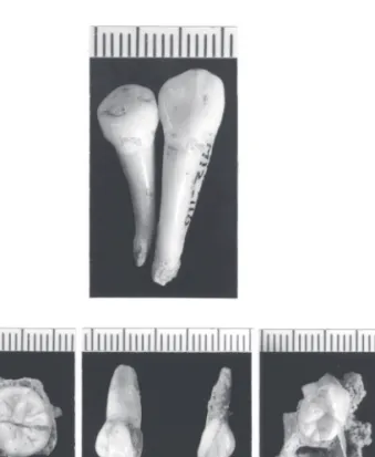
cisterna 4
The bone consists of the diaphysis of an immature metacarpal, from the broken flare for the base to portions of the distal metaphysis (Figure 8; Table 6). The maxi-mum preserved length is 30.3 mm. The immaturity of the bone is based in part on its generally mild cortical bone porosity. In addition, there is a small 4.8 x 2.3 mm section of the distal metaphyseal surface along the dorsal half of the left distal edge.
Cisterna 4 Cisterna 5 Cisterna 6
Immature Metacarpal Manual Proximal Phalanx 5
Pedal Proximal Phalanx 2–4
Articular length –– (25.0) (23.0)
Midshaft height 6.4 4.0 5.5
Midshaft breadth 5.2 7.3 5.8
Distal height –– 4.8 5.6
Distal maximum breadth –– 7.8 8.8
Distal articular breadth –– 7.3 8.3
Table 6 – Osteometrics of the manual and pedal remains. Measurements are in millimeters.
The metaphyseal surface appears straight and perpendicular to the diaphy-seal axis. The right side of the diaphysis remains straight distally, but the left side
flares to the side. Since the distal radial side of immature metacarpals normally flares more than the ulnar side, the bone derives from the right side. There is a strong proximal to dis-tal clockwise torsion of the shaft, between 30° and 33° depending upon whether the dorsal surfaces or the estimated dorsopalmar axes of the base and head are employed. The interos-seous muscle lines are faint to absent along the diaphysis.
The degree of formation of the bone suggests a minimum age of about five years postnatal, whereas the absence of evidence of fusion of the metacarpal head epiphysis sug-gests a maximum age of about twelve years (Greulich & Pyle, 1959). It is difficult to be more precise as to its age–at–death.
cisterna 5
The specimen consists of the complete head and diaphysis plus portions of the base of a fifth proximal hand phalanx (Figure 8; Table 6). The shaft and head are complete, with car-bonate encrustations dorsally and palmarly on the diaphysis. The base retains a small dorsal left corner of the proximal articulation and the
adjacent dorsal bone. The maximum preserved length is 25.4 mm. based on its proximal–to–distal clockwise torsion, it is probably a right bone.
The diaphysis is smooth, with the flexor tendon sheath lines projecting less palmarly than the middle of the palmar diaphysis. The head is smooth with the tubercles projecting only slightly beyond the trochlea. The right (ulnar?) side of the trochlea is larger than the left (radial?) side. An index of robusticity ((shaft height x shaft breadth)1/2 / articular length) for the bone provides a value of ~21.6,
which is similar to the values (19.7, 21.4, 21.5) for three EUP specimens providing sufficiently complete fifth manual proximal phalanges. A recent European sample provides similar values (21.4 ± 2.0, N = 38).
cisterna 6
The Cisterna 6 phalanx is a proximal pedal phalanx from one of the three middle digits (Figure 8; Table 6). It retains the complete head, the complete shaft
Fig. 8 – Palmar/plantar (above) and right lateral (below) views, from left to right, of the Cisterna 4 right metacarpal diaphysis, the Cisterna 5 fifth proximal hand phalanx, and the Cisterna 6 proximal pedal phalanx. Scale in centimeters.
with dorsal encrustations, and the dorsal margin adjacent to the proximal joint capsule attachment area. The preserved length is 23.2 mm, and articular length is estimated at 23.0 mm. Since the head horizontal angle is ~0°, side remains indeterminant.
The diaphysis is smooth with no indications for the flexor tendon sheaths. The head has a larger trochlear height on the right side, but the left side of the tro-chlea flares more than the right side. A robusticity index for the specimen (~24.6) compares to a EUP mean of 23.5 (± 1.9, N = 15 specimens, 6 individuals) and one of 25.5 (± 1.4, N = 6 from 2 individuals) for the available LUP sample. A recent European sample provides a slightly lower mean (21.2 ± 2.3; N = 105 bones, 35 individuals).
NuMbER oF INDIVIDuALS
The dental remains include a young child, a late juvenile and an adolescent/ young adult. The Cisterna 4 metacarpal could well belong to the same individual as the Cisterna 1 dentition, based on age–at–death. Similarly, the Cisterna 5 and 6 phalanges could be associated with the Cisterna 2 teeth. This would place the minimum number of individuals at three.
SuMMARy
The Magdalenian human remains from the Galeria da Cisterna in the Gruta do Almonda represent relatively young individuals, with the oldest (Cisterna 2) probably being little older than the early third decade based on dental attrition. They show no evidence of either developmental or degenerative lesions. Their den-titions are large compared to recent Europeans and among the larger of the Late Upper Paleolithic Europeans. The three postcranial elements are unremarkable.
AcKNowLEDgMENtS
The excavations at the Galeria da Cisterna, Almonda karstic system were car-ried out under the auspices of the Sociedade Torrejana de Espeleologia e Arqueo-logia (STEA) and funded by the Câmara Municipal de Torres Novas, and benefited from precious logistical support provided by Fábrica de Papel A Renova. Analysis of the Cisterna human remains was funded by Washington University (ET) and Sigma Xi Scientific Research Association (SEb). Comparative metric data were col-lected by ET and funded by the Wenner–Gren, L.S.b. Leakey and National Science Foundations. To all of them we are grateful.
REFERENcES
AGUIRRE, E.; bERMÚDEZ DE CASTRO, J.M.; GARRALDA, M.D. (1991) – Spain. In ORbAN, R., ed. – Hominid Remains An Up–Date 4. brus-sels: Université Libre de bruxelles.
ARSUAGA, J. L.; MARTÍNEZ, I.; VILLAVERDE, V.; LORENZO, C.; QUAM, R.; CARRETERO, J.M.; GRACIA, A. (2001). – Fósiles humanos des país valenciano. In VILLAVERDE, V., ed. – De Nean‑
dertales a Cromañones. El Inicío del Poblamiento Humano en las Tierras Valencianas. Valencia:
Uni-versitat de Valencia. p. 265–322.
bAILEY, S. E. (2004) – A morphometric analysis of maxillary molar crowns of Middle–Late Pleis-tocene hominins. Journal of Human Evolution. London. 47, p. 183–198.
bLACK, T. K. III (1978) – Sexual dimorphism in the tooth crown diameters of the deciduous teeth. American Journal of Physical Anthropology. New York. 48, p. 77–82.
bRäUER, G. (1988) – Osteometrie. In Anthro‑
pologie I. Stuttgart: Fischer Verlag. p.160–232.
bURNETT, S. E. (1998) – Maxillary Premolar
Accessory Ridges (MxPAR): Worldwide Occurrence and Utility for Population Differentiation. Tempe:
Department of Anthropology, Arizona State University. MA Thesis.
CHAbAI, V. P.; SITLIVY, V. I.; MARKS, A. E. (2000–2001) – Lower Paleolithic industry of brecha das Lascas, level 7 (Portugal). Préhistoire
Européenne. Liège. 16–17, p. 17–41.
CRUZ, A. J. C. (1993) – Estudo geoquímico de
preenchimentos sedimentares de grutas da Estre‑ madura com vestígios de ocupação humana pré– histórica. Lisboa: University of Lisbon. Ph. D.
dissertation.
DAVIS, S. J. M. (1992) – A rapid method for recording information about mammal bones from archaeological sites. London, Historic buildings and Monuments Commission, Ancient Monuments Laboratory report 19/92. DAVIS, S. J. M. (2002) – The Mammals and birds from the Gruta do Caldeirão, Portugal. Revista
Portuguesa de Arqueologia. Lisboa. 5, p. 29–98.
DRIESCH, A. von den (1976) – A guide to the
measurement of animal bones from archaeological sites. Cambridge, Mass.: Peabody Museum of
Archaeology and Ethnology; Harvard University (Peabody Museum bulletin; 1).
FEREMbACH, D.; ROCHE, J. (1971) – Portugal. In OAKLEY, K. P.; CAMPbELL, b. G.; MOLLE-SON, T. I., eds. – Catalogue of Fossil Hominids II:
Europe. London: british Museum, p. 277–282.
(Natural History).
GAMEIRO, C. ; ALMEIDA, F. (2004) – A ocupação da camada 3 da Lapa dos Coelhos (Casais Mar-tanes, Torres Novas). Novos elementos sobre a produção de suportes lamelares durante o Magdalenense Final da Estremadura Portuguesa.
Promontoria. Faro. 2, p. 193–238.
GREULICH, W. W.; PYLE, S. I. (1959) – Radio‑
graphic Atlas of Skeletal Development of the Hand and Wrist. London: Oxford University Press.
GUILAINE, J.; FERREIRA, O. V. (1970) – Le Néo-lithique ancien au Portugal. Bulletin de la Société
Préhistorique Française. Paris. 67, p. 304–322.
MARKS, A. E.; bRUGAL, J.–Ph.; CHAbAI, V. P.; MONIGAL, K.; GOLDbERG, P.; HOCKETT, b.; PEMAN, E.; ELORZA, M.; MALLOL, C. (2002) – Le gisement Pléistocène Moyen de Galeria Pesada (Estrémadure, Portugal): premiers résultats. Paléo. Les Eyzies. 14, p. 77–99. MARKS, A.; MONIGAL, K.; CHAbAI, V. (1999) – Report on the initial excavation of brecha das Lascas and Galeria Pesada (Almonda, Portu-guese Estremadura). Journal of Iberian Archaeol‑
ogy. Porto. 1, p. 237–250.
MARKS, A.; MONIGAL, K.; ZILHãO, J. (2001) – The lithic assemblages of the Late Mousterian at Gruta da Oliveira, Almonda, Portugal. In ZIL-HãO, J.; AUbRY, Th.; CARVALHO, A. F., eds. – Les
premiers hommes modernes de la Péninsule Ibérique. Actes du Colloque de la Comission VIII de l’UISPP, Vila Nova de Foz Côa, Octobre 1998. Lisboa:
Insti-tuto Português de Arqueologia. p. 145–154. (Trabalhos de Arqueologia; 17).
MAURÍCIO, J. (1988) – Contribuição para o conhecimento da Pré–História de Torres Novas.
MUSGRAVE, J. H. (1970) – An Anatomical Study
of the Hands of Pleistocene and Recent Man.
Cam-bridge: University of Cambridge. Ph.D. Thesis. NOGUEIRA, A. M.; VAULTIER, M.; ZbYSZE-WSKI, G. (1941) – Primeiras pesquisas na Gruta do Almonda. Brotéria. Lisboa. 32: 1, p. 67–68. PAÇO, A.; VAULTIER, M.; ZbYSZEWSKI, G. (1947) – Gruta da Nascente do Rio Almonda.
Trabalhos de Antropologia e Etnologia. Porto. XI:
1–2, p. 171–187.
SMITH, b.H. (1991) – Standards for human tooth formation and dental age assessment. In KELLEY, M.A.; LARSEN, C. S., eds. – Advances
in Dental Anthropology. New York: Wiley–Liss.
p.143–168.
TRINKAUS, E.; bAILEY, S. E.; ZILHãO, J. (2001). – Upper Paleolithic human remains from the Gruta do Caldeirão, Tomar, Portugal. Revista Por‑
tuguesa de Arquelogia. Lisboa. 4, p. 5–17.
TRINKAUS, E.; HILTON, C. E. (1996) – Nean-dertal pedal proximal phalanges: diaphyseal loading pat terns. Journal of Human Evolution. London. 30, p. 399–425.
TRINKAUS, E.; MAKI, J.; ZILHãO, J. (2007) – Middle Paleolithic Human Remains from the Gruta da Oliveira (Torres Novas), Portugal.
American Journal of Physical Anthropology. New
York. 134, p. 263–273.
TRINKAUS, E.; MARKS, A. E.; bRUGAL, J.–PH.; bAILEY, S. E.; RINK, W. J.; RICHTER, D. (2003) – Later Middle Pleistocene human remains from the Almonda Karstic system, Torres Novas, Por-tugal. Journal of Human Evolution. London. 45, p. 219–226.
TURNER, C.; NICHOL, C.; SCOTT, G. (1991) – Scoring procedures for key morphological traits of the permanent dentition: The Arizona State University Dental Anthropology System. In KEL-LEY, M.; LARSEN, C.S., eds. – Advances in Dental
Anthropology. New York: Wiley Liss. p. 13–31.
TWIESSELMANN, F.; bRAbANT, H. (1967) – Nouvelles observations sur les dents et les max-illaires d’une population ancienne d’âge franc de Coxyde (belgique). Bulletin du Groupement
International pour la Recherche Scientifique en Sto‑ matologie. brussels. 10, p. 5–180.
WENINGER, b.; JÖRIS, O. (2004) – Glacial Radiocarbon Calibration. The CalPal Program. In HIGHAM, T.; bRONK RAMSEY, C.; OWEN, C. (eds.) – Radiocarbon and Archaeology. Fourth
International Symposium, Oxford, 2002. Oxford:
Oxbow books. p. 9–15 (Oxford University School of Archaeology Monographs; 62). ZILHãO, J. (1997) – O Paleolítico Superior da
Estremadura Portuguesa. Lisbon: Edições Colibri.
ZILHãO, J.; MAURÍCIO, J.; SOUTO, p. (1991) – A Arqueologia da Gruta do Almonda. Resultados das escavações de 1988–89. In Actas das IV Jorna‑
das Arqueológicas. Lisboa, 1990. Lisboa:
Associ-ação dos Arqueólogos Portugueses. p. 161–171. ZILHãO, J.; MAURÍCIO, J.; SOUTO, p. (1993) – Jazidas arqueológicas do sistema cársico da nas-cente do Almonda. Nova Augusta. Torres Novas. 7, p. 35–54.
ZILHãO, J.; MCKINNEY, C. (1995) – Uranium– Thorium dating of Lower and Middle Paleo-lithic sites in the Almonda karstic system (Torres Novas, Portugal). In Actas da 3.ª Reunião do
Quaternário Ibérico. Coimbra: Universidade de
Coimbra. p. 513–516.
ZILHãO, J.; TRINKAUS, E., eds. (2002) – Portrait
of the Artist as a Child. The Gravettian Human Skel‑ eton from the Abrigo do Lagar Velho and its Archeo‑ logical Context., Lisboa, Instituto Português de
Arqueologia, p. 1–609. (Trabalhos de Arqueo-logia; 22)