The bone marrow micronucleus test and metronidazole genotoxicity
in different strains of mice (
Mus musculus
)
Ximena C. Abrevaya
1, Marta A. Carballo
2and Marta D. Mudry
1 1Grupo de Investigación en Biología Evolutiva, Departamento de Ecología, Genética y Evolución,
Facultad de Ciencias Exactas y Naturales, Universidad de Buenos Aires, Argentina.
2
Citogenética Humana y Genética Toxicológica, Departamento de Bioquímica Clínica,
Facultad de Farmacia y Bioquímica, Universidad de Buenos Aires, Argentina.
Abstract
The mouse (Mus musculus) bone marrow micronucleus test was carried out using 24 outbred National Institutes of Health (NIH) mice, 24 inbred Swiss Webster (CFW) mice and 20 inbred Bagg albino/color locus Jackson (BALB/cJ) mice. The mice in the experimental group (n = 32) were injected intraperitoneally with 133 mg kg-1
of metronidazole parenteral solution and the control group consisted of mice (n = 36) which had not been injected with metronidazole. There was no significant difference (p > 0.05) between the sexes regarding the micronucleus frequency in either the experimental or the control group. When the Mn frequencies of the three strains were compared, the results for the CFW and BALB/cJ strains did not differ statistically (p > 0.05) for either the experimental or control groups but there were significant (p < 0.05) differences between the CFW and NIH strains and the NIH and BALB/cJ strains for the ex-perimental and control groups, with the NIH strain always showing the highest micronucleus frequency. Our results also show that metronidazole was possible genotoxic agent because it produced a significant increase (p < 0.05) in the micronucleus frequency of the experimental group as compared to the control group for all the three mouse strains tested.
Key words:BALB/cJ, bone marrow micronucleus test, CFW, genotoxicity, metronidazole, NIH.
Received: September 6, 2006; Accepted: April 2, 2007.
The micronucleus test is one of the most widely ap-plied short term test used in genetic toxicology and has be-come one of the most important tests implemented by the regulatory entities of different countries to evaluate muta-genicity of, and sensitivity to, xenobiotics (OECD, 1997; EPA, 1998). The experimental models proposed for these evaluations include different strains of inbred, outbred or hybrid mice (Salamone and Mavourin, 1994), transgenic
mice (Recioet al., 2005) and, more recently, wildlife
ani-mal models (Da Silvaet al., 2000 a, b). The species
em-ployed to monitor the potential genotoxic effect must be considered as a source of variability as certain genotoxic agents have been described as species-specific. For exam-ple, the effects of ionizing radiation have been highly vari-able when assayed with different animal species and
laboratory strains (Catena et al., 1994) and differing
re-sponses have occurred between different rat strains when
exposed to chemical agents such as cyclophosphamide
(Hamadaet al., 2001). According to Simula and Priestly,
1992, these effects could be due to the influence of various factors such as the differential distribution of the compound tested within the tissues of the different species and strains of animals used in testing. The importance of strain-de-pendence sensitivity to different agents has been growing
in the last three decades (Styleset al., 1983; Aeschbacher,
1986; Satoet al., 1987, 1993). Satoet al.(1993) analyzed
the micronucleus frequency in the bone marrow of different mice strains treated with base and nucleotide analogues, and found that BALB/c mice were more susceptible to clastogenic effects than C57BL/6 or DBA/2 strains of mice. Similar results were obtained after exposure to radiation
(Bhilwadeet al., 2004).
Erexsonet al., (1991) found no species or strain
dif-ferences in the micronucleus test for rats, mice or humans when investigating the effects of X-ray radiation and this
was supported by Styleset al.(1983), who reported no
dif-ferences in micronucleus test results for different mice strains exposed to genotoxic agents such as cyclophos-phamide and hexamethyl phospharamide. More recently,
www.sbg.org.br
Send correspondence to Ximena C. Abrevaya. Grupo de Investiga-ción en Biología Evolutiva, Departamento de Ecología, Genética y Evolución, Facultad de Ciencias Exactas y Naturales, Universidad de Buenos Aires, Cdad. Universitaria, Pabellón II, Lab 46. 4to Piso, Cdad. Autónoma de Buenos Aires, Argentina. E-mail: abrevaya@ ege.fcen.uba.ar.
however, Salamone and Mavourin (1994) reported that the basal micronucleus frequency of different mice strains di-verge significantly from that of their parental strains.
To clarify this situation and ascertain whether or not the strain of mice used in the micronucleus test affected the results of the test we compared three mice strains fre-quently used in genetic toxicology evaluation protocols in respect to their micronucleus test results after exposure to the genotoxic agent metronidazole, an anti-infective agent mainly used in the treatment of infections caused by proto-zoa and anaerobic bacteria.
The mouse (Mus musculus) bone marrow
micro-nucleus test was carried out according with EPA, 1994, and Schmid, 1975, using 24 outbred National Institute of Health (NIH) mice, 24 inbred Swiss Webster (CFW) mice and 20 inbred Bagg albino/color locus Jackson (BALB/cJ) mice (Schmid, 1975; Potter, 1985; EPA, 1994) (Table 1). CFW and NIH mice were provided by the animal breeding unit of the School of Medicine (FMed, UBA) and the BALB/cJ mice by the animal breeding unit of the School of Exact and Natural Sciences (FCEyN, UBA), both in Bue-nos Aires, Argentina. The mice strains had previously been obtained from National Atomic Energy Comittee (CNEA) (CFW and NIH) and Jackson Laboratory (BALB/cJ). The mice in the experimental group (n = 32) were injected
intraperitoneally with 133 mg kg-1 of metronidazole
parenteral solution, this dose being comparable to that used in humans. The control group consisted of mice (n = 36) which had not been injected with metronidazole. In this ex-perimental design the control group was not injected with the vehicle (physiologic solution) since our previous results
did not show significant differences between experimental mice treated with the vehicle and controls without
treat-ment (Abrevayaet al., 2004). For both the experimental
and control groups of each strain we used equal numbers of adult mice of both sexes (60-days old, mean weight = 30 g ± 5 g) housed in separate same-sex communal cages main-tained at 20 °C ± 2 °C under a 20 lux 12 h day-length and
given ad libitum access to a commercial feed (Nestle
Purina, Argentina) and filtered water acidified to pH 2.5. Sterile wood shavings were used as bedding and the air in the animal house was renewed eight times a day. Both groups were sacrificed by cervical dislocation 30 h after
in-jection with metronidazole (Mudryet al., 1994). This study
as approved by the animal experimentation review board of our institution.
Bone marrow preparations were made and stained ac-cording to the method described in Schmid, 1975. The pres-ence of micronucleated polychromatic erythrocytes was visually scored (at least 1000 per mouse) by optical micros-copy using a Leica bright field microscope. Cells were con-sidered to be micronucleated when they contained neatly defined chromatin corpuscles with a diameter of less than one-third the diameter of the cell nucleus and stained equal or lighter than the nucleus of the cell from which the micronucleated cell had developed (Schmid, 1975). The experimental and control micronucleus frequency for each specimen within and between the different mice strains were compared using the non-parametric two-tailed un-paired Mann-Whitney test, the Kruskal-Wallis and the Chi-Squared tests using the Statistica program V3 (the analysis was performed considering individual value frequencies).
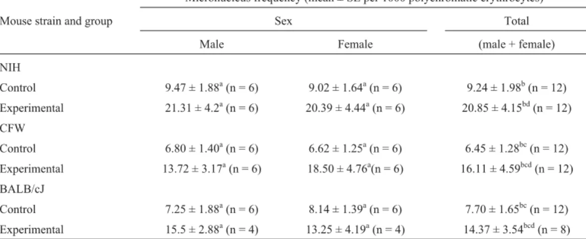
Table 1- Micronucleus frequency (mean ± standard error, SE) for bone marrow polychromatic erythrocytes from three strains of mice treated with
133 mg kg-1of metronidazole (the experimental group) and untreated mice (the control group). We assessed six mice of each sex for both the control and experimental groups, except for the BALB/cJ experimental group for which four mice were assessed for each sex. At least 1000 cells were scored per mouse, although for the BALB/cJ experimental group 1500 cells were scored per mouse.The two-tailed Mann-Whitney test was used to test for differences between frequency values.
Micronucleus frequency (mean ± SE per 1000 polychromatic erythrocytes)
Mouse strain and group Sex Total
Male Female (male + female)
NIH
Control 9.47 ± 1.88a(n = 6) 9.02 ± 1.64a(n = 6) 9.24 ± 1.98b(n = 12)
Experimental 21.31 ± 4.2a(n = 6) 20.39 ± 4.44a(n = 6) 20.85 ± 4.15bd(n = 12)
CFW
Control 6.80 ± 1.40a(n = 6) 6.62 ± 1.25a(n = 6) 6.45 ± 1.28bc(n = 12)
Experimental 13.72 ± 3.17a(n = 6) 18.50 ± 4.76a(n = 6) 16.11 ± 4.59bcd(n = 12)
BALB/cJ
Control 7.25 ± 1.88a(n = 6) 8.14 ± 1.39a(n = 6) 7.70 ± 1.65bc(n = 12)
Experimental 15.5 ± 2.88a(n = 4) 13.25 ± 4.19a(n = 4) 14.37 ± 3.54bcd(n = 8)
a
There was no significant difference (p > 0.05) be-tween the sexes regarding the micronucleus frequency in either the experimental or the control group, because of which the data for the male and female mice of each strain were pooled (Table 1). For all the strains, the micronucleus frequency was, as expected, significantly higher (p < 0.05) in the experimental group than the control group. When the micronucleus frequency of three strains were compared the results for the CFW and BALB/cJ strains did not differ sta-tistically (p > 0.05) when each individual group (experi-mental and control) was considered separately but, how-ever, there were significant (p < 0.05) differences between the CFW and NIH strains and the NIH and BALB/cJ strains for the experimental and control groups when considered separately, with the NIH strain always showing the highest micronucleus frequency.
Since the 1980s our research team has used experi-mental designs based on the micronucleus test using the
CFW mouse strain (Larripaet al., 1984, 1992; Mudryet al.,
1987, 1994) and established an historical mean number of micronuclei for untreated control CFW mice of 9.73 ± 2.53 micronuclei per 1000 polychromatic erythrocytes, the addi-tion of the data from the present study altering this mean value to 9.13 ± 2.58 micronuclei per 1000 polychromatic erythrocytes for this strain.
The micronucleus test is often used to predict the
car-cinogenic potential of compounds (Satoet al., 2001),
al-though the scope of this test is continuously broadening by incorporating new technologies for the detection of
differ-ent genetic alterations (Zuñiga Gonzálezet al., 2001a, b;
Ateeqet al., 2002; Cristaldiet al., 2004). Several authors
have reported modifications to the test, including
alter-ations in culture media,β-cytochalasin concentration,
os-molarity, pH and staining techniques, all of which are capable of introducing artifacts and producing intra- and
inter-laboratory variability (Fenechet al., 2003; Mentières
and Marzin, 2004). Additionally, there have been reported cases of false negative results when subjecting 5-azaci-tidine, diazepam or hydroquinone to the micronucleus test using cultured LUC2 cells (Lynch and Parry, 1993).
Some authors have described sex as an important
variable in the micronucleus test (Fenech et al., 1994;
Zuñigaet al., 2000, 2001a, b), with males generally being
more sensitive than females to the induction of micronuclei
(Hayashi et al., 1982; CSGMT, 1988). However, other
studies have shown no sex-related differences in
micro-nucleus test results (Vanparyset al., 1990; Mudryet al.,
1994; Gimmler-Luzet al., 1997) and although fewer mice
were used in our study our results also showed no signifi-cant differences in micronuclei frequency between the sexes for the mice strains tested.
The role played by the genotype in the response of mice strains to xenobiotics has been discussed by several
authors (Evans et al., 1986; Sato et al., 1987) and it is
known that the genetic background of different mouse lines
results in some lines having special features in their
physio-logical processes (Boggs et al., 1978; Pettersson et al.,
2000; Gutierrez-Enriquez et al., 2004). When different
strains of mice are used, the variability observed in micro-nucleus frequencies could be reflect some of this process
(Bhilwadeet al., 2004).
Our results support the reports of inter-strain differ-ences between mice used in the micronucleus test in agree-ment with previous paragraph (Table 1).
In our study the value for the inbred CFW strain rang-ing from 5.17 to 7.73 (mean 6.45 ± 1.28 for n = 12, Table 1) show partially overlapping within the basal range from 7.50 to 15.00 reported in the literature for this strain (Salamone and Mavourin, 1994). Although a literature search showed no available data for the basal micronucleus frequency of the inbred BALB/cJ strain, our basal value for this strain (mean for 7.70 ± 1.65 for n = 12, Table 1) ranging from 6.05 to 9.35 fell in the superior value of the range between 1.00 to 7.00 reported in the literature for the closely related BALB/c strain (Salamone and Mavourin, 1994). However, our basal range for the outbred NIH strain (mean for 7.70 ± 1.65 for n = 12, Table 1) ranging from 7.26 to 11.22 showed no correspondence with the value of 1.7 to 6.1 re-ported in the literature for this strain (Salamone and Ma-vourin, 1994). Such a discrepancy could be due to breeding inside the NIH strain colonies, which could have lead to di-vergence in different breeding centers due to the degree of homogeneity reached inside the colonies in a relatively re-duced population of mice (Salamone and Mavourin, 1994). However, the difference in the micronucleus frequency be-tween the inbred and outbred strains (CFW or BALB/cJ versus NIH) was significantly higher (p < 0.05) than that observed between the inbred strains alone (CFW versus BALB/cJ).This could be due to the genetic variability gen-erated when performing each type of crossing. Endogamic crossing, can lead to a high degree of genetic homogeneity within each strain, resulting in each inbred line having a high degree of differentiation in regard to other lines (Hartl, 2001). Crosses producing outbred stocks generate high ge-netic variability which explains the genotypic differences among individual mice in such stocks and perhaps pro-duces more resistance to chemically induced toxicity than is the case for inbred strains (Hartl, 2001). Furthermore, even though there may be differences between one outbred line and another, the existence of some genetically hetero-geneous mice in each outbred line may generates a certain degree of population identity that may have statistical sig-nificance (Hartl, 2001). In our study, strain dependent vari-ability of micronucleus frequencies was observed for both the control and experimental groups when the outbred strains were compared with the inbred strain (p < 0.05).
During the last 20 years, the nitroimidazolic geno-toxic agent metronidazole has been studied by several other
authors (Dobiàset al., 1994; Cavas and Ergene-Gozukara,
to the metabolism of this compound and with the genotype of the exposed organisms, or both. When we analyze the potential metronidazole genotoxicity we found differences in micronucleus frequencies between the control groups
and the experimental groups which had received
metronidazole for all the mouse strains used. These find-ings agree with previous reports described in the literature
(Dobiàset al., 1994, Cavas and Ergene-Gozukara, 2005)
and widely studied in other models and experimental
de-signs (Mudry et al., 1994; López Nigro et al., 2003;
Palermoet al., 2004; Mudryet al., 2007).
Acknowledgments
We are grateful to Matias Paczkowski who helped re-vise the English. This work was supported by Marta Do-lores Mudry grants from CONICET: PIP 5012; UBACyT: X107 and Marta Ana Carballo, UBACyT:B034.
References
Abrevaya XC, Mudry MD and Carballo MA (2004) Mouse bone marrow micronucleus test to evaluate strain sensitivity in front of potential genotoxical agents. Basic Appl Genet XVI:60.
Aeschbacher HU (1986) Rates of micronuclei induction in differ-ent mouse strains. Mutat Res 164:109-115.
Ateeq B, Abul Farah M, Ali N and Ahmad W (2002) Induction of micronuclei and erythrocyte alterations in the catfish Clarias batrachus by 2,4-dichlorophenoxyacetic acid and butachlor. Mutat Res 518:135-144.
Bhilwade HN, Chaubey RC and Chauhan PS (2004) Gamma Ray induced bone marrow micronucleated erythrocytes in seven strains of mouse. Mutat Res 560:19-26.
Boggs SS, Boggs DR and Walter MJ (1978) Differng patterns of erythropoiesis following whole-body irradiation in W/W and SI/SId mice. Radiat Res 74:312-322.
Brock WJ, Munley SM, Swanson MS, McGown KM and Hurtt ME (2003) Developmental toxicity and genotoxicity studies of 1,1,1,3,3,3-hexacloropropane (HCC-230 fa) in rats. Toxicol Sci 75:448-457.
Catena C, Conti D, Villani P, Nastasi R, Archilei R and Righi E (1994) Micronuclei and 3AB in human and canine lympho-cytes afterin vitroX-irradiation. Mutat Res 312:1-8. Cavas T and Ergene-Gozukara S (2005) Genotoxicity evaluation
of metronidazole using the piscine micronucleus test by acridine orange staining. Environ Toxicol Pharmacol 1:107-111.
The Collaborative Study Group for the Micronucleus Test (CSGMT) (1988). Strain difference in the micronucleus test. Mutat Res 204:307-16.
Cristaldi M, Ieradi LA, Udroiu I and Zilli R (2004) Comparative evaluation of background micronucleus frequencies in do-mestic mammals. Mutat Res 559:1-9.
Da Silva J, de Freitas TRO, Heuser V, Marinho JR, Bittencourt F, Cerski CTS, Kliemann LM and Erdtmann B (2000a) Effects of chronic exposure to coal in wild rodent (Ctenomys torquatus) evaluated by multiple methods and tissues. Mutat Res 470:35-39.
Da Silva J, de Freitas TRO, Heuser V, Marinho JR and Erdtman B (2000b) Genotoxicity biomonitoring in coal regions using wild rodentCtenomys torquatusby comet assay and micro-nucleus test. Environ and Mol Mutagen 35:270-278. Dobiás L, Cerna M, Rössner P and Srám R (1994) Genotoxicity
and carcinogenicity of Metronidazole. Mutat Res 317:177-194.
Environmental Protection Agency (EPA) (1998) Health Effects Test Guidelines OPPTS 870.5395. Mammalian Erythrocyte Micronucleus Test, USA, pp 1-12.
Erexson GL, Kligerman AD, Bryant MF, Sontag MC and Hel-perin EC (1991) Induction of micronuclei by X-irradiation in human mouse and rat peripheral blood lymphocytes. Mutat Res 253:193-198.
Evans HH, Horng M and Beer JZ (1986) Lethal and mutagenic ef-fects of radiation and alkylating agents on two strains of mouse L5178Y cells. Mutat Res 161:91-97.
Fenech M (1998) Important variables that influence base-line micronucleus frequency in cytokinesis-blocked lympho-cytes: A biomarker for DNA damage in human populations Mutat Res 404:155-165.
Fenech M, Neville S and Rinaldi J (1994) Sex is an important vari-able affecting spontaneous micronucleus frequency in cyto-kinesis-blocked lymphocytes. Mutat Res 313:203-207. Fenech M, Chang WP, Kirsch-Volders M, Holly N, Bonassi S and
Zeiger E (2003a) Intra- and inter-laboratory variation in the scoring of micronuclei and nucleoplasmic bridges in binu-cleated human lymphocytes. Results of an international slide-scoring exercise by the HUMN project. Mutat Res 534:45-64.
Fenech M, Chang WP, Kirsch-Volders M, Holly N, Bonassi S and Zeiger E (2003b) HUMN project: Detailed description of the scoring criteria for the citokinesis block micronucleus assay using isolated human lymphocyte cultures. Mutat Res 534:65-75.
Gimmler-Luz MC, Rodrigues de Andrade HH and Tozzo Mara-fon-Bayer A (1997) Benzidine and diaminobenzidine in-duced micronuclei in mice after intraperitoneal and oral sin-gle or multiple treatment. Braz J Genet 20:247-252. Grahn D and Hamilton CF (1957) Genetic variation in the acute
lethal response of four inbred mouse strains to whole body X-irradiation. Genetics 42:189-198.
Gutierrez-Enriquez S, Fernet M, Dork T, Bremer M, Lauge A, Stoppa-Lyonnet D, Moullan N, Angele S and Hall J (2004) Functional consequences of ATM sequence variants for chromosomal radiosensitivity. Genes Chrom Cancer 40:109-119.
Hamada S, Yamasaki K, Nakanishi S, Omori T, Serikawa T and Hayashi M (2001) Evaluation of general suitability of the rat for the micronucleus assay: The effect of cyclophosphamide in 14 rats. Mutat Res 495:127-134.
Hartl DL (2001) Genetic Management of Outbred Laboratory Ro-dent Populations. Charles River Laboratories, 15 pp. http:// www.criver.com/techdocs/index.html.
Hayashi M, Sofuni T and Ishidate M (1982) High sensitivity in micronucleus induction of a mouse strain (MS). Mutat Res 105:253-256.
Larripa I, Mudry MD, Labal de Vinuesa M, Demattei A and Brieux de Salum S (1984)In vivoandin vitrocytogenetic ef-fects of the anti-tumor agent amsacrina (AMSA). Mutat Res 138:87-91.
Larripa IB, Carballo MA, Mudry MD and Labal de Vinuesa ML (1992) Etoposide and teniposide:In vivoandin vitro geno-toxic studies. Drug Invest 4:365-375.
Lopez Nigro MM, Palermo AM, Mudry MD and Carballo MA (2003) Cytogenetic evaluation of two nitroimidazole deriva-tives. Toxicol in Vitro 17:35-40.
Mentières S and Marzin D (2004) Apoptosis may contribute to false positive results in thein vitromicronucleus test per-formed in exterme osmolality, ionic strength and pH condi-tions. Mutat Res 560:101-118.
Mudry MD, Labal de Vinuesa M and Larripa I (1987) Mutagenic bioassay of certain pharmacological drugs: I. Thiabendazole (TBZ). Mutat Res 188:1-6.
Mudry MD, Carballo M, Labal de Vinuesa ML, González Cid M and Larripa I (1994) Mutagenic bioassay of certain pharma-cological drugs: III. Metronidazole (MTZ). Mutat Res 305:127-132.
Mudry MD, Palermo AM, Merani MS and Carballo MA (2007) Metronidazole induced alterations in murine spermatozoa morphology. Reprod Toxicol 23:246-252.
Organization for Economic Operation and Development (1997) Guidelines for Testing of Chemicals n. 474. Mammalian Erytrhrocyte Micronucleus Test. Paris, pp 1-10.
Palermo AM, Reynoso AS, López Nigro M, Carballo MA and Mudry MD (2004) Teratogenic evaluation of Metronidazole and Ornidazole usingDrosophila melanogasteras an exper-imental model. Birth Def Res (Part A) 70:157-162. Pettersson K, Delaunay F and Gustafsson JA (2000) Estrogen
re-ceptor beta acts a dominant regulator of estrogen signaling. Oncogene 19:4970-4978.
Potter M (ed) (1985) The BALB/c Mouse: Genetics and Immu-nology, Current Topics in Microbiology and Immunology. Springer-Verlag, New York, v. 122. http://jaxmice.jax.org/ library/notes/443a.html.
Recio L, Bauer A and Faiola B (2005) Use of genetically modified mouse models to assess pathways of benzene-induced bone marrow cytotoxicity and genotoxicity. Chem Biol Interact 153-154:159-164.
Salamone MF and Mavourin KH (1994) Bone marrow micro-nucleus assay: A review of the mouse stocks used and their published mean spontaneous micronucleus frequencies. En-viron Mol Mutagen 23:239-273.
Sato S, Kitajima H, Konishi S, Takizawa H and Inui N (1987) Mouse strain differences in the induction of micronuclei by polycyclic aromatic hydrocarbons. Mutat Res 192:185-189. Sato S, Tazikawa H and Inui N (1993) Mouse strain differences in
induction of micronuclei by base analogues and nucleosides. Mutat Res 301:45-49.
Sato S and Tomita I (2001) Short-term screening method for the prediction of carcinogenicity or chemical substances: Cur-rent status and problems of anin vivorodent Micronucleus Assay. J Health Sci 47:1-8.
Schmid W (1975) The micronucleus test. Mutat Res 31:9-15. Simula AP and Priestly BG (1992) Species differences in the
genotoxicity of cyclophosphamide and styrene in threein vivoassays. Mutat Res 271:49-58.
Styles JA, Richardson CR and Burlinson B (1983) A comparison of incidence of micronuclei in blood and bone marrow in 3 strains of mouse dosed with cyclophosphamide or hexa-methylphosphoramide (HMPA). Mutat Res 122:143-147. Vanparys P, Vermeiren F, Sysmans M and Temmerman R (1990)
The micronucleus assay as a test for the detection of aneu-genic activity. Mutat Res 244:95-103.
Zúñiga-González G, Torres-Bugarín O, Luna-Aguirre J, Gonzá-lez-Rodríguez A, Zamora-Perez A, Gómez-Meda BC, Ven-tura-Aguilar AJ, Ramos-Ibarra ML, Ramos-Mora A, Ortíz GG et al. (2000) Spontaneous micronuclei in peripheral blood erythrocytes from 54 animal species (mammals, rep-tiles and birds). Mutat Res 467:99-103.
Zuñiga González G, Torres-Bugarín O, Ramos Ibarra ML, Zamo-ra-Pérez A, Gómez-Meda BC, Ventura-Aguilar A, Ramos-Mora A, Ortiz GG, Alvarez-Moya C, Ontiveros-Lira Det al. (2001a) Variation of micronucleauted erytrocytes in periph-eral blood ofSciurius aureogasterin relation to the age. An increment of micronucleated polychromatic erythrocytes af-ter the administration of colchicine. Environ Mol Mutagen 37:173-177.
Zuñiga González G, Torres-Bugarín O, Zamora-Pérez A, Gó-mez-Meda BC, Ramos Ibarra ML, Martínez-González S, González-Rodríguez A, Luna-Aguirre J, Ramos-Mora A, Ontiveros-Lira Det al.(2001b) Differences in the number of micronucleated erythrocytes among young and adult ani-mals including humans. Spontaneous micronuclei in 43 spe-cies. Mutat Res 494:161-167.