HAL Id: tel-00763493
https://tel.archives-ouvertes.fr/tel-00763493
Submitted on 21 Jan 2013
HAL is a multi-disciplinary open access archive for the deposit and dissemination of sci- entific research documents, whether they are pub- lished or not. The documents may come from teaching and research institutions in France or abroad, or from public or private research centers.
L’archive ouverte pluridisciplinaire HAL, est destinée au dépôt et à la diffusion de documents scientifiques de niveau recherche, publiés ou non, émanant des établissements d’enseignement et de recherche français ou étrangers, des laboratoires publics ou privés.
To cite this version:
van Nga Nguyen. Microsphères résorbables pour embolisation et chimio embolisation. Autre. Univer- sité Paris Sud - Paris XI, 2012. Français. �NNT : 2012PA114808�. �tel-00763493�
ECOLE DOCTORALE :
INNOVATION THÉRAPEUTIQUE : DU FONDAMENTAL A L APPLIQUÉ PÔLE : PHARMACOTECHNIE ET PHYSICO-CHIMIE PHARMACEUTIQUE
DISCIPLINE :
4100684 Pharmacotechnie et biopharmacie
ANNÉE 2011 - 2012 SÉRIE DOCTORAT N° 1138
THÈSE DE DOCTORAT
soutenue le 27/02/2012 par
Van Nga NGUYEN
Microsphères résorbables
pour embolisation et chimio-embolisation
D ir e ct e u r de t h è se : D e n is LABARRE, Professeur, (UMR CNRS 8612 - Univ Paris Sud) Ch r ist in e VAUTH I ER DR CNRS (UMR CNRS 8612 - Univ Paris Sud)
Com posit ion du j u r y :
Rapport eurs : Sandrine CAMMAS-MARION CR CNRS, HDR (UMR CNRS 6226 - ENSC Rennes) Yves FRERE CR CNRS, HDR (UPR22-CNRS - ICS Strasbourg)
à Nessim, à mon grand père, à mes oncles, tantes et cousins,
qui m ont soutenue et encouragée sans réserves. Cette thèse est la leur !
laboratoire UMR CNRS 8612 ainsi que pour leur soutien au cours de ce projet de collaboration ;
au Dr Sandrine Cammas-Marion et au Dr Yves Frère pour avoir accepté de juger ces travaux en tant que rapporteurs ;
au Prof. Dominique Hourdet pour avoir accepté de participer au jury de la thèse ;
à la société Occlugel, au Prof. Denis Labarre et au Dr Laurence Moine pour m avoirpermis de vivre cette aventure enrichissante et inoubliable ;
l cole doctorale ED 425 « Innovation thérapeutique » dirigée par le Prof. Marc Pallardy et le Prof. Catherine Dubernet pour son soutien ;
au Dr Christine Vauthier pour sa grande disponibilité et ses conseils scientifiques qui m ont été précieux pour appréhender les différentes étapes du projet. Je lui suis également très reconnaissante pour sa gentillesse hors du commun, pour la confiance qu elle m a t moign e et pour ses encouragements gr ce auxquels j ai pu surmonter les moments les plus difficiles de cette thèse ;
au Dr Nicolas Huang, au Prof. Jean-Louis Grossiord et au Prof. Florence Agnely pour leur collaboration fructueuse en rhéologie, pour leurs conseils scientifiques, leur disponibilité et leur confiance ;
au Dr Ghozlene Mekhloufi pour sa disponibilité et son aide précieuse dans les mesures de granulométrie ;
au Dr Cinda Lepêtre et au Dr Didier Desmaële pour les discussions enrichissantes et leurs conseils en chimie organique ;
au Dr Christelle Zandanel et au Dr Emile Jubeli pour nos discussions scientifiques et culturelles passionnantes ainsi que pour leur amitié et leur soutien tout au long de la thèse ; aux différents stagiaires qui ont participé à plusieurs parties de ce travail : Audrey Lams, Elisabeth Gaudron et Sabrina Hocine ;
aux membres de l quipe Santé Publique et Risques Environnementaux » pour leur sympathie, leur amitié et pour m avoir permis d utiliser certains mat riels (notamment la centrifugeuse) : Prof. Yves Levi, Dr Sara Karolak, Dr Lucie Oziol, Emilie Bailly, Aurore Collin, Thomas Nefau, Maya Bimbot, Viviane Huteau et Tania Boyer ;
aux amis du laboratoire pour les agréables moments passés ensemble : Dr Mariane Lira, Dr Sajeesh Sankaranarayanan, Marisa Rendina, Guillaume Collin, Anne-Laure Ramon, Guillaume David, Dr Rachel Da Costa Martins, Claire Lageat, Dr Khelil Slimani, Odile Diou, Dr Perrine Pivette, Dr Eléonore Jouanny, Olivier Cauchois, Kuniyuki Hidaka, Alexis Dupuy, Dr Ali Makky, Amélie Dufay-Wojcicki, Dr Rime Skanji, Dr Rachel Diaz Lopez, Dr Donato Teutonico, Cong Tri Truong, Thu Hanh Pham, Dr Lucien Bildstein, Dr Benjamin Le Droumaguet, Dr Franceline Reynaud, Hélène Marie, Floriane Sequier, Silvia Mazzaferro, Davide Brambilla, Nadège Grabowski et Nathalie Ménard ;
tous ceux que j ai côtoyés au laboratoire UMR CNRS 8612, au CR2i et à la faculté (statuaires, étudiants, stagiaires, personnels techniques et administratifs) ;
à mes amis aux quatrecoins du monde qui me soutiennent et m encouragentsans cesse
INTRODUCTION GENERALE 4
ETUDE BIBLIOGRAPHIQUE
Projet d article de review « Biodegradable crosslinkers for hydrogels synthesis in biomedical applications »
1. Introduction
2. General methods for the synthesis of covalently crosslinked hydrogels 3. Chemical nature of hydrolysable covalently crosslinked hydrogels
4. Formulation and applications of hydrolysable covalently crosslinked hydrogels in the biomedical field
5. Conclusion 6. References
11
12 14 18 31 58 67 68
TRAVAUX EXPERIMENTAUX Introduction
Chapitre 1 - Synth se de microsph res constitu es d un hydrogel hydrolysable
1. Introduction
2. Etude des conditions d obtention de microsph res d hydrogel r sorbables par polymérisation en suspension directe
Projet d article 2 « A new class of biodegradable microspheres for therapeutic embolization »
79 80 86
87 90 92
Results and discussions Conclusions
Acknowledgement References
3. Etude des conditions d obtention de microsph res d hydrogel r sorbables par polymérisation en suspension inverse
3.1. Matériels
3.2. Méthodes de synthèses 3.3. Résultats
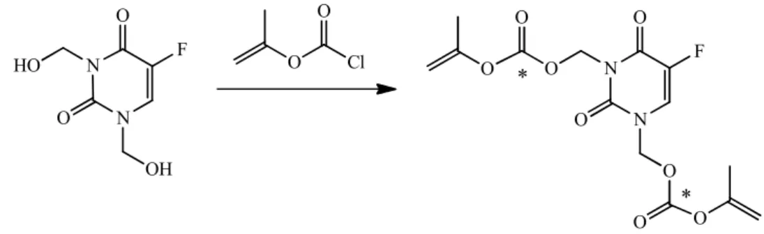
4. Synth se de microsph res d hydrogel hydrolysables charg es en principe actif 4.1. Chargement des microsphères par une méthode de gonflement 4.2. Chargement des microsphères par copolymérisationd une prodrogue
d un agent anti-inflammatoire Bibliographie
Chapitre 2 - Développement de méthodes de caractérisation de la dégradation des microsphères hydrolysables
Introduction
Projet d article 3 « Rheology as a tool for characterisation of mechanical properties of chemically crosslinked microspheres and for monitoring their degradation »
Abstract Introduction
Materials and methods Results and discussions Conclusions
Acknowledgement References
Chapitre 3 - Caractérisation de la dégradation des microsphères constituées d un hydrogel hydrolysable
Introduction
104 117 117 118 121 122 122 125 130 131 135
141
144
145 148 149 150 152 157 168 169 169
173
174
Introduction
Materials and methods Results
Discussions Conclusions Acknowledgement References
178 179 186 196 200 201 201
DISCUSSION GENERALE
1. Synth se des microsph res d hydrogel
2. Synthèse de microsphères pour embolisation chargées en principes actifs 3. Caractérisation des microsphères
3.1. D veloppement d une m thode de mesure des propri t s de résistance mécanique des microsphères basée sur un test de compression
3.2. D veloppement d une m thode de mesure des propri t s
rhéologiques des microsphères et application à la caractérisation de leur dégradation
3.3. D veloppement d une m thode de mesure du gonflement des microsphères et application à la caractérisation de leur dégradation 3.4. Etude de la dégradation des microsphères
Bibliographie
205
208 211 212 213
216
218 219 223
CONCLUSIONS GENERALE ET PERSPECTIVES 224
AIBN Azobisisobutyronitrile
AP Ammonium Peroxodisulfate
DCC N,N'-dicyclohexylcarbodiimide
DMA N-diméthyle acrylamide
DMAP 4-diméthyle aminopyridine
DSP polymérisation en suspension directe
GMA méthacrylate de glycidyle
HEMA (2-hydroxyéthyle) méthacrylate ISP polymérisation en suspension inverse
MBA N,N'-Méthylènebisacrylamide
MS microsphères
PEG1500 poly(éthylène glycol) avec la masse moyenne de 1500 g/mol PEGDMA poly(éthylène glycol diméthacrylate)
PEGMA Poly(éthylène glycol) méthacrylate
PEGMMA Poly(ethylene glycol methyl éther methacrylate) PLGA Poly(acide lactique-co-glycolique)
PVA poly(vinyl alcohol)
RMN Résonance magnétique nucléaire
Rpm Tour par minute
Sn(Oct)2 octoate d'étain Span 80® Sorbitan oleate
TEA triéthyleamine
TEMED N, N, N', N'-tetramethylethylenediamine TEG tétra(éthylène glycol)
Tris N-acryloyle tris(hydroxyméthyle) aminométhane
2005 à une réflexion associant le Département de Neuroradiologie Interventionnelle de l H pital Lariboisi re APHP (Dr. A. Laurent) et le Laboratoire des biomat riaux de l UMR CNRS 8612 et Paris XI (Prof. D. Labarre, Dr. L. Moine).
La solution technique imaginée alors pour répondre au besoin médical a été la synthèse de microsphères par polymérisation en suspension de macromonomères hydrolysables base de PLGA en pr sence d un comonom re de PEG.
Courant 2006-2007, des travaux de faisabilité ont montré la possibilité de synthétiser de telles molécules et de les introduire dans un procédé de polymérisation en suspension. Cela a abouti à une collaboration de trois ans (d but e le 15 octobre 2007) entre l UMR CNRS 8612 et la soci t Occlugel, sous forme d une th se CIFRE r alis e par Melle Nguyen sous la responsabilité du Pr. D. Labarre, puis, après son départ en retraite, sous celle du Dr. C.
Vauthier. Le Dr. L. Moine, collaboratrice proche du Pr. D. Labarre et spécialiste des polymères résorbables et des procédés de polymérisation en suspension, a étroitement collabor aux diff rents travaux men s jusqu fin 2009.
---
Les différents proc d s et applications ont fait l objet du d p t le 10 septembre 2009: d une demande de brevet provisoire am ricaine d pos e l Office am ricain des brevets (USPTO) sous le n° 61/241 183 et intitulée « Implantable bio-resorbable polymer » et
d une demande de brevet européen EP 0930 5830 intitulée « Implantable bio- resorbable polymer », qui a été étendue par voie de PCT en septembre 2010. Son contenu a été porté à la connaissance de la communauté via publication en date du 16 mars 2011.
---
- Monsieur Alexandre LAURENT, MCU-PH, Service de Neuroradiologie de lh pital Lariboisière, AP-HP,
- Monsieur Denis LABARRE, PU, Laboratoire Biomatériaux et Polymères, UPS 11, - Madame Laurence MOINE, CNRS,
- Monsieur Laurent BEDOUET, OCCLUGEL,
- Monsieur Michel WASSEF, MCU-PH, Service de Cytologie et Anatomie Pathologique de l h pital Lariboisi re, AP-HP,
- Mademoiselle Van Nga NGUYEN, OCCLUGEL.
---
L AP-HP, le CNRS, l UPS 11, l UPD 7 et OCCLUGEL sont copropriétaires du Brevet, de certaines inventions et découvertes relatives à des polymères gonflants et résorbables, chargeables pour implantation humaine ou animale.
---
Les quatre articles inclus dans le présent rapport de thèse sont à l'état de projet et n'ont pas encore été validés pour publication par la société OCCLUGEL.
tumeurs hypervascularisés, des hémoragies ainsi que des malformations artériovéneuses [Hovsepian et al. 2004, Syed et al. 2007, Artinyan et al. 2008]. Le but de l intervention est d occlure de mani re s lective les vaisseaux sanguins qui constituent ou qui nourrissent une lésion, ou de boucher une lésion portée par un ou plusieurs vaisseaux.
L acte chirurgical est r alis par un radiologue. L agent embolisant est conduit jusqu l endroit o il doit tre inject l aide d un cath ter sanscompromettre le flot sanguin dans le reste de la circulation. Lem decin observe en temps r el l acheminement du cath ter dans le réseau vasculaire sous rayon X et l aide d un agent de contraste pour l amener jusqu la lésion à traiter. L intervention est dite mini-invasive car elle ne n cessite qu une ponction fémorale, évitant une ouverture importante comme dans les techniques chirurgicales classiques. Elle peut être réalisée sous une simple anesthésie locale et nécessite généralement une courte dur e d hospitalisation. Concernant l agent d embolisation, une d finition en a t proposée par le Docteur Laurent dans un texte publié en 2006. Cette définition est reprise dans la citation suivante « On peut d finir comme agent d embolisation vasculaire tout produit sous une forme quelconque (solide/liquide ) qui peut être largué dans un flux sanguin pour se bloquer dans le vaisseau où il détermine une occlusion temporaire ou durable » [Laurent 2006]. Dans la pratique, les agents d embolisation se pr sentent sous forme de particules de forme et de taille plus ou moins calibrées, de spires métalliques (coils), de solutions gélifiante ou de microsphères [http://www.brainaneurysm.com/aneurysm- treatment.html, http://pennstatehershey.org/web/stroke/patientcare/services/onyx, http://www.ajronline.org/content/178/1/135.full, www.biospheremed.com].
Parmi les particules d embolisation, les microsphères calibrées constituent une classe d agent d embolisation vasculaire particuli rement bien adapt e pour cette application. Leur
particules pour le ciblage de l occlusion.En effet, par le choix de leur taille, on peut prévoir le niveau de p n tration de l embole dans un tissu tumoral par exemple. Cet avantage peut être crucial dans le cas où la microsphère porte une charge de médicament anticancéreux destinée à une délivrance locale intra ou péri-tumorale.
En fonction de l application th rapeutique, il peut tre souhait que l embolisation soit permanente ou temporaire.Quelque soit la qualit de l embolisation recherch e, les matériaux dégradables constituant les emboles s avèrent plus adaptées [Schwarz et al. 2004; Zhang and Schwarz 2006; Moine et al. 2011; Weng et al. 2011]. Ces matériaux sont également intéressantpour d autres applicationsqui nécessitent une implantation in vivo, comme pour la réparation et régénération tissulaire [Alsberg et al. 2003; Tan and Marra 2010], l encapsulation de cellules [Temenoff et al. 2004; Nicodemus and Bryant 2008] ou la formulation de systèmes à libération contrôlée de principe actif [Park et al. 1993; Hatefi and Amsden 2002; Miyata et al. 2002; Hamidi et al. 2008].
Dans beaucoup de cas cliniques comme par exemple une hémorragie, des tumeurs bénignes (fibrome utérin) ou maligne (hépatocarcinome), un agent d embolisation TEMPORAIRE pourrait être plus bénéfique sur le plan thérapeutique tout en apportant plus de confort aux patients. En effet, l utilisation d un agent d embolisation non dégradable pourra entraîner des effets indésirables avec un risque d inflammation importante. En outre, dans le cas du traitement par embolisation des tumeurs ischiémo-sensibles comme le fibrome ut rin, il est inutile de prolonger l occlusion vasculaire au-delà du temps nécessaire pour obtenir la nécrose de la tumeur. Une recanalisation rapidene peut que profiter l organe pour l aider reprendre un fonctionnement physiologique normal et liminer les tumeurs n cros es. Dans d autres cas cliniques comme celui des tumeurs malignes hypervascularisées
l occlusion vasculaire à la d livrance d un principe actif charg dans les microsphères. Cette technique, connue sous le nom de chimio-embolisation, permet de concentrer de manière considérable la charge médicamenteuse au niveau de la tumeur et donc de diminuer la concentration systémique et en même temps les effets indésirables. Au-delà du ciblage permettant la délivrance du principe actif à la tumeur, il est souhaitable d obtenir une recanalisation des vaisseaux nourriciers pour éviter la destruction totale de l organeatteint qui comporte encore du tissu sein et fonctionnel et pour permettrel application denouvelles cures de chimio-embolisationou d autres th rapiespour obtenir la destruction totale de la tumeur.
Actuellement, seuls deux produits commerciaux sont proposés pour réaliser des embolisations temporaires. Ce sont des éponges de gélatines (Gelfoam® de Pfizer, Spongel® de Sanofi-Aventis) et des microsphères à base d amidon (Spherex® de Pharmacia, Embocept® de PharmaCept). L agent biodégradable le plus utilisé est l ponge h mostatique de gélatine. Ce produit, de faible coût, est commercialisé sous forme de plaques ou réglettes (destinées à être découpées, broyées ou grattées) ou bien sous forme de particules sèches tamisés. Malgré le tamisage,aucune calibration n est possibledue à leur forme irrégulière et à la tendance des particules s agglom rer. Les agrégats ainsi formés peuvent parfois conduire au blocage du cathéter. De plus, les éponges de gélatines seules ne produisent qu une occlusion partielle [Laurent 2006] : l embolisation totale n est atteintequ apr s l activation de la coagulation et la formation d un caillot sanguin en compl ment du matériau d embolisation. La d gradation de l embole se produit suite la r action inflammatoire à corps étranger. Il est tr s difficile d avoir des informations pr cises concernant le temps de recanalisation d la variabilit de la compacit de l ponge, de l ampleur de la r action inflammatoire et de l activit enzymatique du patient [Laurent 2006]. Les microsphères biodégradables calibrées seraient une réponse aux limites présentées par ces matériaux
complète et mieux ciblée avec un bien meilleur contrôle du temps de dégradation. Des microsphères biodégradables à base d amidon ont d j fait l objet de recherches pour des applications dans le domaine de l embolisation[Hilger et al. 2008]. Ces microsphères ne sont disponibles que dans une gammede taille d un diam tre inf rieur 100 m cequi compromet leur application pour réaliser des embolisations nécessitant une occlusion des vaisseaux plus grands. Ces particules se dégradent très rapidement lorsqu elles sont isol es: elles disparaissent en quelques dizaines de minutes [Pohlen et al. 2000], ce qui est généralement trop court pour réaliser une embolisation. A l inverse, du fait de l accumulation d agrégats de plusieurs dizaines voire centaines de microsphères, La résorption est alors fortement ralentie (en fonction de la taille des amas) et il devient excessivement difficile de contrôler le temps de dégradation del embole qui s est r ellement form in situ. Il faut également souligner que la dégradation enzymatique de l amidon dépend aussi fortement du site d implantation et des conditions physiologiques du patient. L ensemble de ces consid rations montre clairement que la dégradation des emboles produits par ces microsph res n est pas bien ma tris e. A ce problème s ajoute la difficult d obtenir un véritable ciblage de l occlusion du fait de la formation d agrégats.
Actuellement, il apparait que le d veloppement des techniques d embolisation temporaire est consid rablement frein par le manque de mat riaux d embolisation adapt leur mise en uvre. L exp rience acquise des techniques d embolisation men e avec des matériaux non dégradables montre très clairement que la forme idéale des emboles est une forme sphérique. Pour lever la limite au d veloppement de l embolisation temporaire ilparait nécessaire de développer des emboles sphériques de taille calibrée dans un matériau dont la vitesse de dégradation est parfaitement maîtrisable. Les travaux réalisés sur des microsphères biodégradables d amidon ont mis en vidence une limite l utilisation de matériaux dont la
taille de microsphère trop petite amenait des difficultés pour contrôler le ciblage de la localisation de l embolisation. L objectif de notre travail de thèse a été de rechercher un mat riau dont la d gradation ne d pendrait pas d une activit enzymatique et qui pourrait tre transformé en microsphères résorbables de grande taille pour embolisation. Notre recherche de matériaux dégradables implantables in vivo s est tourn e vers les hydrogels. Les r sultats de l tude bibliographique pr sent s dans la première partie du mémoire de thèse a permis de sélectionner des types d hydrogels hydrolysables répondants a priori aux critères recherchés.
Le travail expérimental présenté dans la deuxième partie du mémoire de thèse rapporte les résultats des études ayant portées sur la synthèse des microsphères, sur le développement d une m thode originale pour suivre leur d gradation in vitro et sur l tude de leur dégradation.
Bibliographie
Alsberg E., H. J. Kong, et al. (2003). "Regulating Bone Formation via Controlled Scaffold Degradation." Journal of Dental Research 82(11): 903-908.
Artinyan A, Nelson R, Soriano P, Chung V, Retseck J, Reynolds J, Marx H, Kim J and Wagman L. (2008). Treatment response to transcatheter arterial embolization and chemoembolization in primary and metastatic tumors of the liver. HPB (Oxford). 10 (6):
396 404.
Hamidi M., A. Azadi, et al. (2008). "Hydrogel nanoparticles in drug delivery." Advanced Drug Delivery Reviews 60(15): 1638-1649.
Hatefi A. and B. Amsden (2002). "Biodegradable injectable in situ forming drug delivery systems." Journal of Controlled Release 80(1-3): 9-28.
Hilger R. A., Richly H., Grubert M., Libicher M., Strumberg D., Ebert J., Hecker D., Zähringer M. (2008). « Pharmacokinetics of intra-arterial applied liposomal encapsulated doxorubicin in liver metastases or hepatocellular carcinoma: A phase I study. » J Clin Oncol 26: 13523.
Omary RA, Pelage JP, Rajan D, Schwartzberg MS, Towbin RB, Walker WJ, Sacks D. (2004).
Quality improvement guidelines for uterine artery embolization for symptomatic leiomyomata. J Vasc Interv Radiol. 15(6): 535-41.
Laurent A. (2006). "Radio diagnostic - Principes et techniques d imagerie - Agents d embolisation." EMC (Elsevier Masson SAS, Paris); 35-140-A-20: 1-10.
Miyata, T., T. Uragami, et al. (2002). "Biomolecule-sensitive hydrogels." Advanced Drug Delivery Reviews 54(1): 79-98.
Moine L., L. Bedouet, et al. (2011). "IMPLANTABLE BIO-RESORBABLE POLYMER "
WO/2011/029867.
Nicodemus G. D. and S. J. Bryant (2008). "Cell Encapsulation in Biodegradable Hydrogels for Tissue Engineering Applications." Tissue Engineering Part B: Reviews 14(2): 149-165.
Park, K., W. S. W. Shalaby, et al. (1993). Biodegradable hydrogels for drug delivery.
Lancaster, PA, Technomic Pub.
Pohlen U., Berger G., Binnenhei M., Reszka R. and Buhr H. J. (2000). Increased Carboplatin Concentration in Liver Tumors through Temporary Flow Retardation with Starch Microspheres (Spherex) and Gelatin Powder (Gelfoam): An Experimental Study in Liver Tumor-Bearing Rabbits. Journal of Surgical Research 92: 165 170.
Schwarz, A., H. Zhang, et al. (2004). "Transcatheter embolization using degradable crosslinked hydrogels." Biomaterials 25(21): 5209-5215.
Syed MI, Chaudhry N, Shaikh A, Morar K, Mukerjee K and Damallie E. (2007). Catheter- directed middle hemorrhoidal artery embolization for life-threatening rectal bleeding. Can J Gastroenterol. 21(2): 117 123.
Tan, H. and K. G. Marra (2010). "Injectable, Biodegradable Hydrogels for Tissue Engineering Applications." Materials 3(3): 1746-1767.
Temenoff, J. S., H. Park, et al. (2004). "In vitro osteogenic differentiation of marrow stromal cells encapsulated in biodegradable hydrogels." Journal of Biomedical Materials Research Part A 70A(2): 235-244.
Weng, L., H. C. Le, et al. (2011). "Doxorubicin loading and eluting characteristics of bioresorbable hydrogel microspheres: In vitro study." International Journal of Pharmaceutics In Press, Corrected Proof.
Zhang, H. and A. Schwarz (2006). Degradable crosslinkers and degradable crosslinked hydrogels comprising them. U. S. Pat. 7135593 B2.
BIODEGRADABLE CROSSLINKERS FOR HYDROGELS SYNTHESIS IN BIOMEDICAL APPLICATIONS
V.N.Nguyen1,2, C. Vauthier1,3* L. Moine1,3and D. Labarre1,3
1. Univ Paris Sud, Physico-chimie, Pharmacotechnie and Biopharmacie, UMR 8612, Chatenay-Malabry, F 92296
2. Occlugel SARL, Paris BioTech, 24 Rue du Faubourg Saint-Jacques, Paris, F-75014 3. CNRS, Chatenay-Malabry, F-92296
Corresponding author:
Christine Vauthier
Physico-chimie, Pharmacotechnie and Biopharmacie UMR CNRS 8612
University Paris Sud, Faculté de Pharmacie 92296 CHATENAY-MALABRY Cedex France Fax : 33 1 46 83 53 12
E-mail: christine.vauthier@u-psud.fr
Abstract
Hydrogels have received increasing attentions for a wide range of biomedical applications including drug delivery and tissue engineering. Progresses in synthetic chemistry make biodegradable hydrogels a very dynamic topic with development potential of new materials resolving drawbacks found with non-degradable gels and improving comfort of patients by developing new techniques of therapeutics. They are versatile materials for which properties can be tuned by the chemistry to fit requirements identified for the desired application. In this review, we describe biodegradable hydrogels and present their potential use in biomedical applications. The review was focussed on synthetic hydrogels including hydrolysable crosslinks.
Key words: hydrogels, biodegradable crosslink, hydrolysis, biomedical applications
Contents:
1. INTRODUCTION
2. General methods for the synthesis of covalently crosslinked hydrogels 3. Chemical nature of hydrolysable covalently crosslinked hydrogels
3.1. Chemical nature of hydrolysable materials
3.2. Design of crosslinkers with hydrolysable segments
4. Formulations and applications of hydrolysable covalently crosslinked hydrogels in the biomedical field
5. CONCLUSIONS 6. References
1. Introduction
Hydrogels are three dimensional polymer networks which absorb and retain large amounts of water. They are universally found in Mother Nature as material of matrices of the living organisms including for instance the interstitial tissue and the cell cytoplasm.
Synthetic hydrogels were first introduced as biomaterial in 1960 by Wichterle and Lim (Wichterle and Lim 1960) as soft contact lenses. Since then, they continue to raise interests among biomaterial scientists because of their high potential of biocompatibility. Indeed, they are a class of materials with the most similar structure to the materials found in the nature.
The three dimensional polymer network which composes hydrogels swells in aqueous medium. It absorbs and retains large amount of water thanks to the high content in hydrophilic groups in the polymer structure (Ratner and Hoffman 1976, Peppas et al. 2000, Kopecek 2002). Thus, hydrogels were extensively studied as cell encapsulation matrices, drug delivery systems, biosensors and as matrix for tissue regeneration (Baldwin and Saltzman 1998, Rosiak and Yoshii 1999, Lee and Mooney 2001, Peppas et al. 2006, Ong et al. 2008, Caldorera-Moore and Peppas 2009). Non degradable hydrogels are, by far, the most abundant types of synthesized hydrogels. They are suitable for many applications such as encapsulation of bulky compounds including proteins and RNA (Monteiro et al. 2004; Leach and Schmidt 2005). However, degradable hydrogels have appeared to be more suitable in applications in which the material is directly implanted in the body and needs to be removed when its action is completed. Such applications include scaffolds for tissue regeneration (Alsberg et al. 2003;
Tan and Marra 2010), cell encapsulation (Temenoff et al. 2004; Nicodemus and Bryant 2008), drug delivery systems with controlled released properties (Park et al. 1993; Hatefi and Amsden 2002; Miyata et al. 2002; Hamidi et al. 2008) and materials for temporary embolisation (Schwarz et al. 2004; Zhang and Schwarz 2006; Weng et al. 2010; Moine et al.
2011; Weng et al. 2011). The degradation time scale of the hydrogel materials used in these applications needs to be very precisely adjusted. This is a property of hydrogels which can be finely tuned by adapting various parameters of synthetic hydrogels. For instance, degradation rate can be controlled by the physical properties of the crosslinkers (e.g. length, rigidity), the chemical nature of the polymer composing the hydrogel and by the characteristics of the tridimensional polymer network, which establish the permeability of water inside the gel (West and Hubbell 1995). Moreover, the morphology of the hydrogels including their porosity, their size and their shape (slide, block, spherical particles, thread) influencing the surface/weight relation and the surface to volume ratio has shown to influence also the degradation rate especially when degradation occurs by hydrolysis (Wu and Ding 2004).
Although all types of hydrogels have a general structure based on a three dimensional polymer network swelled by a large amount of water, the polymer chains forming the network are hold together either by physical interactions including secondary forces such as ionic, H- bonding or hydrophobic forces or by covalent bonds in the case of covalently-crosslinked polymer networks. Hydrogels formed from physical interactions are called physical or reversible hydrogels. One advantage of these gels is that they can be formed without the need of chemical modification or chemical reaction. However, they can be dissolved by simple changes occurring in the physico-chemical conditions of the environment (Xu and Kope ek 2007; He et al. 2008). In comparison, covalently crosslinked hydrogels or chemical hydrogels would be more suitable for applications requiring materials with perfectly controllable degradation rates over time. Indeed, in theory, the degradation rate of these hydrogel can be finely tuned by adjusting many of the parameters characterizing the hydrogels. For instance, the chemical nature of the degradable segment can be used to define the mechanism by which the hydrogel will be degraded. Sedla k and collaborators have designed a hydrogel based on crosslinked synthetic poly( -amino acid)s (Sedla ket al. 2011)
which degrades under the action of enzymes while the PLA-PEG-PLA crosslinked hydrogels proposed by Sawhney and collaborators are simply hydrolysed by water (Sawhney et al.
1993). Hydrogels composed of polyphosphoester can be degraded through both hydrolytic and enzymatic mechanisms (Iwasaki et al. 2004; Wang et al. 2005). As illustrated in these few examples, the nature of the degradable segment is another parameter which influences hydrogel degradation. This degradation depends on the enzyme activity and/or on the susceptibility to hydrolysis. The degree of crosslinking is another important parameter which influences the degradation rate (Kweon et al. 2003, Timmer et al. 2003 a,b, Bencherif et al, 2008, 2009, Wang et al. 2010). It controls the swelling of the hydrogel and the mesh size of the pores which influences the amount of water penetrating the hydrogel. The hydrophilicity of the hydrogel may also modulate the capacity of water absorption hence influence the degradation rate particularly in the case of hydrolysable hydrogels (Sawhney et al. 1993, Poon et al. 2009, Wang et al. 2010). Regarding applications that request a very high degree of degradation rate control, it seems that covalently crosslinked hydrogels degrading through a hydrolytic mechanism are the most suitable. Indeed, their degradation rate will only depend on the chemical structure and characteristics of the hydrogel and will be totally independent on the site of implantation in the body and on the physiology of the patient (Katti et al. 2002).
Their degradation rate can theoretically be adjusted only from the manner the hydrogel was chemically designed and synthesized.
The present reviews aimed to summarize the different types of hydrolysable covalently crosslinked hydrogels developed so far. It first presents general methods that can be used to synthesise hydrogels. Then, it describes the chemical nature of the materials used to produce hydrolysable covalently crosslinked hydrogels. In this part, the different types of hydrolysable chemical compounds and the way they can be used to synthesize crosslinkers will be
described. The different forms that hydrogels can take and their applications in the biomedical field are considered in the last part of the paper.
Abbreviations and Commercial products
4EDMAB ethyl-4-N,N-dimethylaminobenzoate
AIBN aso-bis isobutyronitrile
AP ammonium persulfate
BPO benzoyl peroxide
CQ camphorquinone
Darocur 2959 2-hydroxy-1-(4-(2-hydroxyethoxy)phenyl)-2-methyl-1-propanone
DEAP diethoxyacetophenone
DMAEMA di(methylamino ethyl methacrylate)
DMF Dimethylformamide
DMPA 2,2-dimethoxy-2-phenylacetophenone
DMSO Dimethyl sulfoxide
DMT N,N-dimethyl-p-toluidine
EGDMA ethylene glycol dimethacrylate
FAME fumaric acid monoethyl ester
HEMA hydroxyethyl methacrylate (
Irgacure 184 1-hydroxycyclohexyl phenyl ketone
Irgacure 819 phenyl bis(2,4,6-trimethyl benzoyl) phosphine oxide (BAPO) Irgacure 2959 ((4-(2-hydroxyethoxy)phenyl-(2-propyl)ketone
MACAH methacryloyl hexane-1,6-diol cholanoate
MMA Methyl methacrylate
NIPAM N-isopropylacrylamide
NVP N-vinylpyrolidone
OCMCS O-carboxymethylchitosan
PCL Poly( -caprolactame)
PEG poly(ethylene glycol)
PEGMMA Poly(ethylene glycol) methacrylate
PGA Poly(glycolic acid)
PLA Poly(lactic acid)
PMA poly(malic acid)
PPF poly(propylene fumarate)
TED N,N,N ,N-tetraethyl thiuram disulfide TEMED N,N,N ,N-Tetramethylethylene diamine
TMC trimethylene carbonate
TMPTA trimethylolpropanetriacrylate
2.General methods for the synthesis of covalently crosslinked hydrogels
Different methods can be used to synthesize polymers with three-dimensional network architecture by covalent linkage. This depends whether it is formed from monomer units or from the reticulation of preformed linear chains of polymer (Fig. 1).
In all cases, the nature of the building blocks and links created during assembly are keys to determine the biodegradable character of the resulting hydrogel. Building blocs are assembled together thanks to the presence of chemical functions that allow formation of a three- dimensional polymer network. Two families of chemical functions can be distinguished whether they contain unsaturated carbon-carbon bonds or they include in their structure chemical reactive functions consisting in alcohol, aldehyde, carboxylic acid, amine, hydrazide or thiol groups able to react with each other (Table I).
In the first family, the linkage between building blocs can occur by a simple reaction of polymerization resulting from the opening of the carbon-carbon double bond included in the molecule (Fig. 1C). Such reactions can be initiated by the formation of a radical that allows carbon-carbon double bonds to add to each other based on a chain type reaction (Fig. 2).
Many of the hydrogels developed for biomedical applications are synthesized by radical polymerizations which can be performed under mild conditions. Polymerization initiations is generally based on thermal, red-ox and photochemical methods. The obtaining of polymers with three dimensional network architecture requires that one building bloc named the crosslinking agent contains at least two carbon-carbon double bonds in their structure. Thus, preformed polymers containing a few carbon-carbon double bonds in their structure can be crosslinked by initiating a radical polymerization (Fig. 1B).
A
B
C
D
Figure 1. Scheme of the synthesis of polymer networks from monomer units (A, B) and from a
blend of preformed functionnalized polymer chains with crosslinking agents (C, D). A illustrates the formation of the polymer network from monomers containing unsaturated functions. Mono-unsaturated monomers are included in the linear parts while di-unsaturated monomers form the connections between chains. B illustrates the formation of the polymer network from monomer units containing complementary functions able to react together.
Difunctional monomers are included in the linear segments of the polymer network while tri and tetrafunctional monomers form the connection between chains. C illustrates the formation of polymer network from a blend of polymer chains functionalized with unsaturated carbon carbon bonds. D illustrates formation of polymer network from a blend of polymer chains functionalized with chemical functions complementary to that of the difunctional monomers added as crosslinking agent.
Table I Chemical reactions of functional groups included on buiding blocks
R1 OH O
R2 OH
R1 O O
R2 R1 Cl
O
+ or
R2 NH2
R1 N H O
R2
+
R1 O R1
O O
or
carboxylic acid
acyl chloride
acid anhydride
alcohol
amine
R1 H O
aldehyde
R2 OH
R1 O OH
R2
+
R2 NH2
R1 H NR2
+
alcohol
amine
ester
amide
hemiacetal
R2 N H O
R2
+
hydrazine
R1 H N
HN R2 O imine
hydrazone
R1 OH
+ alcohol
N C O R2
isocyanate R
1
O H
N R2 O
urethane
R1 SH + HS R2
disulf ide thiol
R1 S
S R2 CH2
R2 n
unsaturated C=C bonds radical polymerization
R1 R1 R2 R1 R2 R1 R2 R1 R2
A
A T or UV or Oxydo-reduction reaction
C C Initiation
Propagation
Termination
A
C C
(n times)
A C C
A C C A C C C C
n
A C C C C
n
C C C C A
m
A C C A
n+m+2 A C C C C
n
C C H C
chain transfert
A C C H
n+1
C C C
+
Figure 2. Scheme of the mechanism of radical polymerization.
The swelling characteristics of the hydrogels and their properties will depend on the length of the linear segment separating two connections between chains. In case hydrogels are synthesized from monomers, the polymerization is performed with two types of monomers.
One is mono-unsaturated and will be included in linear segments of the polymer network. A di- or pluri-unsaturated monomer is needed as crosslinking agent to form connections between chains making possible the obtaining of a three-dimensional polymer network (Fig.
1A). The composition in mono-unsaturated and di- or pluri-unsaturated monomers define the degree of reticulation of the polymer network and in turn the swelling properties and the mesh size of the hydrogel (Sawhney et al. 1993; Zhu et al. 2005). It is noteworthy that the first hydrogel synthesized for medical application was prepared according to this scheme by
radical copolymerization of HEMA as the mono-unsaturated monomer with EGDMA as a di- unsaturated monomer (Wichterle and Lim 1960). Although, this hydrogel is not biodegradable because of the nature of the monomers, degradable hydrogels can be obtained by introducing degradable segments in the structure of the building blocks as it will be further discussed in the following parts of this review (Tables II - VII). Many other properties can be incorporated in hydrogels by varying the nature of the building blocks. For instance, building blocks with pH and temperature sensitive properties were incorporated in the structure of hydrogels in order to design stimuli responsive devices (Bettini Colombo et al. 1995, Ciçek and Tuncel 1998). A polymerizable prodrug with a cleavable bond is another example of an interesting building block that we could incorporate in the hydrogel structure in order to design drug delivery systems in which the drug release is controlled by the hydrogel degradation rate (Table VI) (Liu and Rimmer 2002).
In the second family, the building blocs are chosen between chemicals containing functions which can react together and form of a covalent linkage between the parent molecules (Table I). The nature of the covalent bonds which crosslink the parent molecules depends on the nature of the chemical functions reacting together to form the polymer network. In turn, the nature of the linkage defines whether or not the hydrogel is degradable and the mechanism of degradation. Beside chemical reactions, hydrogels can be obtained using biochemical methods in which the crosslinking reaction is catalysed by enzymes. For instance, Sperinde and Griffith used transglutaminase to form peptide based hydrogels (Sperinde and Griffith 2000). It is noteworthy that radical polymerization of C=C double bond containing monomers and macromers is so far the most commonly used method to produce hydrogels for biomedical applications, since the design of suitable monomers is versatile and hydrogel can be formed in various conditions including in vivo in situ if needed.
the hydrogel polymerization Poly( -hydroxyacid)-PEG1000-
20000- Poly( -hydroxyacid) diacrylate
Poly( -hydroxyacid) were composed of PLA and/or PGA
UV irradiation ( 365 nm) with DMPA.
Copolymerization with NVP.
Solution photopolymer- ization, performed in molds to form disks or on a surface to form films
Degradation study in HEPES buffer salin pH 7.3, 37°C.
Time of degradation vary from 1 to 120 days depending on PEG chain length and lactide and glycolide content
Drug delivery system for Macromolecules.
Healing material.
Shawhney et al. 1993
PLA-PEG-PLA diacrylate PLA: 6 or 10 lactides units PEG: 2 or 6 ethylene glycol units.
bulk photopolymerization initiated by visible light( 450 nm) with CQ and 4EDMAB in the presence of cells or in molds
disks (1 x 5 mm) and porous scaffolds obtained by polymeri- zation in molds.
Polymerization can be performed in the presence of cells.
Degradation study in PBS pH 7.4, 37°C Time of degradation (30 to 60% mass loss) in 8 weeks depending on PEG-PLA content
Orthopaedia as scaffolds for bone tissue regenerating applications.
Burdick et al.
2001
Poly( -hydroxyacid)-PEG6000- 10000- Poly( -hydroxyacid) diacrylate
Poly( -hydroxyacid) were PLA and PCL.
redox initiator system composed of AP and TEMED, 37°C
Solution polymeriza- tion in a bath.
Degradation study in PBS pH 7.4, 37°C.
Total degradation was observed in 35 days.
Scaffold for tissue engineering and tissue regeneration.
Zhu and Ding 2006
PLA-PEG4600-PLA diacrylate UV irradiation with Darocur 2959
Solution photopolymer- ization can be
performed in the presence of cells.
Up to 50 % degradation in 3 weeks Encapsulation of cells from Rat brain Evaluation of compatibility with cell cultures and cell growth
Scaffold for tissue engineering and tissue regeneration. Suitable to be used as support for neural cell growth.
Benoit et al.
2006,
Mahoney and Anseth 2007 PCL-PEG1000- PCL diacrylate UV irradiation ( 365 nm)
initiated with DMPA dissolved in TMPTA.
Bulk polymerization in silicone rubber
embossing molds.
Formation of micro- patterned hydrogel films.
No degradation study
Adhesion tests toFibroblasts in cell culture
cell shape control scaffold for tissue- engineering.
Chan-Park et al 2006,
Zhu et al. 2006
Poly( -hydroxyacid) diacrylate Poly( -hydroxyacid) were composed of PLA and PCL.
initiated with CQ and 4EDMAB.
zation in molds Polymerization can be performed in the presence of cells
About 10% mass loss after the first week.
After 8 weeks, the mass loss of the faster degrading hydrogels (poly(2EG6LA), poly(2EG10LA)) ranged from 50 to 60%
while the mass loss of the slowest
degrading hydrogel (poly(8EG6LA)) was lower than 30%.
Cell seeding experiment with primary rat calvarial osteoblasts
Evaluation of neurite outgrowth from retina explant
regeneration.
Implants for controlled drug delivery of neurotrophins to enhance regeneration of damage to the central nervous system
2003,
Burdick et al.
2006
PLA- PEG4600-PLA diacrylate UV irradiation ( 365 nm) initiated with Irgacure 2959.
Solution photopolymer- ization performed in molds
Compares the degradation of the macromer crosslinker in solution to that of the reticulated crosslinker : under the reticulated form, more sensitive
degradation to fluctuations in macromer cross-linker composition and to the environement (water content, pH, ionic strength)
Drug delivery systems and Tissue
engineering scaffold.
potential stimuli- responsive materials
Shah et al.
2006
PEG-PLA-PEG di- (meth)acrylate Biotin functionnalized hydrogels
UV irradiation ( 365 nm) initiated by Irgacure 184, Copolymerised with PEG- Biotin methacrylate
Solution photopolymer- ization performed in molds
-with crosslinkers containing the larger number of lactide units (4 and 6) total degradation required > 3 months.
- Degradation incomplete with the crosslinker containing the lower number of lactide units (2) in the structure.
- Cell attachment test with preosteoblast human palatal mesenchymal cells
- Drug delivery system,
- Biodegradable composite formulation - Scaffold for tissue engineering
Clapper et al.
2007
Clapper et al.
2008
PGA- PEG4000-PGA diacrylate, dimethacrylate and di-urethane methacrylate.
UV irradiation ( 365 nm) initiated by Irgacure-2959.
Solution photopolymer- ization performed in molds to prepare disk
Degradation between 1 to 27 days.
Tuning achieved by controlling
crosslinker chemistry which in turn make
Matrice for control release of drugs;
Degradable scaffolds
Bencherif et al.
2009a,b
co methacryloyl hexane UV irradiation by DMPA.
MACAH as copolymer.
polymerization Study of the morphology and swelling of the hydrogel.
Poly( -hydroxyacid)-PEO- PPO-PEO- Poly( -
hydroxyacid) diacrylate Poly( -hydroxyacid) were composed of PLA and PCL.
Inverse suspension redox polymerization initiated by AP and TEMED.
Inverse suspension polymerization to prepare microspheres (~ 200 µm).
No complete degradation was reached.
Microspheres prepared with PLA
containing crosslinker degrade faster (90- 95% degradation after 56 days) than PCL containing ones (80% degradation after 56 days).
Thermo responsive and degradable
hydrogel microspheres for controlled release of drug.
Zhu et al. 2005
PCL-PEO-PPO-PEO-PCL glycidyl methacrylate
UV irradiation initiated by DMPA
Copolymerised with MMA, PEGMMA
Solution polymerization.
Degradation was monitored over 50 days.
Weight loss varied from 25 to 50% over the 50 day period of time depending on pH of the incubation medium,
composition of the crosslinker and degree of reticulation
Controlled release drug delivery system
Wang et al.
2010
PLGA-glycerol-PLGA diacrylate (star shaped)
initiated by AIBN under heating.
Copolymerisation with N- vinyl pyrrolidone
Solution polymeriza- tion performed in molds to obtain films
no total degradation after 7 weeks incubation in PBS pH 7.4 at 37°C.
Degradation proceeded in two steps: A slow steps occurring up to a mass loss of 25 to 30% over 5 to 7 weeks followed by a faster degradation reaching a maximum of 50% mass loss after 6 weeks for the samples which degraded the faster.
Degradation rate depended on the composition and molecular weight of the crosslinking agent
Tissue engineering scaffolds
Controlled release drug delivery system
Jiao et al. 2006
star-shaped polymeric precursors functionalized with FAME.
The precursor polymers, was based on D,L lactide,
UV irradiation ( 365 nm) with DMPA.
Bulk polymerization in molds to form films.
No degradation study
Characterization of the morphology and of the thermal and physical properties of the networks.
Scaffold for engineering of cardiovascular tissues
Grijpma et al.
2005
hydrogel polymerization PCL diacrylate UV irradiation ( 360 nm)
with DMPA.
Copolymerized with PCL macromer.
Solution polymeriza- tion, obtaining form as the reaction vessel.
Rate of weight loss depended on the rate of reticulation of the hydrogel but no more than 10% mass loss after 42 days in PBS (pH 7.4, 37°C).
Cell proliferation test considering MG-63 osteoblasts
Scaffold for tissue engineering (bone reparing support material)
Kweon et al.
2003
PCL diacrylate intiated by AP and TEMED.
Copolymerisation with NIPAM and methacrylic acid.
Solution polymeriza- tion in DMF, performed in molds to provide with thick films
Total mass loss after 29 hours incubation at pH 12, 25°C when the hydrogel was soaked in acidified water (pH 2) prior to be dried and prior to the start of the experiment.
pH responsive
controlled release drug delivery systems
París and Quijada- Garrido 2009
PCL diacrylate UV irradiation ( 365 nm) with DEAP or initiated by AIBN under heating.
Copolymerised with NIPAM.
Solution polymeriza- tion in DMSO, performed in molds to obtain thick films.
Degradation in PBS pH 7.4 (0.2M) at 37°C in presence of lipase. Less than 20%
mass loss after 50 days of incubation.
Porous gels degraded more rapidly than non porous gels
Thermo-responsive degradable hydrogel for controlled release of drug and scaffold for tissue engineering
Lee and Cheng 2008
PCL diacrylate UV irradiation with Irgacure 819.
Copolymerised with PPF.
Solution
polymerization in CH2Cl2, performed in molds.
No degradation study.
Cytotoxicity test with mouse MC3T3-E1 and rat SpL201 cells,
Cell adhesion test and cell proliferation tests.
Cell behaviour was found to depend on mechanical properties of the crosslinked matrices.
Scaffold for tissue engineering for bone and nerve regeneration
Cai and Wang 2010
hydrogel polymerization PMA-PEG1000-PMA
dimethacrylate
UV irradiation ( 365 nm) with Irgacure 2959.
Copolymerization with methacrylated OCMCS and PEG.
Amount of crosslinker used: 7 to 20% of polymers.
Solution polymeriza- tion performed in molds.
Degradation rate depended on the length of the PMA chain. 80% weight loss max after 50 days incubation in PBS at 37°C.
Evaluation of the compatibility with vascular smooth muscle cells:
PMA improved cellular biocompatibility of the hydrogel. Cell proliferation was further improved by adding Arg-Gly-Asp (RGD) peptides on the hydrogel surface.
Potential materials for tissue engineering applications.
Poon et al.
2009
HEMA (4 to 12 mol %) grafted on PMA .
UV irradiation ( 365 nm) with Irgacure 2959.
Solution polymeriza- tion in water, obtaining form as the reaction vessel.
Degradation in PBS pH 7.4 at 37°C.
Hydrogels were loosing shape after 75 to 80% weight loss (after 2 to 5 days).
Lower PMA content and lower amount of HEMA show shorter degradation times.
Degradation occurred by both bulk and surface mechanism.
Scaffold for engineering tissue.
He et al. 2006
hydrogel polymerization (PPF) diacrylate
Material with low water absoption capacity.
Redox polymerization initiated by BPO and DMT at 37°C.
Copolymerised with PPF.
Bulk polymerization achieved in molds to provide cylinder and film shaped.
Total dissolution of the polymer network occurred in nearly 4 days in NaOH 1M, 70-80°C.
biomaterial scaffolds for orthopedic tissue engineering
He et al. 2001
(PPF)diacrylate
Material with low water absoption capacity.
Polymerization initiated by visible irradiation ( 470 nm) with Irgacure 819 (or BAPO).
Copolymerised with PPF.
Bulk polymerization in molds of different shapes: rod, cylinder, dogbone shapes.
Preparation of
composite material by incorping -tricalcium phosphate (TCP) particles.
No total degradation in most formulations after 52 days incubation in PBS pH 7.5 and in citrate buffer (pH 5.0). Degradation occurred in bulk with minimal changes in mass and geometry prior to sudden fracture. Degradation rate was increased by reducing the crosslinking density and to a lower extend the pH.
Degradation is delayed in composite materials incorporating TCP.
Orthopedic implants Timmer et al.
2003a,b
poly(PEG fumarate) (OPF) modified with a bioactive peptide (GRGD)
UV irradiation with Irgacure 819 (or BAPO).
Copolymerised with PPF.
Solution polymeriza- tion in CH2Cl2in mold to make films of 0.2mm thick.
No degradation study. Functionalized
Scaffolds for tissue regeneration and engineering.
Jo et al. 2001, Dadsetan et al.
2007
PPF-co-PCL Initiated by BPO. Solution polymeriza-
tion in CH2Cl2.
No degradation study. Injectable in situ poly- merization material to be used in bone tissue regeneration.
Yan et al. 2011
PPF-PCL-PEG Thermal polymerization with AP at 70°C
Copolymerised with
acrylamide and ascorbic acid.
Bulk polymerization. Evaluation of fibroblast growth and adhesion.
Scaffold for tissue engineering
Krishna and Jayabalan 2009
oligo(PEG fumarate) (OPF) Copolymerized with acrylated PEG-dithiothreitol (Ac PEG- DTT)
Solution polymeriza- tion in PBS.
Degradable hydrogels
Degradation time can be tuned from a few days to >1 month by varying OPF and Ac PEG-DTT.
In situ tissue regeneration.
Proof of concept evaluated as carrier for marrow stroma cells (MSC), for tendon overuse injuries repair
Qiu et al. 2011
hydrogel polymerization Dicarbonate dimethacrylate UV photopolymerization,
DMPA.
Copolymerised with NVP or with DMAEMA.
Bulk polymerization, performed in molds
In vitro and in vivo degradation study.
Reproducible in vitro degradation.
Degradation can be tuned during the synthesis by the choice of comonomer and composition in the crosslinking reagent. In vivo, rods can be fragmented in less than 2 days after implantation in the eye. No polymer remained after 2 months at the implantation site.
Potential use as drug delivery system in the vitreous body of the eye. Copolymer with NVP showed a good compatibility for applications in oph- thalmology in contrast with copolymer with DMAEMA.
Bruining et al.
1999, 2000
5-Fluororacil dicarbonate dimethacrylate
Thermal polymerization with AIBN.
Copolymerised with NVP.
Polymerization in solution (1,4-dioxane) performed in molds
In vitro degradation study performed by incubation in PBS pH 7.4 over 40 days through the release of the drug and the swelling of the gel. Degradation profiles showed two phases, one rapid at the beginning followed by a much slower process.
Drug delivery system.
Drug release was controlled by the degradation of the device.
Liu and Rimmer 2002
Poly(hexa methylene
carbonate)-co-polyfumarates, Poly(hexa methylene
carbonate) diacrylate, Poly(hexa methylene carbonate)-co-plyethylene glycol-co-polyfumarates
Polymerization initiated with visible light in the blue region with camphorquinone (CQ).
Copolymerised with NVP.
Bulk polymerization in molds
In vitro biocompatibility essai evaluating the cytotoxicity on L-929 fibroblasts cells from mice using the MTT test: no adverse cytotoxicity was found. No degradation study was provided.
Scaffold material for cell delivery, tissue engineering and drug delivery applications
Sharifi et al.
2008
hydrogel polymerization Copolymers of sebacic acid
dimethacrylate with 1,6-bis(carboxyphenoxy) hexane dimethacrylate
UV light (365 nm) and DMPA; or visible light (470- 490 nm) and CQ and
4EDMAB.
Bulk polymerization in molds to provide disks
Entrapment of DNA protected in alginate beads during polymerization
Degradation in PBS/HEPES at 37 °C. Total mass loss can be tuned from 6 to 42 days by varying the composition in hydrophobic comonomer in the composition of the polymer.
Surface erosion mechanism and linear degradation throughout the study period.
Controlled released drug delivery system for genes (DNA must be protected to avoid damage during photoencapsulation
Quick et al.
2004
Copolymers of sebacic dimethacrylate,
Tricarballylic dimethacrylate and
Pyromel