Article
J. Braz. Chem. Soc., Vol. 26, No. 7, 1466-1474, 2015. Printed in Brazil - ©2015 Sociedade Brasileira de Química 0103 - 5053 $6.00+0.00
A
*e-mail: [email protected], [email protected]
Poly(vinyl alcohol)-hyaluronic Acid Membranes for Wound Dressing Applications:
Synthesis and
in vitro
Bio-Evaluations
Alaa Fahmy,a Elbadawy A. Kamoun,*,b Raafat El-Eisawy,a Esmail M. El-Fakharany,c
Tarek H. Taha,d Basem K. El-Damhougya and Farag Abdelhaia
aChemistry Department, Faculty of Science, Al-Azhar University, 11884 Cairo, Egypt
bPolymeric Materials Research Dep., Advanced Technology and New Materials Research Institute
(ATNMRI), City of Scientific Research & Technological Applications (SRTA-City), New Borg Al-Arab City 21934 Alexandria, Egypt
cTherapeutic and Protective Proteins Laboratory, Protein Research Department and dEnvironmental
Biotechnology Dep. Genetic Engineering and Biotechnology Research Institute, City of Scientific Research & Technological Applications (SRTA-City), New Borg Al-Arab 21394 Alexandria, Egypt
Physically crosslinked poly(vinyl alcohol)-hyaluronic acid (PVA-HA) hydrogel membranes composed of different amounts of HA were prepared by freeze-thawing (F-T) method. F-T cycle was repeated for three consecutive cycles. HA was chosen and routinely utilized in the local treatment of chronic wounds, because of its advantages as, HA is endogenous and biodegradable polymer. Physicochemical properties of PVA-HA membranes such as, gel fraction (GF), swelling, mechanical properties, hydrolytic degradation and in vitro bio-evaluation tests were investigated. Results revealed that introducing HA into PVA structure affected significantly the physicochemical properties of membranes than the pristine PVA, because of its crosslinking interaction with PVA. With the increase of HA content in PVA hydrogel membranes, GF and mechanical stability of PVA-HA membranes decreased. However, the swelling behavior, mechanical flexibility, protein adsorption and hydrolytic degradation of PVA membrane increased. The HA content < 20% in PVA hydrogels showed high cell viability (%) and no toxicity was observed using microculture tetrazolium assay (MTT-assay). However, less cell viability was determined with high HA incorporation. PVA-HA-ampicillin free showed antimicrobial activity against Candida albicans
as a result of HA presence.Thus, ampicillin-loaded wound dressing with PVA-HA membranes could be used as promising materials with easy forming and biologically evaluated for wound care.
Keywords: poly(vinyl alcohol), hyaluronic acid, hydrogel membranes, freezing-thawing, wound dressing
Introduction
Hydrogel membranes have been employed previously as essential materials for fabricating wound dressing materials, which were invented in 1989 by Rosiak et al.1
However, some of these membrane materials showed a drawback like low mechanical properties resulting in sticking to the wound surface or damaged under stress or stretching, which did not satisfy as perfect dressing requirements.2 Therefore, the ideal wound dressing
materials should meet the following conditions: (i) maintain a local moist environment, (ii) keep the wound protected
from any side contamination, (iii) good surface absorbance for wound fluids, (iv) reduce the wound surface necrosis, (v) avoid the wound dryness, (vi) stimulate the growth factors, and also be (vii) elastic, non-antigenic and biocompatible/biodegradable material.3-5 Accordingly,
Poly(vinyl alcohol) (PVA) as a hydrophilic polymer is water soluble (Figure 1).2 PVA hydrogels have been
previously utilized intensively for several biological applications, due to its biological advantages such as: nontoxic, non-carcinogenic, biodegradable and bio-adhesive characteristics with the ease of processing.2,6,7
As a result of the latter features, PVA is able to simulate natural tissues and easily to be accepted in the body implantation. PVA gels have been applied in different biomedical application sites, such as contact lenses, the lining for artificial hearts, wound dressing, and drug delivery. Generally, PVA polymer can crystallize upon cooling from the melt, as known crystallization process to form ordered region called lamellae networks.
Although the precise mechanism of gelation in physical PVA gels is still not well understood,6 previous experiments
have used the semi-crystallization process using annealing amorphous PVA film. Thus, the basic source of the entangled PVA stability was the crystalline structure formed during annealing process,8-10 and freeze-thawing (F-T)
repeated cycles method based on the crystallization of PVA molecules.11,12 In recent decades, the need of physical
crosslinked gels has been potentially increased. The reason that behind avoiding the use of chemical crosslinker is the fact that these chemicals are not only predominantly toxic compounds, which can be removed or somewhat extracted from final gels before application, but also can affect the biological integrity of the substances when entrapped (e.g., proteins, drugs, and cells). Accordingly, the physical crosslinking method in particular F-T cycles method based on crystallization, has been preferred for PVA crosslinking as free from solvent or crosslinkers comparable with the common chemical crosslinking method.6,13
Hyaluronic acid (HA) is a high molecular weight biopolysaccharides, it is a natural linear dipolysaccharides consists of β-(1,4)-linked D-glucuronic acid and
β-(1,3) N-acetyl-D-glucosamine units, (Figure 1). This polyanionic polymer has unique physicochemical properties and distinctive biological functions.14 HA is presented in
the human body specifically in neural and epithelial tissues. Thus, HA was chosen as a good blended polymer with PVA, due to its biological, endogenic and natural origin. Some recent biomedical applications of HA included ophthalmic surgery, arthritis treatment, polymeric scaffolds for wound healing, tissue engineering, cartilage repair, and drug delivery,15 and it has been used also as components
for implant or scaffold materials.16-18 Previously, natural
polymers (e.g., alginate, dextran, chitosan, glucan, starch, hydroxyethyl starch, gelatin and their derivatives), and synthetic polymers (e.g., polyethylene glycol, polyvinylpyrrolidone, and poly N-isopropylacrylamide),
were used as blended polymers with PVA for wound dressing applications and they were recently reviewed by Kamoun et al.6 Recently, PVA-HA derived hydrogels
were recently synthesized using thiol-yne click reaction.19
Also, PVA-HA microgels for biomedical applications, were previously synthesized using click chemistry method.20
Herein, the present work is designed to prepare a novel blended hydrogel membranes based on PVA and varied portions of HA contents using F-T consecutive cycles for crosslinking. The designed PVA-HA membranes were tested in terms of their physicochemical and biological properties (e.g., bovine serum albumin (BSA) protein adsorption, cell viability (%), and antimicrobial activity) to be compatible as wound dressing materials in biomedical application.
Experimental
Materials and methods
PVA (typically average Mw = 72,000 g mol
-1; 98.9%
hydrolyzed) was obtained from Biochemica, Germany. HA was purchased from Shanghai Jiaoyuan industry Co., Ltd, China. Ascorbic acid (Mw = 176.13 g mol-1; 99%),
phosphate buffer saline (PBS), pH 7.4 and ampicillin sodium salt were obtained from Sigma-Aldrich Chemie GmbH, Steinheim, Germany. Distilled water was used throughout this research.
Preparation of PVA-HA hydrogel membranes
PVA-HA hydrogel membranes were prepared by F-T cycle according to the reported slightly modified procedure of Peppas and Stauffer.21 Briefly, aqueous solution
containing 5% (m/v) PVA, 1% (m/v) of HA and 0.3% (m/v) of ascorbic acid as a plasticizer was carefully dissolved in distilled water. Different portions of HA contents (0%, 10%, 20%, 30%, 40%, and 50%, m/v), were mixed with calculated amounts of ascorbic acid. The aforementioned polymers solution was mixed thoroughly using ultrasonic water-bath (ultrasonic cleaner water-bath was obtained from Thermoline Scientific, Australia) at 40 oC, for 1 h
O COOH
OH HO
O O
H
O
NH
O
H3C
HO
HO OH
n
PVA HA
and then vortexed for two minutes to ensure the mixture homogeneity. Proper amount of this mixture (ca. 15 mL) is poured in plastic Petri dishes, followed by freezing at −20 °C for 18 h and then thawing for 6 h at 25 °C and eventually the formed membrane was kept unfrozen for 1 h. The F-T cycles were repeated for three consecutive cycles for providing mechanically acceptable membranes are proper for further experiments. The membrane thickness was adjusted by casting 15 mL of polymeric mixture solution. The thickness of the wet membranes was between 200 µm and 300 µm. The obtained PVA-HA membranes were freeze dried under vacuum at room temperature to obtain the final PVA-HA membranes. The thickness of resultant dried membranes was adjusted in the range of 150-200 µm. The resultant PVA-HA membranes are stored in plastic and air-vacuumed pages until use and tests.
Characterizations
Gel fraction
The obtained PVA-HA hydrogel membranes were vacuum-dried at room temperature to avoid the membrane surface shrinkage, for 6 h and weighted (W0), then soaked
in distilled water for 24 h up to an equilibrium swelling weight for removing any leachable soluble HA parts from the membrane. The gel membrane was then dried again and weighted again (We). The gel fraction (GF%) was carried
out according to the method reported by Yang et al.,22 and
calculated using the equation 1.
Gel fraction (GF%) = (We / W0) × 100 (1)
where, (W0) and (We) are the weights of hydrogel samples
dried for 6 h at 50 °C before and after soaking, respectively.
Swelling or water uptake
The swelling degree of PVA-HA hydrogel membranes was carried out in distilled water at 37 oC. The membrane
samples were cut into 2 cm × 2 cm pieces and vacuum-dried at room temperature for 6 h, the weight of dried sample was determined (We). The dried samples were soaked in
distilled water, and incubated at 37 °C, then weighted again (Ws) at specific interval times. The degree of swelling or
water uptake of PVA-HA hydrogel membranes could be described as degree of water absorptivity of the hydrogel membrane (equation 2).22
Water uptake (%) = (Ws − We / We) × 100 (2)
where (Ws) is the weight of swelled sample and (We) is the
weight of dried sample after soaking.
Mechanical strength
The mechanical properties of PVA-HA blend hydrogel membranes (maximum tensile strength and the elongation-at-break) have been conducted using a universal tensile test machine (Universal Testing Machine, model: AG-I /50 N-10 KN, Japan). PVA-HA membranes were cut into specific a dog-bone like-shape demission (5 cm long, 1.5 cm wide at the ends and 1 cm in the middle). The analysis was performed at stretching rate 10 mm min-1. The
thickness of membrane samples were measured using an electronic digital micrometer before examination.23
Hydrolytic degradation
The weight loss (%) against the immersion time in PBS solution was determined by the hydrolytic degradation method.24 This method is based on gravimetric
determination study of the weight loss (%) of the gel. Dried membrane samples with a dimension (2 cm × 2 cm) were weighed and immersed in 10 mL PBS (0.1 mol L-1, pH 7.4,
at 37 °C). The samples were removed at time intervals, and gently wiped with soft paper to remove surfaced water, and gently dried at ambient temperature, then weighed again.
Protein adsorption study
The amount of adsorbed BSA was detected by UV-Vis spectrophotometer at 630 nm, with supporting a standard calibration curve of BSA ranging from 3.1-60 mg mL-1.
Beer’s law was used to determine the exact adsorbed BSA at the membrane surface as follows below in equation 3.
A= acL (3)
where, A is the absorbance, c is the concentration, a is a proportionality constant and L is the path length which is constant.25 Pieces of PVA-HA hydrogel membranes
cut into (1 cm × 1 cm) then were immersed in 10 mL PBS (pH 7.4), and incubated at 37 °C for 24 h until reach to equilibrium swelling weight. The swollen hydrogel pieces were transferred to buffer solution containing BSA (30 mg mL-1) and shacked for 4 h at 37 °C, and then
the hydrogel pieces were gently removed. The protein adsorption was calculated by the difference between protein concentrations before and after immersing hydrogel pieces in protein/phosphate buffer solution using albumin reagent kit (at 630 nm), this procedure has been adapted and slightly modified from the reported procedure of Lin et al.26
In vitro biocompatibility and cell viability testing
(MTT-assay). The cells were cultured and grown in complete RPMI 1640 media supplemented with 10% fetal bovine serum and complete Dulbecco’s Modified Eagle Medium (DMEM) media supplemented with 10% fetal bovine serum for HepG2 and Hela cells, respectively, (at 37 °C in a 5% CO2, 95% humidity atmosphere). Briefly, about 10
4 HepG2
and Hela cells in 200 µL complete media were plated in 96-well micro-titer plates and PVA-HA membranes were added then cultured for 4 days at 37 ºC. Another method, the cells were seeded in 96-well plates at a density of 104 cells per well and incubated overnight at 37 °C and
5% CO2, then the medium was refreshed with new media
containing PVA-HA membranes. The cells were incubated for 4 days at 37 °C. After incubation, the cells were washed twice with PBS or fresh media, and 200 µL of MTT solution (0.5 mg mL-1 in PBS) was added to each well. After incubation
for 6 h at 37 °C, 5% CO2, the media was left-aside and the
wells were dried. Formazan crystals were re-suspended in 200 µL dimethyl sulfoxide (DMSO), and then shacked for 5 min to fully dissolve formazan in the solvent. The optical density was monitored at 570 nm with a reference at 630 nm. Parallel medium containing test substances was treated the same way in the absence of cells to exclude staining effects with adding substances itself. The relative cell viability (%) compared to control wells containing cells without adding other additives was determined, as given in equation 4.27-29
The relative cell viability (%) = [Atest / Acontrol] × 100 (4)
where, Atest is number of cells after incubation with
membranes and Acontrol is number of initial cells before
incubation with membrane samples. The dynamic fluid viscosity of culture medium DMEM was measured before and after cell culture using dynamic fluid viscometer (model: SVM 3000-Stabinger viscometer, Anton Paar, USA).
Antimicrobial activity test
Disc diffusion method
The effect of PVA-HA hydrogel disc membranes (different HA contents, e.g., 0, 10, 20, 30, 40, and 50%, m/m) against human pathogenic bacteria and fungi was examined using the universal agar diffusion method as following. Luria broth (LB) agar medium (Oxoid, England) was prepared according to the manufacture instructions and was sterilized at 1.5 psa and 115 oC for
20 min. Before solidification; the prepared sterilized medium was poured into 9 cm sterile Petri plates and kept to solidify at room temperature. Overnight cultures (18 h) of LB containing single colony of the tested pathogenic
bacteria and fungi tests as follows: Gram-negative bacteria, (e.g., Escherichia coli (E. coli), Klebsiella pneumonia, and Enterobacter sp.), Gram-positive bacteria, e.g., Staphylococcus aureus and fungi, e.g., Candida albicans, respectively were grown at 30 oC
and 200 rpm, then were spread over the LB plates using sterile cotton swaps. Hydrogel disc membranes (0.7 cm diameter) were distributed on the plate’s surfaces under sterile conditions using sterile forceps. For hydrogel disc samples which containing ampicillin as an antibiotic model; 0.5 mg mL-1 of ampicillin sodium salt was mixed
to PVA-HA blended solution, vortexed for 30 min and poured in Petri-dish for conducting F-T cycles before evaluating the antimicrobial test. The plates were then preserved at 4 oC for 1 h and then transferred to 30 oC
incubator for 24 h. Finally, the formed clear zones or microbial inhibition zones were measured in centimeters and recorded precisely. This test has been conducted triplicate to calculate the inhibition zone average.
Results and Discussion
Swelling and gel fraction
Figure 2 shows the water uptake and the gel fraction of PVA-HA hydrogels membrane versus different HA contents using equations 1 and 2. In the light of presented swelling results, the results indicate clearly that, a significant water uptake was noticed by addition of HA (0-20 m/v, %). Moreover, a convergent increase in the water uptake was found with incorporation HA between 30% and 50%. It was found that also the maximum swelling ability of PVA-HA hydrogels increases with an increase of HA contents in PVA hydrogels. These results were attributed to the high hydrophilicity degree of HA that increases the swelling ability of obtained PVA-HA hydrogels as HA is added ceaselessly. These results are coincided with previous reported results in literatures.6,13,30,31 They found
that, the swelling of PVA-hydroxyethyl starch (PVA-HES) and PVA-alginate blended hydrogel membranes increased with increasing the blended polymer, e.g., HES or alginate contents, respectively. On the other side, the gel fraction of the PVA-HA hydrogel membrane decreases progressively with the increase of HA content. Generally, when the gel fraction decrease, the hydrogel strength becomes weaker, however the hydrogel flexibility is increased.32,33
Zhao et al.,35 for PVA-carboxymethylated chitosan blend
hydrogels.
Mechanical properties
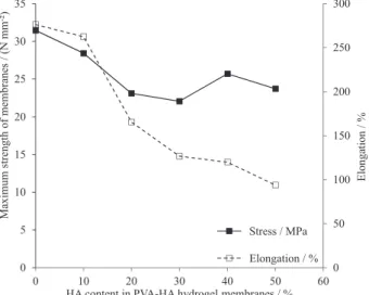
The influence of HA content on the mechanical properties of PVA hydrogel membranes has been estimated in terms of, their tensile strength and elongation-at-break, as presented in Figure 3. The maximum tensile strength and elongation-at-break of PVA-HA hydrogel membranes, decreases clearly with increasing the HA content in membranes. As HA was blended with PVA, the crosslinking density of the gel was decreased and the mechanical flexibility increases. These results are coincided with that of Razzak et al.36 They reported that the maximum tensile
strength of PVA hydrogel deteriorated with addition of the blended polymer, owing to decreasing the crosslinking degree and density. Hwang et al.37 demonstrated that, the
maximum tensile strength of PVA hydrogel has decreased sharply with increasing dextran portions in the hydrogel. Similarly, Kenawy et al.13 reported that the maximum
tensile strength and elongation-at-break of PVA-HES
hydrogel membranes, sharply decreased with increasing HES contents. All these reported contributions refer to the addition of blend polymers into PVA might hinder the entanglement reaction, unlike the mechanical flexibility improved.
Hydrolytic degradation
Gravimetric calculation was performed to study the hydrolytic degradation or the mass loss (%) of PVA-HA hydrogen membranes. The hydrolytic degradation of PVA hydrogel membrane as a function of different hyaluronic acid contents in membranes was conducted in PBS, as shown in Figure 4. The results exhibited that, the constant rate of hydrolytic degradation of PVA hydrogel membrane increases progressively with increasing the HA content in PVA hydrogel membranes, due to increasing of the hydrophilicity of HA parts in membrane composition. Weight loss (%) results of PVA-HA hydrogels as presented in Figure 4 are fully coincided with the swelling results as presented in Figure 2. This phenomenon can be ascribed to the degradation of PVA-HA hydrogel membranes that are mostly caused by the breaking of crosslinking segments between PVA and HA molecular structures which produce low molecular weights and degraded polymers. The weight loss results refer to that, the degradation behavior follows the second order kinetic. This also is owing to the fact that, the degradation of PVA is much slow (ca. 30% within 2 days, Figure 4) as well as previously discussed by Takasu et al.,38 whereas the degradation of PVA-HA is
much higher than the pristine PVA hydrogel (between ca. 40% and 78% within 2 days, Figure 4). In addition, as PVA and HA are nontoxic, this implies that, obtained byproducts of degraded PVA-HA moieties might be hypothesized to be nontoxic too.
0 10 20 30 40 50 60 70 0 200 400 600 800 1000 1200 1400 1600 1800
0 10 20 30 40 50 60
HA content in PVA-HA hydrogel membranes / (m/v, %)
Swelling ratio of PV
A -HA hy drogel membrane s / %
Gel fraction of
PV A -HA h y drogel membrane s / %
Gel fraction / %
Water update / %
Figure 2. Effect of HA contents in PVA hydrogel membranes on water uptake (%) and gel fraction (%).
0
Stress / MPa
Elongation / % 50 100 150 200 250 300 0 5 10 15 20 25 30 35
0 10 20 30 40 50 60
Elonga tion / % Maximum strength of membrane s
/ (N mm
-²)
HA content in PVA-HA hydrogel membranes / %
Figure 3. Mechanical properties of PVA hydrogel membrane with respect to different ratio of HA contents.
0 10 20 30 40 50 60 70 80
0 10 20 30 40
0% HA 10% HA 20% HA 30% HA 40% HA 50% HA W
eight loss of PV
A
HA
hy
d
rogel
membranes / %
time of degradation / h
Figure 4. Weight loss of the PVA-HA hydrogel membranes versus different degrading times in PBS (0.1 mol L-1, pH 7.4, at 37 °C), with
Protein adsorption
The membrane blood compatibility is evaluated by the amount of plasma protein adsorbed onto the membrane surface. The protein adsorption onto PVA-HA blend hydrogel membranes has been conducted via in vitro experiment.Figure 5 shows the protein adsorption onto surface of PVA-HA hydrogel membrane as function of different HA contents in distilled water. The mechanism of protein adsorption on surface of PVA-HA membranes is owing to various types of interaction forces between protein molecules and the membrane surface, such as weak bonding (e.g., Van der Waal interactions, ionic bonding, hydrogen bonding or hydrophobic interactions) or strong chemical bonding due to chemically surface modified membranes.13 Thus, in our study the clearest values of
protein adsorption on PVA-HA surface have been detected with the highest values of hydrophilic surface interaction due to addition of HA portions. The results appeared that the adsorbed protein onto surface of PVA hydrogel membrane increases with increasing HA contents in PVA hydrogel membranes, and the highest values of protein adsorption on PVA-HA surface have been detected with the highest values of hydrophilic surface interaction, due to the addition of HA contents. Interestingly, the protein adsorption onto membrane surface has improved within 200% to 400%, after inclusion the HA with different portions (Figure 5). For further evidences, when the tested membrane is direct contacting with the blood, the protein is adsorbed onto membrane surface resulting in platelet adhesion and activation.39,40 Since, the albumin adsorption
on the synthetic surfaces could inhibit platelet activation, which does not promote clot formation. Therefore, high protein adsorption property of wound dressing materials is of the most required characteristics.41 Thus, HA gave less
adhesion of platelets onto artificial surfaces and high protein
or plasma adsorption property. These results are compatible with reported results by Kim et al.,33 and Hwang et al.37
They proved that, the adsorption of protein increased with increasing blended alginate and dextran polymers in PVA hydrogel membranes, respectively.
In vitro biocompatibility test
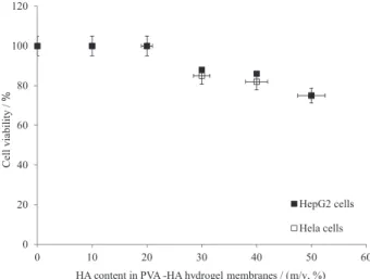
In vitro biocompatibility is principle property of any material being implanted in the biological body. The biocompatibility and cell viability (%) toward the components of PVA-HA membranes were tested in vitro by MTT-assay using HepG2 and Hela human cell lines (Figure 6). As shown, PVA-HA hydrogels with low HA contents (i.e., 0-20%) showed ca. 100 (%) of cell viability and non-toxicity behavior was observed. In the contrast, the cell viability (%) decreased dramatically with increasing HA contents (> 30%) in PVA membranes composition, and relatively less dead cells were observed. These results might be attributed to the released or degraded HA molecules in the culture media assuming an expanded random coil structure in physiological solution which occupy a very long domain and HA forms viscous compounds in DMEM culture media. This high viscous media might inhibit and reduce the migration, movement and cell viability (%). Thus, the fluidic viscosity of DMEM culture media increases as (0.00081, 0.0021, 0.0086, 0.0145, 0.0162, and 0.0187 Pa.s) for (0, 10, 20, 30, 40, and 50 m/v, %) of HA content), respectively. Furthermore, this allows an exceptionally high swelling ability for PVA-HA hydrogels with high HA contents, (Figure 2). These results are fully consistent with results of Schramm et al.42, and
Becker et al.43 They demonstrated that, the high HA content
in DMEM culture media increased the viscosity, and consequently the biocompatibility of the culture media was reduced as well.
0,5 1,0 1,5 2,0 2,5 3,0
0 10 20 30 40 50 60
Concentra
tion
of adsorbed
BSA
on the
membrane surafc
e
of PV
A
-HA
/
(m
g
mL
-1)
HA content in PVA -HA hydrogel membranes / (m/v, %)
Figure 5. Effect of HA content on protein adsorption onto the surface of PVA-HA hydrogel membranes.
0
20 40 60 80 100 120
0 10 20 30 40 50 60
HepG2 cells
Hela cells
Cell viability /
%
HA content in PVA -HA hydrogel membranes / (m/v, %)
Antimicrobial activity
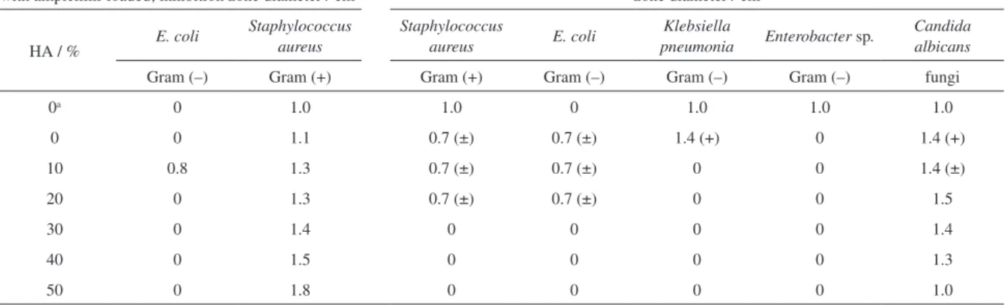
The antimicrobial activity test is a crucial parameter for wound dressing. The antimicrobial assay of PVA-HA membranes with/without ampicillin-loaded against E. coli, Staphylococcus aureus, Klebsiella pneumonia, Enterobacter sp. and Candida albicans was checked. The results of bacterial inhibition zones were listed in Table 1, and their selected photographs were presented in Figure 7. An antibiotic like ampicillin or another antimicrobial drug is urgent loaded onto the dressing material to stop the microbes growth or killing them outright.44
Thus, most dressing materials are derived from polymeric materials loaded-antimicrobial drug, even in case the used polymeric materials have antimicrobial activity themselves, like chitosan-dressing materials which have a dual antimicrobial actions.22,45 Interestingly, the results
in case E. coli, no inhibition zone was formed even with
PVA-HA-ampicillin loaded membranes. This implies that, E. coli has a hard resistant against membrane compositions containing ampicillin. This was clearly evident when the control assay (PVA-ampicillin discs) exhibited also negative results against the microbial growth. Conversely, the control assay sample and PVA-HA-ampicillin-loaded could effectively constrain the growth of Staphylococcus aureus, and the formed inhibition zones were obviously dispersed with HA incorporation as compared with the control assay. However, these inhibition zones do not form in the absence of ampicillin-loaded membranes, (Table 1 and Figure 7). These results might be attributed to Staphylococcus aureus growth inhibition is owing to only the presence of ampicillin antimicrobial inhibitor and the inhibition zones were further improved with HA introduction, which is entirely opposite behavior of E. coli. It is exciting to find that, PVA-HA membranes without ampicillin showed a big resistance for microbial growth against Candida albicans and the inhibition zones were improved with HA incorporation to PVA hydrogel as found with Staphylococcus aureus. This result is regarded as important and remarkable results, because of Candida albicans is well-known the most frequently microbial can infect the body skin wound, which matches the study goal. Overall, the microbial resistance was improved for PVA-HA with certain HA content in membrane composition (up to < 20%), and it was further improved with the membrane ampicillin-loaded. Unexpectedly, in case of Klebsiella pneumonia and Enterobacter sp., PVA-HA membranes in the absence of ampicillin do not show antimicrobial activity and no inhibition zones formed, whereas the inhibition zones were found with the control assay (Table 1). The current antimicrobial results are consistent with our published results of Kamoun et al.31 They showed antimicrobial Table 1. Effect of various HA contents in PVA membranes composition on their antimicrobial activity
Antimicrobial activity of PVA-HA disc membranes with ampicillin-loaded; inhibition zone diameter / cm
Antimicrobial activity of PVA-HA disc membrane without ampicillin-loaded; inhibition zone diameter / cm
HA / % E. coli
Staphylococcus aureus
Staphylococcus
aureus E. coli
Klebsiella
pneumonia Enterobacter sp.
Candida albicans
Gram (–) Gram (+) Gram (+) Gram (–) Gram (–) Gram (–) fungi
0a 0 1.0 1.0 0 1.0 1.0 1.0
0 0 1.1 0.7 (±) 0.7 (±) 1.4 (+) 0 1.4 (+)
10 0.8 1.3 0.7 (±) 0.7 (±) 0 0 1.4 (±)
20 0 1.3 0.7 (±) 0.7 (±) 0 0 1.5
30 0 1.4 0 0 0 0 1.4
40 0 1.5 0 0 0 0 1.3
50 0 1.8 0 0 0 0 1.0
aControl assay is conducted in case PVA-ampicillin-loaded with zero-HA content in all samples.
activities for PVA-alginate membranes ampicillin-loaded against Gram-negative and Gram-positive bacteria after altering the alginate content in PVA membranes composition. Our results conflict slightly with the results of Abd El-Mohdy et al.46 They revealed that the various PVP
contents in PVA hydrogels showed antimicrobial activates against E. coli, Bacillus subtilis and Staphylococcus aureus owing to PVP has antimicrobial activity itself, and no activity against Pseudomonas aeruginosa was observed. Similarly, the antibacterial behavior of hydrogels based on PVA/water-soluble-chitosan was ascribed to their capability to bind the negatively charged bacteria to the positively charged amino groups of polymer in hydrogel.22
Conclusions
In conclusion, the PVA-HA hydrogels have been successfully prepared by F-T as a physical crosslinking method. The physicochemical properties of obtained PVA-HA hydrogels have been assessed as function of different HA contents. The results showed that incorporation of HA in the F-T crosslinked PVA network appreciably affected its physicochemical properties. The swelling behavior, mechanical flexibility, hydrolytic degradation rate and protein adsorption of PVA-HA hydrogel membranes increased with increasing the HA content in hydrogel composition. However, as the gel fraction decreased, the mechanical strength of the gel was weakened but the flexibility was increased. Addition HA into PVA hydrogel membranes improved protein adsorption property onto PVA membrane surface as compared to pristine PVA membranes. Therefore, the HA content in hydrogel composition could be used to control the mechanical strength and flexibility of PVA-HA hydrogels because it reduced the crosslinking reaction and, consequently the gelation process. PVA-HA hydrogel membranes supernatant exhibited nontoxic behavior and higher cell viability with low HA contents (up to < 20%), compared with the high HA content hydrogels. Interestingly, PVA-HA without-ampicillin membranes showed effective antimicrobial activity, particularly against Candida albicans, as a result of the HA presence. However, the same membranes showed similar antibacterial activity against Staphylococcus aureus especially after loading ampicillin. The PVA-HA membranes offered no microbial resistance against E. coli, membranes with/without ampicillin-loaded. Thus, the addition of HA to PVA hydrogels changed and improved the physicochemical properties and biological activity of membranes, compared with the pristine PVA hydrogel for wound dressing applications. Thus, it is a potential wound dressing with easy forming and improved bio-evaluations for wound dressing application.
Acknowledgements
We are indebted to the Department of Macromolecular Chemistry, Institute for Technical Chemistry, Technical University of Braunschweig (TU-BS), Germany for the technical and characterization supports. Authors also gratefully acknowledge Prof Xin Chen, State Key Laboratory of Molecular Engineering of Polymers, Department of Macromolecular Science, Laboratory of Advanced Materials, Fudan University, Shanghai, People’s
Republic of China, for providing some chemicals which have been used to achieve this work.
References
1. Rosiak, J. M.; Rucinska-Rybus, A.; Pekala, W.; US Patent
4,871,490 1989.
2. Yoshii, F.; Zhanshan, Y.; Isobe, K.; Shiozaki, K.; Makunchi, K.;
Radiat. Phys. Chem. 1999, 55, 133.
3. Kannon, G. A.; Garrett, A. B.; Dermatol Surg 1995, 21, 583. 4. Lin, S. Y.; Chen, K. S.; Run-Chu, L.; Biomaterials 2001, 22,
2999.
5. Purna, S. K.; Babu, M.; Burns 2000, 26, 54.
6. Kamoun, E. A.; Chen, X.; Mohy Eldin, M. S.; Kenawy, E. S.;
Arab. J. Chem.2015, 8, 1.
7. Long, Z.; Hiroshi, M.; Maolin, Z.; Fumio, Y.; Naotsugu, N.; Tamikazu, K.; Carbohydr. Polym. 2013, 53, 439.
8. Mallapragada, S. K.; Peppas, N. A.; Polym. Mater. Sci. Eng.
Proceed. 1995, 73, 22.
9. Mallapragada, S. K.; Peppas, N. A.; J. Polym. Sci. Part B Polym.
Phys. 1996, 34, 1339.
10. Mallapragada, S. K.; Peppas, N. A.; AIChE J. 1997, 43, 870.
11. Hassan, C. M.; Peppas, N. A.; Macromolecules 2000, 33, 2472.
12. Papancea, A.; Valente, A. J. M.; Patachia, S.; Miguel, M. G.; Lindman, B.; Langmuir 2008, 24, 273.
13. Kenawy, E.-R.; Kamoun, E. A.; Mohy Eldin, M. S.; El-Meligy, M. A.; Arab. J. Chem. 2014, 7, 372.
14. Laurent, T. C.; Laurent, U. B. G.; Fraser, J. R. E.; Ann. Rheum.
Dis. 1995, 54, 429.
15. Saettone, M. F.; Monti, D.; Torracca, M. T.; Chetoni, P.; J. Ocul.
Pharmacol. Ther. 1994, 10, 83.
16. Candy, T.; Sharma, C. P.; Biomater. Artif. Cells Artif. Organs 1990, 18, 1.
17. Hong, S. R.; Lee, S. J.; Shim, J. W.; Choi, Y. S.; Lee, Y. M.; Song, K. W.; Park, M. H.; Nam, Y. S.; Lee, S. I.; Biomaterials 1993, 14, 2777.
18. Okamoto, Y.; Minami, S.; Matuhashi, A. In Advances in Chitin
and Chitosan Animals; Brine, C. J.; Sandford, C. J.; Zikakis,
19. Li, Y.; Wang, L.; Wu, J.; Ma, Y.; Wang, J.; Wang, Y.; Luo, Y.;
Mater. Letters 2014, 134, 9.
20. Kupal, S. G.; Cerroni, B.; Ghugare, S. V.; Chiessi, E.; Paradossi, G.; Biomacromolecules 2012, 13, 3592.
21. Peppas, N. A.; Stauffer, S. R.; J. Controlled Release 1991, 16, 305.
22. Yang, X.; Liu, Q.; Chen, X.; Yu, F.; Zhu, Z.; Carbohydr. Polym. 2008, 73, 401.
23. Alencar, D. Q. H.; Humberto, G. F.; Gustavo, A. A.; Maria, M. F.; Antonio, L. B.; Julio, S. R.; J. Biomed. Mater. Res. Part A 2003, 64, 147.
24. Xiao, C.; Zhou, G.; Polym. Degrad. Stab. 2003, 81, 297. 25. Queiroz, A. C.; Santos, J. D.; Monteiro, F. J.; Gibson, I. R.;
Knowles, J. C.; Biomaterials 2001, 22, 1393.
26. Lin, W. C.; Yu, D. G.; Yang, M. C.; Colloids Surf., B 2006, 47, 43.
27. El-Fakharany, E. M.; Haroun, B. M.; Ng, T. B.; Redwan, E. R.;
Protein Pept. Lett.. 2010, 17, 1031.
28. El-Fakharany, E. M.; Sánchez, L.; Redwan, N. A.; Redwan, E. M.; J. Virol. 2013, 10, 199.
29. Mosmann, T.; J. Immunol. Methods 1983, 65, 55.
30. Fahmy, A.; Abu-Saied, M. A.; Kamoun, E. A.; Khalil, H. F.; Youssef, M. E.; Attia, A. M.; Esmail, F. A.; J. Adv. Chem.
2015, 11, 3426.
31. Kamoun, E. A.; Kenawy, E. S.; Tamer, M. T.; El-Meligy, M. A.; Mohy Eldin, M. S.; Arab. J. Chem.2015, 8, 38.
32. Ajji, Z.; Othman, I.; Rosiak, J. M.; Nucl. Instr. Meth. Phys. Res:
Sect. B 2005, 229, 375.
33. Kim, J. O.; Park, J. K.; Kim, J. H.; Jin, S. G.; Yong, C. S.; Li, D. X.; Int. J. Pharm. 2008, 359, 79.
34. Zhai, M.; Yoshii, F.; Kume, T.; Hashim, K.; Carbohydr. Polym. 2002, 50, 295.
35. Zhao, L.; Mitomo, H.; Zhai, M.; Yoshii, F.; Nagasawa, N.; Kume, T.; Carbohydr. Polym. 2003, 53, 439.
36. Razzak, M. T.; Darwis, D.; Zainuddin, S.; Radiat. Phys. Chem. 2001, 62, 107.
37. Hwang, M. R.; Kim, J. O.; Lee, J. H.; Kim, Y.; Kim, J. H.; Chang, S. W.; Jin, S. G.; Kim, J.-A.; Lyoo, W. S.; Han, S. S.; Ku, S. K.; Young, C. S.; Choi, H. G.; AAPS Pharm. Sci. Technol. 2010,
11, 1092.
38. Takasu, A.; Itou, H.; Takada, M.; Inai, Y.; Hirabayashi, T.;
Polymer 2002, 43, 227.
39. Burkatovskaya, M.; Tegos, G. P.; Swietlik, E.; Demidova, T. N.; Castano, A.; Hamblin, M. R.; Biomaterials 2006, 27, 4157. 40. Coleman, D. L.; Gregonis, D. E.; Andrade, J. D.; J. Biomed.
Mater. Res. 1982, 16, 381.
41. Dion, I.; Baquey, C.; Havlik, P.; Monties, J. R.; Int. J. Artif.
Organs 1993, 6, 545.
42. Schramm, C.; Spitzer, M. S.; Henke-Fable, S.; Steinmetz, G.; Januschwski, K.; Heidusehka, P.; Geis-Gerstorfer, J.; Biedermann, T.; Bartz-Schmidt, K. U.; Szurmann, P.; Invest.
Ophth. Vis. Sci.2012, 53, 613.
43. Becker, L. C.; Bergfeld, W. F.; Belsito, D. V.; Klaassen, C. D.; Marks Junior, J. G.; Shank, R. C.; Slaga, T. J.; Synder, P. W.; Andersen, F. A.; Inter. J. Toxicol.2009, 28, 5.
44. Chhatri, A.; Bajpai, A. K.; Shandhu, S. S.; Jain, N.; Biswas, J.;
Carbohydr. Polym. 2011, 83, 876.
45. Kanatt, S. R.; Rao, M. S.; Chawla, S. P.; Sharma, A.; Food
Hydrocolloids 2012, 29, 290.
46. Abd El-Mohdy, H. L.; Ghanem, S.; J. Polym. Res. 2009, 16, 1.
Submitted: March 7, 2015