Validation experiments confirmed the protein-protein interaction of IQGAP1 with hnRNPM, as well as RNA-dependent interactions with SRSF1, hnRNPA1, hnRNPA2B1, hnRNPC1/C2, DDX17 and other splicing regulators. To study the role of IQGAP1 depletion on gastric cancer cell phenotype and splicing patterns, CRISPR-Cas9 IQGAP1 knock-out clones of the cell lines of interest were subjected to phenotypic assays. Our current hypothesis is that IQGAP1 plays an essential role in the assembly of RNP complexes that participate in pre-mRNA splicing and post-transcriptional regulation in gastric epithelial and gastric cancer cells, orchestrating patterns of AS that may be associated with phenotypic changes of gastric cancer cell lines.
Interactions of IQGAP1 with post-transcriptional regulators therefore emerge as potential drivers of AS in gastric cancer - a result which, after systematic investigation, may contribute with potential candidates for the development of new therapeutic agents against a rare and fatal disease.
Introduction
- Alternative splicing
- General mechanism
- The spliceosomal proteome
- Regulation of alternative splicing
- General mechanism
- Alternative splicing regulation through post-translational modifications
- Alternative splicing and cancer
- Hallmarks of cancer and alternative splicing
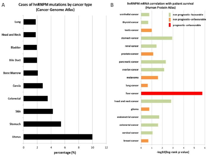
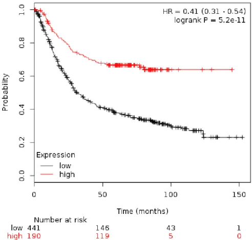
- The role of heterogeneous nuclear ribonucleoprotein M (hnRNPM) in cancer
- IQGAP1
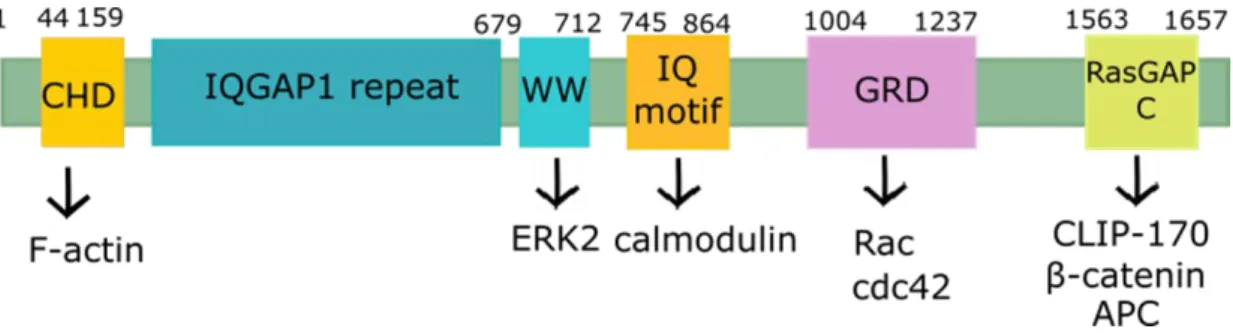
- Function and cellular localization of IQGAP1
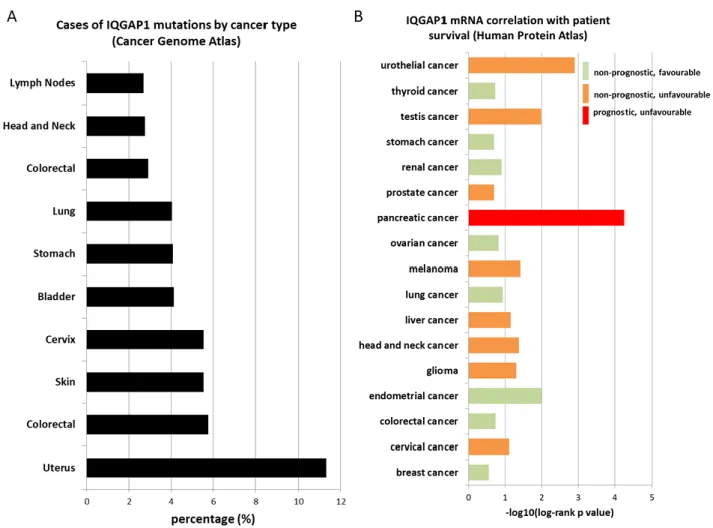
- IQGAP1 and cancer
- Implications of IQGAP1’s interaction with hnRNPM
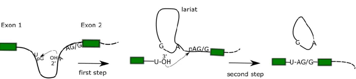
After the lariat formation, the catalytically active complex C52 is formed and proceeds with the second step of the splicing reaction. Biochemical mechanism of pre-mRNA splicing. In the first step, a nucleophilic attack of the 2' OH of the branch site adenine residue to the 5' splice site phosphate (of the conserved GU dinucleotide) forms the intron lariat, linked to the 3' exon and releases the 5' exon. In the second step, the 3' OH of the released exon attacks the phosphate group of the adenine residue in the conserved AG/G 3' sequence of the intron.
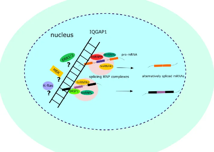
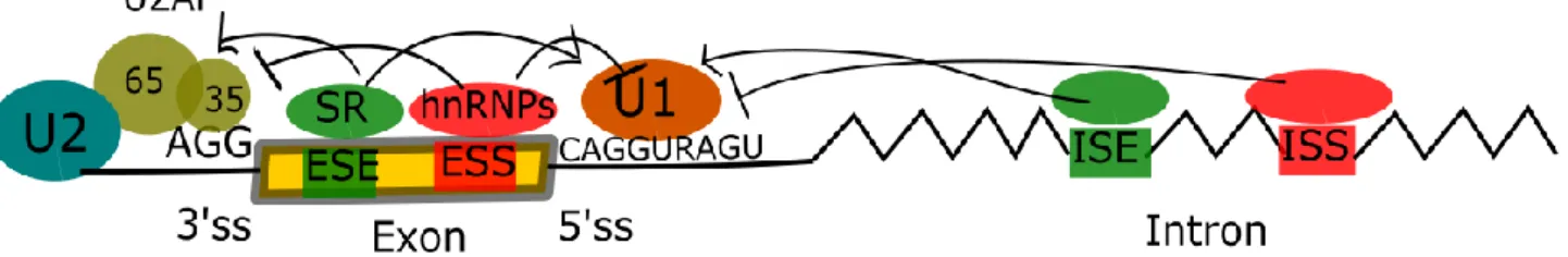
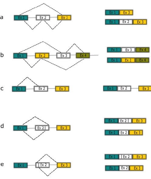
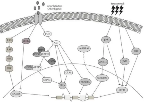
To summarize, enhancer and silencer sequences play a crucial role in alternative splicing regulation by recruiting RBPs that can promote E complex formation and splicing initiation at their adjacent 5' and 3' splice sites (SRSF) or , conversely, prevent The complex is assembled and thus leads to the exclusion of the exon or intron defined by these boundaries from the final transcript (hnRNPs) (Figure 5). The products of the possible AS reactions (indicated by rows above and below the pre-RNA) are shown in the second column (above and below, respectively).a. In addition to its role in cytoskeleton regulation, IQGAP1 acts as a protein scaffold for signaling molecules (reviewed in 207,217).
Aims of the project
Materials and methods
- Cell culture
- Preparation of nuclear and cytoplasmic extracts
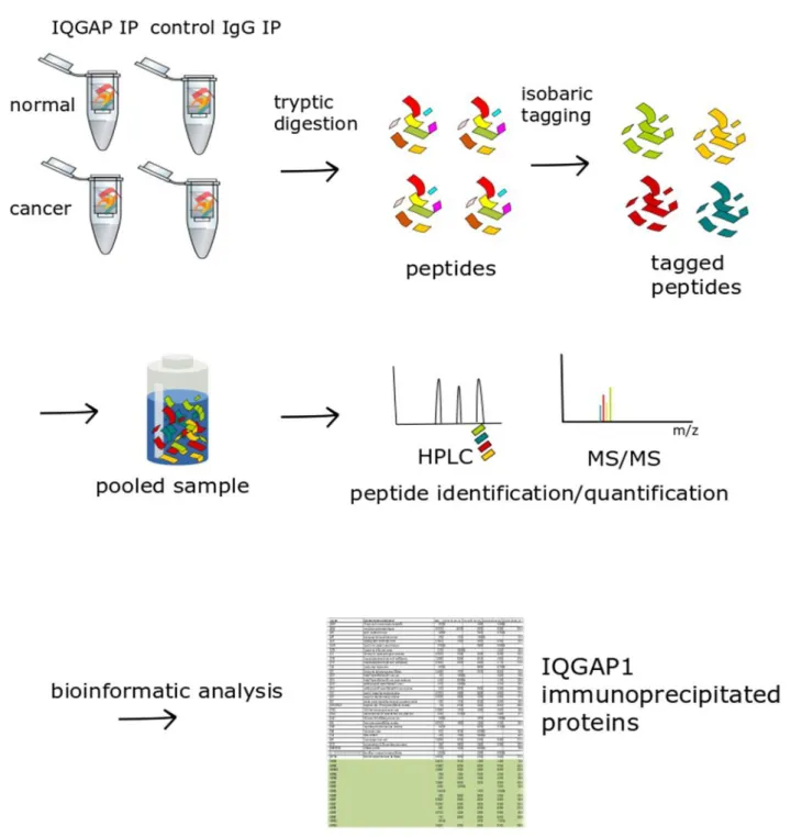
- Immunoprecipitation of proteins
- Western blotting
- Mass spectrometry and Proteomics analysis
- Analysis of phosphorylation in IQGAP1-immunoprecipitated proteins
- Generation of cell lines with stable IQGAP1 knockdown by CRISPR-Cas9
- Colony formation assay
- Cell cycle analysis
- Mini-gene splicing assay
- RNA isolation/Reverse Transcription/PCR
After binding the sample, beads were recovered as before, and an aliquot of the supernatant was retained to monitor sample flow. This allows the simultaneous identification and comparative quantification of identical peptides (and therefore proteins) in each of the multiplexed samples. The binary pumps of the HPLC (RSLCnano, Thermo Scientific) contain solution A (2% (v/v) acetonitrile in 0.1% (v/v) formic acid) and solution B (80% acetonitrile in 0.1% formic acid) .
After annealing, the presence of the PAM allows Cas9 to cleave opposite strands of the target DNA, introducing a double-strand break (DSB) in the target DNA (3–4 nucleotides upstream of the PAM sequence). Repair of a Cas9-induced DSB via the NHEJ pathway should ideally lead to a loss-of-function mutation that “knocks out” the target gene. Use of CRISPR-Cas9 D10 nickase to target Exon 1 of the IQGAP1 gene using two sgRNAs (Assembly 1 and Assembly 2) targeting adjacent regions of the Exon 1 sequence.
The proliferative capacity of the IQGAP1 knockout cell lines was measured using an adherent cell colony formation assay.270 Sub-confluent or confluent cells (controls and stable IQGAP1 knockout clones) were harvested appropriately, resuspended in fresh medium and counted. Flow cytometry was performed using a BD FACS CantoTM II system, with the GO/G1 gate of the PI-A histogram set to 50 units. To complete the RT PCR reaction setup, 2 µl of 5x RT buffer, 0.05 µmol DTT, 20 units of RNase (RNase-out™, Invitrogen) and 100 units of reverse transcriptase (Superscript III Reverse Transcriptase, Invitrogen) were added. added each time. reaction, to a final volume of 10 µl.
The RT reaction was completed in a thermocycler set to the following program: 45' at 50oC, 15' min at 70oC followed by incubation of the cDNA samples on ice. A final extension step of 3 minutes at 72oC is added after completion of the last cycle.
Results
Study of IQGAP1-hnRNPM RBP complex
- IQGAP1 enters the nucleus and interacts with hnRNPM
- Nuclear IQGAP1 interacts with spliceosomal components
- IQGAP1 forms two putative complexes with spliceosomal components
- Study of post-translational modifications in IQGAP1-RBP complex components
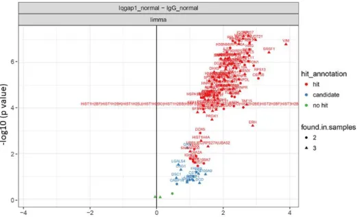
IQGAP1 interacting proteins in HFE-145 (normal) nuclear extracts. A volcano plot of quantitative comparison of proteins in HFE (normal) IQGAP1 IPs against their respective IgG controls is shown. B Representative immunoblots of the cytoplasmic extracts, nuclear extracts (inset), IQGAP1 and rabbit IgG control IP samples used for mass spectrometry analysis. Significantly, 39 of the identified IQGAP1 interactors showed multiple annotations for core (CC), core mRNA splicing via spliceosome (BP), and RNA splicing (BP).
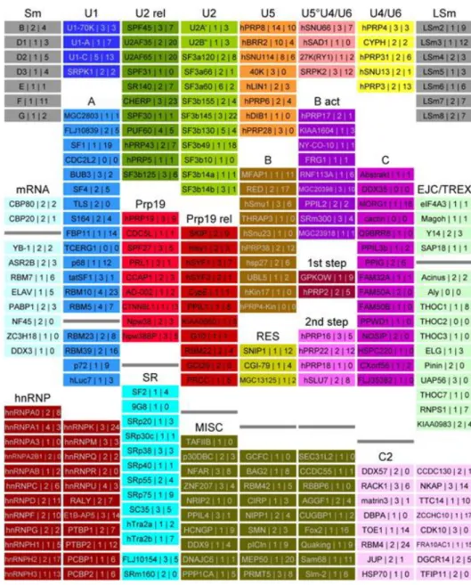
Interestingly, out of the total of 31 proteins identified as ribosomal components, 29 were annotated as components of the cytosolic ribosome. However, closer inspection of these components showed that 20 of the ribosomal proteins were also annotated as having nuclear/nucleolar localization, while all identified ribosomal components were annotated (in Reactome) for participation in nuclear processes such as ribosomal biogenesis, rRNA processing or nonsense - mediated decay (NMD). A number of these have previously been implicated in mRNA splicing regulation/spliceosomal assembly: comparison of our results with spliceosomal components identified in previous mass spectrometry studies of the spliceosome, as compiled in the spliceosome database280, showed that 71 of the identified IQGAP1 core partners have .
Schematic of the proposed IQGAP1 nuclear complexes, based on examination of mass spectrometry identified spliceosomal components. This hypothesis remains to be confirmed through ongoing further investigation of the protein interactions involved, some of which appear to be RNA-dependent (Table 2, data not shown). The next step after identifying the components of the IQGAP1 nuclear RBP complex was to study the potential effect of IQGAP1 on alternative splicing regulation through the induction of post-translational modifications on its spliceosomal partners.
Sample preparation was performed using two methods: Filter-assisted sample preparation (FASP)261, and a modified form of the SP3 sample preparation strategy265, in an attempt to increase peptide yield (see section 3.6). Two of the previously identified IQGAP1-interacting proteins (HSPA8 and PRPF6) were phosphorylated.
Phenotypical study of IQGAP1 knockout cell lines
- Generation of IQGAP1 knockout cell lines
- Effects of IQGAP1 depletion on cell cycle progression on normal gastric epithelial and
- Effects of IQGAP1 depletion on proliferative capacity of normal gastric epithelial and
- Splicing effect of IQGAP1 knockout on hnRNPM-dependent splicing
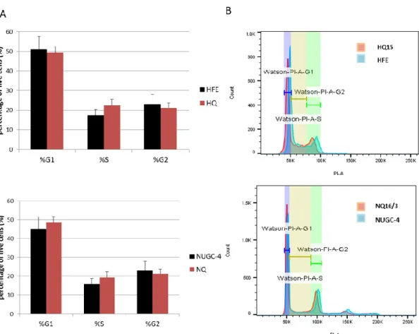
To study the effect of IQGAP1 knockout on the phenotype of cancer cells, we employed strategies that are widely used to study hallmarks of cancer, including adhesion, invasion, proliferative potential, and cell cycle progression.289 The effect of IQGAP1 knockout depletion on Cell cycle progression was examined by fluorescence-assisted cell sorting of cells stained with propidium iodide, which stains DNA, separating cell cycle phases based on their DNA content (see 3.9). The CRISPR-Cas9 D10 IQGAP1-depleted clones of the HFE-145 and NUGC-4 cell lines were grown to 55–75% confluency, in non-synchronized conditions, and stained with propidium iodide after ethanol fixation and washing with phosphate-citrate buffer . Percentage of living cells with DNA content corresponding to each phase of the cell cycle (G1/S/G2-M). The FACS results indicate that depletion of IQGAP1 has no significant effect on the cell cycle progression of normal gastric epithelial HFE-145 cells or NUGC-4 gastric adenocarcinoma cells (Figure 23A).
Side-by-side comparisons of PI staining histograms between control and IQGAP1 depleted cells highlight the fact that the number of cells in each phase of the cell cycle is similar between wild type and IQGAP1 depleted clones (Figure 23B). These results are consistent with what was previously observed in IQGAP1-siRNA silenced NIH3T3 cells, which also did not show a differential cell cycle progression after asynchronous culture. The proliferative capacity of the IQGAP1-depleted clones was assessed using a colony formation assay designed to test the clonogenic capacity of adherent cells as described in 3.8.270.
The survival fraction (SF) of each control was taken as 100%, and the SF of IQGAP1-depleted clones was compared to the control using the Student's t test. The results shown in Figure 24 show that depletion of IQGAP1 results in a decrease in the clonogenic capacity of HFE-145 cells by approximately 50%: it can be seen that HQ clones show a statistically significant decrease in the survival rate compared to parental HFE cells. Since hnRNPM was the first observed spliceosomal component observed to interact with IQGAP1 in the nucleus, and one of the most robust and direct interactions observed in subsequent experiments, we used an assay aimed at investigating hnRNPM-dependent splicing events.
DUP51 is a three-exon-two-intron construct developed for the study of differential exon inclusion,274 which was subsequently adapted for the study of hnRNPM-dependent alternative splicing.49 More specifically, exon 2 of the modified DUP51M construct contains an hnRNPM consensus-binding motif (UGGUGGUG), which acts as an exonic splicing silencer (Figure 25A).273. When the splicing function of hnRNPM is unperturbed, its binding to the exon 2 consensus motif leads to splicing out of exon 2 of the DUP51 construct and the formation of the smaller, two-exon splice product of construct DUP51.
Discussion
Interestingly, IQGAP1 depletion appeared to have opposite effects on the ability of the two cell lines to form colonies in culture. Interestingly, IQGAP1 depletion has opposite effects in regulating clonogenic capacity of the two cell lines studied, indicating a different role of IQGAP1 in regulating clonogenesis in HFE-145 gastric epithelial cells and NUGC-4 gastric adenocarcinoma cells. Functional analysis of the human CDC5L complex and identification of its components by mass spectrometry.
The RNA-binding domain of protein component A of small nuclear ribonucleoprotein U1 analyzed by NMR spectroscopy is structurally similar to ribosomal proteins. Role of SR protein modular domains in subnuclear localization and alternative splicing specificity. The RNA-binding and adapter protein Sam68 modulates signal-dependent binding and transcriptional activity of the androgen receptor.
Arginine methylation controls the subcellular localization and functions of the oncoprotein splicing factor SF2/ASF. The 72/74-kDa polypeptides 70-110 S of the large heterogeneous nuclear ribonucleoprotein complex (LH-nRNP) represent a discrete subset of the hnRNP M protein family. Association of 72/74-kDa proteins, members of the heterogeneous nuclear ribonucleoprotein group M, with pre-mRNA at early stages of spliceosome assembly.
A novel mechanism of ERK1/2 regulation in smooth muscle involving acetylation of the ERK1/2 scaffold IQGAP1. Role of IQGAP1, a target of the small GTPase Cdc42 and Rac1, in the regulation of E-cadherin-mediated cell adhesion. Dephosphorylation of the nuclear factor of activated T cell transcription factor (NFAT) is regulated by an RNA-protein scaffold complex.
Antiapoptotic roles of ceramide synthase-6-generated C16 ceramide via selective regulation of the ATF6/CHOP arm of ER stress response pathways.