Branchial O
2
chemoreceptors in Nile tilapia
Oreochromis niloticus
:
Control of cardiorespiratory function in response to hypoxia
Vivian M. Zeraik
a, Thiago C. Belão
a, Luiz Henrique Florindo
b,c,1, Ana L. Kalinin
a,c, F. Tadeu Rantin
a,c,⁎
aDepartment of Physiological Sciences, Laboratory of Zoophysiology and Comparative Biochemistry, Federal University of São Carlos (UFSCar), P.O. Box 676, Via Washington Luis, km 235,13565–905 São Carlos, SP, Brazil
bDepartment of Zoology and Botany, São Paulo State University
–UNESP/IBILCE, Rua Cristóvão Colombo, 2265, 15054–000 - São José do Rio Preto, SP, Brazil cNational Institute of Science and Technology - Comparative Physiology (FAPESP/CNPq), Brazil
a b s t r a c t
a r t i c l e
i n f o
Article history:
Received 8 March 2013
Received in revised form 24 April 2013 Accepted 29 April 2013
Available online 4 May 2013
Keywords:
Gill chemoreceptors Hypoxia
Hypoxic bradycardia Oreochromis niloticus Ventilation
This study examined the distribution and orientation of gill O2chemoreceptors inOreochromis niloticusand their role in cardiorespiratory responses to graded hypoxia. Intactfish, and a group with thefirst gill arch excised (operated), were submitted to graded hypoxia and their cardiorespiratory responses (oxygen uptake
-_
VO2, breathing frequency -fR, ventilatory stroke volume -VT, gill ventilation -V_G, O2extraction from the ventilatory current -EO2, and heart rate -fH) were compared. Their responses to bolus injections of NaCN into the bloodstream (internal) or ventilatory water stream (external) were also determined. TheV_O2of op-eratedfish was significantly lower at the deepest levels of hypoxia. Neither reflex bradycardia nor ventilatory responses were completely abolished by bilateral excision of thefirst gill arch.EO2of the operated group was consistently lower than the intact group. The responses to internal and external NaCN included transient decreases in fHand increases infRandVamp(ventilation amplitude). These cardiorespiratory responses were attenuated but not abolished in the operated group, indicating that chemoreceptors are not restricted to thefirst gill arch, and are sensitive to oxygen levels in both blood and water.
© 2013 Elsevier Inc. All rights reserved.
1. Introduction
Aquatic environments frequently exhibit daily fluctuations in
dissolved O2concentrations. Such variations can sometimes result in
se-vere O2depletions, known as environmental hypoxia, in particular in
tropical and subtropical regions (Randall et al., 1997). Cardiorespiratory responses to environmental hypoxia have been documented for a
con-siderable number offish species. Hypoxia usually induces increases in
breathing frequency (fR) and ventilatory stroke volume (VT) as well as
a reduction in heart rate (fH). Hyperventilation and bradycardia are
re-flex responses that serve to maximize the effectiveness of oxygen trans-fer from the water across the gill surface and into the blood stream (Taylor et al., 1999; Campbell and Egginton, 2007). They are controlled
by reference to O2chemoreceptors sensitive to O2partial pressures of
the inspired water (PiO2), or arterial blood (PaO2), or both (Burleson
et al., 1992; Burleson and Milsom, 1993; Sundin et al., 1999, 2000;
Leite et al., 2007; Micheli-Campbell et al., 2009; Lopes et al., 2010). Afferent nerves then relay the information from chemoreceptors to the cardiac and respiratory centers in the central nervous system. This
elicits reflex homeostatic cardiorespiratory responses, via efferent
in-puts to the ventilatory muscles and heart (Gilmour and Perry, 2007).
The precise anatomical locations and distributions of O2-sensitive
chemoreceptors infishes are not completely established, but
physiolog-ical and pharmacologphysiolog-ical evidence consistently indicate the gills (in-cluding the pseudobranch in those species that possess this structure) and the oro-branchial cavity as the structures housing these receptors (Laurent and Rouzeau, 1972; Randall and Jones, 1973; Butler et al., 1977; Daxboeck and Holeton, 1978; Smith and Davies, 1984; Smatresk et al., 1986; Burleson and Smatresk, 1990; McKenzie et al., 1991; Burleson and Milsom, 1993; Sundin et al., 1999; Milsom, 2002; Florindo et al., 2006). The receptors located in the oro-branchial cavity are innervated by branches of the cranial nerve V (trigeminal) and/or VII (facial). Those in the pseudobranch are also innervated by the cranial nerve VII, while those on the gill arches are innervated by branches of the IX (glossopharyngeal) and/or X (vagus) (Perry and Reid, 2002).
Histological and neurophysiological evidence suggest that O2
chemore-ception in the gills may arise from neuro-epithelial cells containing ve-sicular serotonin (Jonz and Nurse, 2003; Jonz et al., 2004; Coolidge et al., 2008; Porteus et al., 2012; Zachar and Jonz, 2012). However, the
distri-bution of O2chemoreceptors can vary considerably among different gill
⁎ Corresponding author at: Department of Physiological Sciences, Laboratory of Zoophysiology and Comparative Biochemistry, Federal University of São Carlos (UFSCar), P.O. Box 676, Via Washington Luis, km 235, 13565–905 São Carlos, SP, Brazil. Tel.: +55 16 3351 9774 (office), +55 16 3351 8314 (laboratory); fax: +55 16 3351 8401.
E-mail address:[email protected](F.T. Rantin).
1Tel.: +55 17 3221 2370; fax: +55 17 3221 2374.
1095-6433/$–see front matter © 2013 Elsevier Inc. All rights reserved.
http://dx.doi.org/10.1016/j.cbpa.2013.04.024
Contents lists available atSciVerse ScienceDirect
Comparative Biochemistry and Physiology, Part A
arches and among species (Burleson and Milsom, 1993; Sundin et al., 2000). Furthermore, there is also evidence of O2-chemosensitive sites outside the branchial apparatus (Butler et al., 1977; Barret and Taylor, 1984). The location, distribution, orientation and function of the gill
chemoreceptors in thefish species studied to date have recently been
reviewed byMilsom (2012).
The innervation, location and orientation of the O2
chemorecep-tors triggering reflex responses to hypoxia have also been determined
based on cardiorespiratory responses to different O2levels and/or to
NaCN, which is a potent stimulant of O2receptor activity (Burleson
et al., 1992). These reflexes can be modified or eliminated by section-ing nerves to the gills and oro-branchial cavity (Burleson and Milsom, 1993; Sundin et al., 1999, 2000; Milsom, 2002; Florindo et al., 2006; Leite et al., 2007; Micheli-Campbell et al., 2009) or by the excision of gill arches (Jia and Burggren, 1997; Perry and Reid, 2002).
One main conclusion that can be drawn from all of these studies is that it is impossible to establish a unifying pattern for the location,
dis-tribution pattern and orientation of O2chemoreceptors infishes. For
example, phylogenetically closely related species, such as the characids
pacu,Piaractus mesopotamicus, and tambaqui,Colossoma macropomum
(Sundin et al., 2000; Leite et al., 2007), or species in the same genus
such as the erythrinids traíra,Hoplias malabaricus, and trairão,Hoplias
lacerdae(Sundin et al., 1999; Micheli-Campbell et al., 2009), all exhibit
different O2 chemoreceptor locations, distributions and orientation
patterns.
Nile tilapia,Oreochromis niloticus(Cichlidae), is known for its
tol-erance of environmental factors such as temperature and pH, and low levels of dissolved oxygen (Chervinski, 1982; Fernandes and Rantin, 1986a, 1986b, 1987, 1989; Lague et al., 2012). Although the cardiore-spiratory responses by this species to hypoxia have been extensively studied (Fernandes and Rantin, 1987, 1994; Kalinin et al., 1999; Speers-Roesch et al., 2010), the mechanisms of oxygen sensing and the distribution of chemoreceptors mediating reflex responses still remain to be elucidated. The present study aimed, therefore, to
pro-vide insight into the distribution and orientation of the branchial O2
chemoreceptors involved in reflex hypoxic responses in this species. In particular, to test whether chemoreceptors are restricted to the
first pair of gill arches, or are distributed over all gill arches, thefirst
gill arch was excised bilaterally, as described by Perry and Reid
(2002), and cardioventilatory responses to hypoxia and NaCN
com-pared to those of intactfish.
2. Material and methods
2.1. Collection and maintence
Specimens ofO.niloticus(Mb = 220–260g) were obtained from
Santa Cândidafish farm, Santa Cruz da Conceição, São Paulo State,
Brazil. In the laboratoryfish were acclimated for 40 days prior the
ex-perimentation in 1000 l tanks supplied with recirculated andfiltered
normoxic water (~140 mmHg) at 25 ± 1 °C. During this periodfish
were fedad libitumwith commercial pellets (22% protein diet), but
food was withdrawn 24 h prior to experimentation.
2.2. Surgical procedures
Fish were anesthetized by immersion in a benzocaine solution
(0.1 g l−1) until righting responses were lost. They were then
trans-ferred to a surgical table where their gills were irrigated with a
vigor-ously aerated benzocaine solution at 0.05 g l−1.
Aflared polyethylene cannulae (PE 90) was placed in the buccal
cavity through a hole drilled in the dorsal palate, and secured with a cuff. Subsequently, using the same technique, the borders of both opercula of some individuals (those to be exposed to hypoxia, Hyp) were also cannulated with PE-70. The buccal catheter was used to
monitor buccal pressure variations to determinefR. The same catheter
served to inject NaCN solution into the ventilatory water stream (NaCN group). In the Hyp group, the buccal catheter was used to
col-lect water samples to measure the oxygen partial pressure (PO2) of
inspired water (PiO2), while the opercular catheter was used to
si-phon water samples to measurePO2of the expired water (PeO2).
Fish were allfitted with ECG electrodes to recordfH, as described
byGlass et al. (1991). One electrode (+) was inserted and sutured in a ventral position between the gills and the heart, and a second
(−) in a ventrocaudal position close to the pelvicfins. This
prepara-tion results in ECG recordings equivalent to the bipolar lead I of stan-dard electrocardiography.
The caudal vein was cannulated (PE 50) (Axelsson and Fritsche, 1994) for intravenous injections of saline and cyanide in the NaCN group. The intrabuccal and intravenous injections of NaCN were used
selectively to stimulate external and internal O2chemoreceptors,
re-spectively. To validate that the vein, and not the artery, was cannulated, the catheter was connected to a pressure transducer probe (Deltran® Pressure Transducer, Utah Medical Products, Inc., USA) of a pressure digital amplifier (AVS Projetos, São Carlos, SP, Brazil).
2.2.1. Excision of thefirst gill arch
In studies conducted withH. malabaricus (Sundin et al., 1999),
H. lacerdae (Micheli-Campbell et al., 2009), jeju, Hoplerythrinus unitaeniatus (Lopes et al., 2010), C. macropomum (Sundin et al., 2000) andP. mesopotamicus (Leite et al., 2007) the cranial nerves
(IX to thefirst gill arch and X for thefirst and other arches) could
be assessed by a small incision made in the epithelium at the dorsal end of the gill arches where they meet the roof of the opercular
cav-ity. In theO. niloticus, these cranial nerves cannot be assessed without
damaging the ventilatory muscles.
Fish were anesthetized and placed on an operating table as
de-scribed in 2.2. Thefirst gill arch on either side was ligated dorsally
and ventrally with two hemostatic clamps and then removed with scissors (Perry and Reid, 2002). Subsequently, the stumps were cau-terized to prevent bleeding and treated with an antifungal emulsion (Trok - ketoconazole and betamethasone dipropionate) and antibiotic
spray (Rifampin 10 mg ml−1) to prevent infections. The stumps had
completely scarred over at 72 h after surgery. Thefish were then
allowed to recover a further 24 h (96 h in total), after which catheters
and ECG electrodes were implanted, andfish were allowed to recover
a further 12 h before experiments began. Nile tilapia does not possess a pseudobranch.
Thus, there were four experimental groups, Hyp-ctr (intactfish
ex-posed to hypoxia); NaCN-ctr (intactfish exposed to intrabuccal and
in-travenous injections of NaCN); Hyp-opr (fish with thefirst gill arches
excised and submitted to hypoxia), and NaCN-opr (fish with thefirst
gill arches excised and subject to intrabuccal and intravenous injections of NaCN).
2.3. Cardiorespiratory responses to hypoxia
After 12 h recovery from catheter and ECG surgery,fish were
indi-vidually placed inside a flow-through respirometer (Rantin et al.,
1992; Kalinin et al., 1999) positioned in an experimental tank. The following cardiorespiratory variables were measured: oxygen uptake
(V_O2) gill ventilation (V_G),VT,fR, oxygen extraction from the
ventila-tory current (EO2), andfH.
Oxygen uptake (V_O2- mlO2kg−1h−1) was measured as described
byRantin et al. (1992). The O2tensions of ingoing (PinO2) and outgoing
(PoutO2) water were continuously monitored by siphoning water via PE
catheters through an O2electrode (FAC 001-O2; FAC Instruments, São
Carlos, SP, Brazil) connected to a FAC 204A O2Analyzer. The water
flow through the respirometer (VR - mlH2O min−1) was adjusted
according to thefish size and the difference between thePinO2and
PoutO2which, in this case, was set at approximately 15 mmHg. This
known oxygen levels in the respirometer during graded hypoxia
(Steffensen, 1989).V_O2was calculated as:
_
VO2¼ðPinO2−PoutO2Þ⋅α⋅V
R=Wt
where:αis the solubility coefficient for O2in water and Wt is the body
mass (kg).
Total gill ventilation (V_G- mlH2O kg−1min−1) was measured as
described byHughes (1973). Permanently implanted catheters allowed
continuous measurement of inspired (PiO2- buccal catheter) and
ex-pired (PeO2- opercular catheter) water O2tensions.V_Gwas calculated
according toHughes and Saunders (1970):
_
VG¼VR½ðPinO2–PoutO2Þ=ðPiO2–PeO2Þ=Wt
The breathing frequency (fR- breaths min−1) was measured from
buccal pressure variations. The buccal catheter was connected to a pres-sure transducer (MLT0380/D Reusable BP Transducer - ADInstruments) which was coupled to an amplifier and connected to a data-acquisition system (Quad Bridge Amp ADInstruments model: ML224).
The ventilatory tidal volume (VT- mlH2O kg−1breath−1) was
cal-culated by dividing total gill ventilation by breathing frequency (V_G/fR).
The oxygen extraction from the ventilatory current (EO2- %) was
estimated according to the following equation (Dejours, 1981):
EO2¼100:ðPiO2–PeO2Þ=PiO2
The heart rate (fH- bpm) was measured by connecting the ECG
elec-trode to an amplifier (Animal Bio Amp - ADInstruments) and recorded on a PC using a data acquisition system (Power Lab - ADInstruments).
ThefHwas calculated by counting the QRS complex of the ECG, and
expressed in bpm.
The Hyp groups were subjected to the followingPinO2 tensions:
140 mmHg (normoxia), 100, 70, 50, 30 and 20 mmHg for 40 min at
each tension. The fR and fH were monitored continuously and the
other variables (V_O2,V_G,VTandEO2) were calculated based on the
mea-surements (PinO2,PoutO2,PiO2,PeO2) obtained in the last 3 min of each
tension. Normoxia was maintained by bubbling the water with air and
the different hypoxic levels by bubbling the water with N2at controlled
rates.
2.4. Cardiorespiratory responses to NaCN
To determine the orientation of the chemoreceptors,fish of both
NaCN subgroups were placed individually into a perforated holding chamber and maintained under normoxic conditions for at least 24 h. The buccal catheter and ECG electrodes were then connected to the data acquisition system described above.
After this period, NaCN injections were performed in the following sequence: (i) 0.5 ml bolus intravenous injection of saline (0.9%);
(ii) bolus intravenous injection of 0.5 ml of a 750μg ml−1NaCN
solu-tion in saline); (iii) 1.0 ml bolus of water into the bucal cavity, and
(iv) bolus injection of 1.0 ml of a 750μg ml−1 NaCN solution in
water. After the NaCN injections, the cannulae wereflushed with
0.3 ml of saline (internally) and 1 ml of water (externally) to ensure complete drug administration. These NaCN doses were established by pilot experiments.
The response variables measured were:fR(breaths min−1),
ventila-tion amplitude (Vamp- mmHg) andfH(bpm). These variables were
measured as described for the group exposed to hypoxia and were monitored continuously.
TheVampwas measured as the average difference between the
peak and the trough of each pressure oscillation during breathing, at each time interval post-injection.
The cardio-respiratory variables of these two groups were recorded 30 s before each injection, to determine the pre-injection values, and at
intervals of 10 s in thefirst min and in the last 30 s of the 2nd, 3rd, 4th
and 5th min after each injection.
2.5. Data analysis
Data are presented as means ± standard error (± S.E.M.). Signifi
-cant differences were detected by two-way repeated measures analysis of variance. To compare the cardiorespiratory responses to normoxia at each hypoxic tension, a Dunnet test was employed. Tukey tests were used to detect significant differences between groups.
The Dunnet test was also used to detect significant differences in cardiorespiratory variables before and after NaCN injections. Tukey
test was used to detect significant differences between control and
operated group. Differences were considered significant at Pb0.05.
3. Results
3.1. Responses to hypoxia
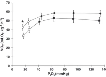
The Hyp-ctr group (intactfish submitted to hypoxia) showed a
relatively constantV_O2(~58 mlO2kg−1h−1) during graded hypoxia.
The Hyp-opr group (fish with thefirst gill arches excised and exposed
to hypoxia) displayed a similar response pattern. When compared to
the Hyp-control, the Hyp-opr showed significantly lowerV_O2(21%)
at the lowestPiO2(17 mmHg)(Fig 1).
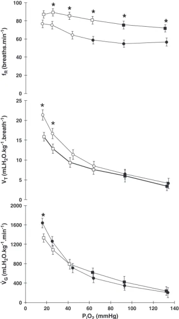
During graded hypoxiaV_Gincreased significantly in both intact and
operated groups, reaching a maximum at aPiO2of 16 mmHg (~696%
-control) and 17 mmHg (~455% - operated). Compared to the normoxic
values, both groups showed significant increases inV_Gstarting from a
PiO2of 44 and 41 mmHg (Hyp-ctr and Hyp-opr, respectively -Fig. 2).
At the lowestPiO2the Hyp-oprfish displayed a significant lowerV_Gin
comparison to the Hyp-ctr animals (Fig. 2).
In both intact and operated groups, the increases in V_G were
achieved by larger increments inVTcompared tofR. Significant
differ-ences inVTwere detected at aPiO2of 25 and 16 mmHg (Hyp-ctr) and
25 and 17 mmHg (Hyp-opr) (Fig. 2).
In the Hyp-ctr group, fR increased gradually from a PiO2 of
44 mmHg to 16 mmHg. Similarly,fish of the Hyp-opr group increased
fRgradually from aPiO2of 63 mmHg to 26 mmHg, below which it
remained nearly constant. The Hyp-oprfish had significant higherfR
0 10 20 30 40 50 60 70
0 20 40 60 80 100 120 140
*
.
PiO2(mmHg)
VO
2
(mLO
2
.kg
-1.h -1)
values (25 to 35%) than the Hyp-ctr, except at the most hypoxicPiO2 (Fig. 2).
The Hyp-ctr group maintainedEO2constant asPiO2was reduced
from normoxia to 44 mmHg, below which a significant decrease was observed. The Hyp-opr group showed a similar pattern by maintaining
a constantEO2(~60 to 65%) from normoxia to the most hypoxicPiO2.
The Hyp-ctr group had significantly higher EO2 than Hyp-opr in
normoxia and at aPiO2of 93 mmHg (Fig. 3).
The effects of graded hypoxia onfHare shown inFig. 4. The Hyp-ctr
group had a constantfH (~45 bpm) at allPiO2. The Hyp-opr fish
displayed constantfHfrom normoxia until aPiO2of 41 mmHg. Below
this oxygen tension the operatedfish developed a significant
bradycar-dia, with a reduction of about 26% infHat the most hypoxic tension
(17 mmHg). In comparison to the control group, the Hyp-opr fish
showed significantly higherfH(~31%) from normoxia until aPiO2of
41 mmHg. Below this tension both groups had practically the samefH
values.
3.2. Responses to NaCN
Both NaCN-ctr (intact fish exposed to NaCN injections) and
NaCN-opr (fish with thefirst gill arches excised and exposed to NaCN
injections) groups showed the same response pattern in relation to
in-ternal and exin-ternal NaCN injections: increase ofV_Gresulting from
in-creases infRandVamp. Responses to internal injections lasted longer
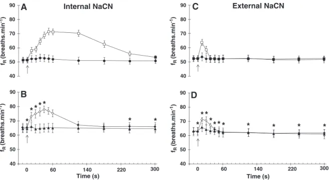
than to external ones in both groups. A 39% increase infRwas detected
in the NaCN-ctr group 50 s after the internal injection of NaCN. This var-iable returned to resting values about 5 min after injection (Fig. 5A). In
the NaCN-opr group thefRincreased approximately 19% (40 s), in
re-sponse to internal NaCN (Fig. 5B) returning to resting values in the 2nd min. The external NaCN injection provoked the largest increase
(21%) infRjust 10 s after the injection in the NaCN-ctr, returning to
pre-injection values before the end of the 1st min (Fig. 5C). Within
the same interval the NaCN-opr exhibited a 13% increase infR, returning
to initial values in the 1st min post-injection (Fig. 5D).
In the NaCN-ctr group,Vampincreased 33% after internal injection
of NaCN, and returned to pre-injection values after the 2nd min
(Fig. 6A). In NaCN-opr group,Vampincreased approximately 18%, in
re-sponse to internal NaCN, returning to resting values in the 2nd min (Fig. 6B).
0 5 10 15 20 25
*
*
0 20 40 60 80 100
*
*
*
*
*
0 400 800 1200 1600 2000
0 20 40 60 80 100 120 140
PiO2 (mmHg)
*
. VT
(mLH
2
O.kg
-1.breath -1) fR
(breaths.min
-1)
VG
(mLH
2
O.kg
-1.min -1)
Fig. 2.The effect of graded hypoxia on total gill ventilation (V_G- mlH2O kg−1min−1),
breathing frequency (fR- breaths min−1) and tidal volume (VT- mlH2O kg−1resp−1)
ofOreochromis niloticusduring graded reduction ofPiO2. Control group -●(n = 10),
operated group -■(n = 10). Open symbols indicate values that are significantly dif-ferent from initial (normoxic) values. * indicates values that are significantly different in relation to the control group (Pb0.05). Values are mean ± S.E.M.
20 40 60 80 100
0 20 40 60 80 100 120 140
*
*
PiO2 (mmHg)
EO
2
(%)
Fig. 3.The effect of graded hypoxia in the O2extraction from the ventilatory current
(EO2- %) ofOreochromis niloticus. Control group -●(n = 10), operated group -■(n = 10). Open symbols indicate values that are significantly different from initial (normoxic) values. * indicates values that are significantly different in relation to the control group (Pb0.05). Points are mean ± S.E.M.
0 10 20 30 40 50 60 70
0 20 40 60 80 100 120 140
PiO2 (mmHg)
*
*
*
*
fH
(bpm)
Fig. 4.The effect of graded hypoxia on heart rate (fH- bpm) ofOreochromis niloticus.
External NaCN injections provoked an increase of 34% inVampjust 10 s after the injection in the NaCN-ctr, and quickly returned to
rest-ing values (Fig. 6C), while in NaCN-opr,Vampincreased by 38% at 10 s,
returning to initial values within the 1st min post-injection (Fig. 6D).
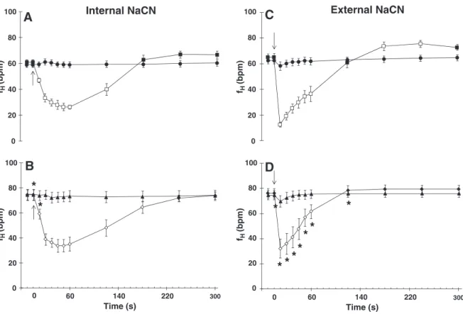
Both internal and external NaCN injections also affectedfH(Fig. 7).
In-ternal NaCN injection evoked pronounced bradycardia in both NaCN-ctr
(56%) and NaCN-opr (46%) groups during the 1st min post-injection. In
both groups,fHreturned to control levels after 4–5 min (Fig. 7A and
7B). In response to external NaCN, the NaCN-ctr group showed a brief
but pronounced bradycardia (reduction of 80% infHduring thefirst
10 s post-injection), then a significant tachycardia after about 2 min.
This tachycardia persisted for the next 2 min and returned to initial fR
(breaths.min
-1)
40 50 60 70 80 90
60 140 220 300
Time (s) fR
(breaths.min
-1)
40 50 60 70 80 90
fR
(breaths.min
-1)
0
B
*
* *
* *
*
*
Time (s)
D
* *
****
*
*
*
*
*
40 50 60 70 80 90
fR
(breaths.min
-1)
40 50 60 70 80 90
A
Internal NaCN
C
External NaCN
60 140 220 300
0
Fig. 5.Changes in breathing frequency (fR- breaths min−1) ofOreochromis niloticusfollowing internal injections of saline (0.5 ml), NaCN (0.5 ml of 750μg ml−1) and external
injections of H2O (1.0 ml) and NaCN (1.0 ml of 750μg ml−1).AandCcontrol (n = 10),BandDoperated group (n = 10). The arrow indicates the moment of injection. Saline
●,▲and NaCN■,♦, H2O●,▲and NaCN■,♦. Open symbols represent significant differences in relation to the initial values (pre-injection). * indicates values that are significantly
different between groups. (Pb0.05). Points are mean ± S.E.M.
0,2 0,4 0,6 0,8
1
Internal NaCN
A
C
External NaCN
D
*
*
*
Time (s)
*
*
B
Time (s) Vamp
(mmHg)
0,2 0,4 0,6 0,8 1
Vamp
(mmHg)
Vamp
(mmHg)
Vamp
(mmHg)
300 220
140 60
0 300
220 140
60 0
0,2 0,4 0,6 0,8 1 0,2 0,4 0,6 0,8 1
Fig. 6.Changes in ventilatory amplitude (Vamp- mmHg) ofOreochromis niloticusfollowing internal injections of saline (●,▲) NaCN (■,♦), and external injections of H2O (●,▲) and
values after 5 min (Fig. 7C). A similar bradycardia was observed in the
NaCN-opr group, but was less pronounced (reduction of 57% infH)
than in the control group (reduction of 80% infH) and there was no
sub-sequent tachycardia (7D). The cardiorespiratory variables were signifi-cantly attenuated after the NaCN injections.
4. Discussion
4.1. Oxygen uptake
Both Hyp-ctr and Hyp-opr groups displayed practically the same _
VO2values in response to graded hypoxia, except at the lowestPiO2
(16 mmHg and 17 mmHg, respectively). Similar results were
ob-served byDuthie and Hughes (1987)in rainbow troutOncorhynchus
mykisswith the 1st or 2nd gill arches cauterized. Significant
differ-ences in the V_O2 of intact and operated O.mykisswere detected
only when thefish were forced to swim. Moreover, in the operated
group theV_O2max(maximumV_O2attained at the highest swimming
speed) was lower than that of the intact group. According to these authors, the reduction of functional gill surface area led to a
propor-tional decrease in V_O2 max, indicating that the fish were unable to
make compensatory adjustments in cardiac performance to enhance
theirV_O2. Under these conditions of maximum aerobic demand, all
available gill area would appear to be fully perfused to ensure O2
sup-ply to the tissues. This highlights the necessity of the entire gill surface area when the recruitment of a larger number of secondary lamellae is required (Duthie and Hughes, 1987), such as in severe hypoxia.
The similar V_O2 values during graded hypoxia in Hyp-ctr and
Hyp-opr groups, except at the lowestPiO2, also indicate that removal
of thefirst gill arch did not compromise the ability of the tilapia to
reg-ulate metabolism through adjustments ofV_Gand gill perfusion.
4.2. Ventilatory responses
Mostfish species studied to date are able to maintain a constantV_O2
in response to graded hypoxia, until a critical O2tension (PcritO2), by
raisingV_G(Randall et al., 1997). This is achieved by increases in both
fRandVT(Randall, 1982). Hypoxia tolerant species, such asO. niloticus,
normally increaseV_Gthrough a more profound increase inVTas
com-pared tofR. This could be a strategy to lower energy costs of the
homeo-static response, assuming that conservation of a constant velocity of muscular contraction is energy saving, whereas a higher frequency of contraction is limited by work against higher internal blood viscosity and high viscosity of water (Johansen et al., 1967; Rantin et al., 1992).
This strategy was observed infish of both experimental groups. The
pat-terns of change inVTandfRare similar to previous studies on this
spe-cies, reported byFernandes and Rantin (1987),Martins et al. (2011)
andThomaz et al. (2009).
Increases inVTwere recorded in both experimental groups. This
re-sponse differs from that observed byPerry and Reid (2002)inO. mykiss
exposed to hypercarbia, where the increase in VT was completely
abolished followingfirst gill arch excision. Denervation of thefirst gill
arch attenuated the increases in bothVTandfRinP. mesopotamicus
exposed to graded hypoxia (Leite et al., 2007). A similar response was
also found in the bowfin,Amia calva, but only after total gill denervation
and excision of the pseudobranch (Mckenzie et al., 1991). The location
and orientation of the chemoreceptors involved in reflex
cardiorespira-tory responses to hypoxia are extremely variable among species of
water and air-breathing fishes, with no unifying pattern (Milsom,
2012). InO. mykiss, ablation of the 1st gill arch abolished reflex
brady-cardia and the increase inVampin response to increased water CO2
levels, hypercarbia (Perry and Reid, 2002). However, other chemore-ceptors are also involved in the cardiorespiratory responses to hypoxia.
In dogfish,Scyliorhinus canicula, receptors mediating bradycardia were
distributed on all gill arches, but were also located throughout the
0 20 40 60 80 100
fH
(bpm)
fH
(bpm)
Internal NaCN
A
0 20 40 60 80 100
0 20 40 60 80 100
fH
(bpm)
fH
(bpm)
0 20 40 60 80 100
60 140 220 300
0
Time (s)
B
*
*
External NaCN
C
D
*
* *
*
*
*
*
*
Time (s)
300
220 140
60 0
Fig. 7.Changes in heart rate (fH- bpm) ofOreochromis niloticusfollowing internal injections of saline (●,▲) NaCN (■,♦), and external injections of H2O (●,▲) and NaCN (■,♦).
oro-branchial cavity, innervated by cranial nerves V (trigeminal) and VII
(facial) (Butler et al., 1977). InP. mesopotamicus, the O2chemoreceptors
mediating reflex bradycardia were located on all gill arches while
the receptors mediating fR responses are possibly located in the
pseudobranch and/or in some extrabranchial sites (Leite et al., 2007). Colossoma macropomumalso possesses extrabranchial O2
chemorecep-tors mediating ventilatory responses (Milsom, 2002).Hoplias lacerdae
possesses receptors triggering alterations infRlocated exclusively in
the 1st gill arch, while those mediatingfHare distributed over all gill
arches (Micheli-Campbell et al., 2009). On the other hand, in the
co-generic species H. malabaricusreflex bradycardia is mediated by
receptors restricted to the 1st gill arch, whereas increases infRare
in-duced by chemoreceptors distributed over the other arches (Sundin et al., 1999).
In the present study, excision of thefirst gill arch did not abolish
ventilatory responses to graded hypoxia. However, theV_GandVTof
opr at 17 mmHg were attenuated when compared to the
Hyp-ctr at 16 mmHg. A decrease in thefRof the Hyp-opr group was also
observed at thisPiO2, accompanied by a ~33% reduction inV_O2.
When comparing both groups, thefRof Hyp-opr was significantly
higher at allPiO2, except at the deepest level of hypoxia. This strategy
was probably needed to compensate the reduction in the respiratory
surface area and ensure the O2uptake required to maintain metabolic
rate.
The cardiorespiratory responses to NaCN are well documented in fishes (Burleson and Smatresk, 1990; Burleson and Milssom, 1995; Mckenzie et al., 1995; Sundin et al., 1999; Milsom, 2002; Leite et al., 2007; Micheli-Campbell et al., 2009). The NaCN acts as a blocker of ox-idative phosphorylation in the mitochondria, thus stimulating the chemoreceptors (Burleson et al., 1992). In the present study gill
exci-sion attenuated, but did not abolish, reflex ventilatory responses in the
NaCN-opr group, except for Vamp following the external injection
(Fig. 6D). A similar attenuation was found in theVampofH. lacerdae
after denervation of thefirst gill arch (Micheli-Campbell et al., 2009)
and in thefRofP. mesopotamicusin response to internal NaCN
injec-tion (Leite et al., 2007).
In both NaCN-ctr and NaCN-opr groups, a larger increase inVamp
was observed compared tofRin response to external NaCN injections,
as observed in hypoxia. However, the internal NaCN injections caused
increases of same magnitude infRand Vamp in both experimental
groups.
Fish species inhabiting hypoxic environments often need more O2
sensitivity to rapidly detect and trigger compensatory mechanisms.
As an example,H. malabaricus, which is found in hypoxic habitats,
presents O2 chemoreceptors mediating the fRand Vamp on all gill
arches, and also at extrabranchial sites (Sundin et al., 1999). By
con-trast,H. lacerdae, a co-generic species that typically inhabits well
ox-ygenated waters, only possesses internally oriented chemoreceptors
mediatingfRin thefirst gill arch (Micheli-Campbell et al., 2009). As
a hypoxic tolerant species, O. niloticuspossess O2 chemoreceptors
mediating the ventilatory variables (fRandVamp) with similar
distri-bution and orientation to those found in the gills ofH. malabaricus.
The highEO2ofO. niloticus(70 to 80%) has been observed
previ-ously in this species exposed to hypoxia (Fernandes and Rantin, 1986a; Kalinin et al., 1999; Thomaz et al., 2009; Martins et al., 2011). Taking into consideration the fundamental equation of
respi-ratory physiology proposed byDejours (1981):
_ VG=V_O2
:EO2:PiO2¼1;
the maintenance of a constant oxygen uptake when the
environ-ment O2concentration declines is possible by means of an increase
in V_G and/or inEO2. Due to a limited capacity to increaseEO2
in-creases infishes, enhancements inV_Gare required to maintain a
con-stantV_O2(Kalinin et al., 1999; Martins et al., 2011). Thus, in hypoxia
the Hyp-ctr group keptEO2constant until aPiO2of 44 mmHg and the
Hyp-oprfish were also able to maintain a constantEO2at all
experi-mentalPiO2.
According toDuthie and Hughes (1987), the secondary lamellae of
thefirst pair of gill arches are responsible for about 30% of the total
respiratory surface area inO. mykiss. Assuming this is also true of
O. niloticus, despite a reduction of this magnitude in the functional
surface area, the Hyp-oprfish were still able to maintain anEO2of
around 65%. This presumably required increased blood perfusion
and lamellar recruitment in the other three gill arches.Fernandes
and Rantin (1986b)reported that the second to fourth gill arches of O. niloticushave a high density of secondary lamellae, which will
have received the entire cardiac output in the absence of thefirst
arch, thus potentially eliciting greater than normal lamellar
recruit-ment in the Hyp-oprfish.
4.3. Cardiac responses
The Hyp-oprfish showed higher restingfHthan the Hyp-ctr. The
same tendency was found inP. mesopotamicus(Leite et al., 2007) and
H. lacerdae(Micheli-Campbell et al., 2009) when these species had
theirfirst gill arch denervated. The increase infHcould be related to
some alteration in the resting vascular tone of the gill vessels or to an alteration of the afferent information from the gills, which modulated cardiac activity (Leite et al., 2007). After atropine injection, normoxic H. malabaricuswith thefirst gill arch denervated increasedfH(Sundin et al., 1999). These authors suggested that cardiac cholinergic modula-tion was altered by denervamodula-tion, although some degree of cardiac control remained even after complete gill denervation. Thus, it is
most likely that the increase infHobserved in the Hyp-opr group
was related to both a decrease of branchial vascular resistance and to the removal of sensory information from the gills, as suggested byLeite et al. (2007).
Fish usually respond to hypoxia with a reduction infH, a hypoxic
bra-dycardia (Randall, 1982; Taylor, 1992). This cardiac response has been documented for many teleosts species, but its physiological significance is not completely understood. Among suggested benefits, the reflex response (Fritsche and Nilsson, 1989; Burleson and Smatresk, 1990) would increase the blood residence time inside the secondary lamellae,
leading to an improvement in O2diffusion from water to the blood, thus
increasing the diffusing capacity and O2extraction by the gills (Farrell
et al., 1980; Gilmour and Perry, 2007). Alternatively, Glass et al. (1991)suggested a causal relationship between cardiac dysfunction
and hypoxic bradycardia in carp,Cyprinus carpio. To date, it is not
en-tirely clear to what extent hypoxic bradycardia infish is a regulatory
response or a consequence of cardiac dysfunction, or both. For example, pharmacological or surgical abolition of hypoxic bradycardia did not
significantly affect branchial gas exchange in O. mykiss (Perry and
Desforges, 2006), or regulation of metabolism during hypoxia in
Atlan-tic cod,Gadus morhua(Mckenzie et al., 2009) and European eel,Anguilla
anguilla(Iversen et al., 2010). The hypoxic bradycardia may improve O2 diffusion to cardiac myocytes by prolonging the diastolic time and, con-sequently, the residence time of the blood inside the heart lumen. Thus, hypoxic bradycardia could be a protective response to the highly aero-bic cardiac muscle (Taylor et al., 1999). In the present study the Hyp-ctr group did not display consistent bradycardia and the Hyp-opr
devel-oped significant bradycardia only at aPiO2of 25 mmHg and below
(Fig. 4).
A different response was found inO. mykiss, in which bradycardia
was abolished after excision of thefirst pair of gill arches (Perry and
Reid, 2002) and inH. malabaricusafter denervation of the first gill
arches (Sundin et al., 1999). Denervation of thefirst gill arch did not,
however, abolish hypoxic bradycardia inP. mesopotamicus(Leite et al.,
2007),H. lacerdae(Micheli-Campbell et al., 2009) andC. macropomum, (Sundin et al., 2000), indicating the presence of chemoreceptors
the hypoxic bradycardia persisted after bilateral ablation of thefirst pair of gill arches.
Regarding O2chemoreceptors orientation, according toBurleson et al.
(1992)the O2chemoreceptors mediatingfHreflexes are externally
orient-ed and locatorient-ed on thefirst gill arch in most water-breathing teleosts, such
asG. morhua(Fritsche and Nilsson, 1989) and sea raven,Hemitripterus americanus(Saunders and Sutterlin, 1971). However, more recent studies
have revealed various different orientations for these receptors.Hoplias
malabaricuspossess chemoreceptors modulating thefHresponses that
are internally oriented, located exclusively in thefirst gill arch (Sundin
et al., 1999). Moreover,C. macropomum(Sundin et al., 2000),M. cephalus
(Shingles et al., 2005),P. mesopotamicus(Leite et al., 2007),H. lacerdae
(Micheli-Campbell et al., 2009) andH. unitaeniatus(Lopes et al., 2010),
exhibit internally and externally oriented chemoreceptors distributed
over all gill arches, that elicit reflex bradycardia.
The attenuated bradycardia in the NaCN-opr is similar to responses
observed in the catfish,Ictalurus punctatus(Burleson and Smatresk,
1990) and after external NaCN injections inP. mesopotamicus(Leite et al., 2007), both with thefirst pair of gill arches denervated. These findings suggest that inO. niloticusthe O2chemoreceptors modulating
thefHresponses are internally and externally oriented, located in the
first gill arch, but also present in other gill arches.
5. Conclusions
In summary, the cardiorespiratory variables involved on the re-sponses to graded hypoxia were not completely abolished by the
ex-cision of thefirst pair of gill arches ofO. niloticusand theV_O2andEO2
were not drastically reduced. Moreover, responses to internal and external injections of NaCN were attenuated, but not abolished, in O. niloticuswith thefirst pair of gill arches excised. The data suggest thatO. niloticuspossess internally and externally oriented O2 chemo-receptors, distributed over all gill arches, mediating reflex homeo-static cardiorespiratory adjustments in response to hypoxia.
Acknowledgments
This research was supported by The São Paulo State Research
Foundation–FAPESP (Proc. 2010/03599-2). The authors are thankful
to Juliana Thomaz and Rafael Zaneli for their technical support and Dr. David J. McKenzie for the text revision.
This study was performed in accordance with the rules of the Brazilian Federal Legislation and was approved by the Ethical Committee on Animal Experimentation, Federal University of São Carlos (Authorization No. 015/2010).
References
Axelsson, M., Fritsche, R., 1994.Cannulation techniques. In: Hochachka, P.W., Mommsen, T.P. (Eds.), Analytical Techniques. Biochemistry and Molecular Biology of Fishes. Elsevier, Amsterdam, pp. 17–36.
Barret, D.J., Taylor, E.W., 1984.Changes in heart rate during progressive hypoxia in the dogfish,Scyliorhinus caniculaL.: evidence for a venous oxygen receptor. Comp. Biochem. Physiol A 78, 697–703.
Burleson, M.L., Milsom, W.K., 1993.Sensory receptors in the 1st gill arch of rainbow-trout. Resp. Physiol. 93, 97–110.
Burleson, M.L., Milsom, W.K., 1995.Cardio-ventilatory control in rainbow trout: I. Pharma-cology of branchial, oxygen-sensitive chemoreceptors. Resp. Physiol. 100, 231–238.
Burleson, M.L., Smatresk, N.J., 1990.Evidence for two oxygen-sensitive chemoreceptor loci in channel catfish,Ictalurus punctatus. Physiol. Zool. 63, 208–221.
Burleson, M.L., Smatresk, N.J., Milsom, W.K., 1992.Afferent inputs associated with cardioventilatory control infish. Fish Physiol 12 B, 389–426.
Butler, P.J., Taylor, E.W., Short, S., 1977.The effect of sectioning cranial nerves V, VII, IX and X on the cardiac response of the dogfishScyliorhinus caniculato environmental hypoxia. J. Exp. Biol. 69, 233–245.
Campbell, H.A., Egginton, S., 2007.The vagus mediates cardiorespiratory coupling that changes with metabolic demand in a temperate nototheniodfish. J. Exp. Biol. 210, 2472–2480.
Chervinsk, J., 1982.Environmental physiology of tilapias. In: Pullin, R.S.V., Lowe-Mcconnel, R.H. (Eds.), The Biology and Culture of Tilapias, ICLARM Conference Proceedings 7.
International Center for Living Aquatic Resources Managements, Manila, Philippines, p. 432.
Coolidge, E.H., Ciuhandu, C.S., Milsom, W.K., 2008.A comparative analysis of putative oxygen-sensing cells in thefish gill. J. Exp. Biol. 211, 1231–1242.
Daxboeck, C., Holeton, G.F., 1978.Oxygen receptors in the rainbow trout,Salmo gairdneri. Can. J. Zool. 56, 1254–1256.
Dejours, P., 1981.Principles of Comparative Respiratory Physiology, 2nd ed. Elsevier, New York.
Duthie, G.G., Hughes, G.M., 1987.The effects of reduced gill area and hyperoxia on the ox-ygen consumption and swimming speed of rainbow trout. J. Exp. Biol. 127, 349–354.
Farrell, A.P., Sobin, S.S., Randall, D.J., Crosby, S., 1980.Intralamellar bloodflow patterns infish gills. Am. J. Physiol. 239, 428–436.
Fernandes, M.N., Rantin, F.T., 1986.Lethal temperatures ofOreochromis niloticus(Pisces, Cichlidae). Rev. Bras. Biol. 46, 589–595.
Fernandes, M.N., Rantin, F.T., 1986.Gill morphometry of cichlidfish,Oreochromis (Sarotherodon) niloticus(Pisces, Teleostei). Ciência. e Cult. 38, 192–198.
Fernandes, M.N., Rantin, F.T., 1987.Respiratory responses ofOreochromis niloticus(Pisces, Cichlidae) to environmental reductions of dissolved oxygen. Bol. Fisiol. Anim. 11, 51–60.
Fernandes, M.N., Rantin, F.T., 1989.Respiratory responses ofOreochromis niloticus
(Pisces, Cichlidae) to environmental hypoxia under different thermal conditions. J. Fish. Biol. 35, 509–519.
Fernandes, M.N., Rantin, F.T., 1994.Relationships between oxygen availability and metabolic cost of breathing in Nile Tilapia (Oreochromis niloticus): aquacultural consequences. Aquacult. 127, 339–346.
Florindo, L.H., Leite, C.A.C., Kalinin, A.L., Reid, S.G., Milsom, W.K., Rantin, F.T., 2006.The role of branchial and orobranchial O2chemoreceptors in the control of aquatic
surface respiration in the neotropicalfish tambaqui (Colossoma macropomum): progressive responses to prolonged hypoxia. J. Exp. Biol. 209, 1709–1715.
Fritsche, R., Nilsson, S., 1989.Cardiovascular responses to hypoxia in the Atlantic cod,
Gadus morhua. Exp. Biol. 48, 153–160.
Gilmour, K.M., Perry, S.F., 2007.Branchial chemoreceptor regulation of cardiorespiratory function. Fish Physiol 25, 97–120.
Glass, M.L., Rantin, F.T., Verzola, R.M.M., Fernandes, M.N., Kalinin, A.L., 1991. Cardio-respiratory synchronization and myocardial function in hypoxic carp,Cyprinus carpio. J. Fish. Biol. 39, 142–149.
Hughes, G.M., 1973.Respiratory responses to hypoxia infish. Am. Zool. 13, 475–489.
Hughes, G.M., Saunders, R.L., 1970.Responses of respiratory pumps to hypoxia in the rainbow trout,Salmo gairdneri. J. Exp. Biol. 53, 529–545.
Iversen, N.K., Mckenzie, D.J., Malte, H., Wang, T., 2010.Reflex bradycardia does not infl u-ence oxygen consumption during hypoxia in the European eel (Anguilla anguilla). J. Comp. Physiol. B 180, 495–502.
Jia, X.X., Burggren, W.W., 1997.Developmental changes in chemoreceptive control of gill ventilation in larval bullfrogs (Rana Catesbeiana). J. Exp. Biol. 200, 2237–2248.
Johansen, K., Lenfant, C., Grigg, G.C., 1967.Respiratory control in lungfish. Comp. Biochem. Physiol. 20, 835–854.
Jonz, M.G., Nurse, C.A., 2003.Neuroepithelial cells and associated innervation of the zebrafish gill: a confocal immunofluorescence study. J. Comp. Neurol. 461, 1–17.
Jonz, M.G., Fearon, I.M., Nurse, C.A., 2004.Neuroepithelial oxygen chemoreceptors of the zebrafish gill. J. Physiol. 560, 737–752.
Kalinin, A.L., Glass, M.L., Rantin, F.T., 1999.A comparison of directly measured and es-timated gill ventilation in the Nile tilapia, Oreochromis niloticus. Comp. Biochem. Physiol. A 122, 207–211.
Lague, S.L., Speers-Roesch, B., Richards, J.G., Farrell, A.P., 2012.Exceptional cardiac anoxia tolerance in tilapia (Oreochromis hybrid). J. Exp. Biol. 215, 1354–1365.
Laurent, P., Rouzeau, J.D., 1972.Afferent neural activity from the pseudobranch of teleost. Effects of PO2, pH, osmotic pressure and Na+ions. Resp. Physiol. 14, 307–331.
Leite, C.A.C., Florindo, L.H., Kalinin, A.L., Milsom, W.K., Rantin, F.T., 2007.Gill chemore-ceptors and cardio-respiratory reflexes in the neotropical teleost pacu, Piaractus mesopotamicus. J. Comp. Physiol. A 193, 1001–1011.
Lopes, J.M., Boijink, C.H., Florindo, L.H., Leite, C.A.C., Kalinin, A.L., Milsom, W.K., Rantin, F.T., 2010.Hypoxic cardiorespiratory reflexes in the facultative air-breathingfish jeju (Hoplerythrinus unitaeniatus): role of branchial O2chemoreceptors. J. Comp.
Physiol. B 180, 797–811.
Martins, N.D., Colvara, W.A., Rantin, F.T., Kalinin, A.K., 2011.Microcystin-LR: How it affects the cardio-respiratory responses to hypoxia in Nile tilapia,Oreochromis niloticus. Chemosphere 84, 154–159.
McKenzie, D.J., Burleson, M.L., Randall, D.J., 1991.The effects of branchial denervation and pseudobranch ablation on cardio-ventilatory control on an air-breathingfish. J. Exp. Biol. 161, 347–365.
McKenzie, D.J., Taylor, E.W., Bronzi, P., Bolis, C.L., 1995.Aspects of cardioventilatory control in the adriatic sturgeon (Acipenser naccarii). Resp. Physiol. 100, 45–53.
McKenzie, D.J., Skov, P.T., Taylor, E.W., Wang, T., Steffensen, J.F., 2009.Abolition of re-flex bradycardia by cardiac vagotomy has no effect on the regulation of oxygen uptake by Atlantic cod in progressive hypoxia. Comp. Biochem. Physiol. A 153, 332–338.
Micheli-Campbell, M.A., Campbell, H.A., Kalinin, A.L., Rantin, F.T., 2009.The relation-ship between O2chemoreceptors, cardio-respiratory reflex and hypoxia
toler-ance in the neotropicalfishHoplias lacerdae. Comp. Biochem. Physiol. A 154, 224–232.
Milsom, W.K., 2002.Phylogeny of CO2and pH chemoreception in vertebrates. Respir.
Physiol. Neurobiol. 131, 29–41.
Milsom, W.K., 2012.New insights into gill chemoreception: receptor distribution and roles in water and air breathingfish. Respir. Physiol. Neurobiol. 184, 326–339.
Perry, S.F., Reid, S.G., 2002.Cardiorespiratory adjustments during hypercarbia in rain-bow troutOncorhynchus mykissare initiated by external CO2receptors on the
first gill arch. J. Exp. Biol. 205, 3357–3365.
Porteus, C.S., Brink, D.L., Milsom, W.K., 2012.Neurotransmitter profiles infish gills: putative gill oxygen chemoreceptors. Respir. Physiol. Neurobiol. 184, 316–325.
Randall, D.J., 1982.The control of respiration and circulation infish during exercise and hypoxia. J. Exp. Biol. 100, 275–288.
Randall, D.J., Jones, D.R., 1973.The effects of deafferenation of the pseudobranch on the respiratory response to hypoxia and hyperoxia in the trout (Salmo gairdneri). Resp. Physiol. 17, 291–302.
Randall, D.J., Burggren, W., French, K., 1997.Gas exchange and acid–base balance. In: Randall, D.J., Burggren, W., French, K. (Eds.), Animal Physiology–Mechanisms and Adaptations. WH Freeman and Co., New York, pp. 517–570.
Rantin, F.T., Kalinin, A.L., Glass, M.L., Fernandes, M.N., 1992.Respiratory responses to hypoxia in relation to mode of life of two erythrinid species (Hoplias malabaricus
andHoplias lacerdae). J. Fish. Biol. 41, 805–812.
Saunders, R.L., Sutterlin, A.M., 1971.Cardiac and respiratory responses to hypoxia in the sea raven,Hemitripterus americanus, and an investigation of possible control mechanisms. J. Fish. Res. Board. Can. 28, 491–503.
Shingles, A., McKenzie, D.J., Claireaux, G., Domenici, P., 2005.Reflex cardioventilatory responses to hypoxia in theflathead grey mullet (Mugil cephalus) and their behav-ioural modulation by perceived threat of predation and water turbidity. Physiol. Biochem. Zool. 78, 744–755.
Smatresk, N.J., Burleson, M.L., Azizi, S.Q., 1986.Chemoreflexive responses to hypoxia and NaCN in longnose gar: evidence for two chemoreceptive loci. Am. J. Physiol. 251, 116–125.
Smith, F.M., Davies, P.S., 1984.Effects of sectioning cranial nerves IX and X on the cardiac response to hypoxia in the coho salmon,Oncorhynchus kisutch. Can. J. Zool. 62, 766–768.
Speers-Roesch, B., Sandblom, E., Lau, G.Y., Farrell, A.P., Richards, J.G., 2010.Effects of environmental hypoxia on cardiac energy metabolism and performance in tilapia Am. J. Physiol. Regul. Integr. Comp. Physiol. 298, 104–119.
Steffensen, J.F., 1989.Some errors in respirometry of aquatic breathers: how to avoid and correct for them. Fish Physiol. Biochem. 6, 49–59.
Sundin, L.I., Reid, S.G., Kalinin, A.L., Rantin, F.T., Milsom, W.K., 1999.Cardiovascular and re-spiratory reflexes: the tropicalfish, traira (Hoplias malabaricus) O2chemoresponses.
Resp. Physiol. 116, 181–199.
Sundin, L., Reid, S.G., Rantin, F.T., Milsom, W.K., 2000.Branchial receptors and cardiore-spiratory reflexes in a neotropicalfish, the tambaqui (Colossoma macropomum). J. Exp. Biol. 203, 1225–1239.
Taylor, E.W., 1992.Nervous control of the heart and cardiorespiratory interactions. Fish Physiol 12B, 343–387.
Taylor, E.W., Jordan, D., Coote, J.H., 1999.Central control of the cardiovascular and re-spiratory systems and their interactions in vertebrates. Physiol. Revs. 79, 855–916.
Thomaz, J.M., Martins, N.D., Monteiro, D.A., Rantin, F.T., Kalinin, A.L., 2009. Cardio-respiratory function and oxidative stress biomarkers in Nile tilapia exposed to the organophosphate insecticide trichlorfon (NEGUVON). Ecotoxicol. Environ. Saf. 72, 1413–1424.