UNIVERSIDADE FEDERAL DE MINAS GERAIS
INSTITUTO DE CIÊNCIAS BIOLÓGICAS
DEPARTAMENTO DE BIOLOGIA GERAL
PROGRAMA DE PÓS-GRADUAÇÃO EM GENÉTICA
PhD Thesis
Pan-genomic analyses of
Corynebacterium pseudotuberculosis
and
characterization of the biovars
ovis
and
equi
through comparative
genomics
ORIENTADO:
Siomar de Castro Soares
SUPERVISOR
: Prof. Dr. Vasco Ariston de Carvalho Azevedo
CO-
SUPERVISOR
:
PD. Dr. Andreas Tauch
BELO HORIZONTE
Siomar de Castro Soares
Pan-genomic analyses of
Corynebacterium pseudotuberculosis
and
characterization of the biovars
ovis
and
equi
through comparative
genomics
.
SUPERVISOR: Prof. Dr. Vasco Ariston de Carvalho Azevedo
CO-SUPERVISOR: PD. Dr. Andreas Tauch
BELO HORIZONTE
August - 2013
Thesis
presented
as
partial
ACKNOWLEDGEMENTS
I would like to thank everybody who worked with or stood by me since 2007, when I first decided to begin my research way of life.
Collaborators:
I would like to thank:
Prof. Dr. Vasco Ariston de Carvalho Azevedo, for the supervision during the
development of my work; for the non-academic advices; for the time expended during
my own development; for seeking the best for all of your students, no matter who;
and, also, for the friendship.
PD. Dr. Andreas Tauch, for the receptivity when I arrived in Germany; for the time
expended, and also patience, when reviewing every section of the works we have
developed in Germany; for giving me the opportunity to take part of CLIB; and, also,
for coming to Belo Horizonte for my Ph.D. defense.
Prof. Dr. Artur Silva, for the great collaboration during the development of the work;
and, for the all opportunities to give talks and conferences in Pará, together with the
LPDNA group, which helped improving my knowledge.
Prof. Dr. Anderson Miyoshi, for the supervision during my master's, which helped in
my own development, organization and writing skills; and, for the advices in all
presentations and manuscripts during the Ph.D.
Programs of Post-graduation in Genetics and Bioinformatics, for all the disciplines;
and, also, for the cooperation and support, mainly in the last steps, when trying to
organize flights and accommodations.
Laboratory of cellular and molecular Genetics, specially the long-date friends
Fernanda, Thiago, Luís, Marcela, Wanderson and several other friends, for all the
knowledge I have acquired from all of you; for the experience and maturity; and, of
course, for all the non-academic time we have expended together.
Center for Biotechnology and Cluster Industrial Biotechnology, specially Iris, Karina,
Arwa, Helena, Eva, Eugenie, Vimac, Anh, Mari and Fabian, for all academic and
non-academic support and friendship.
Laboratory of DNA polymorphism, specially Rommel, Adriana, Diego, Rafael, Hivana
and Leonardo, for the receptivity in Pará, collaboration, support and friendship.
Laboratory of Nanobiotechnology, specially Paula, Juliana, Lara, Galber, Fabiana and
Professors Carlos Ueira and Luiz Goulart, for the opportunity to work with this great
Family:
I would also like to thank:
My parents, for the faith you have in my future, since I was a kid; and, for the
comprehension, even when you do not understand why someone needs to be a
student for so long.
My brothers and sister for the support and great time I have every time I go back to
my hometown. Believe me, to be with you for one weekend is like one year anti-stress
therapy and it makes all the difference.
My nephews, the simple fact that you exist is enough to give strength to everybody on
the family and I work hoping that I may be a good reference for you in future the same
way you are my strength.
My wife, Letícia, only you know the backstage. In the words of Vasco, you give me
stability. I avoid explaining everything you represent because there are no words that
could explain all support, comprehension and peace. And, even if I would to try, half of
this thesis would not be enough. I hope I can do for you, during your newly started
“The only reason for time is
so that everything does not
happen at once.”
(Albert Einstein)
“If I have seen further it is
by standing on the shoulders
of Giants.”
Table of contents
List of Figures ... i
List of Tables ... ii
Abbreviations ... iii
Abstract ... 1
I. Presentation... 2
I.1 Collaborators ... 3
II. Preface ... 4
II.1 C. pseudotuberculosis - state of the art ... 5
II.1.1 Biovars of C. pseudotuberculosis ... 5
II.1.1.1 Biovar ovis ... 5
II.1.1.2 Biovar equi ... 8
II.2 Manuscript Structure and author's contributions ... 10
III. Introduction ... 12
III.1 Corynebacterium pathogenic species in next-generation genomic era: the use of EDGAR and PIPS software and the importance of pathogenicity islands identification in pan-genomic analyses of pathogenic species ... 13
IV. Goals ... 30
IV.1 Main goal ... 31
IV.2 Specific goals ... 31
V. Research Articles ... 32
V.1 Chapter I. PIPS: Pathogenicity Island Prediction Software ... 33
V.1.1 Appendix S1 ... 44
V.1.2 Figure S1 ... 45
V.1.3 Figure S2 ... 46
V.1.4 Table S1 ... 47
V.1.5 Discussion... 49
V.2.1 Discussion... 59
V.3 Chapter III. The Pan-Genome of the Animal Pathogen Corynebacterium pseudotuberculosis Reveals Differences in Genome Plasticity between the Biovar ovis and equi Strains ... 60
V.3.1 Figure S1 ... 75
V.3.2 Discussion... 76
VI. General Discussion ... 77
VII. Conclusions ... 79
VIII. Bibliography ... 81
IX. Appendices... 87
List of Figures
Figure 1. The whole genome of Corynebacterium pseudotuberculosis. ... 6
Figure 2. Gene regions encoding adhesive pili of C. pseudotuberculosis FRC41... 7
Figure 3. Model of pilus biogenesis... 8
Figure 4. Genomic map comparing strains of Corynebacterium pseudotuberculosis,
Corynebacterium ulcerans and Corynebacterium diphtheriae. ... 9
Figure S1. Prediction of PICD12 of C. diphtheriae with a different size than the
literature prediction. ... 45
Figure S2. Graphic representation of PAI features in the genome (A) and in the pathogenicity islands (B) of C. pseudotuberculosis and C. diphtheriae. ... 46
Figura 5. Heatmap showing the presence/absence of PAIs identified by PIPS in 13 strains of C. diphtheriae. ... 50
List of Tables
Abbreviations
BRIG CAPES
CDS CeBiTec CLA CLIB CMNR
CNPq
DNA Fapemig
G+C GEI LGCM
LPDNA MHC PAI PIPS RGMG RNA RPGP
rRNA tRNA UFMG
UFPA
Blast Ring Image Generator
Coordenação de Aperfeiçoamento de Pessoal de Nível Superior (Coordination for the Improvement of Higher Education Personnel)
Coding Sequence Center for Biotechnology Caseous Lymph Adenitis Cluster Industrial Biotechnology
Group composed of Corynebacterium, Mycobacterium, Nocardia and Rhodococcus
Conselho Nacional de Desenvolvimento Científico e Tecnológico (National Counsel of Technological and Scientific Development)
Deoxyribonucleic Acid
Fundação de Amparo à Pesquisa do Estado de Minas Gerais (Foundation for Research Support of the State of Minas Gerais)
Guanine + Thymine Genomic Island
Laboratório de Genética Celular e Molecular (Laboratory of Cellular and Molecular Genetics)
Laboratório de Polimorfismo de DNA (Laboratory of DNA Polimorphism) Major Histocompatibility Complex
Pathogenicity Island
Pathogenicity Island Prediction Software
Rede Genoma de Minas Gerais (Minas Gerais Genome Network) Ribonucleic Acid
Rede Paraense de Genômica e Proteômica (The Genomics and Proteomics Network of the State of Pará)
Ribosomal ribonucleic acid Transporter ribonucleic acid
Universidade Federal de Minas Gerais (Federal University of Minas Gerais)
Abstract
Corynebacterium pseudotuberculosis is the causative agent of diverse communicable
diseases in small ruminants (biovar ovis), horses, camels, buffalo and other animals
(biovar equi), which mainly differ in symptoms and site of infection. Additionally, the
diseases present a highly important economic problem worldwide and there is still a lack of efficient treatments against C. pseudotuberculosis. In this work, we describe the
steps from the first genome sequencing of a strain of C. pseudotuberculosis to the
pan-genomic analyses of 15 strains isolated from different hosts and countries with diverse symptoms. Briefly, we introduce the genus Corynebacterium and the in silico analyses
performed in pathogenic species of this genus to date. Then, we describe the implementation of a software for the prediction of pathogenicity islands (PAIs) in bacteria (PIPS), which outperformed the other available software, and identified 7 PAIs with important virulence factors in C. pseudotuberculosis biovar ovis. Moreover, we
extend the analyses of PAIs to strains of C. pseudotuberculosis biovar equi and predict
49 putative vaccine targets, in silico, which are commonly shared by both biovars, ovis
and equi. Finally, we present the phylogenomic, pan-genomic, core genomic,
singletons and genomic plasticity analyses of the 15 strains of C. pseudotuberculosis,
from both biovars. All the analyses performed here point for a clonal-like behavior of C.
pseudotuberculosis, which could be the result of the facultative intracellular behavior of
the species. Moreover, the biovar equi presents a higher variability in gene content
when compared to biovar ovis, specially in PAI regions. Noteworthy, the strains from
biovar ovis present a high degree of similarity in pili clusters of genes, whereas the
biovar equi strains are very variable. The conservation of pili clusters of genes in biovar ovis could account for the ability of these strains to spread inside host tissues and
I.1 Collaborators
This work was performed on the Laboratories of Molecullar and Celullar Genetics (LGCM) and DNA Polimorphism (LPDNA), at Federal University of Minas Gerais (UFMG) and Federal University of Pará, respectively, and the Center for Biotechnology (CeBiTec), at the Bielefeld University, in a collaboration between the following researchers in alphabetic order:
Profª. Drª. Ana Luiza de Mattos Guaraldi, Researcher and Professor from UERJ, Brazil;
Prof. Dr. Anderson Miyoshi, Researcher and Professor from LGCM-UFMG, Brazil;
PD. Dr. Andreas Tauch, Researcher from CeBiTec and member of the Graduate Cluster Industrial Biotechnology (CLIB), Germany.
Prof. Dr. Artur Silva, Researcher and Professor from LPDNA-UFPA and member of The Genomics and Proteomics Network of the State of Pará (RPGP), Brazil;
Prof. Dr. Raphael Hirata Jr., Researcher and Professor from UERJ, Brazil;
Prof. Dr. Robert Moore, Researcher from CSIRO, Australia;
Prof. Dr. Vasco Ariston de Carvalho Azevedo, Researcher and Professor from LGCM-UFMG and member of the Minas Gerais Genomics Network (RGMG), Brazil;
II.1
C. pseudotuberculosis
- state of the art
Corynebacterium species are members of the CMNR group, which also includes Mycobacterium, Nocardia and Rhodococcus and are mainly characterized by: (i) high
G+C content and (ii) a specific cell wall structure. Corynebacterium genus harbours
several bacteria of high biotechnological, medical and veterinary relevance (Dorella et
al.,2006). C. pseudotuberculosis, the main subject of this work, is closely related to the
pathogenic species C. diphtheriae and C. ulcerans, which share several virulence
genes and present a high degree of genomic sinteny (Buck et al.,1985; Groman et
al.,1984; Ruiz et al.,2011).
II.1.1 Biovars
of C. pseudotuberculosis
II.1.1.1 Biovar
ovis
C. pseudotuberculosis presents two biovars, ovis (nitrate negative reduction) and equi
(nitrate positive reduction), where the former is mainly associated to the worldwide distributed disease Caseous Lymph Adenitis (CLA), which affects lymph nodes and visceral organs of goat and sheep and causes several economic losses by compromising the animal skin, weight, milk and meat production, and causing carcass condemnation and death (Biberstein et al.,1971). Finally, although many vaccines do
exist, they are mainly intended to sheep and goat and provide variable protection levels (Williamson,2001).
In order to better understand the pathogenic mechanisms underlying CLA, the genome sequencing of C. pseudotuberculosis 1002 biovar ovis, isolated from goat in Bahia,
was initially proposed by our group in 2006. The genome sequencing was finished alongside with another biovar ovis strains, C231, which was firstly sequenced in
Australia, by Prof. Robert Moore, and later finished and analyzed by 3 collaborating groups from UFMG, UFPA and CeBiTec (Ruiz et al.,2011). Concomitantly, the genome
sequence of the strain FRC41 isolated from human, biovar ovis, was also finished by
the group of PD. Dr. Andreas Tauch (CeBiTec) in collaboration with the Brazilian's groups (UFMG and UFPA) (Trost et al.,2010). Finally, all genome sequences were
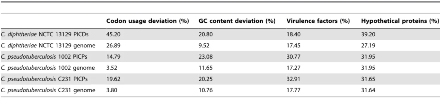
A common feature of virulence factors is their high concentration inside Pathogenicity Islands (PAIs), a class of Genomic Islands (GEIs). PAIs are large genomic regions acquired through horizontal gene transfer, which have in common: deviations in G+C content and codon usage; the presence of transposase and virulence factors; flanking insertion sequences and/or tRNA genes; and the absence in non-pathogenic organism of the same genus or related species (Azevedo et al.,2011). In order to predict PAIs in
the genome sequences of C. pseudotuberculosis, our group has developed a software
named PIPS (Pathogenicity Island Prediction Software), which predicts PAIs taking into account the concentration of the before mentioned features along the genome sequence (Soares et al.,2012). In analyses of C. pseudotuberculosis 1002 and C231,
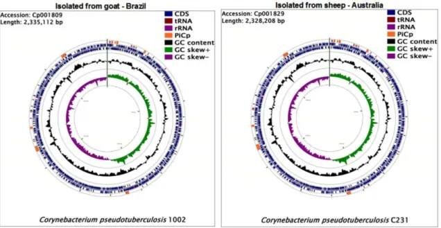
PIPS has identified 7 PAIs (Figure 1), which harbour: the pld gene that codes for the
exotoxin Phospholipase D; the fagABC operon and fagD gene that codes for iron
uptake proteins; and, several other virulence factors and hypothetical proteins (Ruiz et
al.,2011; Soares et al.,2012).
Figure 1. The whole genome of Corynebacterium pseudotuberculosis.
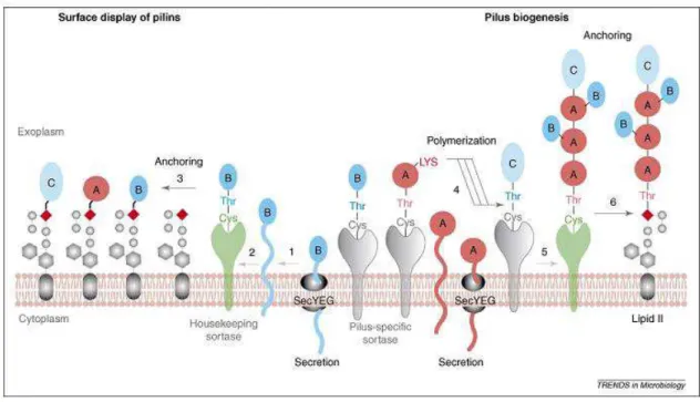
Additionally, in analyses of C. pseudotuberculosis FRC41, it was reported the presence
of 2 clusters of pili genes (Figure 2), which could contribute to the facultative intracellular behavior of this species by coding proteins with roles in adhesion and internalization mechanisms. The pili clusters of genes are named accordingly to their major pilin gene as follow: the spaA (srtB-spaA-srtA-spaB-spaX-spaC) and spaD (srtC
-spaD-spaY-spaE-spaF) clusters, where srtA and srtB are the specific sortases of the spaA cluster; spaA, spaB and spaC encode the major, base and tip pilin proteins,
respectively, of the spaA cluster; srtC is the specific sortase of the spaD cluster; spaD, spaE and spaF encode the major, base and tip pilin proteins, respectively, of the spaD
cluster; and spaX and spaY have currently unknown functions. Additionally, a
housekeeping sortase (srtD) is likely responsible for anchoring the pili to the cellwall
(Trost et al.,2010).
Figure 2. Gene regions encoding adhesive pili of C. pseudotuberculosis FRC41.
The gene clusters involved in the synthesis of adhesive (Spa- like) pili of C. pseudotuberculosis FRC41 are shown. The gene clusters encode sortases required for the assembly of the pilus (blue), major pilins (red), minor pilins (yellow), pilus tip proteins (green), and proteins of unknown function (grey). The detected sorting (LPxTG) signals are indicated. Specifically marked in the major pilin proteins are the characteristic pilin boxes (blue) and E-boxes (white). The predicted binding of the transcription regulator GlxR in the spaA-srtB intergenic region is shown. (Figure from doi: 10.1186/1471-2164-11-728).
The polymerization of pili structures on C. pseudotuberculosis has not been deeply
studied yet, however, there are several studies on the closely related species, C.
diphtheriae (Mandlik et al.,2007). Briefly, in C. diphtheriae, the housekeeping sortase
Figure 3. Model of pilus biogenesis.
Pilin precursors (SpaA, denoted by pink circles; SpaB, denoted by dark-aqua ovals; and SpaC, denoted by light-aqua ovals) are synthesized in the cytoplasm and translocated across the membrane by the Sec machinery. (Figure from doi: 10.1016/j.tim.2007.10.010).
II.1.1.2 Biovar
equi
After achieving a better view of the genome sequences of C. pseudotuberculosis biovar ovis strains isolated from sheep, human and goat, our groups began a great effort to
sequence other biovar ovis strains isolated from other hosts and also biovar equi
strains from different hosts and countries. C. pseudotuberculosis biovar equi strains
were isolated from horses, camels and buffalos where the disease symptons are very variable and visceral commitment is rare (Cerdeira et al.,2011; Lopes et al.,2012;
Pethick et al.,2012; Pethick et al.,2012; Ramos et al.,2012; Ramos et al.,2013; Silva et
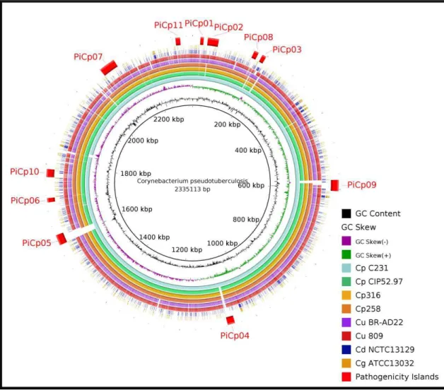
After finishing the sequences of biovar equi genomes, we were able to identify 4
additional PAIs in C. pseudotuberculosis 316 and 258 (PICPs 8-11), both isolated from
horses (Figure 4) (Ramos et al.,2013; Soares et al.,2012). Moreover, further reverse
vaccinology based analyses were performed in C. pseudotuberculosis 258, biovar equi,
in order to find new vaccine candidates that could possibly elicit immune response against this organism. Finally, we have accomplished the genome sequencing of 15 strains of C. pseudotuberculosis from both biovars, isolated from different countries
and hosts, and performed pan-genomics analyses on the whole species aiming to correlate regions of genome plasticity with the disease patterns and host-preference (Soares et al.,2013).
Figure 4. Genomic map comparing strains of Corynebacterium pseudotuberculosis, Corynebacterium ulcerans
and Corynebacterium diphtheriae.
Comparative genomic analyses between: Corynebacterium pseudotuberculosis strains 1002, C231,CIP52.97 and 316; Corynebacterium ulcerans strains BR-AD22 and 809; Corynebacterium diphtheriae NCTC 13129; Corynebacterium
II.2 Manuscript Structure and author's contributions
The thesis is divided into Introduction and 3 chapters based on 1 book chapter and 3 research articles, as follow:
a. The Introduction, presented as a book chapter, shows an overview of in silico
studies performed in pathogenic Corynebacterium species to date, showing the
importance of PAIs, reverse vaccinology and pan-genomics analyses in pathogenic species and providing tables with putative PAIs and vaccine targets. In this work, I have written the whole manuscript with scientific support from all the co-authors. Additionally, I have also performed the identification of PAIs of
C. ulcerans and created the tables and figures;
b. The first chapter presents a research article showing the implementation of the software "PIPS: Pathogenicity Island Prediction Software" and a comparison between this software and other previously available programs. For this matter, analyses were performed using previously described PAIs of C. diphtheriae
NCTC 13129 and Escherichia coli CFT 073. Finally, the article shows data on
analyses performed in C. pseudotuberculosis 1002 and C231 as a case study.
In this work, I have created all the scripts, except for two of them that were kindly provided and one that was implemented by Dr. Rommel Ramos (the credits were added to the specific scripts). I also had support from the co-authors in news ideas for predicting transposases and writing the manuscript; c. The article in the second chapter describes the identification of PAIs in C.
pseudotuberculosis 258, biovar equi, using PIPS, in order to identify regions of
plasticity between both biovars. Furthermore, we applied the reverse vaccinology's theory in C. pseudotuberculosis 258 in comparison with other C.
pseudotuberculosis strains, aiming to identify new putative vaccine targets that
d. The third chapter presents the pan-genomics article, where all 15 genome sequences of different hosts and strains were used. In this article, we review basic concepts about the biovar ovis and equi and create a phylogenomics tree
to find the evolutionary relationship between species of the genus
Corynebacterium. Moreover, we assess the pan-genome, core genome and
singletons subsets of C. pseudotuberculosis and perform comparisons between
both biovars according to these datasets and the PAIs content. In order to acomplish this work, I have participated in specific tasks on all previous steps of genome sequencing, annotation and comparative analyses of all 15 strains. In this work, the retrieving of data from Genbank by GeneDB, the incorporation of data into EDGAR and the statistical analyses performed by R package were triggered by Dr. Jochen Blom. Finally, I have performed all analyses, data interpretation, figure creation and manuscript writing with support from the other authors.
III.1
Corynebacterium
pathogenic species in next-generation genomic era: the
use of EDGAR and PIPS software and the importance of pathogenicity islands
identification in pan-genomic analyses of pathogenic species
S. C. Soares, R. T. J. Ramos, W. M. Silva, L. C. Oliveira, L. G. Amorim, R. Hirata Jr, A. L.
Mattos-Guaraldi, A. Miyoshi, A. Silva, V. Azevedo
Recently, our group has been invited to write a book chapter for "Microbial pathogens and strategies for combating them: science, technology and education". In this book chapter, we review the pathogenic species C. pseudotuberculosis, C. diphtheriae and C. ulcerans, highlighting the in
silico studies performed in these organisms. Additionally, we also review Edgar, PIPS and other
software that were used in our previous works. Furthermore, we summarize potential vaccine targets and PAIs identified in those analyses in a compendium-like work. The description of such data will be helpful in driving future in vitro studies performed by our group and the analyses and
Corynebacterium pathogenic species in next-generation genomic era: the
use of EDGAR and PIPS software and the importance of pathogenicity
islands identification in pan-genomic analyses of pathogenic species
S. C. Soares1, R. T. J. Ramos2, W. M. Silva1, L. C. Oliveira1, L. G. Amorim1, R. Hirata Jr3, A. L. Mattos-Guaraldi3, A. Miyoshi1, A. Silva2, V. Azevedo*,1
1 Laboratory of Cellular and Molecular Genetics, Federal University of Minas Gerais, Belo Horizonte, Minas Gerais, Brazil
2
Microbiology and Immunology Discipline, Medical Sciences Faculty, State University of Rio de Janeiro, Rio de Janeiro, Brazil
3
Department of Genetics, Federal University of Pará, Belém, Pará, Brazil
Corynebacterium genus presents several opportunistic, non-pathogenic, and pathogenic species of high industrial, medical
and veterinary importance. Between Corynebacterium pathogenic species, 3 highly virulent organisms deserve higher attention as the causative agents of the worldwide distributed and communicable diseases diphtheria, caseous lymphadenitis and diphtheria-like, caused by Corynebacterium diphtheriae, Corynebacterium pseudotuberculosis and
Corynebacterium ulcerans, respectively. In order to better understand the virulence mechanisms underlying the diseases
caused by those organisms, several in silico studies have been performed, focusing in: phylogenetics analyses and how those species correlate with each other; pan-genomics analyses and the degree of variability within the species; pathogenicity island identification, commonly shared virulence factors and how genome plasticity may influence the genomes of those species; and, reverse vaccinology and the identification of new canditate targets for future vaccine developments. In this chapter, we review the disease patterns of each species according to their hosts, the high potential of the methodologies and their resulting data, and the putative pathogenicity islands and candidate targets identified in C.
diphtheriae, C. pseudotuberculosis and C. ulcerans to date.
Keywords: PIPS, EDGAR, genomics, Phylogenomics, Reverse Vaccinology, Subtractive Genomics, Pan-Exoproteome, Pathogenicity Islands, Vaccine Targets.
1 Corynebacterium genus
Corynebacterium genus is part of the CMNR group, a suprageneric group of the Actinomycetales family, which includes several genera with high medical, veterinary and biotechnological importance, like: Corynebacterium, Mycobacterium, Nocardia and Rhodococcus. Bacteria from the CMNR group have in common: (i) high G+C content and (ii) a specific cell wall structure composed of mycolic acid, peptidoglycan and arabinogalactan [1].
Corynebacterium genus was first created to harbour Corynebacterium diphtheriae and other pathogenic species [2]. Later on, other species were included, which differed in shape, pathogenicity and sporulation [3]. Nowadays, the genus is mainly composed of: the non-pathogenic species Corynebacterium glutamicum and Corynebacterium efficiens, which are of great biotechnological interest in amino acid production [4,5], and Corynebacterium variabile, a bacterium isolated from the microflora contributing to the development of flavour and texture in cheese ripening [6]; the opportunistic species Corynebacterium jeikeium, Corynebacterium urealyticum and Corynebacterium resistens, which are frequently associated with nosocomial infections [7-9], and the opportunistic and potentially pathogenic Corynebacterium aurimucosum, which is mainly isolated from women with urogenital infections and appears associated with complications in pregnancy [10]; and, the pathogenic species Corynebacterium pseudotuberculosis, C. diphtheriae and Corynebacterium ulcerans, of high veterinary and medical relevance, and a low pathogenic potential bacterium, Corynebacterium kroppenstedtii, which is associated with pulmonary disease and cases of mastitis [9,11-14].
1.1 Corynebacterium pseudotuberculosis
about the diseases caused by C. pseudotuberculosis and the underlying pathogenic mechanisms and virulence factors [1,25,26].
CLA is characterized by the presence of caseous necrosis in lymphatic glands or abscess formation in superficial lymph nodes and subcutaneous tissues of sheep and goats [27], compromising the animal skin, weight, milk and meat production, and causing carcass condemnation and death [1]. The disease has a worldwide distribution and was already reported in several countries, like Australia, New Zealand, South Africa, United States, Canada and Brazil, where sheep and goat farming are very intense [1,28-32]. In Brazil, epidemiologic studies report that a high number of the animals are infected, where the states from the North-East region are the most affected and the underlying losses in this region are highly significant [33,34]. Besides, in the state of Minas Gerais, 78.9% of goats are seropositive for C. pseudotuberculosis infection [35]. The treatment of CLA infected animals is normally performed by draining infected superficial lymph nodes, however, this treatment does not eliminate 100% of the bacteria, it is not viable when visceral organs have been affected, and it may also contaminate the soil [1]. Moreover, although C. pseudotuberculosis is susceptible to a broad range of antibiotics in vitro, the inefficacy of antibiotics in penetrating the abscess capsule and the highly expensive treatment make the antibiotic therapy not applicable [36]. Finally, the licensed vaccines intended for use in sheep present variable efficacy in goat immunization [21] and this scenario is much worst when other hosts and diseases are considered.
1.2 Corynebacterium diphtheriae
C. diphtheriae is a gram-positive, aerobic, non-motile, rod-shaped and pathogenic bacterium [11]. This bacterium is mainly isolated from humans, although other hosts have already been reported, like horses [37], domestic cats [38] and dogs [39]. In humans, C. diphtheriae is responsible for causing the diphtheria disease, an acute upper respiratory tract communicable disease [40,41], and, based on the severity of the infection along with the biochemical profile, the strains are classified under 4 biovars: mitis, gravis, intermedius and belfanti [42]. The cases of diphtheria over the world have decreased drastically since the development of a vaccine based on the inactivation of diphtheria toxin (DT), coded by the tox gene [41]. However, despite the existence of DTP vaccine (diphtheria-tetanus-pertussis) and the decrease in cases worldwide, the disease remains endemic in several regions including Africa, Bangladesh, Vietnam, the tropics and areas of South America, including Brazil [43]. Moreover, more than 150,000 cases have been reported in the former Soviet Union in the 1990s and there are several reports of either re-emergence or persistence of diphtheria in Indian states from 1998-2008 [41,43-46].
The reasons for the reemergence of diphtheria remain to be fully elucidated, however, factors mainly point to: an increased susceptibility of both children and adults; and to the inefficacy of control measures due to shortages of vaccine and deteriorating health infrastructure [41,43,45,47]. Besides, the fact that the tox gene is harboured by a pathogenicity island (PAI), which was horizontally acquired from corynephage, accounts for the emergence of new non-toxigenic strains to which the immune response elicited by the toxoid-based vaccine is not effective [11,48]. The non-toxigenic strains of C. diphtheriae cause infectious diseases varying from cutaneous lesions and pharyngitis to severe invasive commitments, which are characterized by bacteraemia and endocarditis in the absence of toxin mediated lesions [49].
1.3 Corynebacterium ulcerans
2. Comparative genomics in Corynebacterium pathogenic species
2.1 Phylogenomics - Corynebacterium genus
In past, evolutionary reconstructions of the tree of life were mainly performed based in identification of the point of divergence between species solely based in shared homologous characters. However, this methodology could be very trick due to convergent and divergent evolution. With the advent of molecular techniques, phylogenetics was greatly improved by the use of nucleotidic differences in universal reference markers, creating the area of phylogenomics [58]. In the post-genomic era, a second wave of changes brought new approaches to phylogenomics, which now infers the evolutionary divergence by taking advantage of whole-genome data, like: gene content and gene order; orthology; and, DNA string or DNA signature [58,59]. In this sense, phylogenomics may be defined as the junction of phylogenetics and genomics for reconstructing reliable species trees, analysing the distribution and spread of bacterial pathogenicity and predicting orthologous and paralogous genes [60,61].
An approach for the reconstruction of phylogenomics trees and inference of evolutionary divergences is Gegenees, a software that splits the genome data of a group of strains or species in small sequences using pre-defined sizes, performs similarity searches using BLAST, identifies genes commonly shared between the genomes and creates a distance matrix based on the percentage of similarity between the variable contents of the underlying genomes [62]. From the heatmap and phylogenomics tree generated by Gegenees, although C. ulcerans appears more related to C. pseudotuberculosis than to C. diphtheriae, all 3 pathogenic species, C. ulcerans, C. pseudotuberculosis and C. diphtheriae, cluster together, whereas the non-pathogenic and opportunistic species appear separately [63]. This close relationship was already described in previous works [64] and is probably due to the presence of commonly shared virulence factors between those 3 species.
2.2 Pan-genomics analyses using EDGAR - C. diphtheriae and C. pseudotuberculosis
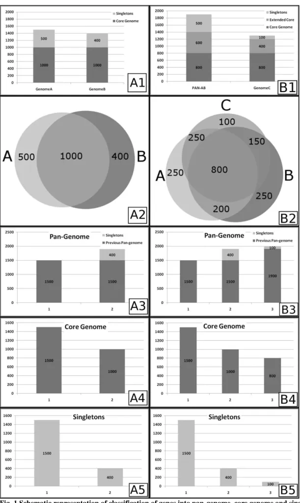
In the last step of pan-genomics approach, a curve fitting is performed to correct the whole curve and also the initial number of genes in pan-genome, core genome and singletons (1500 genes in the example). In order to perform this step, a permutation of all genomes in each position is made and the mean or median number of genes is used for curve fitting of the pan-genome, using Power Law or Heap's Law, and the core genome and singletons, using least-squares fit of the exponential regression decay [65-67,72]. The resulting fitted curves are represented by the formulas n = k*N-α and n = k*exp[-x/τ]+tg(θ) for the Heap' Law and least-squares fit of the exponential regression decay, respectively, where n is the number of genes for a given number of genomes, N is the number of genomes in pan-genome extrapolations, x is the number of genomes in core genome and singletons extrapolations, exp is Euler's number and the other terms are constants defined to fit the specific curve. Interestingly, an α≤1 in Heap's Law represents an open pan-genome,
constantly increasing by the addition of newly sequenced genomes, whereas an α>1 is representative of a closed pan -genome, i.e., no substantial change will be seen in the number of genes in the pan-genome with the addition of newly sequenced genomes [63]. Moreover, the formula for least-squares fit of the exponential regression decay may also be used to achieve a development prospect, where tg(θ) in core genome analysis is representative of the number of genes found in the stabilized core genome after a given number of genomes are sequenced and added to the analysis, whereas in singletons analysis, it is the approximate number of genes which will be added to the pan-genome by each newly sequenced genome [66].
In C. pseudotuberculosis and C. diphtheriae, the software EDGAR was used to perform pan-genome analysis. This software identifies orthologous and paralogous genes by performing all-versus-all BLAST searches and using the score from the alignments to define orthology. As the BLAST score is a very variable metric, EDGAR normalizes the value by using the score rate value (SRV). SRV is calculated as the division of the bit score of a protein B against a protein A by the highest bit score against protein A, i.e., the bit score of protein A against itself. The resulting value, in the range from 0 to 1, is then rounded and multiplied by 100 to represent the percentage of homology [67]. The next steps in pan-genome analysis were performed as described above, using Heap's Law and least-squares fit to the exponential
regression decay, and the α and tg(θ) were calculated for both species, C. pseudotuberculosis and C. diphtheriae, and for both biovars of C. pseudotuberculosis, ovis and equi. According to the results, C. diphtheriae presents an α of 0.69,
whereas the α value of C. pseudotuberculosis is 0.89 [63,69]. Besides, in singletons analysis, the tg(θ) of C. diphtheriae and C. pseudotuberculosis are ~65 and ~19 genes, respectively. Altogether, the findings show that both pan-genomes are open, although the genome of C. pseudotuberculosis is growing at slower rates when compared with the pan-genome of C. diphtheriae, given that C. pseudotuberculosis presents a α value that is closer to 1 and a lower number of genes will be added to the pan-genome for each newly sequenced genome. Finally, according to α value of the pan -genome and tg(θ) of the singletons of C. pseudotuberculosis, the pan-genome of the biovar ovis strains (α = 0.94 and tg(θ) = 16.811) is also growing at slower rates when compared with the pan-genome of the biovar equi strains (α = 0.89
and tg(θ) = 34.533) [63,69]. These examples ilustrate how powerful the use of pan-genome may be in comparative analysis of bacterial species and give new directions on possible targets for genome sequencing, e.g., the choice of a higher number of strains from C. pseudotuberculosis biovar equi, opposing to biovar ovis, for future sequencing projects due to its higher variability.
2.3 Pathogenicity Islands identification using PIPS - C. diphtheriae, C. ulcerans and C. pseudotuberculosis
In pathogenic bacteria, the prediction of PAIs may be performed by the identification of the common features described earlier, such as: deviant genomic signature, i.e., G+C content and codon usage deviation; presence of transposases and flanking tRNA and/or IS; high concentrarion of virulence factors; and, absence in non-pathogenic organism of the same genus or related species. In C. pseudotuberculosis, C. diphtheriae and C. ulcerans, those analyses were performed by the software PIPS: pathogenicity island prediction software [79] using C. glutamicum ATCC 13032 as non-pathogenic organism of the same genus. In C. diphtheriae, 13 PAIs (PICDs1-13) were firstly described in the strain NCTC 13129 [11]. Later, PIPS has identified 11 new putative PAIs (PICDs14-24)(Figure 2A) [79,80]. Finally, using information from the pan-genome of the species, 57 putative GEIs were identified through the whole species [69].
In C. pseudotuberculosis, PIPS has firstly identified 7 putative PAIs (PICPs 1-7) [12]. In a more recent work, 4 additional putative PAIs were identified (PICPs8-11), which presented variations in gene content in comparisons using biovar ovis and equi strains [81,82]. Finally, in pan-genomics analysis, 5 additional putative PAIs were identified, where several PAIs had already been predicted on the first work, but were discarded as the prediction force was low, and revalidated after manual curation during pan-genomics analyses (Figure 2B) [63]. In the last case, the higher number of genomes (15), isolated from different hosts, biovars and locations, gave a better view of regions of plasticity inside the PAIs, which helped in a better classification.
Finally, in C. ulcerans, PIPS has identified 16 putative PAIs (Figure 2C)(this work).
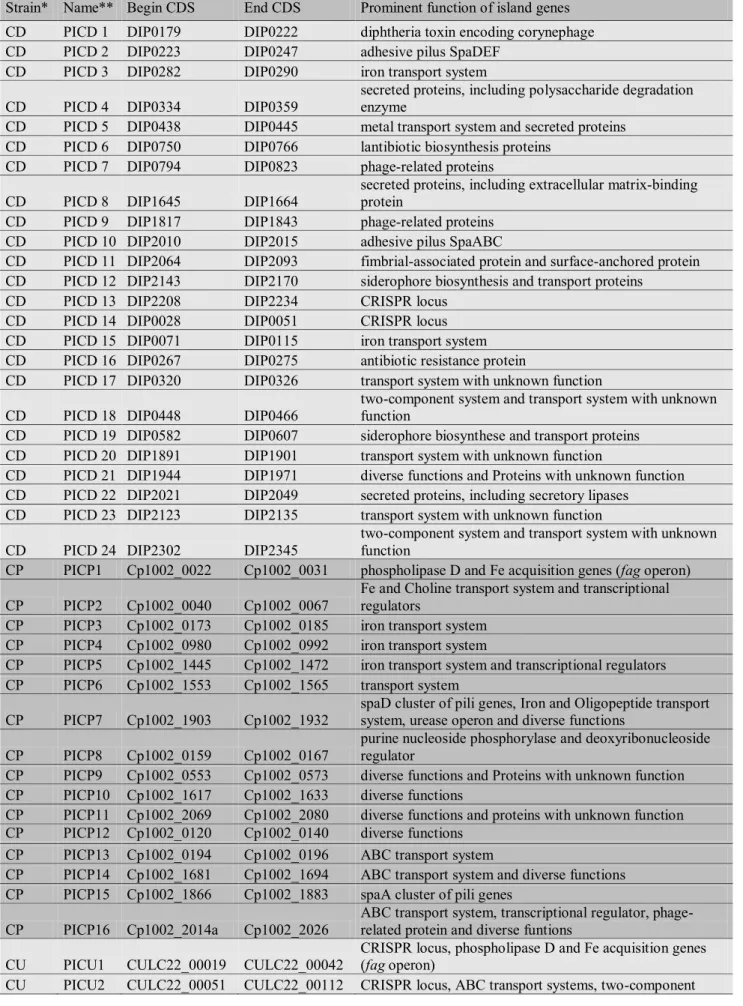
Table 1 Gene content of PAIs identified by PIPS in C. pseudotuberculosis 1002, C. diphtheriae NCTC13129 and C. ulcerans BR-AD22.
Strain* Name** Begin CDS End CDS Prominent function of island genes
CD PICD 1 DIP0179 DIP0222 diphtheria toxin encoding corynephage
CD PICD 2 DIP0223 DIP0247 adhesive pilus SpaDEF
CD PICD 3 DIP0282 DIP0290 iron transport system
CD PICD 4 DIP0334 DIP0359
secreted proteins, including polysaccharide degradation enzyme
CD PICD 5 DIP0438 DIP0445 metal transport system and secreted proteins CD PICD 6 DIP0750 DIP0766 lantibiotic biosynthesis proteins
CD PICD 7 DIP0794 DIP0823 phage-related proteins
CD PICD 8 DIP1645 DIP1664
secreted proteins, including extracellular matrix-binding protein
CD PICD 9 DIP1817 DIP1843 phage-related proteins
CD PICD 10 DIP2010 DIP2015 adhesive pilus SpaABC
CD PICD 11 DIP2064 DIP2093 fimbrial-associated protein and surface-anchored protein CD PICD 12 DIP2143 DIP2170 siderophore biosynthesis and transport proteins
CD PICD 13 DIP2208 DIP2234 CRISPR locus
CD PICD 14 DIP0028 DIP0051 CRISPR locus
CD PICD 15 DIP0071 DIP0115 iron transport system CD PICD 16 DIP0267 DIP0275 antibiotic resistance protein
CD PICD 17 DIP0320 DIP0326 transport system with unknown function
CD PICD 18 DIP0448 DIP0466
two-component system and transport system with unknown function
CD PICD 19 DIP0582 DIP0607 siderophore biosynthese and transport proteins CD PICD 20 DIP1891 DIP1901 transport system with unknown function
CD PICD 21 DIP1944 DIP1971 diverse functions and Proteins with unknown function CD PICD 22 DIP2021 DIP2049 secreted proteins, including secretory lipases
CD PICD 23 DIP2123 DIP2135 transport system with unknown function
CD PICD 24 DIP2302 DIP2345
two-component system and transport system with unknown function
CP PICP1 Cp1002_0022 Cp1002_0031 phospholipase D and Fe acquisition genes (fag operon)
CP PICP2 Cp1002_0040 Cp1002_0067
Fe and Choline transport system and transcriptional regulators
CP PICP3 Cp1002_0173 Cp1002_0185 iron transport system CP PICP4 Cp1002_0980 Cp1002_0992 iron transport system
CP PICP5 Cp1002_1445 Cp1002_1472 iron transport system and transcriptional regulators CP PICP6 Cp1002_1553 Cp1002_1565 transport system
CP PICP7 Cp1002_1903 Cp1002_1932
spaD cluster of pili genes, Iron and Oligopeptide transport system, urease operon and diverse functions
CP PICP8 Cp1002_0159 Cp1002_0167
purine nucleoside phosphorylase and deoxyribonucleoside regulator
CP PICP9 Cp1002_0553 Cp1002_0573 diverse functions and Proteins with unknown function CP PICP10 Cp1002_1617 Cp1002_1633 diverse functions
CP PICP11 Cp1002_2069 Cp1002_2080 diverse functions and proteins with unknown function CP PICP12 Cp1002_0120 Cp1002_0140 diverse functions
CP PICP13 Cp1002_0194 Cp1002_0196 ABC transport system
CP PICP14 Cp1002_1681 Cp1002_1694 ABC transport system and diverse functions CP PICP15 Cp1002_1866 Cp1002_1883 spaA cluster of pili genes
CP PICP16 Cp1002_2014a Cp1002_2026
ABC transport system, transcriptional regulator, phage-related protein and diverse funtions
CU PICU1 CULC22_00019 CULC22_00042
CRISPR locus, phospholipase D and Fe acquisition genes (fag operon)
systems and transcriptional regulators
CU PICU3 CULC22_00166 CULC22_00182 diverse functions and proteins with unknown function CU PICU4 CULC22_00224 CULC22_00236 diverse functions and proteins with unknown function CU PICU5 CULC22_00667 CULC22_00683 Putrescine synthesis and ABC transport proteins CU PICU6 CULC22_01054 CULC22_01061 iron transport system
CU PICU7 CULC22_01155 CULC22_01200 phage-related proteins
CU PICU8 CULC22_01654 CULC22_01724 diverse functions and proteins with unknown function CU PICU9 CULC22_01773 CULC22_01788 secreted proteins and proteins with unknown function CU PICU10 CULC22_01794 CULC22_01816 diverse functions and proteins with unknown function CU PICU11 CULC22_01853 CULC22_01866 diverse functions
CU PICU12 CULC22_01921 CULC22_01985 diverse functions; proteins with unknown function CU PICU13 CULC22_02033 CULC22_02044 chaperone and proteins with unknown function
CU PICU14 CULC22_02071 CULC22_02085
cytochrome C biosynthesis and proteins with unknown function
CU PICU15 CULC22_02101 CULC22_02116 spaDEF cluster of pili genes
CU PICU16 CULC22_02130 CULC22_02168
spaBC cluster of pili genes; iron and oligopeptide transport system; urease operon; diverse functions
CU PICU17 CULC22_02256 CULC22_02267
ABC transport system; transcriptional regulator; diverse functions
CU PICU18 CULC22_02307 CULC22_02325 diverse functions; proteins with unknown function Abbreviations:
* CD, C. diphtheriae NCTC 13129; CP, C. pseudotuberculosis 1002; and, CU, C. ulcerans BR-AD22.
** PICD, putative PAI of C. diphtheriae; PICP, putative PAI of C. pseudotuberculosis; and, PICU, putative PAI of C. ulcerans.
2.4 Reverse vaccinology, pan-exoproteome and subtractive genomics analyses for identifying vaccine targets - C. pseudotuberculosis
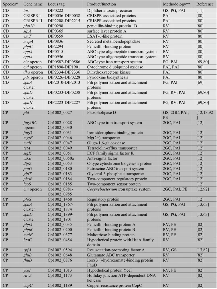
The idea of reverse vaccinology has been initially proposed by Rappuoli in 2000 [89] and relies on the identification of putative vaccine targets using the genome sequence of the pathogen and assaying the chosen targets in vitro, rather than cultivating the pathogen and isolating putative targets one by one, like in conventional vaccine development methodology [90,91]. The reverse vaccinology approach considers proteins that are somehow exposed to the host as putative targets for vaccine development as they are promptly recognized by the immune system. In view of this, proteins from the exoproteome and membrane proteins are considered good targets to elicit immune response [91]; where the exoproteome of an organism is defined as the total repertoire of exported proteins, consisting of 2 main classes: secreted proteins, which are those cleaved on the cell wall, releasing the mature portion into the extracellular space; and, surface exposed proteins that, after cleavage, remain anchored to the cell wall [92].
Subtractive genomics may take advantage of the exoproteome, the idea of comparative genomics, or pan-genomics, and the main concepts of reverse vaccinology to search for vaccine candidates from genome sequences in a subtractive way [93]. Firstly, all gene sequences are considered for their presence in all genome sequences of a given species, because commonly shared genes (core genome) are better targets for vaccine development as they could possibly elicit immune response against all strains. Secondly, the commonly shared genes are analysed for their putative subcellular location (pan-exoproteome and membrane proteins). Next, adhesion probabilities and MHC I and II binding properties analyses are performed in the resulting dataset. Finally, a search for the presence of virulence factors or pathogenicity associated proteins (e.g., genes harboured by pathogenicity islands) may be performed in order to find better targets, although it does not exclude the targets from the previous step [82]. In C. pseudotuberculosis and C. diphtheriae, several in silico strategies have been performed in order to identify new virulence factors and candidate vaccine targets using the ideas of reverse vaccinology [80,82], pan-exoproteome [92] and subtractive genomics [93], summarized at Table 2. In those works, the most recurrent functions are related to maltose transport system (malE and malL), penicillin binding proteins (pbpA, pbpB and pbpC) and resuscitation-promoting factors (rpfA, rpfB and rpfI). Briefly, penicilin-binding proteins are
Table 2 Virulence associated and candidate vaccine targets identified in silico for C. pseudotuberculosis, C. diphtheriae and C.
ulcerans to date.
Species* Gene name Locus tag Product/function Methodology** Reference
CD tox DIP0222 Diphtheria toxin precursor GS, PG, PAI [11]
CD CRISPR I DIP0036-DIP0038 CRISPR-associated proteins PAI [80]
CD CRISPR II DIP2208-DIP2215 CRISPR-associated proteins PAI [80]
CD pbp1B DIP0298 penicillin-binding protein 1B RV [80]
CD slpA DIP0365 surface layer protein A RV [80]
CD esxT DIP0559 ESAT-6-like protein RV [80]
CD mepA DIP0836 Secreted metalloendopeptidase RV [80]
CD pbpC DIP2294 Penicillin-binding protein RV [80]
CD oppA DIP0515 ABC-type oligopeptide transport system RV [80]
CD oppA DIP0956 ABC-type oligopeptide transport system RV [80]
CD ciu operon DIP0582-DIP0586 ABC-type iron transport system PG, PAI [69,80] CD cid operon DIP1898-DIP1901 Cytochrome d ubiquinol oxidase PAI, PAI [80]
CD dha operon DIP2334-DIP2336 Dihydroxyacetone kinase PAI [80]
CD pdx operon DIP0226-DIP0228 Pyridoxine biosynthesis PAI [80]
CD spaA
cluster
DIP2010-DIP2013 Pili polymerization and attachment proteins
PG, PAI [69]
CD spaD
cluster
DIP0233-DIP0238 Pili polymerization and attachment proteins
PG, RV, PAI [69,80]
CD spaH
cluster
DIP2223-DIP2227 Pili polymerization and attachment proteins
PG, RV, PAI [69,80]
CP pld Cp1002_0027 Phospholipase D GS, 2GC, PAI,
PE
[12,13,92 ]
CP fagABC
operon
Cp1002_0028-Cp1002_0030
ABC-type iron transport system 2GC, PAI [12]
CP fagD Cp1002_0031 Iron siderophore binding protein 2GC, PAI [12]
CP mgtE Cp1002_0046 Mg(2+) transporter 2GC, PAI [12]
CP malL Cp1002_0047 Oligo-1,6-glucosidase 2GC, PAI [12]
CP tetA Cp1002_0049 Tetraciclin-efflux transporter 2GC, PAI [12]
CP sigK Cp1002_0051 ECF family sigma factor K 2GC, PAI [12]
CP cskE Cp1002_0050a Anti-sigma factor 2GC, PAI [12]
CP dipZ Cp1002_0053 C-type cytochrome biogenesis protein 2GC, PAI [12]
CP potG Cp1002_0180 Putrescine ABC transport system 2GC, PAI [12]
CP glpT Cp1002_0183 Glycerol-3-phosphate transporter 2GC, PAI [12]
CP phoB Cp1002_0184 Two-component regulatory protein 2GC, PAI [12]
CP lcoS Cp1002_0185 Two-component sensor protein [12]
CP ciu operon Cp1002_0981-Cp1002_0985
Corynebacterium iron uptake system 2GC, PAI, PE [12,92]
CP pfoS Cp1002_1468 Regulatory protein 2GC, PAI [12]
CP spaA
cluster
Cp1002_1867- Cp1002_1874
Pili polymerization and attachment proteins
GS, PG, PAI [13,63]
CP spaD
cluster
Cp1002_1899-Cp1002_1901
Pili polymerization and attachment proteins
GS, PG, PAI [13,63]
CP pbpA Cp1002_0035 Penicillin-binding protein A RV, PE [82]
CP pbpB Cp1002_0200 Penicillin-binding protein B RV, PE [82]
CP malE Cp1002_0377 Maltotriose-binding protein RV, PE [82]
CP htaC Cp1002_0454 Hypothetical protein with HtaA family domain
RV [82]
CP rpfA Cp1002_0594 Resuscitation-promoting factor A RV, GS [13,82]
CP gluB Cp1002_0648 Glutamate ABC transporter RV [82]
CP fhuD Cp1002_0876 Iron(3+)-hydroxamate-binding protein FhuD
RV [82]
CP yceI Cp1002_1013 Hypothetical protein YceI RV, PE [82]
CP ruvA Cp1002_1173 Holliday junction ATP-dependent DNA
helicase
RV [82]
CP thiX Cp1002_1503 Thiamine biosynthesis protein ThiX RV [82]
CP lpqE Cp1002_1763 Lipoprotein LpqE RV [82]
CP nrfC Cp1002_1848 Cytocrome c nitrite reductase RV [82]
CP slpA Cp1002_0237 Surface layer protein A PE [92]
CP malE Cp1002_0497 Maltose transport system PE [92]
CP sprT Cp1002_0562 Trypsin PE, PAI [92]
CP cynT Cp1002_0584 Carbonic anhydrase PE [92]
CP rpfB Cp1002_0681 Resuscitation-promoting factor B PE, GS [13,92]
CP yceG Cp1002_1144 Amino deoxychorismate lyase PE [92]
CP ctaC Cp1002_1425 Cytochrome c oxidade PE [92]
CP lipY Cp1002_1802 Secretory lipase PE [92]
CP oppA1 Cp1002_0357 Oligopeptide binding protein PE [92]
CP lytR Cp1002_0499 Transcriptional regulator PE [92]
CP pknL Cp1002_1409 Serine/Threonine protein kinase PE [92]
CP glpQ Cp1002_1965 Glycerophosphoryl diester
phosphodiesterase
PE [92]
CP hemE Cp1002_0279 Uroporphyrinogen decarboxylase PE [92]
CP accD5 Cp1002_0486 Propionyl-CoA carboxylase PE [92]
CP manB Cp1002_0502 D-alpha-D-mannose-1-phosphate
guanylyltransferase
PE [92]
CP glmU Cp1002_0705 Bifunctional N-acetylglucosamine-1-phosphate
uridyltransferase/glucosaminte-1-phosphate acetyltransferase
PE [92]
CP rpmI Cp1002_0940 50S ribosomal protein L35 PE [92]
CP ndhA Cp1002_1009 Membrane NADH dehydrogenase PE [92]
CP dfp Cp1002_1122 Bifunctional
phosphopantothenoylcysteine decarboxylase/phosphopantothenate synthase
PE [92]
CP qcrA Cp1002_1421 Rieske iron-sulfur protein PE [92]
CP dctA Cp1002_1800 C4-dicarboxylate-transport protein PE [92]
CP ponA1 Cp1002_2034 Penicillin-binding protein PE [92]
CP cwlM Cp1002_2102 Hydrolase PE [92]
CP dcd Cp1002_1931 Deoxycytidine triphosphate deaminase 2CG, SG, PAI [12,96]
CP folP1 Cp1002_1792 Dihydropteroate synthase SG [96]
CP nrdH Cp1002_1677 Glutaredoxin-like protein SG [96]
CP nrdI Cp1002_1676 Ribonucleotide-diphosphate reductase SG [96]
CP murA Cp1002_1695 UDP-N-acetyl glucosamine
1-carboxyvinyltransferase
SG [96]
CP murE Cp1002_1396 UDP-N-acetyl muramoyl
alanyl-D-glutamate--2,6-diamino pimelate ligase
SG [96]
CP nanH Cp1002_0387 Neuraminidase H (sialidase) GS [13]
CP rpfI Cp1002_1072a Resuscitation-promoting factor interacting protein
GS [13]
CP nor Cp1002_0125 Nitric oxide reductase GS, PAI [13]
CP nrpS1 Cp1002_0565 Nonribosomal peptide synthetase 1 GS, PAI [13]
CP nrpS2 Cp1002_1804 Nonribosomal peptide synthetase 2 GS [13]
CP dtsR1 Cp1002_0487 Acetyl-CoA carboxylase β-subunit GS [13]
CP dtsR2 Cp1002_0486 Acetyl-CoA carboxylase β-subunit GS [13]
CP accD3 Cp1002_1950 Acetyl-CoA carboxylase β-subunit GS [13]
CU cpp/ndoE CULC22_02125 Corynebacterial protease CP40/endoglycosidade
2GC [14]
CU pld CULC22_00038 Phospholipase D 2GC, PAI [14]
CU spaBC
cluster
CULC22_02130-CULC22-02131
Pili polymerization and attachment proteins
2GC, PAI [14]
cluster CULC22_02106 proteins
CU rpfI CULC22_01148 Rpf interacting protein 2GC [14]
CU cwlH CULC22_01537 Cell wall-associated hydrolase 2GC [14]
CU nanH CULC22_00437 Sialidase precursor (neuraminidase H) 2GC [14]
CU vsp1 CULC22_00515 Venome serine protease 2GC [14]
CU tspA CULC22_02007 Trypsin-like serine protease 2GC [14]
CU CRISPR I CULC22_00029- CULC22_00032
CRISPR-associated proteins 2GC, PAI [14]
CU CRISPR II CULC22_00106- CULC22_00111
CRISPR-associated proteins 2GC, PAI [14]
Abbreviations:
* CD, C. diphtheriae NCTC 13129; CP, C. pseudotuberculosis 1002; and, CU, C. ulcerans BR-AD22.
** PAI, pathogenicity islands identification; GS, genome sequence and annotation; 2GC, 2 genomes comparison; PG, pan-genomics; SG, subtractive genomics; and, RV, reverse vaccinology.
3. Conclusions
References
[1] Dorella, F.A., Pacheco, L.G.C., Oliveira, S.C., Miyoshi, A. & Azevedo, V. Corynebacterium pseudotuberculosis: microbiology, biochemical properties, pathogenesis and molecular studies of virulence. Vet Res. 2006;37:201-218.
[2] Lehman, K.B. & Neumann, R. Atlas und Grundriss der Bakeriologie und Lehrbuch der speziellen bakteriologischen Diagnositk. 1st Ed. . 1896;:.
[3] Pascual, C., Lawson, P.A., Farrow, J.A., Gimenez, M.N. & Collins, M.D. Phylogenetic analysis of the genus Corynebacterium based on 16S rRNA gene sequences. Int J Syst Bacteriol. 1995;45:724-728.
[4] Kalinowski, J., Bathe, B., Bartels, D., Bischoff, N., Bott, M., Burkovski, A., Dusch, N., Eggeling, L., Eikmanns, B.J., Gaigalat, L. et al. The complete Corynebacterium glutamicum ATCC 13032 genome sequence and its impact on the production of L-aspartate-derived amino acids and vitamins. J Biotechnol. 2003;104:5-25.
[5] Nishio, Y., Nakamura, Y., Kawarabayasi, Y., Usuda, Y., Kimura, E., Sugimoto, S., Matsui, K., Yamagishi, A., Kikuchi, H., Ikeo, K. et al. Comparative complete genome sequence analysis of the amino acid replacements responsible for the thermostability of Corynebacterium efficiens. Genome Res. 2003;13:1572-1579.
[6] Schröder, J., Maus, I., Trost, E. & Tauch, A. Complete genome sequence of Corynebacterium variabile DSM 44702 isolated from the surface of smear-ripened cheeses and insights into cheese ripening and flavor generation. BMC Genomics. 2011;12:545.
[7] Schröder, J., Maus, I., Meyer, K., Wördemann, S., Blom, J., Jaenicke, S., Schneider, J., Trost, E. & Tauch, A. Complete genome sequence, lifestyle, and multi-drug resistance of the human pathogen Corynebacterium resistens DSM 45100 isolated from blood samples of a leukemia patient. BMC Genomics. 2012;13:141.
[8] Tauch, A., Kaiser, O., Hain, T., Goesmann, A., Weisshaar, B., Albersmeier, A., Bekel, T., Bischoff, N., Brune, I., Chakrabort y, T. et al. Complete genome sequence and analysis of the multiresistant nosocomial pathogen Corynebacterium jeikeium K411, a lipid-requiring bacterium of the human skin flora. J Bacteriol. 2005;187:4671-4682.
[9] Tauch, A., Schneider, J., Szczepanowski, R., Tilker, A., Viehoever, P., Gartemann, K., Arnold, W., Blom, J., Brinkrolf, K., Brune, I. et al. Ultrafast pyrosequencing of Corynebacterium kroppenstedtii DSM44385 revealed insights into the physiology of a lipophilic corynebacterium that lacks mycolic acids. J Biotechnol. 2008;136:22-30.
[10] Trost, E., Götker, S., Schneider, J., Schneiker-Bekel, S., Szczepanowski, R., Tilker, A., Viehoever, P., Arnold, W., Bekel, T., Blom, J. et al. Complete genome sequence and lifestyle of black-pigmented Corynebacterium aurimucosum ATCC 700975 (formerly C. nigricans CN-1) isolated from a vaginal swab of a woman with spontaneous abortion. BMC Genomics. 2010;11:91.
[11] Cerdeño-Tárraga, A.M., Efstratiou, A., Dover, L.G., Holden, M.T.G., Pallen, M., Bentley, S.D., Besra, G.S., Churcher, C., James, K.D., De Zoysa, A. et al. The complete genome sequence and analysis of Corynebacterium diphtheriae NCTC13129.
Nucleic Acids Res. 2003;31:6516-6523.
[12] Ruiz, J.C., D'Afonseca, V., Silva, A., Ali, A., Pinto, A.C., Santos, A.R., Rocha, A.A.M.C., Lopes, D.O., Dorella, F.A., Pacheco, L.G.C. et al. Evidence for reductive genome evolution and lateral acquisition of virulence functions in two Corynebacterium pseudotuberculosis strains. PLoS One. 2011;6:e18551.
[13] Trost, E., Ott, L., Schneider, J., Schröder, J., Jaenicke, S., Goesmann, A., Husemann, P., Stoye, J., Dorella, F.A., Rocha, F.S. et al. The complete genome sequence of Corynebacterium pseudotuberculosis FRC41 isolated from a 12-year-old girl with necrotizing lymphadenitis reveals insights into gene-regulatory networks contributing to virulence. BMC Genomics. 2010;11:728.
[14] Trost, E., Al-Dilaimi, A., Papavasiliou, P., Schneider, J., Viehoever, P., Burkovski, A., Soares, S.C., Almeida, S.S., Dorella, F.A., Miyoshi, A. et al. Comparative analysis of two complete Corynebacterium ulcerans genomes and detection of candidate virulence factors. BMC Genomics. 2011;12:383.
[15] Jones, D. & Collins, M.D. Irregular, nonsporing Gram-positive rods, Section 15. Pages 1261–1579 in Bergey’s Manual of Systematic Bacteriology. . 1986;:.
[16] Biberstein, E.L., Knight, H.D. & Jang, S. Two biotypes of Corynebacterium pseudotuberculosis. Vet Rec. 1971;89:691-692. [17] Dorella, F.A., Estevam, E.M., Pacheco, L.G.C., Guimarães, C.T., Lana, U.G.P., Gomes, E.A., Barsante, M.M., Oliveira, S.C.,
Meyer, R., Miyoshi, A. et al. In vivo insertional mutagenesis in Corynebacterium pseudotuberculosis: an efficient means to identify DNA sequences encoding exported proteins. Appl Environ Microbiol. 2006;72:7368-7372.
[18] Liu, D.T.L., Chan, W., Fan, D.S.P. & Lam, D.S.C. An infected hydrogel buckle with Corynebacterium pseudotuberculosis. Br J
Ophthalmol. 2005;89:245-246.
[19] Mills, A.E., Mitchell, R.D. & Lim, E.K. Corynebacterium pseudotuberculosis is a cause of human necrotising granulomatous lymphadenitis. Pathology. 1997;29:231-233.
[20] Peel, M.M., Palmer, G.G., Stacpoole, A.M. & Kerr, T.G. Human lymphadenitis due to Corynebacterium pseudotuberculosis: report of ten cases from Australia and review. Clin Infect Dis. 1997;24:185-191.
[21] Barakat, A.A., Selim, S.A., Atef, A., Saber, M.S., Nafie, E.K. & El-Edeeby, A.A. Two serotypes of Corynebacterium pseudotuberculosis isolated from different animal species. Revue Scientifique et Technique de l'OIE. 1984;3(1):151-163. [22] Yeruham, I., Elad, D., Friedman, S. & Perl, S. Corynebacterium pseudotuberculosis infection in Israeli dairy cattle. Epidemiol
Infect. 2003;131:947-955.
[23] Aleman, M., Spier, S.J., Wilson, W.D. & Doherr, M. Corynebacterium pseudotuberculosis infection in horses: 538 cases (1982-1993). J Am Vet Med Assoc. 1996;209:804-809.
[25] McKean, S., Davies, J. & Moore, R. Identification of macrophage induced genes of Corynebacterium pseudotuberculosis by differential fluorescence induction. Microbes Infect. 2005;7:1352-1363.
[26] McKean, S.C., Davies, J.K. & Moore, R.J. Expression of phospholipase D, the major virulence factor of Corynebacterium pseudotuberculosis, is regulated by multiple environmental factors and plays a role in macrophage death. Microbiology. 2007;153:2203-2211.
[27] Ayers, J.L. Caseous lymphadenitis in goat and sheep: Review of diagnosis, pathogenesis, and immunity. JAVMA. 1977;n. 171:1251-1254.
[28] Arsenault, J., Girard, C., Dubreuil, P., Daignault, D., Galarneau, J.R., Boisclair, J., Simard, C. & Bélanger, D. Prevalence of and carcass condemnation from maedi-visna, paratuberculosis and caseous lymphadenitis in culled sheep from Quebec, Canada.
Prev Vet Med. 2003;59:67-81.
[29] Ben Saïd, M.S., Ben Maitigue, H., Benzarti, M., Messadi, L., Rejeb, A. & Amara, A. [Epidemiological and clinical studies of ovine caseous lymphadenitis]. Arch Inst Pasteur Tunis. 2002;79:51-57.
[30] Binns, S.H., Bailey, M. & Green, L.E. Postal survey of ovine caseous lymphadenitis in the United Kingdom between 1990 and 1999. Vet Rec. 2002;150:263-268.
[31] Connor, K.M., Quirie, M.M., Baird, G. & Donachie, W. Characterization of United Kingdom isolates of Corynebacterium pseudotuberculosis using pulsed-field gel electrophoresis. J Clin Microbiol. 2000;38:2633-2637.
[32] Paton, M.W., Walker, S.B., Rose, I.R. & Watt, G.F. Prevalence of caseous lymphadenitis and usage of caseous lymphadenitis vaccines in sheep flocks. Aust Vet J. 2003;81:91-95.
[33] Brown, C.C., Olander, H.J., Zometa, C. & Alves, S.F. Serodiagnosis of inapparent caseous lymphadenitis in goats and sheep, using the synergistic hemolysis-inhibition test. Am J Vet Res. 1986;47:1461-1463.
[34] Brown, C.C., Olander, H.J. & Alves, S.F. Synergistic hemolysis-inhibition titers associated with caseous lymphadenitis in a slaughterhouse survey of goats and sheep in Northeastern Brazil. Can J Vet Res. 1987;51:46-49.
[35] Seyffert, N., Guimarães, A.S., Pacheco, L.G.C., Portela, R.W., Bastos, B.L., Dorella, F.A., Heinemann, M.B., Lage, A.P., Gouveia, A.M.G., Meyer, R. et al. High seroprevalence of caseous lymphadenitis in Brazilian goat herds revealed by Corynebacterium pseudotuberculosis secreted proteins-based ELISA. Res Vet Sci. 2010;88:50-55.
[36] Collett, M.G., Bath, G.F. & Cameron, C.M. Corynebacterium pseudotuberculosis infections. In: Infections diseases of livestock with special reference to Southern Africa. Oxford University Press. 1994;2:1387-1395.
[37] Leggett, B.A., De Zoysa, A., Abbott, Y.E., Leonard, N., Markey, B. & Efstratiou, A. Toxigenic Corynebacterium diphtheriae isolated from a wound in a horse. Vet Rec. 2010;166:656-657.
[38] Hall, A.J., Cassiday, P.K., Bernard, K.A., Bolt, F., Steigerwalt, A.G., Bixler, D., Pawloski, L.C., Whitney, A.M., Iwaki, M., Baldwin, A. et al. Novel Corynebacterium diphtheriae in domestic cats. Emerg Infect Dis. 2010;16:688-691.
[39] Dixon, B. Sick as a dog. Lancet Infect Dis. 2010;10:73.
[40] Popovic, T., Kombarova, S.Y., Reeves, M.W., Nakao, H., Mazurova, I.K., Wharton, M., Wachsmuth, I.K. & Wenger, J.D. Molecular epidemiology of diphtheria in Russia, 1985-1994. J Infect Dis. 1996;174:1064-1072.
[41] Popovic, T., Mazurova, I.K., Efstratiou, A., Vuopio-Varkila, J., Reeves, M.W., De Zoysa, A., Glushkevich, T. & Grimont, P. Molecular epidemiology of diphtheria. J Infect Dis. 2000;181 Suppl 1:S168-77.
[42] Efstratiou, A., Engler, K.H., Mazurova, I.K., Glushkevich, T., Vuopio-Varkila, J. & Popovic, T. Current approaches to the laboratory diagnosis of diphtheria. J Infect Dis. 2000;181 Suppl 1:S138-45.
[43] Mattos-Guaraldi, A.L., Moreira, L.O., Damasco, P.V. & Hirata Júnior, R. Diphtheria remains a threat to health in the developing world--an overview. Mem Inst Oswaldo Cruz. 2003;98:987-993.
[44] Murhekar, M.V. & Bitragunta, S. Persistence of diphtheria in India. Indian J Community Med. 2011;36:164-165.
[45] Nakao, H., Pruckler, J.M., Mazurova, I.K., Narvskaia, O.V., Glushkevich, T., Marijevski, V.F., Kravetz, A.N., Fields, B.S., Wachsmuth, I.K. & Popovic, T. Heterogeneity of diphtheria toxin gene, tox, and its regulatory element, dtxR, in Corynebacterium diphtheriae strains causing epidemic diphtheria in Russia and Ukraine. J Clin Microbiol. 1996;34:1711-1716. [46] Sharma, N.C., Banavaliker, J.N., Ranjan, R. & Kumar, R. Bacteriological & epidemiological characteristics of diphtheria cases
in & around Delhi -a retrospective study. Indian J Med Res. 2007;126:545-552.
[47] Dittmann, S., Wharton, M., Vitek, C., Ciotti, M., Galazka, A., Guichard, S., Hardy, I., Kartoglu, U., Koyama, S., Kreysler, J. et al. Successful control of epidemic diphtheria in the states of the Former Union of Soviet Socialist Republics: lessons learned. J
Infect Dis. 2000;181 Suppl 1:S10-22.
[48] Viguetti, S.Z., Pacheco, L.G.C., Santos, L.S., Soares, S.C., Bolt, F., Baldwin, A., Dowson, C.G., Rosso, M.L., Guiso, N., Miyoshi, A. et al. Multilocus sequence types of invasive Corynebacterium diphtheriae isolated in the Rio de Janeiro urban area, Brazil. Epidemiol Infect. 2012;140:617-620.
[49] Mattos-Guaraldi, A.L., Duarte Formiga, L.C. & Pereira, G.A. Cell surface components and adhesion in Corynebacterium diphtheriae. Microbes Infect. 2000;2:1507-1512.
[50] Wagner, K.S., White, J.M., Crowcroft, N.S., De Martin, S., Mann, G. & Efstratiou, A. Diphtheria in the United Kingdom, 1986-2008: the increasing role of Corynebacterium ulcerans. Epidemiol Infect. 2010;138:1519-1530.
[51] Bernard, K. The genus corynebacterium and other medically relevant coryneform-like bacteria. J Clin Microbiol. 2012;50:3152-3158.
[52] Hatanaka, A., Tsunoda, A., Okamoto, M., Ooe, K., Nakamura, A., Miyakoshi, M., Komiya, T. & Takahashi, M. Corynebacterium ulcerans Diphtheria in Japan. Emerg Infect Dis. 2003;9:752-753.
[53] Schuhegger, R., Lindermayer, M., Kugler, R., Heesemann, J., Busch, U. & Sing, A. Detection of toxigenic Corynebacterium diphtheriae and Corynebacterium ulcerans strains by a novel real-time PCR. J Clin Microbiol. 2008;46:2822-2823.