Evaluation of hemodynamic effects of xenon in dogs
undergoing hemorrhagic shock
Ruben C. Franceschi,ILuiz Malbouisson,IEduardo Yoshinaga,IJose´ Otavio Costa Auler Jr.,I Luiz Francisco Poli de Figueiredo (in memoriam),IIMaria Jose´ C. CarmonaI
IHospital das Clı´nicas da Faculdade de Medicina da Universidade de Sa˜o Paulo, Anesthesiology, Sa˜o Paulo/ SP, Brazil.IIFaculdade de Medicina da Universidade de Sa˜o Paulo, Surgery, Sa˜o Paulo/SP, Brazil.
OBJECTIVES:The anesthetic gas xenon is reported to preserve hemodynamic stability during general anesthesia. However, the effects of the gas during shock are unclear. The objective of this study was to evaluate the effect of Xe on hemodynamic stability and tissue perfusion in a canine model of hemorrhagic shock.
METHOD:Twenty-six dogs, mechanically ventilated with a fraction of inspired oxygen of 21% and anesthetized with etomidate and vecuronium, were randomized into Xenon (Xe; n = 13) or Control (C; n = 13) groups. Following hemodynamic monitoring, a pressure-driven shock was induced to reach an arterial pressure of 40 mmHg. Hemodynamic data and blood samples were collected prior to bleeding, immediately after bleeding and 5, 20 and 40 minutes following shock. The Xe group was treated with 79% Xe diluted in ambient air, inhaled for 20 minutes after shock.
RESULT: The mean bleeding volume was 44 mL.kg21 in the C group and 40 mL.kg21 in the Xe group.
Hemorrhage promoted a decrease in both the cardiac index (p,0.001) and mean arterial pressure (p,0.001). These changes were associated with an increase in lactate levels and worsening of oxygen transport variables in both groups (p,0.05). Inhalation of xenon did not cause further worsening of hemodynamics or tissue perfusion markers.
CONCLUSIONS:Xenon did not alter hemodynamic stability or tissue perfusion in an experimentally controlled hemorrhagic shock model. However, further studies are necessary to validate this drug in other contexts.
KEYWORDS: Xenon; Hemorrhagic Shock; General Anesthesia.
Franceschi RC, Malbouisson L, Yoshinaga E, Auler Jr. JO, Poli de Figueiredo LF, Carmona MJ. Evaluation of hemodynamic effects of xenon in dogs undergoing hemorrhagic shock. Clinics. 2013;68(2):231-237.
Received for publication onApril 26, 2012;First review completed onJuly 13, 2012;Accepted for publication onSeptember 25, 2012 E-mail: [email protected]
Tel.: 55 11 2661-6335
& INTRODUCTION
Xenon (Xe) is an inert gas that is present in the atmosphere at very low concentrations and has potent hypnotic and analgesic properties that have been known for several decades (1-4). This anesthetic gas is neither toxic nor metabolized within the body, as it is quickly eliminated through the lungs (4-6) and can be used in patients with impaired renal or hepatic function with minimal or no side effects (5). In heart failure conditions, a number of publications have shown that inhaled xenon is an adequate alternative to other inhaled anesthetics, as it does not interfere with cardiac contractility, cardiac autonomic control or vascular tonic regulation (7-11).
However, few studies have evaluated the impact of xenon on hemodynamics, oxygen transport and tissue perfusion during circulatory shock (12,13). It is well known that most inhalational anesthetics used in clinical practice may hinder hemodynamic responses to shock, either by directly inter-fering with cardiac function or the vascular contractile response or by indirectly decreasing sympathetic activity. Due to the absence of hemodynamic effects caused by xenon, this gas is a suitable option in severe shock conditions under which the cardiovascular depressant effects of anesthesia would further worsen tissue hypoper-fusion.
The aim of this study was to assess the impact of xenon on hemodynamics, oxygen transport and tissue perfusion markers in dogs undergoing severe hemorrhagic shock.
& MATERIALS AND METHODS
This research project was approved by the Ethics Committee of the Hospital das Clı´nicas da Faculdade de Medicina da Universidade de Sa˜o Paulo (process CAPPesq 655/97) and was funded through a research grant from Copyrightß2013CLINICS– This is an Open Access article distributed under
the terms of the Creative Commons Attribution Non-Commercial License (http:// creativecommons.org/licenses/by-nc/3.0/) which permits unrestricted non-commercial use, distribution, and reproduction in any medium, provided the original work is properly cited.
No potential conflict of interest was reported.
FAPESP (Fundac¸a˜o de Amparo a` Pesquisa do Estado Sa˜o Paulo; Process 00/10122-6). The study included 26 mixed-breed dogs weighing between 10 and 20 kg. The study met the international criteria for animal care and abided by the rules of COBEA (The Brazilian College of Animal Experimentation).
Animal preparation and measurements
A clinical veterinarian evaluated the health status of the animals following their arrival at the laboratory. Food was withheld from the animals for 12 hours prior to the experi-ment and free access to water was available at all times. Following the arrival of the animals to the research laboratory, a venous line was inserted into one limb using a 20-G Teflon catheter. General anesthesia was induced using etomidate
(2 mg.kg21
), followed by tracheal intubation with an 8.0-mm ID cuffed cannula. A continuous infusion of etomidate
(50mg.kg21.min21) and vecuronium (1mg.kg21.min21) was
initiated to facilitate pressure-controlled ventilation and to enable the installation of vascular lines and was maintained throughout the study. Mechanical ventilation was performed using a Cicero EM (Drager, Lu¨beck, Germany). The inspira-tory pressure was set at 15 cmH2O and the respirainspira-tory rate was set at 20 breaths per minute. The fraction of inspired
oxygen (FiO2) was set at 21% (room air) in a semi-closed
circuit with a CO2absorber. Respiratory system compliance;
tidal volume; and peak, plateau and end-expiratory pressures were directly monitored with the anesthesia equipment after pneumotachometer calibration.
After the induction of anesthesia, small-bore polyethylene catheters (P260) were inserted by dissection into both femoral arteries. One of the arterial catheters was used exclusively to measure systemic arterial pressure, whereas the other catheter was used to remove blood and collect arterial blood samples. The right internal jugular vein was dissected and a pulmonary artery catheter (Edwards Lifesciences, Irvine, CA, USA) was inserted. The position was confirmed by obtaining pulmonary artery pressure (PAP) and pulmonary artery occlusion pressure (PAOP) curves. Heart rate, systolic arterial pressure (SAP), diastolic arterial pressure (DAP), mean arterial pressure (MAP), mean PAP and PAOP were measured using a multi-parameter monitor (Viridia 885, Hewlett-Packard, Palo Alto, CA, USA). All data from the monitor were stored on a personal computer. Intravascular pressures were com-puted using disposable transducers (Edwards Lifesciences, Irvine, CA, USA), which were filled with a 0.9% sodium chloride solution and zeroed at the mid-axillary line. Mixed central venous blood samples were collected from the distal port of the pulmonary artery catheter. Cardiac output (CO) was measured using the standard thermodilution techni-que. The measurement was repeated three times and the average value was divided by body surface area (BSA) to
obtain the cardiac index (CI). Body surface area was calculated using the following formula: BSA = 0.1226
weight2/3. Systemic and pulmonary hemodynamic
calcula-tions, arterial and mixed venous blood oxygen contents (CaO2 and CvO2), the venous admixture and oxygen transport variables were calculated using standard equa-tion. A pulse oximeter probe was applied to the tongue of the animal and a gas analyzer (Criticare Systems Inc., Waukesha, WI, USA) was placed between the endotracheal cannula and respiratory circuit Y piece to continuously
monitor FiO2and ETCO2levels.
Arterial and mixed venous oxygen (PaO2, PvO2), carbon
dioxide (PaCO2, PvCO2) tensions, arterial lactate
concentra-tion and pH were measured using a blood-gas analyzer (Radiometer ABL Gas Analyzer, Copenhagen, Denmark). All results were corrected for body temperature. The bicarbonate concentration, hemoglobin (Hb) concentration and base excess were also calculated.
Study protocol and data collection
Animals were randomly allocated to the Control or Xenon group. A random treatment sequence was obtained using the Stata 11 statistical software (StataCorp, College Station, TX, USA). The allocation of each animal was enclosed in a brown numbered envelope and revealed at the start of each experiment. Following animal preparation, blood samples
Table 1 -Characteristics of the groups.
Control Group Xenon Group p-value
Weight (kg) 15¡2 15¡2 NS
BSA (m2) 0.71¡0.07 0.72¡0.07 NS
Volume removed (ml) 670¡155 602¡156 NS
BSA – body surface area. NS indicates no significant difference between groups. Data are expressed as the mean¡SD. n = 13 for both the Control and Xenon groups.
and respiratory and hemodynamic data were collected before the induction of shock, immediately after induction and 5, 20 and 40 minutes after induction.
The hemorrhagic shock model used in this study is described elsewhere (14). Briefly, the animals were
sub-jected to continuous bleeding (25 mL.min21
) to achieve a MAP of 40 mmHg. When the MAP reached 40 mmHg, continuous bleeding was interrupted and small aliquots of blood were removed to maintain the target pressure for 2 minutes. Individual variations in the total amount of blood volume lost were not expected to significantly alter the MAP. Immediately after establishing circulatory shock in the Experimental group, xenon inhalation was initiated. First, the breathing circuit was saturated with a specially prepared gas mixture (White Martins, Sa˜o Paulo, Brazil) containing 79% xenon and 21% oxygen, with a flow rate of
10 L.min21
. Following initial saturation of the breathing circuit, the animals were ventilated for 20 minutes at a
constant 1-L.min21
flow rate. An additional variable flow
rate of O2 was used as needed to maintain the inspired
fraction of O2 at 21% according to the gas analyzer. The
Control group received room air ventilation at a flow rate of
1 L.min21
and additional O2 ventilation to maintain the
FiO2at 21%. After data were collected at the 20-minute time
point, the configuration of the respiratory circuit was changed from closed to open to allow for the rapid clearance of xenon. The fresh gas flow was then adjusted to
10 L.min21
for both groups. The ventilatory settings were kept constant throughout the experiment. After the end of the experiment, the animals were euthanized by injecting
KCl (25 mEq.L21
) intravenously and sent to the animal care facility for adequate disposal.
Statistical analysis
The distribution of the data was tested for normality using the Kolmogorov-Smirnov test. Weight, body surface area and blood volume lost were compared between groups using Student’s t-test for unpaired samples. The temporal behavior of hemodynamic, respiratory, metabolic and oxygen transport variables was evaluated using a two-way analysis of variance for repeated measures, followed by a
Newman-Keuls post-hoc test when indicated.P-values less
than 0.05 were considered statistically significant. The
results are expressed as the mean ¡ standard deviation
(SD). All statistical analyses were performed using the statistical packages Aabel 3.0.5 (Gigawiz Ltd. Co., Tulsa, OK, USA) and G Power 3 (Heinrich-Heine-Universita¨t, Dusseldorf, Germany).
& RESULTS
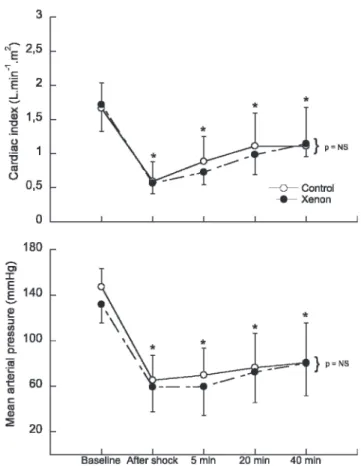
The Control and Experimental groups were similar with respect to weight, BSA and blood volume losses, as indicated in Table 1. The volume of blood removed was
44 mL.kg21 in the Control group and 40 mL.kg21 in the
Xenon group and the blood removal caused a significant reduction in the cardiac index in both groups (64 and 67%, respectively), measured immediately after shock (upper panel of Figure 1). Following the shock induction phase, a non-significant increase in the cardiac index was observed that was similar in both groups. At 20 minutes post-blood loss, the cardiac indexes were 33 and 42% lower than baseline measurements in the control and Xenon groups,
respectively (p,0.05). The removal of xenon inhalation at 20
minutes post-shock did not alter the cardiac index. The mean arterial pressure decreased in both groups by 55% following the blood loss phase compared with baseline values. These values remained stable throughout the remainder of the experiment (lower panel of Figure 1). As seen in Table 2, there was a significant and persistent increase in heart rate and pulmonary vascular resistance index after shock that was similar in both groups. The systemic vascular resistance index transiently increased after shock, returning to baseline levels during the observa-tion period, but there was no significant difference between the groups. The mean pulmonary arterial pressure, cardiac filling pressures and left and right ventricular systolic work indexes exhibited a marked reduction until the last measurement was taken. The interruption of xenon inhalation did not produce any significant alterations or improvement in hemodynamics compared with measurements taken at the 20-minute time point.
Table 2 -Hemodynamic behavior of the animals in the Control and Xenon groups during the study.
Group Baseline After shock 5 min 20 min 40 min p-value
HR (bpm) Control 75 ¡ 19 123 ¡ 38* 124 ¡ 37* 133 ¡ 31* 140 ¡ 28* NS
Xenon 73 ¡ 18 137 ¡ 32* 139 ¡ 34* 149 ¡ 35* 142 ¡ 35*
CVP (mmHg) Control 7 ¡ 2 3 ¡ 1* 3 ¡ 1* 3 ¡ 1* 3 ¡ 1* NS
Xenon 7 ¡ 1 3 ¡ 1* 3 ¡ 1* 4 ¡ 1* 3 ¡ 2*
mPAP (mmHg) Control 18 ¡ 3 10 ¡ 3*! 11 ¡ 2*! 11 ¡ 3*! 14 ¡ 3* NS
Xenon 17 ¡ 4 9 ¡ 2*! 11 ¡ 3*! 12 ¡ 3*! 13 ¡ 4*
POAP (mmHg) Control 11 ¡ 3 5 ¡ 2* 5 ¡ 2* 4 ¡ 2* 6 ¡ 2* NS
Xenon 10 ¡ 2 4 ¡ 2* 5 ¡ 2* 6 ¡ 3* 5 ¡ 2*
SVRi (dyne.s.cm25.m22) Control 7067 ¡ 1675 9924 ¡ 4464* 6651 ¡ 2723 5574 ¡ 2156 6080 ¡ 1971 NS
Xenon 6108 ¡ 1565 8202 ¡ 3298* 6206 ¡ 2311 5659 ¡ 1571 5370 ¡ 1935
PVRi (dyne.s.cm25.m22) Control 366 ¡ 97 676 ¡ 332* 553 ¡ 161* 553 ¡ 232* 729 ¡ 478* NS
Xenon 304 ¡ 158 672 ¡ 246* 629 ¡ 244* 502 ¡ 228* 594 ¡ 289*
LVSWi (g?m.m22) Control 45.8 ¡ 11.8 5.3 ¡ 5.3* 8.5 ¡ 7.9* 9.8 ¡ 7.6* 10.3 ¡ 7.5* NS
Xenon 43.4 ¡ 11.8 3.7 ¡ 2.7* 4.8 ¡ 3.4* 7.5 ¡ 5.2* 9.3 ¡ 4.3*
RVSWi (g?m.m22) Control 5.6 ¡ 1.4 0.7 ¡ 0.7* 1.3 ¡ 1* 1.4 ¡ 0.9* 1.5 ¡ 1* NS
Xenon 5.5 ¡ 2.1 0.5 ¡ 0.3* 0.8 ¡ 0.4* 1.1 ¡ 0.6* 1.5 ¡ 0.7*
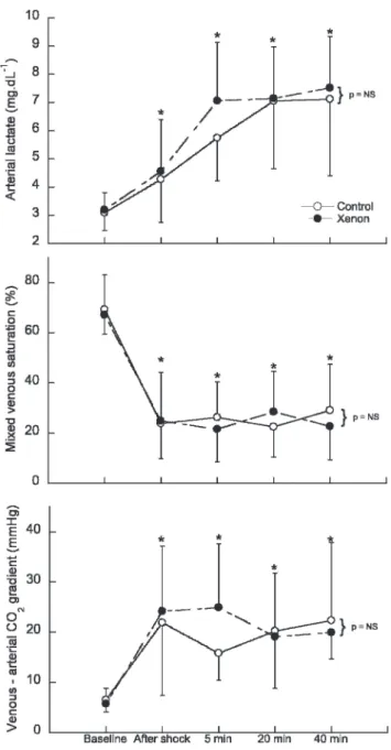
The induction of shock promoted a significant and progressive increase in lactate levels compared with the baseline levels that was similar in both groups (upper panel of Figure 2). The mixed central venous saturation decreased from 69 and 67% to 23 and 24% in the Control and Xenon groups, respectively. Saturation remained low in both groups through the end of the study (middle panel of
Figure 2). The veno-arterial CO2difference increased
four-fold in both groups compared with baseline values and remained elevated throughout the study (lower panel of Figure 2). As noted in Table 3, there was a similar and
significant four-fold reduction in oxygen delivery, accom-panied by an important and sustained augmentation of the oxygen extraction rate, from 29 and 30% to 76 and 74% in the Control and Xenon groups, respectively. The initiation of shock induced a significant decrease in arterial pH, arterial base excess and bicarbonate concentrations that were similar in both groups (Table 3). The removal of xenon from the respiratory mixture did not result in any changes in tissue perfusion markers. No changes in other metabolic parameters were observed. Hemoglobin levels did not change throughout the study, as shown in Table 3.
Table 4 shows the values of the gas exchange and respiratory mechanics variables observed during the study. As seen in the table, none of the studied variables were affected by the induction of shock or the initiation or cessation of xenon inhalation.
& DISCUSSION
In this study, we observed that xenon inhalational anesthesia did not decrease hemodynamic variables or tissue perfusion markers in dogs experiencing severe
hemorrhagic shock. Gas exchange and respiratory
mechanics were not affected by the initiation of inhaled xenon. In addition, the removal of xenon from the respiratory mixture did not produce any improvement in systemic hemodynamics or tissue perfusion.
The properties of xenon anesthesia have been well established (3). The gas has been shown to cause minimal to no cardiovascular instability in experimental and clinical studies in healthy subjects (15,16). Luttropp et al. studied the effects of xenon anesthesia (65% xenon in oxygen) on myocardial function. In their study, the cerebral blood flow velocities in 17 ASA 1 patients undergoing open abdominal surgery were investigated using transesophageal echocar-diography and transcranial Doppler sonography (17). The authors did not observe any adverse effects of xenon anesthesia on myocardial function. However, they observed increased blood flow in cerebral arteries 15 and 30 minutes after the inhalation of xenon. Recently, a multicenter study was conducted to investigate left ventricular function during anesthesia with xenon compared with isoflurane. Using transesophageal echocardiography, 252 patients were evaluated. It was observed that xenon did not reduce contractility; however, isoflurane did decrease the contrac-tile index in patients even in the absence of cardiac dysfunction (18). Even in patients with heart failure under-going implantation of a cardioverter-defibrillator, xenon use during anesthesia has been proven to better maintain mean arterial pressure and the left ventricular ejection fraction compared with propofol/remifentanil anesthesia (10). In recent years, several other studies have been published that have associated xenon inhalation with cardiac precondition-ing and myocardial protection in patients with ischemia and myocardial infarction (8,19-22).
Despite the many studies that have been conducted on the hemodynamic safety profile of xenon anesthesia in healthy subjects and those with cardiac dysfunction, very few studies have tested the impact of xenon on hemodynamic stability and tissue perfusion in the context of severe hemorrhagic shock. Baumert et al. investigated the hemo-dynamic variables in pigs anesthetized with
xenon/remi-fentanil or isoflurane/remifentanil and subjected to
hemorrhagic shock (20 mL.kg21
bleeding volume), followed
by reinfusion (12). These authors observed that the hemodynamic response to acute hemorrhage appeared faster and was more severe with xenon/remifentanil than with isoflurane/remifentanil anesthesia. Despite the fact that the model we used in this study was different from that used by Baumert et al., our results point in the opposite direction of their observations. In our study, the hemor-rhagic shock was more profound and the bleeding volume
was 44 mL.kg21
in the Control group and 40 mL.kg21
in the Xenon group, corresponding to 46 and 42% of the total blood volume, respectively (23). Second, the maximum possible concentration of xenon (inhalation of 79% Xe in room air) was selected to elicit the maximum hemodynamic interference during severe shock conditions. Finally, because our objective was to evaluate the impact of xenon
anesthesia during shock, we did not test the hemodynamic behavior of other inhalational anesthetics.
In the Experimental group, xenon anesthesia was intro-duced when the dogs were in shock and it did not induce any further deterioration in hemodynamics or tissue oxygenation compared with the Control group. One possible explanation for this absence of hemodynamic interference is that the actual autonomic nervous system activity was preserved during xenon inhalation. This is suggested by the absence of a difference in heart rate and either pulmonary or systemic vascular resistance between the groups. According to Baumert et al., autonomic beat-to-beat HR modulation was better preserved with xenon-based anesthesia than with propofol-based anesthesia (9). The authors believe this was a result of xenon’s limited effects on
Table 4 -Values of the gas exchange and respiratory mechanics variables during the study in the Control and Xenon groups.
Group Baseline After shock 5 min 20 min 40 min p-value
PaO2(mmHg) Control 97 ¡ 16 110 ¡ 16 98 ¡ 15 94 ¡ 11 99 ¡ 14 NS
Xenon 91 ¡ 7 108 ¡ 18 103 ¡ 15 103 ¡ 14 87 ¡ 9
PaCO2(mmHg) Control 33 ¡ 3 23 ¡ 5 29 ¡ 7 33 ¡ 7 34 ¡ 7 NS
Xenon 34 ¡ 4 28 ¡ 4 31 ¡ 5 34 ¡ 5 34 ¡ 5
ETCO2(mmHg) Control 30 ¡ 3 18 ¡ 4 19 ¡ 8 22 ¡ 9 24 ¡ 10 NS
Xenon 29 ¡ 10 17 ¡ 6 23 ¡ 4 24 ¡ 7 29 ¡ 5
GAaO2(mmHg) Control 10.8 ¡ 9.3 9.4 ¡ 9.9 13.7 ¡ 11.3 12 ¡ 8.7 8.7 ¡ 7.7 NS
Xenon 12.8 ¡ 7.8 10.2 ¡ 15.9 6.9 ¡ 5.3 6.8 ¡ 5.6 15.4 ¡ 7.2
Shunt (%) Control 11.4 ¡ 5.4 3.3 ¡ 2.2 6.5 ¡ 3.9 7.8 ¡ 3.5 7.5 ¡ 4.3 NS
Xenon 15.8 ¡ 11.8 4.5 ¡ 5.2 5.6 ¡ 3.5 6.5 ¡ 4.8 9.7 ¡ 4.3
Peak pressure (cmH2O) Control 13.5 ¡ 2.4 14.3 ¡ 1.3 14.8 ¡ 2.7 15.5 ¡ 2.6 14.7 ¡ 2.4 NS
Xenon 12.6 ¡ 1.2 15.1 ¡ 2.8 15.6 ¡ 1.6 15.3 ¡ 2.3 14.6 ¡ 2.1
Plateau pressure (cmH2O) Control 14.3 ¡ 5.4 14.6 ¡ 5.1 15 ¡ 5.3 14.3 ¡ 2.6 15.3 ¡ 4.9 NS
Xenon 11.6 ¡ 0.9 14 ¡ 2.5 14.3 ¡ 1.7 15.4 ¡ 5.2 13.8 ¡ 2.3
PEEP (cmH2O) Control 1.7 ¡ 0.7 1.6 ¡ 0.6 1.8 ¡ 0.6 1.6 ¡ 0.7 1.9 ¡ 0.6 NS
Xenon 1.7 ¡ 0.4 1.5 ¡ 0.6 1.9 ¡ 0.4 2 ¡ 0.6 1.5 ¡ 0.6
VTexp (mL) Control 278 ¡ 44 274 ¡ 39 275 ¡ 42 285 ¡ 64 285 ¡ 39 NS
Xenon 282 ¡ 51 290 ¡ 53 305 ¡ 67 304 ¡ 76 279 ¡ 53
Cstat (cmH2O.L21) Control 46 ¡ 21 48 ¡ 18 49 ¡ 20 46 ¡ 15 48 ¡ 18 NS
Xenon 36 ¡ 8 45 ¡ 16 43 ¡ 13 46 ¡ 21 46 ¡ 15
ETCO2 – end-tidal CO2; GAaO2 – alveolar-arterial oxygen gradient; VTexp – expired tidal volume; Cstat – static compliance of the respiratory system. NS indicates no significant difference. Data are expressed as the mean¡SD. n = 13 for both the Control and Xenon groups.
Table 3 -Values of the metabolic, hemoglobin and oxygen transport variables during the study in the Control and Xenon groups.
Group Baseline After shock 5 min 20 min 40 min p-value
Arterial pH Control 7.36 ¡ 0.05 7.39 ¡ 0.09 7.26 ¡ 0.09* 7.15 ¡ 0.11* 7.15 ¡ 0.11* NS
Xenon 7.35 ¡ 0.08 7.35 ¡ 0.08 7.2 ¡ 0.1* 7.19 ¡ 0.08* 7.15 ¡ 0.06*
Arterial HCO3 Control 18.1 ¡ 1.4 13.7 ¡ 1.9* 12.5 ¡ 1.8* 11.7 ¡ 2.6*! 11.8 ¡ 3.8*! NS
(mmol.L21) Xenon 19.2
¡ 2.2 14.6 ¡ 3* 12.2 ¡ 3.2* 12.6 ¡ 2.5*! 11.8 ¡ 2.2*!
Arterial base excess Control -5.5 ¡ 2.1 -9 ¡ 2.7 -13 ¡ 3 -16.3 ¡ 4.7 -13.7 ¡ 5.8 NS
(mmol.L21) Xenon -4.6
¡ 2.6 -9.1 ¡ 4.2 -14.6 ¡ 5 -14.6 ¡ 4 -16 ¡ 3.3
Hemoglobin Control 12.3 ¡ 1.8 11.4 ¡ 1.8 11.1 ¡ 1.9 10.4 ¡ 1.9 10.3 ¡ 2.1 NS
(g.dL21) Xenon 11.6 ¡ 2.2 11.6 ¡ 2.5 10.6 ¡ 1.8 10.7 ¡ 1.7 10.7 ¡ 1.7
DO2i Control 266 ¡ 54 88 ¡ 38 125 ¡ 46 147 ¡ 66 151 ¡ 78 NS
(mL.min21.m22) Xenon 265 ¡ 85 86 ¡ 23 102 ¡ 33 140 ¡ 47 158 ¡ 45
VO2i Control 75 ¡ 22 67 ¡ 33 89 ¡ 34 107 ¡ 34 93 ¡ 40 NS
(mL.min21.m22) Xenon 72 ¡ 34 63 ¡ 22 78 ¡ 26 94 ¡ 30 117 ¡ 36
O2ER Control 29 ¡ 10 76 ¡ 14 75 ¡ 14 76 ¡ 12 69 ¡ 19 NS
(%) Xenon 3 ¡ 16 74 ¡ 2 77 ¡ 14 70 ¡ 17 75 ¡ 14
sympathetic-vagal activity (12). On the other hand, Hanss et al. showed that xenon increased parasympathetic activity and decreased sympathetic activity, in contrast to the effects of total intravenous anesthesia (24). Marx et al. observed that plasma adrenaline concentrations were decreased in pigs anesthetized with xenon anesthesia compared with the Control group, a finding that is potentially explained by the analgesic properties of xenon. Although adrenaline
concen-trations were decreased, circulating catecholamines
remained at normal levels (25). Based on these studies and our results, we hypothesize that in subjects submitted to circulatory shock, the minimal sympathetic inhibition induced by xenon in high inspiratory concentrations is not capable of offsetting the intense autonomic response to the shock. Therefore, hemodynamic variables and tissue perfu-sion were not negatively affected by the administration of xenon.
A second point that may also contribute to the well-tolerated hemodynamic profile of xenon during shock is that xenon inhalation does not hamper myocardial contrac-tility or its response to inotropic stimuli (18). Using isolated guinea pig ventricular muscle bundles, Schroth et al. observed that, in contrast to isoflurane, xenon did not alter the force of myocardial contractions or their response to inotropic stimuli, such as calcium, isoproterenol and increased pacing frequency (15). As described above, several other authors also described the absence of significant effects of xenon on myocardial contractility in
either experimental or clinical contexts (7,17,26).
Experimentally, xenon has a minimal impact on autonomic nervous system activity and myocardial function, making it a suitable choice for general anesthesia in subjects in shock. An important point about this study is that splenectomy was not performed to simulate a real trauma situation, in which patients with shock are immediately transported to the operating room to undergo damage control surgery (27). As a consequence, there was a partial recovery in mean artery pressure values within the 1 to 2 minutes necessary to collect blood samples for laboratory analysis and perform hemodynamic measurements after interrupting blood with-drawal. As the magnitude of shock was comparable in both groups, as can be observed from the similar MAP values after shock and blood volume removed (Control group,
40 mL.kg21
; Experimental group, 44 mL.kg21
), this partial recovery of MAP values does not alter the results of the study.
In conclusion, the use of xenon at the maximum inhaled concentration in a canine model of severe hemorrhagic shock did not promote a decrease in hemodynamic stability, oxygen transport or tissue perfusion. However, clinical studies are necessary to confirm the applicability of xenon-based anesthesia in hemorrhagic shock patients.
& AUTHOR CONTRIBUTIONS
Franceschi RC contributed to the study design, data collection and manuscript drafting. Malbouisson LM contributed to the data analysis, manuscript drafting and reviewing. Yoshinaga EM performed data collection. Auler Jr JO contributed to the study design. Carmona MJ contributed to the study design, data collection and manuscript drafting and reviewing.
& REFERENCES
1. Lachmann B, Armbruster S, Schairer W, Landstra M, Trouwborst A, Van Daal GJ, et al. Safety and efficacy of xenon in routine use as an
inhalational anaesthetic. Lancet. 1990;335(8703):1413-5, http://dx.doi. org/10.1016/0140-6736(90)91444-F.
2. Franks NP, Dickinson R, de Sousa SL, Hall AC, Lieb WR. How does xenon produce anaesthesia? Nature. 1998;396(6709):324, http://dx.doi. org/10.1038/24525.
3. Cullen SC, Gross EG. The anesthetic properties of xenon in animals and human beings, with additional observations on krypton. Science. 1951;113(2942):580-2, http://dx.doi.org/10.1126/science.113.2942.580. 4. Pittinger CB, Moyers J, Cullen SC, Featherstone RM, Gross EG.
Clinicopathologic studies associated with xenon anesthesia. Anesthesiology. 1953;14(1):10-7, http://dx.doi.org/10.1097/00000542-195301000-00002.
5. Lane GA, Nahrwold ML, Tait AR, Taylor-Busch M, Cohen PJ, Beaudoin AR. Anesthetics as teratogens: nitrous oxide is fetotoxic, xenon is not. Science. 1980;210(4472):899-901, http://dx.doi.org/10.1126/science. 7434002.
6. Nalos M, Wachter U, Pittner A, Georgieff M, Radermacher P, Froeba G. Arterial and mixed venous xenon blood concentrations in pigs during wash-in of inhalational anaesthesia. Br J Anaesth. 2001;87(3):497-8. 7. Stowe DF, Rehmert GC, Kwok WM, Weigt HU, Georgieff M, Bosnjak ZJ.
Xenon does not alter cardiac function or major cation currents in isolated guinea pig hearts or myocytes. Anesthesiology. 2000;92(2):516-22, http://dx.doi.org/10.1097/00000542-200002000-00035.
8. Hein M, Roehl AB, Baumert JH, Bleilevens C, Fischer S, Steendijk P, et al. Xenon and isoflurane improved biventricular function during right ventricular ischemia and reperfusion. Acta Anaesthesiol Scand. 2010;54(4):470-8, http://dx.doi.org/10.1111/j.1399-6576.2009.02116.x. 9. Baumert JH, Hein M, Hecker KE, Satlow S, Schnoor J, Rossaint R.
Autonomic cardiac control with xenon anaesthesia in patients at cardiovascular risk. Br J Anaesth. 2007;98(6):722-7.
10. Baumert JH, Falter F, Eletr D, Hecker KE, Reyle-Hahn M, Rossaint R. Xenon anaesthesia may preserve cardiovascular function in patients with heart failure. Acta Anaesthesiol Scand. 2005;49(6):743-9, http://dx.doi. org/10.1111/j.1399-6576.2005.00662.x.
11. Baumert JH, Hein M, Hecker KE, Satlow S, Neef P, Rossaint R. Xenon or propofol anaesthesia for patients at cardiovascular risk in non-cardiac surgery. Br J Anaesth. 2008;100(5):605-11.
12. Baumert JH, Hecker KE, Hein M, Reyle-Hahn SM, Horn NA, Rossaint R. Haemodynamic effects of haemorrhage during xenon anaesthesia in pigs. Br J Anaesth. 2005;94(6):727-32.
13. Francis RC, Philippi-Hohne C, Klein A, Pickerodt PA, Reyle-Hahn MS, Boemke W. Xenon/remifentanil anesthesia protects against adverse effects of losartan on hemodynamic challenges induced by anesthesia and acute blood loss. Shock. 2010;34(6):628-35, http://dx.doi.org/10. 1097/SHK.0b013e3181e682f9.
14. Prist R, Rocha e Silva M, Velasco IT, Loureiro MI. Pressure-driven hemorrhage: a new experimental design for the study of crystalloid and small-volume hypertonic resuscitation in anesthetized dogs. Circ Shock. 1992;36(1):13-20.
15. Schroth SC, Schotten U, Alkanoglu O, Reyle-Hahn MS, Hanrath P, Rossaint R. Xenon does not impair the responsiveness of cardiac muscle bundles to positive inotropic and chronotropic stimulation. Anesthesiology. 2002;96(2):422-7, http://dx.doi.org/10.1097/00000542-200202000-00030.
16. Coburn M, Kunitz O, Baumert JH, Hecker K, Haaf S, Zuhlsdorff A, et al. Randomized controlled trial of the haemodynamic and recovery effects of xenon or propofol anesthesia. Br J Anaesth. 2005;94(2):198-202. 17. Luttropp HH, Romner B, Perhag L, Eskilsson J, Fredriksen S, Werner O.
Left ventricular performance and cerebral hemodynamics during xenon anesthesia. A transoesophageal echocardiography and transcranial Doppler sonography study. Anesthesia. 1993;48(12):1045-9.
18. Wappler F, Rossaint R, Baumert J, Scholz J, Tonner PH, van Aken H, et al. Multicenter randomized comparison of xenon and isoflurane on left ventricular function in patients undergoing elective surgery. Anesthesiology. 2007;106(3):463-71, http://dx.doi.org/10.1097/00000542-200703000-00010.
19. Weber NC, Frassdorf J, Ratajczak C, Grueber Y, Schlack W, Hollmann MW, et al. Xenon induces late cardiac preconditioning in vivo: a role for cyclooxygenase 2? Anesth Analg. 2008;107(6):1807-13, http://dx.doi. org/10.1213/ane.Ob013e31818874bf.
20. Baumert JH, Hein M, Gerets C, Baltus T, Hecker KE, Rossaint R. The effect of xenon on isoflurane protection against experimental myocardial infarction. J Cardiothorac Vasc Anesth. 2009;23(5):614-8, http://dx.doi. org/10.1053/j.jvca.2009.01.028.
21. Mio Y, Shim YH, Richards E, Bosnjak ZJ, Pagel PS, Bienengraeber M. Xenon preconditioning: the role of prosurvival signaling, mitochondrial permeability transition and bioenergetics in rats. Anesth Analg. 2009;108(3):858-66, http://dx.doi.org/10.1213/ane.0b013e318192a520. 22. Schwiebert C, Huhn R, Heinen A, Weber NC, Hollmann MW, Schlack W,
et al. Postconditioning by xenon and hypothermia in the rat heartin vivo. Eur J Anaesthesiol. 2010;27(8):734-9.
by means of carbon monoxide. I. Normal subjects. J Clin Invest. 1944;23(5):628-35, http://dx.doi.org/10.1172/JCI101533.
24. Hanss R, Bein B, Turowski P, Cavus E, Bauer M, Andretzke M, et al. The influence of xenon on regulation of the autonomic nervous system in patients at high risk of perioperative cardiac complications. Br J Anaesth. 2006;96(4):427-36.
25. Marx T, Froeba G, Wagner D, Baeder S, Goertz A, Georgieff M. Effects on haemodynamics and catecholamine release of xenon anaesthesia
compared with total i.v. anaesthesia in the pig. Br J Anaesth. 1997;78(3):326-7.
26. Hartlage MA, Berendes E, Van Aken H, Fobker M, Theisen M, Weber TP. Xenon improves recovery from myocardial stunning in chronically instrumented dogs. Anesth Analg. 2004;99(3):655-64, http://dx.doi.org/ 10.1213/01.ANE.0000129999.74324.4E.