Activated Receptor Delta (PPAR-Delta) Selective Ligand
Binding
Fernanda A. H. Batista1., Daniela B. B. Trivella1.¤, Amanda Bernardes1, Joyce Gratieri1, Paulo S. L. Oliveira2, Ana Carolina M. Figueira2, Paul Webb3, Igor Polikarpov1*
1Instituto de Fı´sica de Sa˜o Carlos, Universidade de Sa˜o Paulo, Sa˜o Carlos, Sao Paulo, Brazil,2Laborato´rio Nacional de Biocieˆncias, Centro Nacional de Pesquisas em Energia e Materiais (CNPEM/ABTLUS) Laborato´rio Nacional de Biociencias (LNBio), Campinas, Sao Paulo, Brazil,3Diabetes Center and Cancer Research Unit, The Methodist Hospital Research Institute, Houston, Texas, United States of America
Abstract
Peroxisome proliferator activated receptors (PPARsd,a andc) are closely related transcription factors that exert distinct effects on fatty acid and glucose metabolism, cardiac disease, inflammatory response and other processes. Several groups developed PPAR subtype specific modulators to trigger desirable effects of particular PPARs without harmful side effects associated with activation of other subtypes. Presently, however, many compounds that bind to one of the PPARs cross-react with others and rational strategies to obtain highly selective PPAR modulators are far from clear. GW0742 is a synthetic ligand that binds PPARdmore than 300-fold more tightly than PPARaor PPARcbut the structural basis of PPARd:GW0742 interactions and reasons for strong selectivity are not clear. Here we report the crystal structure of the PPARd:GW0742 complex. Comparisons of the PPARd:GW0742 complex with published structures of PPARs in complex withaandcselective agonists and pan agonists suggests that two residues (Val312 and Ile328) in the buried hormone binding pocket play special roles in PPARd selective binding and experimental and computational analysis of effects of mutations in these residues confirms this and suggests that bulky substituents that line the PPARaandcligand binding pockets as structural barriers for GW0742 binding. This analysis suggests general strategies for selective PPARdligand design.
Citation:Batista FAH, Trivella DBB, Bernardes A, Gratieri J, Oliveira PSL, et al. (2012) Structural Insights into Human Peroxisome Proliferator Activated Receptor Delta (PPAR-Delta) Selective Ligand Binding. PLoS ONE 7(5): e33643. doi:10.1371/journal.pone.0033643
Editor:Annalisa Pastore, National Institute for Medical Research, United Kingdom
ReceivedJanuary 4, 2012;AcceptedFebruary 14, 2012;PublishedMay 11, 2012
Copyright:ß2012 Batista et al. This is an open-access article distributed under the terms of the Creative Commons Attribution License, which permits unrestricted use, distribution, and reproduction in any medium, provided the original author and source are credited.
Funding:The authors are grateful for the utilization of the high-performance computing multi-user facility supported by FAPESP (Fundac¸a˜o de Amparo a` Pesquisa do Estado de Sa˜o Paulo) grant 2009/53853-5, hosted by Ludwig Institute for Cancer Research (Sa˜o Paulo, Brazil). This work was supported by CNPq (Conselho Nacional de Desenvolvimento Cientı´fico e Tecnolo´gico) and FAPESP (2006/00182-8, 2010/17048-8). The funders had no role in study design, data collection and analysis, decision to publish, or preparation of the manuscript.
Competing Interests:The authors have declared that no competing interests exist.
* E-mail: [email protected]
¤ Current address: Instituto de Quı´mica, Universidade de Campinas, Campinas, Sao Paulo, Brazil
.These authors contributed equally to this work.
Introduction
It is important to develop rational strategies for development of highly selective nuclear hormone receptor (NR) ligands; homology between closely related family members means that drugs which activate particular NRs can cross-react with others, often triggering undesirable side effects. There are three peroxisome proliferator activated receptor (PPAR) subtypes termed PPARb/d
(hereafterd), PPARaand PPARcwith different expression profiles and actions [1]. PPARd activation improves overall metabolic profile. While no PPARdagonists are yet approved for human use, they have been shown to enhance fatty acid oxidation in skeletal muscle, reduce serum triglycerides, increase serum high density lipoprotein (HDL) cholesterol and stimulate aspects of reverse cholesterol transport, improve glucose homeostasis, and trigger thermogenesis and weight loss [2,3,4]. Additionally, PPARd
ligands even enhance metabolic benefits of exercise training and can act as an exercise mimetics in their own right. Whereas agonists that activate other PPARs exert beneficial effects, these
actions are tempered by deleterious side effects. PPARcagonists (thiazolidinediones, TZDs) are potent insulin sensitizers [5,6] but cause edema, gain in fat mass, increased bone fractures and elevated risk of heart attack which have led to restrictions in their use. Fibrates that activate PPARa [6] reduce serum triglycerides and increase HDL but PPARa agonists are carcinogenic in rodents. Dual specificity ligands (glitazars) that simultaneously activate PPARa and PPARc elicit significant improvements in insulin sensitivity and atherogenic serum lipid profiles in humans, but were discontinued because of cardiovascular events and increased death rate, carcinogenicity in rodents, liver toxicity and kidney damage. Current indications suggest that desirable PPARd
agonists should not cross-react with other PPARs.
PPARs exhibit complex ligand binding modes. PPAR C-terminal ligand binding domains (LBDs) are 60–70% homolo-gous [7] with large (<1300A˚3) Y-shaped ligand binding pockets
line C-terminal activation helix 12 (H12rs) [8,9,10]. Arms II and Arm III are predominantly hydrophobic and less well conserved among PPARs [9,11]. All three PPARs bind a variety of natural and synthetic ligands, none of which completely fills the LBP and PPAR ligands can adopt different binding modes [9]. Many agonists, however, conform to a standard pharma-cophoric model [12] in which ligands comprise a hydrophilic head group that binds Arm I and a hydrophobic tail that binds Arm II and/or Arm III.
GW0742 (Fig. 1) was developed using standard medicinal chemistry and conforms to the pharmacophoric model of PPAR ligands, yet displays 300–1,000 fold selectivity for PPARdversus other PPARs [13] and full PPARdagonist actions in cell culture and animal models [14,15,16,17]. Presently, however, the structural basis for this high selectivity is not obvious. Whereas X-ray structures of PPARd in complex with PPARd-specific partial agonists are reported and reveal ligand binding within parts of Arms II and III far from H12, X-ray structures of PPARdin complex with GW0742 or other PPARdselective agonists are not publicly reported.
Here, we report the resolution of the structure of the PPARd
LBD in complex with GW0742 to gain insights into selective binding of this ligand and methods to improve PPARd-selective binding of agonists that conform to the standard pharmacophoric model. Comparisons of the docking mode of this GW0742 with those of highly hPPARaand hPPARcselective agonists with their respective receptors and a pan agonist with all three PPARs coupled to mutational and computational analysis of effects of PPARdmutants identifies two LBP residues (Val312 and Ile328) that are crucial for specificity, pinpointing regions of the LBP that could be explored in new ligand development.
Results and Discussion
hPPARd-LBD:GW0742 Complex Structure
The crystal structure of hPPARd-LBD with GW0742 was determined in the P212121 space group, at 1.95 A˚ resolution
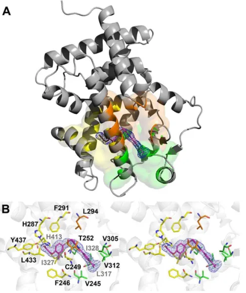
(Fig. 2A). The final model consists of a monomer in the asymmetric unit, composed of residues Gln171 to Tyr441 (hPPARd numbering). One molecule of GW0742, 185 water molecules and one glycerol molecule were also resolved in the structure. All protein residues occupy favorable regions of the Ramachandran plot; data collection statistics are given in Table 1. Overall folding resembles previous PPARdLBD structures and is not further described.
GW0742 occupied the Y-shaped LBP and adopted a position predicted by the pharmacophoric model of PPAR ligands
[8,9,10,18] (Fig. 2B). The hydrophilic head group interacts with arm I and the hydrophobic tail, comprising the thiazole and the fluorine substituted phenyl ring, is positioned mostly in arm II. The linker connecting the head and tail groups lies close to H3 (Fig. 2B). In total, GW0742 made 29 ligand interactions with PPARdpocket, including three polar interactions and 26 apolar interactions (Table S1).
Polar interactions mostly involve the ligand hydrophilic head group and residues in Arm I and appear similar to other PPAR agonists with their respective PPARs [9]. By analogy, these interactions are probably responsible for maintaining the locked agonist conformation of activation helix 12. One ligand carbox-ylate oxygen engages in hydrogen bonds with the side chains of residues His413 (helix 10/11) and Tyr437 (helix 12) - Figure 2B. The other carboxylate oxygen contacts the His287 side chain from PPARdhelix 7.
Apolar interactions involved residues in all three Arms. In Arm I Phe246, Phe291, His413, Ile327, Leu433 and Cys249 side chains contact ligand. In arm II, Val245, Val305, Val312, Leu317 and Ile328 side chains bind ligand and two residues that lie within Arm III, Thr252 and Leu294, are also engaged in ligand contact.
We were not able to discern any GW0742 contacts with amino acids that were completely unique to PPARdand could account for selective ligand binding (Fig. 3) [18]. Of 12 Arm I amino acids (Fig. 3A); eight (Phe246, Cys249, His287, Phe291, Ile327, His413, Leu433 and Tyr437) contact GW0742. Of these, His287, Phe291 and Ile327 vary between PPARs and none are exclusive to PPARd; Phe291 and Ile327 are conserved in PPARaand His287 is conserved in PPARc. Of 12 Arm II residues (Fig. 3B), five (Val245, Val305, Val312, Leu317 and Ile328) are involved in ligand contact. Of these; Leu317 is identical in all subtypes and there are conserved substitutions at the other four positions. Of nine Arm III residues (Fig. 3C), only two contact ligand; Leu294 is conserved in the three PPAR subtypes and Thr252 is conserved in PPARawith a non-conserved substitution in PPARc.
Potential Steric Hindrance to GW0742 Binding in PPARa
andc
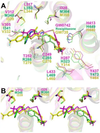
We next compared the PPARd:GW0742 structure with analogous structures of PPARa and c LBDs in complex with representative selective agonists (GW735 and Rosiglitazone) and the three PPARs with a pan agonist, indeglitazar (PDB ids: 2P54 [19], 2PRG [20] and 3ET2, 3ET3 and 3ET1, respectively [21]. All four ligands conform to the standard PPAR ligand pharma-cophoric model [22] and adopt a similar position in the pocket (Fig. 4). However, GW0742 binding exhibited two features that were unique. First, the linker group is displaced from H3 relative to other PPAR subtype selective ligands (Fig. 4A). This shift was also seen in the PPARdstructure with the non-selective agonist indeglitazar (not shown), suggesting that it does not account for selectivity. More interestingly, the GW0742 hydrophobic tail occupies the entrance to Arm II, unlike GW735 and Rosiglitazone tails which are directed towards Arm III between the helices 3 and 2` (Fig. 4A and B).
Comparison of amino acids that form the PPARd Arm II entrance with equivalent regions of PPARaand PPARcrevealed two substitutions which could potentially form barriers to GW0742 binding and could block access to PPARcand PPARa
Arm II;dVal312 is replaced by bulkier side chainsaIle339 and
cMet376 anddIle328 is substituted by the bulkier methionine in both PPARs (aMet355 and cMet392) (Fig. 4B). Other nearby substitutions do not exhibit similar potential to block GW0742 binding. Some introduce similarly sized amino acids (dHis287/
aTyr314/cHis351; dPhe291/aPhe318/cTyr355; dVal245/
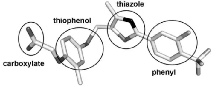
Figure 1. Three-dimensional structure of PPARd-selective agonist GW0742, as found in our hPPARd:GW0742 crystal structure (PDB id 3TKM).Typical structural features of PPAR agonists are displayed. Carbon, fluoride, sulfur, oxygen and nitrogen atoms are colored white, light grey, grey, dark grey and black, respectively. doi:10.1371/journal.pone.0033643.g001
aIle272/cIle309 and dVal305/aVal332/cIle369). Nearby PPARc-specific substitutions (dIle327/aIle354/cPhe391 and
dThr252/aThr279/cArg316) introduce residues with flexible side chains that are not likely to block GW0742 binding; cPhe391 contacts Rosiglitazone (PDB id 2PRG), but faces away from ligand in the PPARc:Indeglitazar structure (PDB id 3ET3) (Figure S1) andcArg316 faces away from both ligands.
Site Directed Mutagenesis Confirms Key Roles for
dVal312 anddIle328 in GW0742 Binding
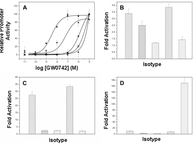
To determine whetherdVal312 anddIle328 are important for PPARd selective activation by GW0742, we introduced Met substitutions at both positions: PPARd-LBD/Val312Met and PPARd-LBD/Ile328Met and determined effects of mutations on responses to different ligands. As expected [13], GW0742 was a potent activator of PPARd(EC50= 3.25 nM) relative to PPARaor
PPARc; it was not possible to derive accurate EC50values for the
latter curves. Both PPARd mutants displayed similar levels of
activation at very high GW0742 concentrations, but EC50values
were greatly increased relative to wild type receptor, indicative of reduced potency (Fig. 5A). Half-maximal responses were one order of magnitude higher (66.0 nM) for PPARdIle328Met relative to wild type receptor and EC50values for PPARdVal312Met mutant
were even higher, it was not possible to achieve an adequate estimate of EC50values similar to wild type PPARaand PPARd.
The met substituents did not completely change overall PPARd
ligand binding profile. The pan-PPAR agonist benzafibrate [23]; activated PPARa, PPARd and PPARcwith descending efficacy (Fig. 5B) and PPARd Val312Met activation was about 2.5 fold, similar to wild type PPARdand PPARdIle328Met was similar to that of PPARc(1.5 times of activation). Neither met substituent enhanced activation by the PPARa selective agonist GW7647 (Fig. 5C) or the PPARcselective agonist rosiglitazone (Fig. 5D). Thus, the presence of bulky residues at positions 312 and 328 reduces PPARd activation by GW0742 but does not permit PPARdactivation by ligands that bind other subtypes.
Figure 2. Crystallographic structure of the complex hPPARd-LBD:GW0742.(A)The ligand (magenta sticks) occupies the PPARd-LBD (grey cartoon) and performs interactions with residues belonging to the arm I (yellow), arm II (green) and arm III (orange).(B)Stereo view of the binding site, showing the electron density calculated for the ligand (omit map, contoured ats= 1.0) and the PPARdresidues that stabilize the ligand. Polar interactions between hPPARd-LBD and the GW0742 ligand are shown as dashed lines. Nitrogen, oxygen, sulfur and fluoride atoms are colored blue, red, yellow and light blue, respectively. The residues from arms I, II and III are colored in yellow, green and orange, respectively. Figures were generated with the Pymol software (Schro¨dinger).
Docking and Molecular Dynamics Simulation
We next modeled PPARd-LBD mutants, docking the ligand inside the structures. After docking, energy minimization and molecular dynamics simulation, all the PPARs (a, c, d and the mutants dVal312M and dIle328M) showed accommodation of their main chains, with trajectory root mean square deviation (RMSDs) ranging from 1.3 A˚ to 1.65 A˚. Docking analysis revealed that for all PPARs, GW0742 was able to accommodate itself in the ligand binding pocket, but considerable conformational changes of the side chains, which corresponds to the Val312 and Ile328 substitutions, and also in the ligand were observed (data not shown).
Analysis of RMSDs of Met312 and Ile328, after simulations, shows that PPARd presents smaller conformational changes in comparison to the other PPARs (Table 2), clearly revealing necessity of large side chain adjustments by PPARa, c and the mutants, in order to accommodate GW0742 ligand.
In summary, we have solved the PPARd-LBD structure in complex with GW0742, a high potent and selective PPARd
agonist. The ligand follows the binding model predicted to other PPAR ligands based on the same pharmacoforic groups. The carboxylate group occupies arm I of the binding pocket while the hydrophobic tail occupies arm II. Comparison of the structures of the three PPARs isotypes with agonists allowed us to observe some subtle differences that could explain the isotype delta ligand selectivity to GW0742. Specifically, the hydrophobic tail of GW0742 occupies part of Arm II, unlike equivalent PPARaand PPARcagonists which dock into Arm III and we propose that the presence of two residues in PPARd-LBD, Val312 and Ile328, is intimately related with selectivity. Here, both of these residues are replaced by amino acids with bulkier side chains in PPARaand PPARc, and it is likely that these would occlude the entrance to ArmII in the context of these PPAR subtypes and prevent the GW0742 hydrophobic tail from docking into its preferred position. To validate this hypothesis, we performed two single point mutations, Val312Met and Ile328Met, and conducted cell activation assays and docking analyses of PPAR isotypes and mutants using selective ligands for each isotype and confirmed that
Table 1.Crystallographic data collection statistics.
Parameter
Wavelenght (A˚) 1.46
Space Group P212121
Unit Cell Dimensions (A˚) 35.466 41.766 96.287
Resolution Range (A˚) 24.4 (1.95)
Reflections at working set 19134 (2511)
Reflections at test set 978 (135)
Redundancy 5.8 (4.7)
Completeness (%) 99.28 (99.1)
I/d 17.3 (2.6)
Rfree 24.5 (30.2)
Rfactor 19.5 (25.6)
RMSD bond lengths (A˚) 0.004
RMSD bond angles (degrees.) 1.006
Average B-factor 24.85
Ramachandran outliers 0/303
Values in parentheses indicate the high-resolution shell. doi:10.1371/journal.pone.0033643.t001
Figure 3. Alignment of amino acid residues forming the binding site of the different human PPAR isotypes.Residues placed in arm I (A), arm II (B) and arm III (C) are shown. Residues involved in the hPPARd-LBD:GW0742 interactions are underscored. Residues in black, bold and gray represent identical residues, residues with same chemical character and residues with different chemical character, respectively.
doi:10.1371/journal.pone.0033643.g003
Figure 4. Crystallographic structure superposition of selective ligands to each PPAR isotype.Helices from PPAR are shown as yellow, magenta and green cartoons for PPARa,dandc, respectively. Theaselective ligand, GW735 (PDBid: 2P54), the dselective ligand, GW0742, and thecselective ligand, rosiglitazone (PDBid: 2PRG), are shown as yellow, magenta and green sticks, respectively. Oxygen, nitrogen, sulfur and fluoride atoms are shown in red, blue, yellow and light blue, respectively. A) Upper vision of the binding site. B) Stereoscopic view of the PPAR binding sites, highlighting the importance of Val312 and Ile328 in GW0742 accommodation and GW735 and rosiglitazone displacement, presumably due to the presence of bulky substitutions. Ligands GW735, GW0742, rosiglitazone are painted in yellow, magenta and green, respectively.
doi:10.1371/journal.pone.0033643.g004
introduction of substituents that resemble other PPARs at these positions reduces activation of PPARdby GW0742 but not other non-selective PPAR ligands. Our results indicate that ligands carrying short linkers and large and rigid hydrophobic tails find difficulties in being accommodated into PPARaand PPARcarm II, probably as a consequence of the bulky amino acid substitution found in these isotypes. We propose that this hypothesis brings some light to the understanding of the molecular basis of PPAR
selective ligands mode of interaction and may be helpful in further rational design of PPAR selective agonists.
Our results agree with previous studies which link effects of amino acid substitution in PPARs binding sites upon ligand binding to the binding site shape, which, in turn, limits ligand entry and accommodation [8,9,10,18,24]. PPARd presents the smaller arm I as a consequence of the presence of Met417 in the place of the Val residue present in PPARa, what explains the relative low affinity of this isotype for some fibrates and other
Figure 5. PPAR transactivation assays.PPAR activation induced by(A)thed-selective agonist GW0742;(B)the pan-agonist benzafibrate;(C)the a-selective agonist GW7647 and(D)thec-selective agonist rosiglitazone. All data were normalized by the level ofRenillaluciferase activity.&/# wtPPARd,m/xx PPARdVal312Met,
N
///PPARdIle328M, / = PPARaandb/ PPARc. doi:10.1371/journal.pone.0033643.g005Table 2.RMSD values of the residuesdMet312,dIle328 and its corresponding residues from PPARa,cand mutants after GW0742
docking and molecular dynamic simulations.
model RMSD ofdMet312 position(A˚ ) RMSD ofdIle328 position (A˚ )
PPARd 0.3 0.6
PPARa 0.6 1.1
PPARc 1 1.8
PPARdV312M 0.9 0.7
PPARdI328M 0.3 0.9
ligands with large groups linked to the hydrophilic head [18,24]. In the same direction, the presence of Tyr344 in PPARaarm III reduces the size of the binding site entrance, causing steric restrictions to ligand entry [8]. Substitutions in arm II were mainly related to change the accommodation of the main hydrophobic part of the ligands [8,18]. This mode of selectivity is very different from that of other NRs, such as thyroid hormone and estrogen receptors, where selectivity often relates to enhanced contacts between ligand and specific amino acids within the pocket. It will be important to understand the rules that link pocket shape to ligand position in PPARs to better develop new selective ligands.
Methods
Protein Expression and Purification
The human PPARdLBD plasmid (amino acids 171–441) with cDNA inserted into pET15 vector (Novagen, USA) was trans-formed in BL21(DE3) Escherichia coli. Protein expression was performed in LB culture, induced with 1 mM IPTG, at 18uC for 12 h. Cells were harvested and ressuspended in a 20 ml of buffer A (20 mM Hepes pH 7.5, 300 mM NaCl, 5% glycerol, 10 mMb -mercaptoethanol, 10 mM PMSF and 250mg/mL lysozyme) per
liter of culture. The lysate was sonicated, clarified by centrifuga-tion and loaded onto a Talon Superflow Metal Affinity Resin (BD Biosciences Clontech, Palo Alto, CA), and eluted with an imidazol gradient (0–300 mM). The fractions containing the purified protein were pooled and washed, using centrifugal concentrators (Amicon, 10 MW cutoff), to remove imidazol. The His-tag was cleaved with trombin (7 U/mg), at 18uC, overnight. Protein purity was checked by Coomassie blue-stained SDS-PAGE. Protein concentrations were determined using the Bradford dye assay (Bio-Rad, Hercules, CA).
Crystallization
Protein buffer was changed to 20 mM Hepes (pH 7.5), 500 mM ammonium acetate, 10 mMb- mercaptoethanol, according to [8]. Prior to crystallization, PPARd-LBD (256mM) was incubated for 4 h with GW0742 (Tocris Bioscience) (1:4 protein:ligand molar ratio) in DMSO (DMSO final concentration equals to 5%), at 4uC. The sitting-drop vapor diffusion method was used, with drops containing 2ml of protein:ligand complex, 0.5ml of the detergent n-Octyl-b-D-thioglucoside and 2.5ml of the reservoir solution made of 14% (w/v) polyethylene glycol (PEG) 8000, 200 mM KCl, 40 mM bis-Tris-propane (pH 9.5), 6% propanol, 1 mM CaCl2. hPPARd-LBD:ligand co-crystals were grown at 18uC and
appeared after 3 days, showing a well-defined geometric form.
Data Collection, Model Refinement and Analysis
Crystals were transferred to a cryo-protecting solution, contain-ing the well solution plus 10% glycerol, and immediately flash cooled to 100 K in a nitrogen stream prior to data collection. The X-ray diffraction data collection was performed at the MX-2 beamline of the Brazilian National Synchrotron Light Laboratory (LNLS, Campinas, Brazil) [25] using synchrotron radiation of wavelength 1.459 A˚ to optimize crystal diffraction efficiency and the synchrotron-radiation flux of the LNLS storage ring [26]. The diffraction images were registered on a MAR225 mosaic detector, with an oscillation of 1uper image. Data reduction was performed using HKL200/Scalepack package [27].
The X-ray structure of PPARd-LBD (PDB ID: 3ET2) [21] was used as an initial model for molecular replacement using the program PHASER [28]. The protein atomic model was improved through alternated cycles of real space refinement using COOT [29] and maximum likelihood minimization using PHENIX [30].
Ligand and solvent molecules were included in the last steps of refinement.
Protein:ligand contacts were analyzed using the Ligplot software [31], followed by visual inspection using the program COOT [29]. A hydrogen bond distance cutoff of 3.4 A˚ was applied. Superpo-sition of different PPAR crystal structures was performed with the Superpose software [32] and analyzed using the Pymol software [33].
PDB Accession Code
The atomic coordinates and structure factors of the hPPARd -LBD:GW0742 crystal complex reported here are deposited in the Protein Data Bank under code 3TKM.
Site Directed Mutagenesis and Transactivation Assay
Mutations in the hPPARd-LBD were introduced by PCR in an existing vector PPARdGAL4 [34] with overlapping of mutated primers and vector using the QuikChange site-directed mutagen-esis kit (Stratagene). All mutated constructs were verified by sequencing. The reporter plasmid pGRE-LUC (GAL4 responsive element,Fireflyluciferase reporter vector) and PPARdLBDGal4 inserted in pBIND (Promega). The pRL-TK, that containsRenilla
luciferase, was purchased from Promega (Dual-Luciferase Report Assay systemPromega, Madison, WI).
HepG2 cells were cultured in Dulbecco’s Modified Eagle’s Medium (DEMEM) supplemented with 10% heat-inactivated fetal bovine serum, 2 mM glutamin, 50 UI/mL penicillin/streptomy-cin under 95% air and 5% CO2 at 37uC. For transactivation
assays, the cells were removed by trypsinization and replated in 24 wells plate at density of 1,26105cells/well. Cell transfections were performed using FuGENE 6 transfection reagent (Roche, Swiss) with 100gg of plasmids containing wild-type PPARa,douc-LBD or PPARd-LBD mutants, DBD Gal-4, 50gg of luciferase reporter plasmid and 1gg ofRenillaluciferase plasmid per well. Cells were treated with different concentrations of agonists of PPARa -GW7647, PPARd - GW0742 and PPARc - Roziglitazone, in triplicate 24 h after transfection and incubated for additional 24 h. Cell lysates were prepared and luciferase assay was performed using the Dual-Luciferase Report Assay system (Promega, Madison, WI), following manufacturer instructions. Light emission was measured by integration over 5 seconds of reaction in a Safire luminescent counter (Tecan, Tecan US, NC, USA). Firefly luciferase activity was normalized by the level ofRenillaluciferase activity, as recommended by manufacturersDual-Luciferase Report Assay system. Data were fitted using a sigmoidal dose-response function with corresponding EC50 determination according to GraphPad Prism software (version 5.0).
Docking and Molecular Dynamics Simulation
The molecular complexes for PPARa,c, mutants and GW0742 were built using the ligand conformation obtained from crystal-lographic structure of PPARdLBD:GW0742 complex (PDBid 3TKM). PPARaandcLBD structures (PDBid 3ET1 and 3ET3 respectively) were superposed to PPARdcomplex and coordinates of the ligand were copied to the PPARaandcstructures. Mutant PPARd-LBD models V312M and I328M were built using the YASARA software. All structures were submitted to energy minimization and molecular dynamics simulation using YASARA. For that, all hydrogen atoms and other missing atoms from the model were created using force field parameters, obtained from YAMBER3. A simulation box was defined at 15 A˚ around all atoms of each complex. Protonation was performed based on the pH 7. Cell neutralization was reached filling the box with water molecules and Na+
/Cl2counter ions. A short molecular dynamics
(MD) simulation was performed for the solvent adjust, deleting water until the density of 0.997 g/ml was reached. A short steepest descent energy minimization was carried until the maximum atom speed dropped below 2200 m/s. Then 500 steps of simulated annealing were performed with a temperature of 0 K. Finally, a 4 ns (nanosecond) simulation at 298 K and a non-bonded cutoff of 7.86 A was performed. A snapshot was saved every 25 ps (picosecond). Simulation time was adjusted to stabilize the contacts between protein and ligand.
Supporting Information
Figure S1 Observation of phenylalanine flexibility on PPARc
structures. Superposition of the c-selective ligand rosiglitazone (green stick), pan-agonist ligand indeglitazar (blue sticks) and the
cPhe391 residue from the respective crystallographic structures for PDB id 2PRG (green lines) and 3ET3 (green lines). Helix 3 is shown as a blue and green cartoon. Oxygen, nitrogen, sulfur and
fluoride atoms are shown in red, blue, yellow and light blue, respectively.
(DOC)
Table S1 Atoms involved in interactions between the GW0742 ligand and hPPARd-LBD, as found in our hPPARd-LBD: GW0742 crystal structure.
(DOC)
Acknowledgments
We thank William N. Hunter for the hPPARd-LBD clone.
Author Contributions
Conceived and designed the experiments: FAHB DBBT ACMF PW IP. Performed the experiments: FAHB DBBT JG AB PSLO. Analyzed the data: FAHB DBBT ACMF PW IP. Contributed reagents/materials/ analysis tools: PW IP. Wrote the paper: FAHB DBBT ACMF.
References
1. Tenenbaum A, Motro M, Fisman EZ (2005) Dual and pan-peroxisome proliferator-activated receptors (PPAR) co-agonism: the bezafibrate lessons. Cardiovasc Diabetol 4: 14.
2. Desvergne B, Wahli W (1999) Peroxisome proliferator-activated receptors: nuclear control of metabolism. Endocr Rev 20: 649–688.
3. Kota BP, Huang TH, Roufogalis BD (2005) An overview on biological mechanisms of PPARs. Pharmacol Res 51: 85–94.
4. Wagner KD, Wagner N (2010) Peroxisome proliferator-activated receptor beta/ delta (PPARbeta/delta) acts as regulator of metabolism linked to multiple cellular functions. Pharmacol Ther 125: 423–435.
5. Heikkinen S, Auwerx J, Argmann CA (2007) PPARgamma in human and mouse physiology. Biochim Biophys Acta 1771: 999–1013.
6. Kersten S, Desvergne B, Wahli W (2000) Roles of PPARs in health and disease. Nature 405: 421–424.
7. Xu HE, Lambert MH, Montana VG, Plunket KD, Moore LB, et al. (2001) Structural determinants of ligand binding selectivity between the peroxisome proliferator-activated receptors. Proc Natl Acad Sci U S A 98: 13919–13924. 8. Fyffe SA, Alphey MS, Buetow L, Smith TK, Ferguson MA, et al. (2006)
Recombinant human PPAR-beta/delta ligand-binding domain is locked in an activated conformation by endogenous fatty acids. J Mol Biol 356: 1005–1013. 9. Markt P, Schuster D, Kirchmair J, Laggner C, Langer T (2007) Pharmacophore modeling and parallel screening for PPAR ligands. J Comput Aided Mol Des 21: 575–590.
10. Xu HE, Lambert MH, Montana VG, Parks DJ, Blanchard SG, et al. (1999) Molecular recognition of fatty acids by peroxisome proliferator-activated receptors. Mol Cell 3: 397–403.
11. Zoete V, Grosdidier A, Michielin O (2007) Peroxisome proliferator-activated receptor structures: ligand specificity, molecular switch and interactions with regulators. Biochim Biophys Acta 1771: 915–925.
12. Sundriyal S, Bharatam PV (2009) Important pharmacophoric features of pan PPAR agonists: common chemical feature analysis and virtual screening. Eur J Med Chem 44: 3488–3495.
13. Sznaidman ML, Haffner CD, Maloney PR, Fivush A, Chao E, et al. (2003) Novel selective small molecule agonists for peroxisome proliferator-activated receptor delta (PPARdelta)–synthesis and biological activity. Bioorg Med Chem Lett 13: 1517–1521.
14. Bility MT, Devlin-Durante MK, Blazanin N, Glick AB, Ward JM, et al. (2008) Ligand activation of peroxisome proliferator-activated receptor beta/delta (PPAR beta/delta) inhibits chemically induced skin tumorigenesis. Carcinogen-esis 29: 2406–2414.
15. Bility MT, Zhu B, Kang BH, Gonzalez FJ, Peters JM (2010) Ligand activation of peroxisome proliferator-activated receptor-beta/delta and inhibition of cyclo-oxygenase-2 enhances inhibition of skin tumorigenesis. Toxicol Sci 113: 27–36. 16. Gaudel C, Schwartz C, Giordano C, Abumrad NA, Grimaldi PA (2008) Pharmacological activation of PPARbeta promotes rapid and calcineurin-dependent fiber remodeling and angiogenesis in mouse skeletal muscle. Am J Physiol Endocrinol Metab 295: E297–304.
17. Wagner N, Jehl-Pie´tri C, Lopez P, Murdaca J, Giordano C, et al. (2009) Peroxisome proliferator-activated receptor beta stimulation induces rapid
cardiac growth and angiogenesis via direct activation of calcineurin. Cardiovasc Res 83: 61–71.
18. Zoete V, Grosdidier A, Michielin O (2007) Peroxisome proliferator-activated receptor structures: Ligand specificity, molecular switch and interactions with regulators. Biochimica et Biophysica Acta (BBA) - Molecular and Cell Biology of Lipids 1771: 915–925.
19. Sierra ML, Beneton V, Boullay AB, Boyer T, Brewster AG, et al. (2007) Substituted 2-[(4-aminomethyl)phenoxy]-2-methylpropionic acid PPARalpha agonists. 1. Discovery of a novel series of potent HDLc raising agents. J Med Chem 50: 685–695.
20. Nolte RT, Wisely GB, Westin S, Cobb JE, Lambert MH, et al. (1998) Ligand binding and co-activator assembly of the peroxisome proliferator-activated receptor-gamma. Nature 395: 137–143.
21. Artis DR, Lin JJ, Zhang C, Wang W, Mehra U, et al. (2009) Scaffold-based discovery of indeglitazar, a PPAR pan-active anti-diabetic agent. Proc Natl Acad Sci U S A 106: 262–267.
22. Jain E, Bairoch A, Duvaud S, Phan I, Redaschi N, et al. (2009) Infrastructure for the life sciences: design and implementation of the UniProt website. BMC Bioinformatics 10: 136.
23. Willson TM, Brown PJ, Sternbach DD, Henke BR (2000) The PPARs: From orphan receptors to drug discovery. J Med Chem 43: 528–550.
24. Takada I, Yu RT, Xu HE, Lambert MH, Montana VG, et al. (2000) Alteration of a single amino acid in peroxisome proliferator-activated receptor-alpha (PPAR alpha) generates a PPAR delta phenotype. Mol Endocrinol 14: 733–740. 25. Guimara˜es BG, Sanfelici L, Neuenschwander RT, Rodrigues F, Grizolli WC, et al (2009) The MX2 macromolecular crystallography beamline: a wiggler X-ray source at the LNLS. J Synchrotron Radiat 16: 69–75.
26. Polikarpov I, Perles LA, de Oliveira RT, Oliva G, Castellano EE, et al. (1998) Set-up and experimental parameters of the protein crystallography beamline at the Brazilian National Synchrotron Laboratory. J Synchrotron Radiat 5: 72–76. 27. Otwinowski Z, Minor W (1997) Processing of X-ray Diffraction Data Collected
in Oscillation Mode. Methods in Enzymology 276: 307–326.
28. McCoy AJ, Grosse-Kunstleve RW, Adams PD, Winn MD, Storoni LC, et al. (2007) Phaser crystallographic software. J Appl Crystallogr 40: 658–674. 29. Emsley P, Cowtan K (2004) Coot: model-building tools for molecular graphics.
Acta Crystallogr D Biol Crystallogr 60: 2126–2132.
30. Adams PD, Afonine PV, Bunko´czi G, Chen VB, Davis IW, et al. (2010) PHENIX: a comprehensive Python-based system for macromolecular structure solution. Acta Crystallogr D Biol Crystallogr 66: 213–221.
31. Wallace AC, Laskowski RA, Thornton JM (1995) LIGPLOT: a program to generate schematic diagrams of protein-ligand interactions. Protein Eng 8: 127–134.