Article
Regulation of Adipocyte Differentiation via MicroRNAs
You Hwa Son1,*, Sojeong Ka2, A Young Kim2, Jae Bum Kim1,2
1Seoul National University School of Biological Sciences; 2Institute of Molecular Biology and Genetics, Seoul National
University, Seoul, Korea
Adipocyte differentiation, termed adipogenesis, is a complicated process in which pluripotent mesenchymal stem cells differenti-ate into mature adipocytes. The process of adipocyte differentiation is tightly reguldifferenti-ated by a number of transcription factors, hor-mones and signaling pathway molecules. Recent studies have demonstrated that microRNAs, which belong to small noncoding RNA species, are also involved in adipocyte differentiation. In vivo and in vitro studies have revealed that various microRNAs af-fect adipogenesis by targeting several adipogenic transcription factors and key signaling molecules. In this review, we will sum-marize the roles of microRNAs in adipogenesis and their target genes associated with each stage of adipocyte differentiation.
Keywords: MicroRNAs; Adipocyte differentiation; Obesity
INTRODUCTION
Adipose tissue plays key roles in energy storage, regulation of body temperature and absorption from mechanical collision. In a state of obesity, adipose tissue expands due to an increase in adipocyte size (hypertrophy) and/or number (hyperplasia), thereby inducing dysregulation of glucose and lipid metabo-lism. As a result, increased adipose tissue destroys whole body energy balance and enhances the risk of insulin resistance, hy-pertension, and dyslipidemia. Therefore, proper understanding of adipocyte differentiation would provide valuable informa-tion for designing comprehensive and effective therapeutic strategies against obesity.
Adipocyte differentiation occurs in several stages, involves many signaling pathways, and progress depends on various stimuli such as nutrients and hormones. For example, adipo-genesis is tightly controlled by a cascade of several
transcrip-tion factors such as CCAAT/enhancer-binding proteins (C/
EBPs) and peroxisome proliferator-activated receptor γ (PPAR-γ). Additionally, several signaling molecules, including wing -less and INT-1 proteins (Wnts) and insulin, modulate adipo-genesis. It has also been demonstrated that microRNAs are in-volved in adipocyte differentiation [1].
MicroRNAs (miRNA) are 19 to 22 nucleotide fragments of noncoding RNA that play important roles in various cellular processes through posttranscriptional regulation of target genes. As many groups have reported that miRNAs actively participate in cell proliferation and differentiation [2-4], knowl-edge is constantly increasing related to miRNAs involvement in adipogenesis and fat metabolism. Although several reviews have recently been published on related topics [1,5,6], this re-view article will specifically describe the miRNA-mediated regulatory mechanism in adipocyte differentiation, particularly by updating and integrating the most recent information.
Corresponding author: Jae Bum Kim
School of Biological Sciences and Institute of Molecular Biology and Genetics, Seoul National University,1 Gwanak-ro, Gwanak-gu, Seoul 151-742, Korea
Tel: +82-2-880-5852, Fax: +82-2-878-5852, E-mail: [email protected] *Current affiliation: Division of Drug Discovery Research, Pharmacological Research Center, Korea Research Institute of Chemical Technology, Daejeon, Korea
Copyright © 2014 Korean Endocrine Society
MOLECULAR EVENTS DURING
ADIPOCYTE DIFFERENTIATION
In vitro studies have suggested that the progression of adipo-cyte differentiation has, at least, two key steps: commitment and differentiation. Adipocytes are derived from pluripotent mesenchymal stem cells (MSCs) that have the capacity to de-velop into several cell types, including adipocytes and osteo-blasts. Commitment or determination of MSCs’ fate to differ-entiate into preadipocytes is caused by adipogenic differentia-tion signaling cues that have not yet been indentified [7]. Fol-lowing this step, committed MSCs are specified for an adipo-genic lineage and often lose their ability to differentiate into other cell lineages.
In the differentiation step, committed preadipocytes derived from MSCs (e.g., 3T3-L1 cells) are differentiated into adipo-cytes after exposure to hormone cocktails such as insulin, dexamethasone and cyclic adenosine monophosphate (cAMP) activators [8]. Contact with these chemicals induces G1 phase-arrested 3T3-L1 cells to synchronously undergo, on average, two cycles of cell division, so called mitotic clonal expansion. During the cell cycle, clonal expansion is regulated by the Rb-E2F pathway, which is responsible for the G1-to-S transition. Rb inhibits the cell cycle by binding to, and repressing, the transcriptional activity of E2F. Upon hyperphosphorylation of Rb by cyclin-dependent kinases, E2F is released and promotes transcriptional activation of genes that encode cell-cycle regu-lators required for S phase entry; a process that initiates clonal expansion [9]. In growth-arrested preadipocytes, there are sig-nificant levels of Rb family p130 (pRB/p130). Inactivation of Rb2/p130 by phosphorylation enables clonal expansion. When cells exit the cell cycle, E2F loses its activity and terminal dif-ferentiation is initiated [10].
Several signaling pathways highlight molecules such as bone morphogenic protein (BMP) and Wnt, which have been shown to be key molecules in the regulation of MSC commit-ment to adipocyte lineage and the differentiation of a subset of
adipocytes. BMPs belong to the transforming growth factor β (TGF-β) family of growth factors, which consists of 14 family
members. BMP-2 and BMP-4 have been implicated in adipo-genesis and are thought to promote commitment of cells to ad-ipogenic lineages [11-14]. The positive role of BMP-4 in adi-pocyte commitment has been demonstrated with several estab-lished cell lines. In C3H10T1/2 cells, exogenous BMP-4 acti-vation induces potent adipocyte differentiation. In addition, a committed preadipocyte A33 cell line derived from C3H10T1/
2 stem cells expresses and secretes BMP-4 at the same time point when exogenous BMP-4 is added to C3H10T1/2 cells for adipogenic differentiation. Furthermore, exposure of A33 cells to noggin, a naturally occurring BMP-4-binding antagonist, during this critical time window blocks subsequent differentia-tion [11]. The effect of BMP-2 is more complex. BMP-2 can enhance adipogenesis of C3H10T1/2 cells at low concentra-tions, but stimulates chondrocyte and osteoblast development at higher concentrations [14]. In preadipocytes, BMPs activate Sma and Mad related protein (Smad) signaling and regulate many target genes including cytoskeleton-associated proteins [12]. BMPs are also known as powerful cytokines that induce bone and cartilage formation. BMP-Smad signaling in this de-velopmental context can activate runt-related transcription fac-tor 2 (Runx2), osterix, Dlx5/6, and Sox9, which are essential transcription factors for osteogenesis and chondrogenesis [13].
In addition to BMPs, the β superfamily member, TGF-β, is also involved in adipogenesis. In general, TGF-β signals
through two types of transmembrane serine/threonine kinase
receptors, type I and type II TGF-β receptors, and signaling effector Smads. Activation of Smad2 or Smad3 by TGF-β re -ceptors results in heterodimerization with Smad4 and stimu-lates nuclear translocation of Smad complexes. In the nucleus, Smad proteins regulate transcription by binding to DNA and interacting with other transcription factors. During
adipogene-sis, TGF-β phosphorylates only Smad3, which then binds to
C/EBPs and inhibits their transcriptional activity, including
the ability to transactivate PPARγ [15]. Consistently, it has been demonstrated that TGF-β1 inhibits the early stages of
3T3-L1 differentiation [16] by promoting the proliferation of progenitor cells and hampering lipid accumulation [17].
More-over, transgenic overexpression of TGF-β in adipose tissue in -hibits differentiation in vivo [18].
The Wnt family is made up of secreted glycoproteins that influence cell fate and development. Wnt proteins bind to
friz-zled receptors to stimulate signaling cascades through β-catenin-dependent (Wnt/β-catenin) and -inβ-catenin-dependent pathways. The Wnt/β-catenin signaling pathway is often activated in preadi -pocytes and expression of Wnts declines after induction of
dif-ferentiation [19,20]. It has been suggested that Wnt/β-catenin
signaling enhances proliferation during commitment and
mi-totic clonal expansion [20]. Activated Wnt/β-catenin signaling
enables the lymphoid-enhancer-binding factor/T-cell-specific transcription factor (LEF/TCF) family of transcription factors to activate Wnt target genes. In late stages of adipogenesis,
is known about how Wnt inhibits adipogenesis through TCF/ LEF [21].
Many signaling pathways influence adipocyte differentia-tion. For instance, mitogen-activated protein kinase (MAPK) pathways may enhance adipogenesis through extracellular sig-nal-regulated kinases (ERKs) and p38. Although it has been suggested that activation of ERK1 positively regulates adipo-genesis during clonal expansion, activity has to be reduced af-ter proliferation in order for adipogenesis to proceed [22]. Among various hormones, insulin plays a key role in adipo-genesis. Insulin functions predominantly through insulin growth factor-1 receptor signaling, and the downstream signal-ing involves insulin receptor substrate (IRS), phosphoinositide 3-kinase (PI3K), PDK1, and AKT/protein kinase B (PKB) sig-naling. Insulin signaling can be transmitted to the adipogenic cascades in several different ways. IRS signaling is known to promote cAMP response element-binding (CREB) phosphory-lation, thereby influencing adipogenesis [23]. Another way for insulin to deliver its signal is through inhibition of antiadipo-genic forkhead box protein O (FOXO) transcription factors af-ter AKT/PKB-mediated phosphorylation [24].
Induction of adipogenesis by differentiation cocktails im-mediately phosphorylates and activates CREB, which in turn
transcriptionally activates the C/EBPβ gene. Elevated C/EBPβ
is somewhat inactive until it acquires DNA-binding activity through a series of phosphorylaton events by MAPK and
GSK3β. Early activation of C/EBPβ, together with C/EBPδ, promotes gene expression of C/EBPα and PPARγ through C/ EBP regulatory elements in their proximal promoters. C/EBPα
induces adipogenic genes, and in vivo studies indicate an im-portant role for this factor in the development of adipose
tis-sue. PPARγ is the ‘master regulator’ of adipogenesis and is a
necessary and sufficient factor for adipogenesis. Even though the C/EBPs are considered to be important transcription fac-tors in adipogenesis, they cannot function efficiently without
PPARγ [25]. PPARγ and C/EBPα coordinately control expres -sion of a large group of genes that are required for adipocyte
phenotypes. During adipogenesis, PPARγ and C/EBPα posi -tively cross-activate each other through their respective C/ EBP regulatory elements [25,26].
Other key transcriptional factors are the Kruppel-like fac-tors (KLFs). The KLFs are a large family of C2H2 zinc-finger proteins that regulate apoptosis, proliferation, and differentia-tion. This family of transcription factors contains both repres-sors and activators of transcription. Seven of the 17 known KLF proteins have been shown to be involved in different
phases of adipocyte differentiation (i.e., KLF2 and 3 inhibit differentiation, whereas KLF4, 5, 6, 9, and 15 stimulate differ-entiation) [27].
ROLES OF miRNAs IN ADIPOGENESIS
A number of miRNAs and their targets have been implicated in adipogenesis. As summarized in Table 1 [1,2,28-68], some appear to enhance adipocyte differentiation while others in-hibit adipogenesis in various model systems. An integrative model displaying the function of each miRNA in the progres-sion of adipogenesis is depicted in Fig. 1.
LINEAGE DETERMINATION
(COMMITMENT)
Adipocytes and osteoblasts originate from common MSCs. The differentiation of MSCs into adipocytes and osteoblasts is not only dependent on the mechanisms that determine a spe-cific cell lineage, but also on the mechanisms that suppress the development of other lineages [69]. In mesenchymal progeni-tors, the selection between adipogenesis and osteogenesis is influenced by reciprocal regulation of different intracellular signals and transcription factors. Furthermore, previous stud-ies have suggested that miRNAs are involved in both the lin-eage fate of MSCs and osteoblast differentiation [70].
PPARγ regulates the whole process of adipogenesis includ -ing lineage commitment and differentiation. Thus, it appears
that miRNAs specifically targeting PPARγ would also be
modulators of adipogenesis. Several computational prediction programs have proposed that miR-27a and miR-130a may
tar-get PPARγ 3’-untranslated region (UTR) in a sequence-specif -ic manner. In accordance with those pred-ictions, it has been reported that both miR-27a and miR-130a indeed suppress
ad-ipocyte differentiation through PPARγ downregulation [28,
29]. In 3T3-L1 cells, the levels of miR-27a and miR-130a are gradually decreased during adipogenesis, which is inversely
correlated with expression levels of PPARγ. Furthermore,
overexpression of miR-27a and miR-130a evidently
suppress-es adipocyte differentiation, concomitantly with PPARγ pro -tein expression. These findings have suggested that both
miR-27a and miR-130a negatively regulate PPARγ expression,
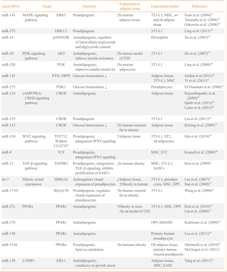
Table 1. MicroRNAs Related to Adipogenesis
micro RNA Target Function Expression in
adipose tissue Experiment model Reference
miR-143 MAPK signaling pathway
ERK5 Proadipogenic ↑In murine adipose tissue
3T3-L1, MSC, wt and ob adipose tissue
Esau et al. (2004)43
Takanabe et al. (2008)47
Oskowitz et al. (2008)48
miR-375 ERK1/2 Proadipogenic 3T3-L1 Ling et al. (2011)36
miR-14 p38MAPK Antiadipogenic, regulator of intracellular triglyceride and diglyceride content
Drosophila Xu et al. (2003)34
miR-29 PI3K signaling pathway
AKT Antiadipogenic,
induces insulin resistance
↑In mouse model
of T2D
3T3-L1 He et al. (2007)37
miR-320 PI3K Antiadipogenic,
improves insulin sensitivity
↑In murine
adipocytes
3T3-L1 Ling et al. (2009)49
miR-143 PTN, ORP8 Glucose homeotatsis ↓ Adipose tissue, 3T3-L1, MSC
Jordan et al (2011)50
Yi et al. (2011)51
miR-375 PDK1 Glucose homeotatsis ↓ Preadipocytes El Ouaamari et al. (2008)52
miR-124 cAMP/PKA/ CREB signaling pathway
CREB Antiadipogenic Adipose tissue Rajasethupathy et al. (2009)53
Qadir et al. (2013)30
Laine et al. (2012)54
miR-155 CREB Proadipogenic 3T3-L1 Liu et al. (2011)40
miR-132 CREB Glucose homeotatsis ↓ ↑In human omental fat in obesity
Adipose tissue Kloting et al. (2009)55
miR-210 WNT signaling pathway
TCF712, Wntless CG32767
Proadipogenic,
antagonizes WNT signaling
↑Adipose tissue 3T3-L1, ST2, ob adipocytes
Qin et al. (2010)56
miR-8 TCF Proadipogenic,
antagonizes WNT signaling
MSC, ST2 Kennell et al. (2008)44
miR-21 TGF-β signaling pathway
TGFBR2 Proadipogenic, antagonizes
TGF-β signaling, inhibits
proliferation of hASCs
↓In human obesity MSC, 3T3-L1, hASCs
Kim et al. (2009)2
let-7 Mitotic clonal expransion
HMGA2 Antiregulates clonal expansion of preadipocytes
↓Adipose tissue,
↑Obesity in human 3T3-L1, preadipo cytes, MSC, OP9
Lee et al. (2007)57
Sun et al. (2009)33
miR-17-92 Rb2/p130 Proadipogenic, regulates clonal expansion of preadipocytes
↑In human omental
fat in obesity
3T3-L1 Wang et al. (2008)32
miR-27a PPARs PPARγ Antiadipogenic ↑Obesity in mice
↑In rat model of T2D3T3-L1, MSC, OP9 Kim et al. (2010)
28
Lin et al. (2009)35
miR-27b PPARγ Antiadipogenic OP9, hMADS Karbiener et al. (2009)58
miR-130 PPARγ Antiadipogenic Primary human preadipocyte
Lee et al. (2011)29
miR-519d PPARα Proadipogenic, lipid accumulation
↑In humans obesity Ob adipose tissue, primary human visceral preadipocyte
Martinelli et al. (2010)45
McGregor et al. (2011)1
miR-138 C/EBPs EID-1 Antiadipogenic,
conducive to growth arrest
Adipose tissue, MSC, hASC
Yang et al. (2011)42
in the differentiation of MSCs to osteoblasts in bone tissue for therapeutic purposes.
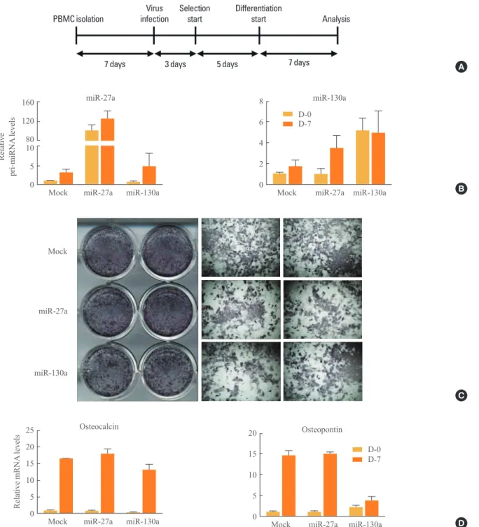
To investigate whether miR-27a and miR-130a may regulate osteogenesis, isolated primary bone marrow cells (PBMCs) were retrovirally infected with miR-27a and miR-130a (Fig.
2A, B). After infection, PBMCs were maintained under osteo-genic differentiation conditions. Alkaline phosphatase (ALP) staining showed that osteoblast differentiation was not influ-enced by miR-27a or miR-130a (Fig. 2C). Further, in PBMCs overexpressing miR-27a and miR-130a, mRNA expression
Table 1.Continued
micro RNA Target Function Expression in
adipose tissue Experiment model Reference
miR-31 C/EBPα Antiadipogenic ↓Adipose tissue MSC, hASC, Ob adipocytes
Sun et al. (2009)39
Tang et al. (2009)38
miR-326 C/EBPα Antiadipogenic MSC Tang et al. (2009)38
miR-155 C/EBPβ Antiadipogenic Adipose tissue, macrophages
Lin et al. (2011)40
miR-448 KLF5 Proadipogenic 3T3-L1 Kinoshita et al. (2010)41
miR-204 miR-211
Runx2 Proadipogenic, blocks osteogenesis in MSCs
C3H10T1/2, BMSC Huang et al. (2010)31
miR-199a Smad1 Proadipogenic, blocks chondrogenesis
C3H10T1/2, ATDC5 Lin et al. (2009)35
miR-103 PDK1 Proadipogenic 3T3-L1, MSC, preadipocytes, Ob adipose tissue
Wilfred et al. (2007)59
Xie et al. (2009)46
miR-15a DLK1 O/E decreases number but increase size
↑Adipose tissue 3T3-L1, Adipose tissue
Andersen et al. (2010)60
miR-24 Proadipogenic Gu et al. (2007)61
miR-150 Proadipogenic ↑Adipose tissue 3T3-L1, ob adipose tissue
Estep et al. (2010)62
miR-221 miR-222
Antiadipogenic ↑In mice obesity,
↓In human obesity 3T3-L1, ob adipose tissue, preadipocytes
Meerson et al. (2013)63
Parra et al. (2010)64
miR-326 Antiadipogenic ↑Adipose tissue Preadipocytes, ob adipose tissue
Tang et al. (2009)38
miR-335 Proadipogenic ↑Adipose tissue 3T3-L1, MSC, ob adipose tissue
Zhu et al. (2013)65
miR-378 Proadipogenic, enhance lipid accumulation in adipocytes
3T3-L1, ST2, MSC Carrer et al. (2012)66
Jin et al. (2010)67
miR-278 Involved in insulin vity & energy homeostasis
Drosophila Teleman et al. (2006)68
miR-200 Proadipogenic MSC, ST2 McGregor et al. (2011)1
Kennell et al. (2008)44
miR-107 Proadipogenic, predicted to regulate acetyl-coA
↓Adipose tissue 3T3-L1, MSC, preadipocytes, ob adipose tissue
Wilfred et al. (2007)59
MAPK, mitogen-activated protein kinase; ERK, extracellular signal-regulated kinase; MSC, mesenchymal stem cell; PI3K, phosphoinositide 3-kinase; T2D, type 2 diabetes; PTN, pleiotrophin; ORP, oxysterol-binding protein-related protein; PDK, phosphoinositide-dependent kinase; cAMP, cyclic ade-nosine monophosphate; PKA, protein kinase A; CREB, cAMP response element-binding; WNT, wingless and INT-1; TCF, T-cell-specific transcription
factor; TGF-β, transforming growth factor β; TGFBR2, TGF-β receptor 2; hASC, human adipose tissue-derived stem cell; HMGA2, high mobility
levels of osteogenic marker genes such as osteopontin and os-teocalcin were not systematically changed (Fig. 2D). These results reveal that miR-27a and miR-130a do not have signifi-cant effects on osteogenesis, implying that 27a and miR-130a may selectively affect adipogenesis, not osteogenesis. There are other known miRNAs that can control lineage de-termination. miR-124 is gradually upregulated during adipo-cyte differentiation of human bone marrow-derived mesen-chymal stem cells (hMSCs). In 3T3-L1 cells, miR-124 over-expression promotes adipocyte differentiation via the suppres-sion of Dlx5, accompanied by elevated fatty acid binding
pro-tein 4 (FABP4). Dlx5 is known as a pro-osteogenic transcrip-tion factor that determines cell fate in MSCs [71]. Thus, miR-124 has a proadipogenic effect by targeting Dlx5 [30]. Recent studies have demonstrated that miR-204, and its homolog miR-211, promote the induction of bone marrow stromal cell differentiation into adipocytes [31]. As shown by reporter as-says, miR-211 directly targets 3’UTR of Runx2, which is a key transcription factor for osteoblast differentiation. Ectopic expression of miR-204 decreased Runx2 protein levels, where-as miR-204 knockdown significantly increwhere-ased Runx2 protein levels. These data imply that miR-204 would suppress the
ac-Fig. 1. Signals and microRNAs involved in adipogenesis. HMGA2, high mobility group AT-hook2; CREB, cAMP response
element-binding; MAPK, mitogen-activated protein kinase; WNT, wingless and INT-1; TGF-β, transforming growth factor β; TGFBR, TGF-β
PBMC isolation
Virus infection
Selection start
Differentiation
start Analysis
7 days 3 days 5 days 7 days A
Relative
pri-miRNA
levels
10 5 0 160 120 80
Mock miR-27a miR-27a
miR-130a
8 6 4 2 0
Mock miR-27a miR-130a
miR-130a D-0
D-7
B
Mock
miR-27a
miR-130a
C
25 20 15 10 5 0
Mock miR-27a Osteocalcin
Relat
ive mRNA
l
evels
miR-130a
20 15 10 5 0
Mock miR-27a Osteopontin
miR-130a D-0 D-7
D
Fig. 2. Regulation of osteogenesis by microRNA (miR)-27a and miR-130a. (A) Experimental scheme. primary bone marrow cell (PBMCs) were isolated from the femur and tibia of 4-week-old C56BL/6J mice. After 7 days, PBMCs were infected with retrovirus con-taining mock, full-length of pri-miR-27a or pri-miR-130a. Retrovirus-infected PBMCs were selected with puromycin for 5 days and
dif-ferentiated for an additional 7 days. For osteoblast differentiation, PBMCs were cultured in α-modified essential medium with 10% fetal bovine serum, 50 μM ascorbic acid, and 10 mM β-glycerophosphate. All experiments with mice were approved by the Institute of Labo -ratory Animal Resources at Seoul National University. (B) Overexpression of miR-27a or miR-130a. The relative amounts of pri-miR-27a and pri-miR-130a against cyclophilin were measured with quantitative reverse transcription polymerase chain reaction (q-RT-PCR). Results are expressed as mean±standard error of mean (SEM). (C) Microscopic view of differentiated PBMCs after alkaline
tion of Runx2 in osteoblast differentiation and enhance adipo-cyte differentiation.
miRNAs REGULATING MITOTIC CLONAL
EXPANSION
Some miRNAs are involved in adipogenesis by regulating the RB-E2F pathway that controls mitotic clonal expansion. The miR-17-92 cluster has been reported to be upregulated during the clonal expansion stage of adipocyte differentiation, posi-tively regulating adipogenesis [32]. Overexpression of miR-17-92 accelerates adipocyte differentiation and directly re-presses RB family Rb2/p130. On the contrary, miR-363 inhib-its adipocyte differentiation by targeting E2F family member, E2F3 [72]. miR-363 overexpression downregulates expression
levels of C/EBPα and PPARγ.
In addition, let-7 negatively regulates adipogenesis by regu-lating the expression of high mobility group AT-hook2 (HMGA- 2) [33]. Ectopic introduction of let-7 in 3T3-L1 and 3T3-F442A cells decreases mRNA levels of mature adipocyte markers
such as FABP4 and PPARγ. Among several genes that have
been previously shown to be targets of let-7, the most affected in 3T3-L1 is HMGA2, a transcription factor. Expression of HMGA2 is high during clonal expansion but turns off at termi-nal differentiation. HMGA2-deficient mice exhibit a reduction in adipose tissue [73], whereas overexpression in transgenic mice leads to an increase in fatty tissue [74]. This result depicts let-7 as an anti-adipogenic regulator that regulates the transi-tion from clonal expansion to terminal differentiatransi-tion.
ANTIADIPOGENIC miRNAs
Some miRNAs control adipogenesis by blocking signal trans-duction pathways such as MAPK. miR-14 has a suppressive effect on fat metabolism by targeting p38 and MAPK in dro-sophila [34]. In flies, deletion of miR-14 increases the number of lipid droplets and the concentration of triacylglycerol. Simi-larly, miR-27a and miR-27b are involved in adipocyte differ-entiation. Computational prediction and luciferase assays have shown that miR-27a and miR-27b target prohibitin (PHB) [75], which is highly expressed in cells that rely heavily on mitochondrial function and has recently been implicated in adipogenesis [76]. In 3T3-L1 cells, overexpression of PHB in-hibits insulin-induced adipogenesis, whereas in the absence of insulin, PHB facilitates adipogenesis through upregulation of MAPK/ERK signaling. PHB silencing by transfection with
miR-27 impairs adipocyte differentiation in adipose tissue-de-rived stem cells (ASC). Likewise, protein levels of PHB, C/
EBPβ, PPARγ, and aP2 are reduced in miR-27a- or miR-27b
transfected ASCs. Moreover, ectopic expression of miR-27a or miR-27b attenuates lipid accumulation [35]. Considering all of these factors, miR-27a and miR-27b are therefore nega-tive modulators of adipocyte differentiation by suppressing PHB.
Many miRNAs have been reported to negatively regulate adipocyte differentiation by targeting insulin signaling and C/ EBPs. In 3T3-L1 adipocytes, miR-320 and miR-29 decrease insulin activity by targeting PI3K directly [36] and AKT indi-rectly [37], respectively. However, it is unclear whether they can influence adipogenesis. Tang et al. [38] identified signifi-cant downregulation of miR-31 and miR-326 during adipo-genesis of ADSCs. In silico prediction of target genes suggest-ed that miR-31 may target phosphoinositide-3-kinase, class 2,
alpha polypeptide (PIK3C2A) and C/EBPα, while miR-326
targets Ras association domain family member 1 (RASSF1) and AP2 associated kinase 1 (AAK1) [38]. In addition, expres-sion of these predicted target genes is correlated with adipo-genic differentiation. Another study has demonstrated that C/
EBPα protein levels are downregulated by miR-31 expression
in rats [39]. A recent report also demonstrated that miR-155 is an inhibitor of adipocyte differentiation. Ectopic expression of miR-155 decreased lipid accumulation as assessed by oil-red O staining in 3T3-L1 cells [40] and adipose tissue-derived preadipocytes [77]. Overexpression of miR-155 is associated
with reduced protein levels of CREB and C/EBPβ, which are
essential regulators during the early stages of terminal differ-entiation.
Transcriptional or posttranscriptional processes related to adipogenesis are also controlled by many miRNAs. A bioin-formatics approach has indicated that miR-448 targets KLF5 [41], and miR-448 is considered to be an inhibitor of adipo-cyte differentiation. Overexpression of miR-448 reduces KLF5 and inhibits adipocyte differentiation. mRNA levels of
adipo-genic marker genes such as C/EBPα and PPARγ, which are
through C/EBPβ-dependent and -independent mechanisms.
miR-224-5p is able to regulate fatty acid metabolism by di-rectly targeting acyl-CoA synthetase long chain family mem-ber 4 (ACSL4). miR-138 has been implicated as an inhibitor of adipocyte differentiation through posttranscriptional regula-tion of EP300 interacting inhibitor of differentiaregula-tion 1 (EID-1), which can promote adipocyte differentiation [42]. The role of miR-138 in human multipotent MSCs has been demonstrated by gain of function experiments. When miR-138 is overex-pressed in hMSCs, lipid droplets are significantly reduced and
the expression levels of C/EBPα and PPARγ decrease.
PROADIPOGENIC miRNAs
It has been reported that several miRNAs may promote adipo-cyte differentiation by targeting MAPK signaling. In 3T3-L1 cells, expression of miR-143 increases during adipogenesis, and thereby stimulates adipocyte differentiation through inhibi-tion of ERK5 via binding to the 3’-UTR of ERK5 mRNA [43]. Similarly, miR-375 overexpression increases the mRNA
ex-pression levels of C/EBPα, PPARγ, and adipocyte FABP (aP2)
[36]. The number of lipid droplets is also increased by ectopic expression of miR-375 in 3T3-L1 cells. Furthermore, induction of miR-375 represses phosphorylation levels of ERK1/2. In 3T3-L1 cells, miR-375 knockdown elevates the phosphoryla-tion level of ERK1/2 and decreases mRNA expression levels of
C/EBPα, PPARγ, and aP2.
Recent findings have suggested that miR-26b inhibits adipo-genic differentiation in human preadipocytes [3]. Bioinformat-ics approaches and luciferase assays have revealed that the 3’UTR of the phosphatase and tensin homolog gene is a target of miR-26b. miR-26b overexpression enforces downregulation
of adipogenic marker genes including aP2, C/EBPα and PPARγ. In addition, the abundance of lipid droplets and the tri -glyceride concentration are lowered in miR-26b-overexpress-ing cells. However, knockdown of miR-26b enhances adipo-cyte differentiation, accompanied with an increase in both adi-pocyte-specific marker genes and lipid droplets.
Kennell et al. [44] have reported that miR-8 can inhibit the evolutionarily-conserved Wnt/Wingless pathway in drosophila. This regulation has been recapitulated with ST2 marrow stro-mal cells during adipogenesis. Overexpression of the miR-8 family (miR-200c-141 and miR-200b-200a-429 clusters) in-creases adipogenesis, FABP4, and lipid accumulation [44]. miR-8 indirectly targets the TCF transcription factor in Wnt signaling and directly targets CG32767, a zinc finger protein
identified as a positive regulator of Wnt signaling in drosophi-la. Furthermore, miR-210 has been reported as a proadipogenic miRNA [79]. In 3T3-L1 cells, lentiviral overexpression of miR-210 enhances adipocyte differentiation, whereas inhibi-tion of miR-210 suppresses expression of adipogenic marker genes. miR-210 enhances adipogenesis by inhibiting TCF7L2, which is a key transcription factor in Wnt signaling.
According to the microarray results, the expression of miR-21 changes during adipocyte differentiation [80]. Oil-red O staining has demonstrated that miR-21 overexpression increas-es the abundance of lipid droplets and the size of adipocytincreas-es. In 3T3-L1 cells, miR-21 stimulates an increase in mRNA and protein levels of adiponectin. In addition, regulation of human ASC (hASC) differentiation by miR-21 is mediated by direct
inhibition of TGF-β receptor 2 (TGFBR2) expression, which is closely related to the TGF-β signaling pathway. Recent find -ings suggest that miR-21 may enhance adipocyte differentia-tion of hASCs via inhibidifferentia-tion of TGFBR2 [2]. miR-21 is known
to have an adipogenic effect by targeting TGFBR2 in TGF-β
signaling.
In addition, miR-146b has been identified as a positive mod-ulator of adipocyte differentiation via suppression of sirtuin 1 (SIRT1) [81]. SIRT1, a target of miR-146b, works as an inhibi-tor of adipocyte differentiation by inducing deacetylation of FOXO1. Expression patterns of miR-146b change significantly during adipocyte differentiation. In 3T3-L1 cells, ectopic
ex-pression of miR-146b increases exex-pression of C/EBPα, PPARγ,
and aP2. Furthermore, knockdown of miR-146b significantly decreases expression of adipogenic markers, body weight and visceral fat. Interestingly, miR-146b is highly expressed in the adipose tissue of obese mouse models such as DIO, diet-in-duced obesity, ob/ob, and db/db. This finding has implicated miR-146b as a positive regulator of accelerated adipocyte dif-ferentiation through modulation of SIRT1.
It has also been shown that miR-519d is downregulated in obese individuals. In human subcutaneous adipose tissue, miR-519d overexpression results in an increase in adipocyte
differ-entiation by targeting PPARα [45]. PPARα plays a crucial role
in fatty acid oxidation in the liver, muscles and heart. There-fore, it is likely that miR-519d enhances adipogenesis by
inhib-iting fat burning through the downregulation of PPARα.
Some miRNAs exhibit clear association with adipogenesis al-though their target genes or processes are unknown. For exam-ple, gain of function experiments have revealed that overex-pression of miR-103 in preadipocytes upregulates exoverex-pression
have been reported; however, there are inconsistencies be-tween the miRNA profiles of mice and humans [46]. Thus, further research is necessary to delineate the role of miR-103 in adipocyte differentiation. miR-378 is also involved in adi-pocyte development, and is highly induced during adiadi-pocyte differentiation of ST2 cells [82]. Overexpression of miR-378 increases the size of lipid droplets and seems to promote the
transcriptional activity of C/EBPα and C/EBPβ. Conversely,
knockdown of miR-378 decreases the rate of triglyceride ac-cumulation in ST2 cells. miR-132 has been identified as a bio-marker of obesity.
Numerous studies have revealed that miRNAs are associat-ed with key metabolic parameters in obese patients. Microar-ray expression profiles have shown different circulating levels of miR-132 between obese and nonobese omental fat. Analy-sis of 132 has revealed that altered expression of miR-132 in blood is correlated with body mass index, fasting glu-cose, and glycosylated hemoglobin [6]. Among the targets of miR-132, CREB has been shown to be involved in regulation of appetite and glucose level. Thus, miR-132 expression may contribute to regulation of metabolic actions including energy homeostasis.
CONCLUSIONS
Because obesity-related hyperplasia and hypertrophy are often associated with various metabolic disorders, detailed under standings of the molecular events regulating adipogenesis and lipid metabolism are important. Emerging evidence shows that miRNAs may directly or indirectly modulate adipocyte different iation [6]. The regulation of adipogenesis by miRNAs includes altering the expression of genes associated with each stage of adipocyte differentiation. In this regard, miRNAs can act on adipogenic factors in either positive or negative manners. Moreover, recent findings have proposed the idea that certain miRNAs could be biomarkers of obesity. However, many challenges remain. For example, a complex redundancy of miRNAs and target interactions may exist during the com plicated processes of adipogenesis, and the nature of these interactions has not been fully characterized. Therefore, further investigations are definitely required to elucidate the precise roles of these miRNAs and their regulatory mechanisms in adipogenesis. In conclusion, more research concerning the miRNAs involved in adipocyte differentiation would provide pathophysiological roles for these miRNAs and novel insight into obesity and its related metabolic diseases.
CONFLICTS OF INTEREST
No potential conflict of interest relevant to this article was re-ported.
ACKNOWLEDGMENTS
This work was supported by the National Creative Research Initiative Program (2011-0018312), funded by the Ministry of Education, Science and Technology (MEST).
REFERENCES
1. McGregor RA, Choi MS. microRNAs in the regulation of adipogenesis and obesity. Curr Mol Med 2011;11:304-16.
2. Kim YJ, Hwang SJ, Bae YC, Jung JS. MiR-21 regulates adipogenic differentiation through the modulation of TGF-beta signaling in mesenchymal stem cells derived from hu-man adipose tissue. Stem Cells 2009;27:3093-102.
3. Song G, Xu G, Ji C, Shi C, Shen Y, Chen L, Zhu L, Yang L, Zhao Y, Guo X. The role of microRNA-26b in human adi-pocyte differentiation and proliferation. Gene 2014;533: 481-7.
4. Vimalraj S, Selvamurugan N. MicroRNAs: synthesis, gene regulation and osteoblast differentiation. Curr Issues Mol Biol 2012;15:7-18.
5. Alexander R, Lodish H, Sun L. MicroRNAs in adipogene-sis and as therapeutic targets for obesity. Expert Opin Ther Targets 2011;15:623-36.
6. Heneghan HM, Miller N, Kerin MJ. Role of microRNAs in obesity and the metabolic syndrome. Obes Rev 2010;11: 354-61.
7. Tang QQ, Lane MD. Adipogenesis: from stem cell to adi-pocyte. Annu Rev Biochem 2012;81:715-36.
8. MacDougald OA, Lane MD. Transcriptional regulation of gene expression during adipocyte differentiation. Annu Rev Biochem 1995;64:345-73.
9. Burkhart DL, Sage J. Cellular mechanisms of tumour sup-pression by the retinoblastoma gene. Nat Rev Cancer 2008; 8:671-82.
10. Fajas L, Debril MB, Auwerx J. Peroxisome proliferator-activated receptor-gamma: from adipogenesis to carcino-genesis. J Mol Endocrinol 2001;27:1-9.
U S A 2006;103:13022-7.
12. Huang HY, Hu LL, Song TJ, Li X, He Q, Sun X, Li YM, Lu HJ, Yang PY, Tang QQ. Involvement of cytoskeleton-asso-ciated proteins in the commitment of C3H10T1/2 pluripo-tent stem cells to adipocyte lineage induced by BMP2/4. Mol Cell Proteomics 2011;10:M110.002691.
13. Nishimura R, Hata K, Ikeda F, Ichida F, Shimoyama A, Matsubara T, Wada M, Amano K, Yoneda T. Signal trans-duction and transcriptional regulation during mesenchymal cell differentiation. J Bone Miner Metab 2008;26:203-12.
14. Wang EA, Israel DI, Kelly S, Luxenberg DP. Bone mor-phogenetic protein-2 causes commitment and differentia-tion in C3H10T1/2 and 3T3 cells. Growth Factors 1993;9: 57-71.
15. Choy L, Derynck R. Transforming growth factor-beta in-hibits adipocyte differentiation by Smad3 interacting with CCAAT/enhancer-binding protein (C/EBP) and repressing C/EBP transactivation function. J Biol Chem 2003;278: 9609-19.
16. Zamani N, Brown CW. Emerging roles for the transform-ing growth factor-{beta} superfamily in regulattransform-ing adipos-ity and energy expenditure. Endocr Rev 2011;32:387-403.
17. Tsurutani Y, Fujimoto M, Takemoto M, Irisuna H, Ko-shizaka M, Onishi S, Ishikawa T, Mezawa M, He P, Honjo S, Maezawa Y, Saito Y, Yokote K. The roles of transform-ing growth factor-beta and Smad3 signaltransform-ing in adipocyte differentiation and obesity. Biochem Biophys Res Com-mun 2011;407:68-73.
18. Clouthier DE, Comerford SA, Hammer RE. Hepatic fibro-sis, glomerulosclerofibro-sis, and a lipodystrophy-like syndrome in PEPCK-TGF-beta1 transgenic mice. J Clin Invest 1997; 100:2697-713.
19. Bennett CN, Ross SE, Longo KA, Bajnok L, Hemati N, Johnson KW, Harrison SD, MacDougald OA. Regulation of Wnt signaling during adipogenesis. J Biol Chem 2002; 277:30998-1004.
20. Bowers RR, Lane MD. Wnt signaling and adipocyte lin-eage commitment. Cell Cycle 2008;7:1191-6.
21. Singh R, Artaza JN, Taylor WE, Braga M, Yuan X, Gonza-lez-Cadavid NF, Bhasin S. Testosterone inhibits adipogenic differentiation in 3T3-L1 cells: nuclear translocation of an-drogen receptor complex with beta-catenin and T-cell fac-tor 4 may bypass canonical Wnt signaling to down-regulate adipogenic transcription factors. Endocrinology 2006;147: 141-54.
22. Bost F, Aouadi M, Caron L, Binetruy B. The role of
MAPKs in adipocyte differentiation and obesity. Biochim-ie 2005;87:51-6.
23. Tseng YH, Butte AJ, Kokkotou E, Yechoor VK, Taniguchi CM, Kriauciunas KM, Cypess AM, Niinobe M, Yoshikawa K, Patti ME, Kahn CR. Prediction of preadipocyte differ-entiation by gene expression reveals role of insulin recep-tor substrates and necdin. Nat Cell Biol 2005;7:601-11.
24. Nakae J, Kitamura T, Kitamura Y, Biggs WH 3rd, Arden KC, Accili D. The forkhead transcription factor Foxo1 reg-ulates adipocyte differentiation. Dev Cell 2003;4:119-29.
25. Rosen ED, MacDougald OA. Adipocyte differentiation from the inside out. Nat Rev Mol Cell Biol 2006;7:885-96.
26. Elberg G, Gimble JM, Tsai SY. Modulation of the murine peroxisome proliferator-activated receptor gamma 2 pro-moter activity by CCAAT/enhancer-binding proteins. J Biol Chem 2000;275:27815-22.
27. Siersbaek R, Nielsen R, Mandrup S. Transcriptional net-works and chromatin remodeling controlling adipogenesis. Trends Endocrinol Metab 2012;23:56-64.
28. Kim SY, Kim AY, Lee HW, Son YH, Lee GY, Lee JW, Lee YS, Kim JB. miR-27a is a negative regulator of adipocyte differentiation via suppressing PPARgamma expression. Biochem Biophys Res Commun 2010;392:323-8.
29. Lee EK, Lee MJ, Abdelmohsen K, Kim W, Kim MM, Sri-kantan S, Martindale JL, Hutchison ER, Kim HH, Marasa BS, Selimyan R, Egan JM, Smith SR, Fried SK, Gorospe M. miR-130 suppresses adipogenesis by inhibiting peroxi-some proliferator-activated receptor gamma expression. Mol Cell Biol 2011;31:626-38.
30. Qadir AS, Woo KM, Ryoo HM, Baek JH. Insulin spresses distal-less homeobox 5 expression through the up-regulation of microRNA-124 in 3T3-L1 cells. Exp Cell Res 2013;319:2125-34.
31. Huang J, Zhao L, Xing L, Chen D. MicroRNA-204 regu-lates Runx2 protein expression and mesenchymal progeni-tor cell differentiation. Stem Cells 2010;28:357-64.
32. Wang Q, Li YC, Wang J, Kong J, Qi Y, Quigg RJ, Li X. miR-17-92 cluster accelerates adipocyte differentiation by negatively regulating tumor-suppressor Rb2/p130. Proc Natl Acad Sci U S A 2008;105:2889-94.
33. Sun T, Fu M, Bookout AL, Kliewer SA, Mangelsdorf DJ. MicroRNA let-7 regulates 3T3-L1 adipogenesis. Mol En-docrinol 2009;23:925-31.
35. Lin Q, Gao Z, Alarcon RM, Ye J, Yun Z. A role of miR-27 in the regulation of adipogenesis. FEBS J 2009;276:2348-58.
36. Ling HY, Wen GB, Feng SD, Tuo QH, Ou HS, Yao CH, Zhu BY, Gao ZP, Zhang L, Liao DF. MicroRNA-375 pro-motes 3T3-L1 adipocyte differentiation through modula-tion of extracellular signal-regulated kinase signalling. Clin Exp Pharmacol Physiol 2011;38:239-46.
37. He A, Zhu L, Gupta N, Chang Y, Fang F. Overexpression of micro ribonucleic acid 29, highly up-regulated in dia-betic rats, leads to insulin resistance in 3T3-L1 adipocytes. Mol Endocrinol 2007;21:2785-94.
38. Tang YF, Zhang Y, Li XY, Li C, Tian W, Liu L. Expression of miR-31, miR-125b-5p, and miR-326 in the adipogenic differentiation process of adipose-derived stem cells. OMICS 2009;13:331-6.
39. Sun F, Wang J, Pan Q, Yu Y, Zhang Y, Wan Y, Wang J, Li X, Hong A. Characterization of function and regulation of miR-24-1 and miR-31. Biochem Biophys Res Commun 2009;380:660-5.
40. Liu S, Yang Y, Wu J. TNFalpha-induced up-regulation of miR-155 inhibits adipogenesis by down-regulating early adipogenic transcription factors. Biochem Biophys Res Commun 2011;414:618-24.
41. Kinoshita M, Ono K, Horie T, Nagao K, Nishi H, Kuwaba-ra Y, Takanabe-Mori R, Hasegawa K, Kita T, KimuKuwaba-ra T. Regulation of adipocyte differentiation by activation of se-rotonin (5-HT) receptors 5-HT2AR and 5-HT2CR and in-volvement of microRNA-448-mediated repression of KLF5. Mol Endocrinol 2010;24:1978-87.
42. Yang Z, Bian C, Zhou H, Huang S, Wang S, Liao L, Zhao RC. MicroRNA hsa-miR-138 inhibits adipogenic differen-tiation of human adipose tissue-derived mesenchymal stem cells through adenovirus EID-1. Stem Cells Dev 2011;20: 259-67.
43. Esau C, Kang X, Peralta E, Hanson E, Marcusson EG, Ravichandran LV, Sun Y, Koo S, Perera RJ, Jain R, Dean NM, Freier SM, Bennett CF, Lollo B, Griffey R. MicroR-NA-143 regulates adipocyte differentiation. J Biol Chem 2004;279:52361-5.
44. Kennell JA, Gerin I, MacDougald OA, Cadigan KM. The microRNA miR-8 is a conserved negative regulator of Wnt signaling. Proc Natl Acad Sci U S A 2008;105:15417-22.
45. Martinelli R, Nardelli C, Pilone V, Buonomo T, Liguori R, Castano I, Buono P, Masone S, Persico G, Forestieri P, Pastore L, Sacchetti L. miR-519d overexpression is
associ-ated with human obesity. Obesity (Silver Spring) 2010;18: 2170-6.
46. Xie H, Lim B, Lodish HF. MicroRNAs induced during ad-ipogenesis that accelerate fat cell development are down-regulated in obesity. Diabetes 2009;58:1050-7.
47. Takanabe R, Ono K, Abe Y, Takaya T, Horie T, Wada H, Kita T, Satoh N, Shimatsu A, Hasegawa K. Up-regulated expression of microRNA-143 in association with obesity in adipose tissue of mice fed high-fat diet. Biochem Bio-phys Res Commun 2008;376:728-32.
48. Oskowitz AZ, Lu J, Penfornis P, Ylostalo J, McBride J, Flemington EK, Prockop DJ, Pochampally R. Human mul-tipotent stromal cells from bone marrow and microRNA: regulation of differentiation and leukemia inhibitory factor expression. Proc Natl Acad Sci U S A 2008;105:18372-7.
49. Ling HY, Ou HS, Feng SD, Zhang XY, Tuo QH, Chen LX, Zhu BY, Gao ZP, Tang CK, Yin WD, Zhang L, Liao DF. CHANGES IN microRNA (miR) profile and effects of miR-320 in insulin-resistant 3T3-L1 adipocytes. Clin Exp Pharmacol Physiol 2009;36:e32-9.
50. Jordan SD, Kruger M, Willmes DM, Redemann N, Wun-derlich FT, Bronneke HS, Merkwirth C, Kashkar H, Olk-konen VM, Bottger T, Braun T, Seibler J, Bruning JC. Obesity-induced overexpression of miRNA-143 inhibits insulin-stimulated AKT activation and impairs glucose metabolism. Nat Cell Biol 2011;13:434-46.
51. Yi C, Xie WD, Li F, Lv Q, He J, Wu J, Gu D, Xu N, Zhang Y. MiR-143 enhances adipogenic differentiation of 3T3-L1 cells through targeting the coding region of mouse pleiotrophin. FEBS Lett 2011;585:3303-9.
52. El Ouaamari A, Baroukh N, Martens GA, Lebrun P, Pipel-eers D, van Obberghen E. miR-375 targets 3’-phos-phoinositide-dependent protein kinase-1 and regulates glu-cose-induced biological responses in pancreatic beta-cells. Diabetes 2008;57:2708-17.
53. Rajasethupathy P, Fiumara F, Sheridan R, Betel D, Puthan-veettil SV, Russo JJ, Sander C, Tuschl T, Kandel E. Charac-terization of small RNAs in Aplysia reveals a role for miR-124 in constraining synaptic plasticity through CREB. Neuron 2009;63:803-17.
54. Laine SK, Alm JJ, Virtanen SP, Aro HT, Laitala-Leinonen TK. MicroRNAs miR-96, miR-124, and miR-199a regu-late gene expression in human bone marrow-derived mes-enchymal stem cells. J Cell Biochem 2012;113:2687-95.
expres-sion in human omental and subcutaneous adipose tissue. PLoS One 2009;4:e4699.
56. Qin L, Chen Y, Niu Y, Chen W, Wang Q, Xiao S, Li A, Xie Y, Li J, Zhao X, He Z, Mo D. A deep investigation into the adipogenesis mechanism: profile of microRNAs regulating adipogenesis by modulating the canonical Wnt/beta-catenin signaling pathway. BMC Genomics 2010;11:320.
57. Lee YS, Dutta A. The tumor suppressor microRNA let-7 re-presses the HMGA2 oncogene. Genes Dev 2007;21:1025- 30.
58. Karbiener M, Fischer C, Nowitsch S, Opriessnig P, Papak C, Ailhaud G, Dani C, Amri EZ, Scheideler M. microRNA miR-27b impairs human adipocyte differentiation and tar-gets PPARgamma. Biochem Biophys Res Commun 2009; 390:247-51.
59. Wilfred BR, Wang WX, Nelson PT. Energizing miRNA research: a review of the role of miRNAs in lipid metabo-lism, with a prediction that miR-103/107 regulates human metabolic pathways. Mol Genet Metab 2007;91:209-17.
60. Andersen DC, Jensen CH, Schneider M, Nossent AY, Es-kildsen T, Hansen JL, Teisner B, Sheikh SP. MicroRNA-15a fine-tunes the level of Delta-like 1 homolog (DLK1) in proliferating 3T3-L1 preadipocytes. Exp Cell Res 2010; 316:1681-91.
61. Gu Z, Eleswarapu S, Jiang H. Identification and character-ization of microRNAs from the bovine adipose tissue and mammary gland. FEBS Lett 2007;581:981-8.
62. Estep M, Armistead D, Hossain N, Elarainy H, Goodman Z, Baranova A, Chandhoke V, Younossi ZM. Differential expression of miRNAs in the visceral adipose tissue of pa-tients with non-alcoholic fatty liver disease. Aliment Phar-macol Ther 2010;32:487-97.
63. Meerson A, Traurig M, Ossowski V, Fleming JM, Mullins M, Baier LJ. Human adipose microRNA-221 is upregulat-ed in obesity and affects fat metabolism downstream of
leptin and TNF-α. Diabetologia 2013;56:1971-9.
64. Parra P, Serra F, Palou A. Expression of adipose microR-NAs is sensitive to dietary conjugated linoleic acid treat-ment in mice. PLoS One 2010;5:e13005.
65. Zhu L, Chen L, Shi CM, Xu GF, Xu LL, Zhu LL, Guo XR, Ni Y, Cui Y, Ji C. MiR-335, an adipogenesis-related mi-croRNA, is involved in adipose tissue inflammation. Cell Biochem Biophys 2014;68:283-90.
66. Carrer M, Liu N, Grueter CE, Williams AH, Frisard MI, Hulver MW, Bassel-Duby R, Olson EN. Control of mito-chondrial metabolism and systemic energy homeostasis by
microRNAs 378 and 378*. Proc Natl Acad Sci U S A 2012; 109:15330-5.
67. Jin W, Dodson MV, Moore SS, Basarab JA, Guan LL. Characterization of microRNA expression in bovine adi-pose tissues: a potential regulatory mechanism of subcuta-neous adipose tissue development. BMC Mol Biol 2010; 11:29.
68. Teleman AA, Maitra S, Cohen SM. Drosophila lacking mi-croRNA miR-278 are defective in energy homeostasis. Genes Dev 2006;20:417-22.
69. Muruganandan S, Roman AA, Sinal CJ. Adipocyte differ-entiation of bone marrow-derived mesenchymal stem cells: cross talk with the osteoblastogenic program. Cell Mol Life Sci 2009;66:236-53.
70. Guo L, Zhao RC, Wu Y. The role of microRNAs in self-re-newal and differentiation of mesenchymal stem cells. Exp Hematol 2011;39:608-16.
71. Holleville N, Mateos S, Bontoux M, Bollerot K, Monsoro-Burq AH. Dlx5 drives Runx2 expression and osteogenic differentiation in developing cranial suture mesenchyme. Dev Biol 2007;304:860-74.
72. Chen L, Cui J, Hou J, Long J, Li C, Liu L. A Novel Nega-tive Regulator of Adipogenesis: MicroRNA-363. Stem Cells 2013.
73. Zhou X, Benson KF, Ashar HR, Chada K. Mutation respon-sible for the mouse pygmy phenotype in the developmen-tally regulated factor HMGI-C. Nature 1995;376:771-4.
74. Arlotta P, Tai AK, Manfioletti G, Clifford C, Jay G, Ono SJ. Transgenic mice expressing a truncated form of the high mobility group I-C protein develop adiposity and an abnormally high prevalence of lipomas. J Biol Chem 2000; 275:14394-400.
75. Kang T, Lu W, Xu W, Anderson L, Bacanamwo M, Thompson W, Chen YE, Liu D. MicroRNA-27 (miR-27) targets prohibitin and impairs adipocyte differentiation and mitochondrial function in human adipose-derived stem cells. J Biol Chem 2013;288:34394-402.
76. Ande SR, Xu Z, Gu Y, Mishra S. Prohibitin has an impor-tant role in adipocyte differentiation. Int J Obes (Lond) 2012;36:1236-44.
77. Chen Y, Siegel F, Kipschull S, Haas B, Frohlich H, Meister G, Pfeifer A. miR-155 regulates differentiation of brown and beige adipocytes via a bistable circuit. Nat Commun 2013;4:1769.
regu-lates fatty acid metabolism. Int J Biochem Cell Biol 2013; 45:1585-93.
79. Liang WC, Wang Y, Wan DC, Yeung VS, Waye MM. Characterization of miR-210 in 3T3-L1 adipogenesis. J Cell Biochem 2013;114:2699-707.
80. Kang M, Yan LM, Zhang WY, Li YM, Tang AZ, Ou HS. Role of microRNA-21 in regulating 3T3-L1 adipocyte dif-ferentiation and adiponectin expression. Mol Biol Rep 2013;
40:5027-34.
81. Ahn J, Lee H, Jung CH, Jeon TI, Ha TY. MicroRNA-146b promotes adipogenesis by suppressing the SIRT1-FOXO1 cascade. EMBO Mol Med 2013;5:1602-12.