Protective Effects of N-Acetyl Cysteine
against Diesel Exhaust Particles-Induced
Intracellular ROS Generates
Pro-Inflammatory Cytokines to Mediate the
Vascular Permeability of Capillary-Like
Endothelial Tubes
Chia-Yi Tseng1,2, Jing-Fen Chang3, Jhih-Syuan Wang3, Yu-Jung Chang3, Marion K. Gordon4, Ming-Wei Chao2,3*
1Department of Biomedical Engineering, College of Engineering, Chung Yuan Christian University, Zhongli district, Taoyuan city, Taiwan,2Center of Nanotechnology, Chung Yuan Christian University, Zhongli district, Taoyuan city, Taiwan,3Department of Bioscience Technology, College of Science, Chung Yuan Christian University, Zhongli district, Taoyuan city, Taiwan,4Joint program of Toxicology, Rutgers University, Piscataway, New Jersey, United States of America
Abstract
Exposure to diesel exhaust particles (DEP) is associated with pulmonary and cardiovascu-lar diseases. Previous studies usingin vitroendothelial tubes as a simplified model of capillaries have found that DEP-induced ROS increase vascular permeability with rear-rangement or internalization of adherens junctional VE-cadherin away from the plasma membrane. This allows DEPs to penetrate into the cell and capillary lumen. In addition, pro-inflammatory cytokines are up-regulated and mediate vascular permeability in response to DEP. However, the mechanisms through which these DEP-induced pro-inflammatory cyto-kines increase vascular permeability remain unknown. Hence, we examined the ability of DEP to induce permeability of human umbilical vein endothelial cell tube cells to investigate these mechanisms. Furthermore, supplementation with NAC reduces ROS production fol-lowing exposure to DEP. HUVEC tube cells contributed to a pro-inflammatory response to DEP-induced intracellular ROS generation. Endothelial oxidative stress induced the release of TNF-αand IL-6 from tube cells, subsequently stimulating the secretion of VEGF-A inde-pendent of HO-1. Our data suggests that DEP-induced intracellular ROS and release of the pro-inflammatory cytokines TNF-αand IL-6, which would contribute to VEGF-A secretion and disrupt cell-cell borders and increase vasculature permeability. Addition of NAC sup-presses DEP-induced ROS efficiently and reduces subsequent damages by increasing endogenous glutathione.
a11111
OPEN ACCESS
Citation:Tseng C-Y, Chang J-F, Wang J-S, Chang Y-J, Gordon MK, Chao M-W (2015) Protective Effects of N-Acetyl Cysteine against Diesel Exhaust Particles-Induced Intracellular ROS Generates Pro-Inflammatory Cytokines to Mediate the Vascular Permeability of Capillary-Like Endothelial Tubes. PLoS ONE 10(7): e0131911. doi:10.1371/journal. pone.0131911
Editor:Masuko Ushio-Fukai, University of Illinois at Chicago, UNITED STATES
Received:March 4, 2015
Accepted:June 8, 2015
Published:July 6, 2015
Copyright:© 2015 Tseng et al. This is an open access article distributed under the terms of the
Creative Commons Attribution License, which permits unrestricted use, distribution, and reproduction in any medium, provided the original author and source are credited.
Data Availability Statement:All relevant data are within the paper and its Supporting Information files.
Funding:This work was supported by a grant from Ministry of Science and Technology (NSC-102-2320-B-033-001-MY3) in Taiwan.
Introduction
Diesel exhaust particles (DEP) are associated with induction and exacerbation of cardiovascular disorders through the production of harmful free radicals and initiation of pro-inflammatory responses [1–3]. These events contribute to the resulting adverse health effects of DEP. Endo-thelial dysfunction can be caused by diesel-induced mechanisms [4,5]. Meanwhile, vasocon-striction has been found in humans who inhaled ambient PM2.5(particulate matter2.5μm)
for 2 h [6], and impaired vasodilatation appears 24 h after 1 h of DEP inhalation [5]. Moreover, the water content of the mouse lung which causes epithelial fibrosis and disrupts vascular func-tion (increased 24 h after intratracheal instillafunc-tion of DEPs or DEP extracts), which has been suggested to cause an increase in pulmonary endothelial permeability [7].
DEP have been found to enter into the circulation and may have a direct effect on endothe-lial permeability.In vitro-assembled capillary-like endothelial tubes are used to model the translocation of particles into capillaries [8]. A previous study demonstrated, using confocal microscopy, that DEP affect the permeability of endothelial tubes. The adherens junction com-ponent, VE-cadherin, moves from cell-cell junctions to intracellular locations, and DEP deposit in the cytoplasm of cells and the luminal space of tubes. Disruption of VE-cadherin endothelial barrier integrity has also been shown to alter vascular permeability [9,10]. The pulmonary endothelium acts as a semipermeable barrier, and the integrity of the barrier is necessary for efficient pulmonary function [11]. Furthermore, recent results have suggested that DEPs func-tion by changing the levels of its effector, H2O2, which triggers Nrf2 translocation from the
cytoplasm to the nucleus [12,13]. Downstream heme oxygenase (HO)-1 is then upregulated to facilitate the anti-oxidative stress response in the endothelium [14–17]. HO-1 also functions to induce vascular permeability and contributes to the secretion of vascular endothelial growth factor A (VEGF-A) [18]. VEGF-A, also called vascular permeability factor (VPF), has been shown to induce vascular permeability [19,20]. Upon exposure ofin vitrocapillary tube cells to DEPs, the VE-cadherin/VEGF receptor 2 (VEGF-R2) complex on the cell membrane disso-ciates [12]. Partial internalization of VE-cadherin and discontinuity of the cell-cell border are also induced following these junctional alternations [12,21]. Moreover, these events cause endothelial junctions to become disrupted and may explain how VEGF-A initiates vascular permeability following inhalation of DEP.
DEP exposure damages the alveolar endothelia and sequentially induces pro-inflammatory responses [22,23], in which cytokines function to enhance vascular permeability and cause transmigration of inflammatory cells [24,25]. Among these pro-inflammatory cytokines, tumor necrosis factor (TNF)-αand interleukin (IL)-6 have been investigated most widely in studies of DEPs [26,27]. Inhalation of DEPs for 24 h upregulates TNF-αand leads to accumu-lation of large amounts of TNF-αin human plasma [5]. This changes the expression of adhe-sion molecules on endothelial cells, facilitating the transmigration of neutrophils and thereby leading to changes in vascular permeability [28]. Furthermore, there is a positive correlation between vascular permeability and adherens junction integrity [22]. Nwariakuet al(2002) found that TNF-α-induced tyrosine phosphorylation of VE-cadherin, which permits regula-tion of microvascular permeability, increases the formaregula-tion of intercellular gap formaregula-tion [29]. IL-6 has also been shown to be directly involved in increasing monolayer endothelial perme-ability [30]. Maruoet al(1992) suggested that IL-6 induces increased endothelial permeability by rearranging VE-cadherin and altering the shape of endothelial cells. This implies that the endothelial cell-cell barrier may also be altered [31]. Importantly, when IL-6 is knocked down, vascular permeability is restored.
tube cells, a biologically relevant model. Our group has reported that ROS is an inducer to con-tribute VEGF-A releasing [12]. Then DEP initiates mitochondrial oxidative stress generation, leading to cause ATP depletion and depolymerization of actin cytoskeleton [32]. Subsequently the particle might slip between the cells via redistributed VE-cadherin network, and travel in the circulation system [8]. The pathway through which DEP-induced oxidative stress triggers VEGF-A secretion and facilitates permeability has been observed in various cell types [23,33– 35]; however, whether pro-inflammatory cytokines also function in induction of VEGF-A in the tube-like endothelial model remains unknown. Therefore, in this study, we used an endo-thelial tube model, a system free of the confounding effects of inflammatory cells, to examine how cytokines might modulate endothelial permeability. We also evaluated the mechanisms mediating intracellular oxidative stress induction, endothelial pro-inflammatory responses, VEGF-A secretion, VE-cadherin distribution, and vascular permeability, and the protection of ROS scavenger N-acetyl cysteine (NAC).
Materials and Methods
Cell culture
HUVECs were cultured in Medium 199 with endothelial cell growth supplement (Millipore, Billerica, MA, USA), heparin (Sigma-Aldrich, St. Louis, MO, USA), and 10% FBS. HUVECs at passages 5–15 were used to assemble tubes on 10 mg/mL Matrigel, LDEV-free (BD Biosci-ences). Endothelial tubes were allowed to form for 12 h prior to adding DEPs. Well-character-ized DEPs were collected from Dr. Masaru Sagai, Aomori University of Health and Welfare, Japan [1,36]. Particles were prepared and used as previously described to create a suspension resulting in sizes of PM2.5, of which a portion was PM0.1[8].
Reagents
NAC was obtained from Sigma-Aldrich. TNF-αand IL-6 were purchased from R&D Systems, Inc (Minneapolis, MN, USA). SnPP (protoporphyrin IX) was purchased from Frontier Scien-tific, Inc. (Logan, UT, USA).
Determination of total, reduced and oxidized glutathione levels
Total glutathione content of the cell extracts was assessed using a“total glutathione assay kit”
(CS0260-1KT, Sigma-Aldrich) based on a kinetic assay in which catalytic amounts of GSH caused a continuous reduction of 5,5-dithiobis-(2-nitrobenzoic) acid (DTNB) at 412 nm. For oxidized glutathione (GSSG) determination, reduced glutathione (GSH) was inactivated by the addition of 2-vinylpyridine in the presence of triethanolamine. GSH content was calculated using the equation of GSH = (Total GSH–2 x GSSG). Quantification was achieved by parallel measurements of GSH or GSSG standards, and results were expressed as nmol/ mg protein [37].
Quantification of extracellular and intracellular ROS generation
Extracellular and intracellular ROS detection studies were performed using a Cm-H2DCFDA ROS detection kit (Invitrogen, Grand Island, NY, USA). For cell-free ROS detection, aliquots of 100μL serum-free medium containing each concentration of DEPs with or without NAC were pipetted into 96-well plates and mixed with 10μL HBSS containing Cm-H2DCFDA (final
concentration 25μM), activated by pre-incubation at 37°C for 30 min [37]. ROS generation was measured every 10 min up to 2 h with a microreader at 490/530 nm. Samples treated with 100μM H2O2were used as positive controls. For detection of intracellular ROS, after the
Cm-H2DCFDA (final concentration 25μM). Samples were then measured for 60 min after
Cm-H2DCFDA was activated. ROS levels generated by 1.5 × 10⁵viable treated cells were
expressed as the percentage of ROS produced by an equal number of viable negative control cells.
Determination of Cytotoxicity
DEP-induced cytotoxicity in endothelial tubes was evaluated using the CytoTox- Homoge-neous Integrity Assay Kit (Promega), a method that measures cytosolic lactate dehydrogenase (LDH) released into medium when cells are lysed. After the treatments, cells were collected by centrifugation and lysed for 1 h in 200μL lysis solution following the manufacturer’s instruc-tions. The value of LDH was measured at 490 nm. Untreated cultures were presented as the negative control, defining the maximum amount of LDH potentially released.
Measurement of IL-6 and TNF-
α
levels
TNF-αand IL-6 were evaluated using a Human TNF-α, IL-6, and IL-1βReady-SET-Go! ELISA Kits (eBioscience, San Diego, CA, USA), following the manufacturer’s instructions. Briefly, triplicate 6-well plates were seeded with 1.5 × 10⁵HUVECs/well, and after tubes were formed, various concentrations of DEPs (0, 1, 10, or 100μg/mL) with or without NAC (10 mM) were added for 24 h. Next, 200μL medium was collected and added to the wells, which were precoated with cytokine“capture”antibodies. The plates were incubated overnight at 4°C. The next day, the medium was aspirated, and wells were washed with 2% normal goat serum containing 0.02% NaN3in PBS for 1 h at room temperature. After aspirating off the
wash solution, a biotin-conjugated“detecting”primary antibody targeting either IL-6 or TNF-αwas added, and plates were incubated at room temperature for 1 h. The supernatant was then aspirated off, and the wells were washed several times. Avidin conjugated-horse rad-ish peroxidase (HRP) was added, and plates were incubated for 1 h at room temperature. Finally, substrate solution (tetramethylbenzidine) was added for 15 min at room temperature, and the plates were read at 450 nm. The levels of IL-6 and TNF-αwere determined by com-parison to a standard curve, which was prepared by analyzing 2-fold serial dilutions of each cytokine. For this, plates whose wells were precoated with capture antibodies were used, and the same procedure as outlined above was carried out using the serial dilutions rather than the sample medium.
Vascular permeability assay
Total RNA extraction and quantitative polymerase chain reaction
(qPCR) analysis
Total RNA was isolated fromin vitroHUVEC capillary tubes using TRIzol reagent (Sigma-Aldrich). RNA was quantified and reverse transcribed into cDNA using oligo-dTs and a Super-script Synthesis system kit (Invitrogen). To assess the quantity of endogenous HO-1 and VEGFR2 inin vitroHUVEC capillary tubes, the primers used for the amplification were as
fol-lows:HO-1,50-AGGCCAAGACTGCGTTCCT-30and50-GGTGTCATGGGTCAGCAGC-30
(Hiraiet al., 2003);VEGFR2,5’-GCTCAAAAAGATTGCCCAGA -3’(forward) and5’ -GCGGTAGAGCTGCTTGAACT-3’(reverse). The primers were used at a final concentration of
300 nM. SYBR Green (Applied Biosystems, Foster City, CA, USA) was employed for fluores-cence-based quantification of cDNA by real-time qPCR using an ABI Prism 7000 thermal cycler. The qPCR product was evaluated and normalized to 18S ribosomal mRNA levels and the negative control.
Western blotting
For preparation of whole cell lysates, tube cells were collected and sonicated for 1 min in 1 mL lysis buffer. The cell lysates were cleaned by centrifuging at 10,000 ×gfor 10 min, and the supernatant was collected. Nuclear protein extracts were prepared from endothelial tube cells using an adapted 1-h minipreparation technique [12]. Briefly, cells were collected and soni-cated for 1 min in a relatively low-salt lysis buffer and then centrifuged for 30 s at 2500 rpm. The supernatant, which contained the nuclei, was next incubated for 5 min on ice and then centrifuged for 5 min at 5000 rpm. The pelleted nuclei were resuspended in a higher-salt lysis solution and incubated on ice for 20 min. Insoluble nuclear debris was pelleted by centrifuga-tion for 30 s. The protein concentracentrifuga-tions of the whole cell extract and nuclear extract were determined using BCA assays (Pierce), and the samples were stored at−80°C. Sodium dodecyl sulfate polyacrylamide gel electrophoresis (SDS-PAGE) was then carried out using 20μg pro-tein per lane.
For detection of secreted proteins, the supernatant/medium of HUVEC capillary tubes was collected after treatment with DEPs. A total of 40μL per sample of supernatant/medium was loaded onto SDS gels for electrophoresis after boiling at 95°C for 5 min. Coomassie blue was used as the loading control, and fresh medium was loaded as the negative control. Protein expression was normalized to the 150-kDa band in the loading control. Proteins separated elec-trophorectically on SDS gels were transferred to nitrocellulose membranes. Nonspecific reac-tivity of the membranes was blocked and the primary HO-1, TNF-α, IL-6, anti-IL-1β, anti-IκB, anti-nuclear p65, anti-lamin B1, anti-VEGF-A, anti-VEGF-R2, anti-VE-cad-herin, anti-β-actin, and anti-GAPDH antibodies were applied at appropriate dilutions at 4°C overnight. Secondary goat anti-mouse IgG or anti-rabbit IgG antibodies conjugated with HRP (Bio-Rad, Hercules, CA, USA) were used at 1:5000 dilution. Finally, the protein products were visualized on X-ray film using with chemiluminescent reagent containing luminol, a substrate of HRP.
Immunofluorescence microscopy
anti-fade mounting media (Invitrogen) with DAPI and stored at 4°C overnight. Images were observed at 400× magnification on an IX51 Olympus Microscope.
Statistics
For statistical analysis, each experiment was performed at least 3 times, and samples were always run in triplicate. The results were expressed as means ± standard deviations (SDs) and were analyzed using multiple t-tests. Differences withP-values of less than 0.05 were consid-ered statistically significant.
Results
DEP-induced intracellular ROS generation and cytotoxicity
Chaoet al(2012) demonstrated that tube cells exposed to 1μg/mL DEPs generates H2O2(0.18
nmol). At 10 and 100μg/mL concentrations of DEPs, production of H2O2in tube cells was
increased to 0.51 and 2.05 nmol, respectively [12]. Thus, cytotoxicity might be a consequence of exposure to DEP-induced oxidative stress. However, whether these free radicals are gener-ated from DEPs before they contact the cells or after they are taken up by cells and induce sequential intracellular oxidative damage remains unclear. Therefore, we used Cm-H2DCFDA
to evaluate this pathway. As shown inFig 1A, changes in relatively fluorescence units (RFU) could be used to demonstrate the level of DEP-induced ROS in the cell-free model. From these data, the increased RFU was observed only in the H2O2-containing sample with a
time-depen-dent manner. Other samples containing DEPs (10 or 100μg/mL) ± NAC (10 mM) showed no detectable changes in RFU. In contrast, Cm-H2DCFDA analysis showed that DEP induced
intracellular ROS generation in tube cells in a dose-dependent manner (Fig 1B). Addition of NAC, oxidative stress was completely blocked.Fig 1Cshows cytotoxicity curves for HUVEC tube cells treated with DEPs (1, 5, 10, 50, or 100μg/mL) ± NAC. Cytotoxicity in tube cells exposed to 1μg/mL DEP reached 12%; additionally, 5 and 10μg/mL DEPs induced cell death in 19% and 22% of cells, respectively. Higher doses of DEPs (50 and 100μg/mL) also induced greater cell death (45% and 49%, respectively). Thus, cytotoxicity was increased in a dose-dependent manner, similar to the results shown inFig 1B, suggesting that intracellular ROS might affect cell survival. These experiments not only confirm the results generated by Chao et al(2012), but also suggest that cytotoxicity is directly caused by the free radicals generated in tube cells [12].
NAC provides increase of GSH/GSSG redox ratio in capillary-like tube
cells
DEP-induced ROS trigger secretion of the endogenous cytokines TNF-
α
and IL-6
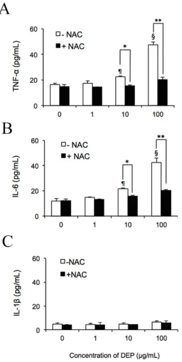
The respiratory system and capillaries have been shown to respond to DEP exposure by secret-ing the pro-inflammatory cytokines TNF-αand IL-6 [7,38,39]. To examine whether DEP-induced ROS triggered the release of these cytokines fromin vitroendothelial tubes and NAC reduces this activity, cultures were treated for 24 h with 1, 10, or 100μg/mL DEPs ± NAC. As determined by comparison with an enzyme-linked immunosorbent assay (ELISA)-generated standard curve, negative control HUVEC tubes released 16.6 pg/mL TNF-α(Fig 2A) and 11.8 pg/mL IL-6 (Fig 2B) into the medium. No significant difference was observed in cytokine levels when cells were treated with 1μg/mL DEPs. However, at 10μg/mL DEPs, 22.7 ± 0.6 pg/mL TNF-αand 21.8 ± 0.6 pg/mL IL-6 was found in the medium. Moreover, after a 24-h exposure to 100μg/mL DEPs, tube cells released 47.5 ± 2.3 pg/mL TNF-αand 42.4 ± 3.6 pg/mL IL-6, about 4 times that of the untreated control. The presence of NAC significantly suppressed the release of both cytokines. With this ROS scavenger, secretion was reduced to 20.4 ± 5.2 pg/mL TNF-αand 20.2 ± 0.84 pg/mL IL-6 in samples treated with 100μg/mL DEPs, comparable to Fig 1. DEPs induced intracellular ROS generation in capillary-like tube cells and caused cytotoxicity.
(A) DEPs were incubated in the cell-free system for 2 h. Cm-H2DCFDA assays were then carried out. ROS production is shown for each sample. (B) HUVECs were exposed to DEPs±NAC for 24 h. Intracellular ROS is shown. (C) Tube cells were exposed to DEPs with or without the addition of NAC. Cytotoxicity was measured as described in the Methods.
doi:10.1371/journal.pone.0131911.g001
Table 1. Cytoplasmic Total, Oxidized and Reduced Glutathione Levels and GSH/GSSG Redox Ratio in the tube cells treated with DEP±NAC.Total GSH, total glutathione; GSSG, oxidized glutathione; GSH, reduced glutathione. Values are given as mean±SD of n = 3 experiments and duplicate measure-ments. The concentrations of DEP used are 1, 10, and 100μg/mL, NAC is 10 mM. DEP (0μg/mL) presents as the negative control.
Total GSH GSSG GSH GSH/GSSG
(U/mg protein) (U/mg protein) (U/mg protein) Redox Ratio
DEP (μg/mL) 0 15.2±0.86 1.5±0.06 13.4±1.27 8.93
1 16.5±0.95 3.6±0.12a 12.5±1.43 3.47a
10 18.6±1.23a 4.6±0.48a 11.8±1.89 2.57a
100 21.8±1.91a 7.6±0.8a 13.1±1.15 1.72a
DEP (μg/mL) 0 20.1±2.09b 0.9±0.01b 17.1±1.63b 19ab
plus NAC 1 21.4±1.89b 1.3±0.11b 18.4±1.41b 14.15ab
10 21.9±2.11 1.7±0.09b 18.9±1.26b 11.12ab
100 22.8±2.47 2.3±0.07ab 20.3±1.71b 8.82
ais signi
ficantly different from the samples to DEP (0μg/mL) in upper column or to NAC+DEP (0μg/mL) in lower column (p<0.05) bis signi
ficant difference between DEP and NAC+DEP samples (p<0.05).
levels of these cytokines in cells treated with 10μg/mL DEPs without the addition of NAC, strongly indicating that DEP-induced ROS mediated the secretion of these cytokines from endothelial tube cells. However, as shown inFig 2C, no significant upregulation of IL-1βwas observed in response to DEPs. Furthermore, as shown inFig 3, immunoblotting results Fig 2. DEP-induced ROS promoted secretion of TNF-αand IL-6 by capillary-like tube culturesin vitro. After exposing endothelial tubes to 1, 10, or 100μg/mL DEPs with or without NAC, medium was collected for evaluation by ELISA. Concentrations of TNF-α(A), IL-6 (B), and IL-1β(C) were measured and were quantified according to comparison with a standard curve. ¶ and § indicate significant differences (P<0.01 andP<0.001) between DEP-treated samples and the untreated control (0μg/mL). Significance was reached at*p<0.05 and**p<0.01 in samples treated with 10 and 100μg/mL DEPs compared to the same amount of DEPs plus NAC. Values represent means±SDs (n = 6). Statistical analysis was carried out using the Student’s t-test.
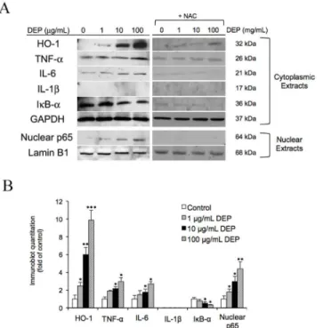
supported that DEPs dramatically increased levels of cytoplasmic HO-1 and slightly increased levels of TNF-αand IL-6. IκB-αwas reduced in a dose-dependent manner following treatment with DEPs, while IL-1βwas not observed. In nuclear extracts, nuclear p65 was elevated in response to DEP exposure. Addition of NAC significantly reduces HO-1, TNF-α, IL-6, IκB-α, and p65 expression. Quantification of the blots of DEP treated samples is shown inFig 3B.
TNF-
α
and IL-6 stimulated the release of vascular permeability factor
(VPF)/VEGFA
HO-1 has previously been shown to induce the secretion of VPF/VEGF-A [18,40,41]. Chao et al(2012) demonstrated that DEPs upregulate HO-1 and stimulate VEGFA secretion into the culture media [12]. Because our previous experiment only investigated whether TNF-αand IL-6 were upregulated, we wondered whether these cytokines affected permeability by directly influencing VEGFA levels. Therefore, we treated tube cells with exogenous TNF-α(47.5 pg/ mL) and IL-6 (42.4 pg/mL) for 24 h to mimic the effects of 24-h exposure to 100μg/mL DEPs. As seen inFig 4A, the combination treatment with both cytokines for 24 h further increased the level of VEGF-A. Addition of Tin protoporphyrin IX (SnPP) (HO-1 inhibitor, 25μM) might not affect the VEGF-A secretion. Quantification of the results is shown inFig 4B, indi-cating that VEGF-A secretion was associated with a DEP-induced pro-inflammatory response but not directly correlated to HO-1.
On the other hand, our previous investigation suggested that DEPs upregulated HO-1 and induced VEGFA secretion in a dose-dependent manner. Moreover, Terryet al(1998 and 1999) Fig 3. DEPs affected the expression of intracellular proteins.(A) After a 24-h exposure to DEPs±NAC, endothelial tube cells were lysed, and cytoplasmic and nuclear proteins were extracted for western blot analysis. The 8 lanes of the immunoblot represent samples exposed to different concentrations of DEPs and DEPs+NAC. Equal loading of cytoplasmic and nuclear proteins was confirmed with GAPDH and Lamin B1 immunoreactivities, respectively. (B) Semiquantitative analysis of the band intensities. Significance was reached at*p<0.05,**p<0.01, and***p<0.001, as analyzed using Student’s t-test.
suggested that TNF-αinduces HO-1 expression in HUVEC monolayer cultures [42,43]. Therefore, we further examined whether TNF-α+ IL-6 would stimulate VEGF-A secretion through the same mechanism by measuring changes in HO-1 expression in this more biologi-cal relevant model following treatment with the cytokines. Here TNF-αplus IL-6 was added to endothelial tubes ± SnPP, and NAC + DEP for 24 h, and the level of HO-1 was assessed. Tube cells treated with 100μg/mL DEP was used as a comparison. Interestingly, treatment of both cytokines did not change the level in HO-1 expression, but NAC significantly reduced DEP-upregulated HO-1 (Fig 4C). Quantification of these results is shown inFig 4D. Moreover, as seen inFig 4E,HO-1mRNA levels were not changed following exposure to TNF-αand IL-6.
TNF-
α
and IL-6 induced vascular permeability independently of HO-1
activation
Since TNF-αand IL-6 stimulated VPF/VEGF-A, further investigations were required to dem-onstrate a link with vasculature permeability. Although HO-1 has been shown to associate with VEGF-A-induced permeability, whether TNF-αand IL-6 are involved in the permeability mechanism of HO-1 activation remains unclear. To assess this issue, we evaluated the amount of dextran-FITC taken up by endothelial tubes exposed to TNF-α, IL-6, TNF-αplus IL-6 (with or without SnPP), and DEPs (10 or 100μg/mL, with or without SnPP or NAC). Samples left untreated or treated with VEGF-A (100 ng/mL) were used as negative and positive controls, respectively. As shown inFig 5, cytokine treatment induced changes in dextran uptake from 5237 ± 145 (RFU) (negative control) to 5747 ± 40 α), 5723 ± 39 (IL-6), 5797 ± 95 (TNF-αplus IL-6), and 5781 ± 60 (TNF-αplus IL-6 and SnPP). Tube cell permeability induced by DEPs (10μg/mL) was restored after the addition of SnPP or NAC. Moreover, 100μg/mL DEPs and 100μg/mL DEPs plus NAC reduced the amount of dextran-FITC taken up by the cells, while cotreatment with SnPP elevated dextran-FITC uptake. Although these values were not significantly different, there was a correlative trend between exposure of endothelial tubes to both cytokines and the amount of fluorescence taken up by the tubes, indicating changes in permeability.
Fig 4. Addition of TNF-αand IL-6 triggered release of VEGF-A from endothelial tube cells.(A) The tube cells were treated with 47.5 and 42.4 pg/mL TNF-αand VEGF-A, respectively, and incubated for a 24 h. The medium was collected and subjected to SDS-PAGE. Half the gel was used for western blot analysis, probing with anti-VEGF-A antibodies, and the other half was identically loaded as a loading control, stained with Coomassie blue. (B) Semiquantitative analysis of the intensity of VEGF-A bands from the blot. Furthermore, TNF-αplus IL-6 independently stimulated HO-1 expression. (C) Analysis of HO-1 expression in HUVEC tube lysates was performed using immunoblotting. Equal protein loading was confirmed with GAPDH
immunoreactivity. (D) Semiquantitative analysis of the intensities of HO-1 bands from the blot. § indicates p<0.05 significance decrease from DEP (100 g/mL) to NAC+DEP (100 g/mL). (E)HO-1mRNA expression response to TNF-αand IL-6 was quantified using qPCR, with normalization to 18S. HO-1 expressed in the negative control was defined as 1. Values are expressed as the fold-change compared to the negative control (n = 3). Significance was reached at*p<0.05 as analyzed using Student’s t-test.
DEP-induced cytokines caused endothelial VE-cadherin redistribution
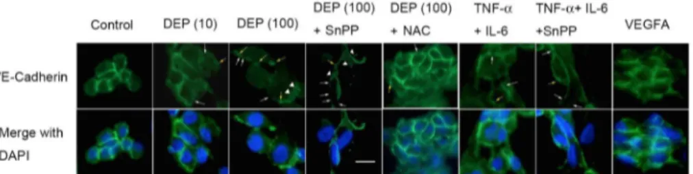
In contrast to VEGFA-dependent permeability, VE-cadherin-dependent cell-cell barriers are crucial to preventing the permeability of endothelial cells [8,44–46]. The effects of TNF-αand IL-6 on VE-cadherin distribution in endothelial tube cells are shown inFig 6. Discontinuity of cadherin localization on the cell-cell border was observed (white arrows), and some VE-cadherin internalization was present, as indicated by punctate staining in the cytoplasm (trian-gular arrows). Some VE-cadherin was pulled from the cell surface into globules under the cell membrane (yellow arrows). To confirm whether TNF-αand IL-6 had the ability to rearrange the VE-cadherin pattern without HO-1 activation, cells were exposed to cytokines plus SnPP. Furthermore, to assess whether NAC restore the VE-cadherin redistribution, NAC was used with DEPs (10 or 100μg/mL). VEGF-A was used as controls. Interestingly, endothelial tube structures were altered with increasing concentration of DEPs. At 100μg/mL, cells were rounded up, and pulling and thinning of the cytoplasm between the cells were observed. Addi-tionally, redistribution of the globules of submembrane VE-cadherin was found, while little discontinuity of VE-cadherin was observed at 10μg/mL. Additionally, most of the cells exhib-ited regular separations from each other, and cells exposed to TNF-αand IL-6 along with the HO-1 inhibitor SnPP exhibited strong distribution of VE-cadherin on the cell border, indicat-ing reduced permeability. These data suggested that TNF-α+IL-6 may contribute to the perme-ability of cell junctions, similar to VEGF-A exposure.
Fig 5. DEPs and the induced cytokines causedin vitrocapillary permeability independently of HO-1 activation.FITC-dextran was added to endothelial tubes for 4 h after a 24-h exposure to different treatments. The RFU was determined for each sample after washing of the endothelial tubes.
doi:10.1371/journal.pone.0131911.g005
Fig 6. DEPs and the induced cytokines caused capillary VE-cadherin redistribution.Endothelial tube cells were treated for 24 h with DEPs±SnPP or TNF-α+IL-6±SnPP. An endothelial tube cell culture treated for 24 h with 100 ng/mL VEGFA was the positive control. VE-cadherin localization was then determined by epifluorescence imaging. White arrows indicate areas where VE-cadherin showed discontinuity around the cell membrane. Arrowheads indicate instances where VE-cadherin moved from the cell membrane to the interior and intracellular submembrane regions. Yellow arrows indicate cell-cell separations and globules of VE-cadherin that accumulated intracellularly near the cell membrane.
DEP-caused VEGFR2 rearrangement
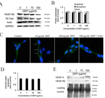
The cytoplasmic tail of VE-cadherin is known to interact with VEGF-R2 andβ-catenin and form a complex linking the actin cytoskeleton to the cell membrane to stabilize cell shape and adherens junctions [47]. Depolarization of actin cytoskeleton network and disruption of VEGF-R2-VE-cadherin formed complex might not only affect endothelial cell-cell contact, but also regulate cell survivability [12,47–49]. Hence, here we assess whether VEGF-R2 and other junctional proteins change in response to DEP. As shown inFig 7A, VE-cadherin and actin are unaffected by DEP exposure, but only VEGF-R2 is decreased slightly. According to a quantita-tion analysis (Fig 7B), VEGF-R2 expresses at 87.5% and 75% intensity of control at 10 and 100μg/mL respectively, but remains unchanged at 1μg/mL. Furthermore, fluorescence images show the morphology of VEGF-R2 that is outlining the cell junctions fairly regularly in the negative control. Image of DEP (10μg/mL) treated tube cells indicate a loss in sharpness of border (arrows) to the VEGF-R2 staining. At 100μg/mL DEP treated tube cells, which have a similar VEGF-R2 performance to 10μg/mL, but has more discontinuity patterns on the cell-cell boarder. Addition of NAC mitigates this VEGF-R2 alternation. On the other hand, our qPCR result reveals thatVEGF-R2mRNA level is unaffected (Fig 7D). This interesting result implies that VEGF-R2 may be secreted to the supernatant/medium. As shown inFig 7E, west-ern blot indicated that sVEGF-R2 (soluble VEGF-R2) and VEGF-A in the medium are dramat-ically increased in a DEP dose dependent manner.
Discussion
The cellular production of ROS is a well-known physiological process. Several studies have vided evidence that free radical-induced oxidative damage of cell membranes, DNA, and pro-teins might be the cause of several diseases including cardiovascular events [50,51]. In previous studies, we have proposed and supported the hypothesis that DEPs affect vascular permeability through VPF/VEGF-A release by production of ROS. This has been demonstrated by experi-ments showing that networks of the adherens junctional protein VE-cadherin become disrupted when cultured cells are treated with DEPs [8]. In the current work, we performed a more exten-sive characterization of the pro-inflammatory response to DEP exposure. The goals of this study were to investigate the induction of extracellular ROS generation, secretion of the cytokine-gen-erating moiety, the mechanism leading to increased vascular permeability, and the influence of VE-cadherin relocalization, and the mitigation of NAC for these disorders described above. We also sought to clarify whether a system without the confounding effects of inflammatory cells could be used to examine how DEPs might modulate endothelial permeability.
Three different methods have been used to demonstrate that DEP exposure is able to gener-ate oxidative stress [12]. However, whether these free radical species are generated by DEPs themselves, before they contact the cell/the inside of the cell, or after they are taken up remains unclear. Therefore, we evaluated both extracellular and intracellular ROS with the Cm-H2DCFDA assay (Fig 1A and 1B). Interestingly, only H2O2sample increased ROS levels
fol-lowing a 2-h incubation, and DEPs did not induce extracellular ROS. However, in contrast to this cell-free result, Cm-H2DCFDA assays showed that DEP induced intracellular ROS
genera-tion in a dose-dependent manner in tube cells. Furthermore, the cytotoxicity curves showed a pattern that was quite similar to the intracellular ROS data, implying that DEP-induced intra-cellular ROS should be a factor in determining cell survival. These experiments not only con-firmed the results reported by Chaoet al(2012), but also provided direct evidence that DEPs induced cytotoxicity through generation of free radicals in tube cells [12].
oxidation [52–54]. After 24 h of supplementation, NAC provided remarkable changes in anti-oxidant/oxidant status of the DEP treated tube cells including extracellular and intracellular ROS (Fig 1AandTable 1). Cellular thiols play central roles in maintaining the cellular redox status [50,51,55]. The increases observed in total GSH levels might be an adaptive response toward an oxidative insult. The elevation in GSSG levels indicates the ability of DEP to interfere with GSH and cause its oxidation. In addition, GSH/GSSG redox ratio decreased significantly with DEP treatment. Administration of NAC with DEP exposure provided significant increases in the redox ratio (Table 1). Recently, we showed that NAC protectsin vitroAS52 CHO cells from the oxidative damage caused by 3,5-dimethylaminophenol [37]. It is well known that
•OH is formed from H2O2and it is the most detrimental ROS, due to its high interaction with
nucleic acids, proteins, and lipids. NAC terminates these chain radical reactions by transferring a single electron, owing to the stability of its own radical ion. Thus, the results suggest that NAC might suppress DEP-induced ROS efficiently and reduce sequential damages due to its effect on cellular redox equilibrium [56].
Promotion of inflammation is considered a key step in the adverse health effects associated with PM exposure [57,58]. Additionally, DEPs cause the release of pro-inflammatory
Fig 7. DEP dissociate VEGF-R2 from adherens junction connection.(A) After 24 h DEP (1, 10 and 100 g/ mL) exposure, the tube cells were lysed and harvested. The untreated cells (0 g/mL) were defined as negative control. The level of adheren junctional proteins VEGF-R2, VE-cadherin (VE-Cad), -catenin and cytoskeleton actin were determined by Western blot. Equal amount of protein loading was confirmed based on total GAPDH expression. (B) Quantification showed VEGF-R2 decrease in a dose dependent manner as the relative fold change to the control. Again, the level of VE-cadherin, -catenin and cytoskeleton actin activities are unaffected by DEP. (C) Images show the distribution of VEGF-R2 in response to DEP (10 and 100 g/mL) and DEP (100 g/mL) plus NAC exposure for 24 h. Nuclei were stained with 10 mM DAPI. Shown is a representative image from three independent experiments. Scale bar = 10 m. Magnification is 400X. (D) To confirm whether DEP changeVEGF-R2expression, the cDNA were probed with VEGFR2 primers and the activity were analyzed using QPCR after exposure with DEP for 24 h. The level of mRNA normalized to 18s gene expression was presented as the fold change relative to the untreated control (0μg/mL). Means±SD, n = 3. (E) Effects of DEP on the secretion of VEGF-R2. Secretion of VEGF-R2 of capillary tube cells related to the DEP exposure. After 24 h DEP (1, 10 and 100 g/mL) exposure, the supernatant/medium were collected and loaded (40 L/well) onto the SDS-PAGE. The expression of secreted VEGF-R2 was tested and measured by using western blot. Coomassie blue stain was defined as the loading control. The supernatant collected from the cells treated without DEP were defined as negative control.
cytokines (e.g., TNF-α, IL-6, and IL-1β) bothin vitroandin vivo[27,59–63]. Our results showed that intracellular ROS were also able to contribute to the release of pro-inflammatory cytokines (Fig 2). However, only TNF-αand IL-6 were secreted from the capillary-tube cells following 24-h exposure to DEPs. No IL-1βwas detected, even at the highest dose of DEP, although many studies in monolayer endothelia have demonstrated the opposite results [43, 64–66]. Our immunoblotting results also confirmed this result. Krishnanet al(2013) suggested that IL-1βmight be increased within 7 h after inhalation of diesel exhaust (DE) [67], and the inactive proform of IL-1βmay be cleaved rapidly, enabling an immediate response to occur [27]. These reasons may explain the lack of IL-1βexpression in our experiment.
Obviously, VEGF-A promotes vascular permeability [68–71]. In addition, TNF-αand IL-6 have been shown to stimulate permeability; however, this effect has only been observed in endothelial monolayer culture [29,31]. Therefore, we sought to identify the mechanisms medi-ating vascular permeability in our model (Fig 5). Our data demonstrated that both cytokines were independently capable of enhancing VEGF-A secretion in the absence of DEPs. While the effects were highest at 24 h, no significant difference was observed at different time points. In addition, VEGFA is modulated by HO-1 during the stimulation of vascular permeability [18, 72,73]. Our previous research also supported the link between HO-1 and VEGF-A secretion in response to DEP exposure and demonstrated that VEFG-A levels were reduced by the addition of NAC or SnPP [12]. Although the functions of TNF-αand IL-6 have been investigated widely, the mechanism through which they contribute to vascular permeability following HO-1 upregulation is still unknown. Therefore, we treated the tube cells with TNF-α+IL-6±SnPP and compared the results to those of samples exposed to DEP. While DEP induced HO-1 expression, no significant differences were detected among samples treated with TNF-α+IL-6 +SnPP, TNF-α+IL-6, and the control. Additionally, we found thatHO-1mRNA was not altered in response to treatment with any of these cytokines, suggesting while TNF-αand IL-6 elevated VEGF-A, they did not enhance HO-1 expression. This may be because endothelial permeability was facilitated by ROS-dependent induction of TNF-αand IL-6, which possibly upregulated VEGF-A independently of HO-1 stimulation. However, times shorter than 24 h have not been tested.
Furthermore, we found that the localization of VE-cadherin to the endothelial cell-cell bor-der became disrupted, with highly irregularly shaped cells displaying large gap formations, sug-gesting that the permeability of the endothelial layer was increased in response to cytokines. These results were consistent with our observation that TNF-αand IL-6 induced VEGF-A expression to stimulate vasculature leakiness. Although the localization patterns of VE-cad-herin included linear discontinuity and globular formation, no dramatic differences in the shape were detected between exposure to DEPs (10μg/mL), TNF-α+IL-6, and TNF-α+IL-6 +SnPP. At 100μg/mL DEPs, VE-cadherin was internalized in endothelial tube cell cultures, causing cell-cell gaps that led to vascular permeability, NAC is able to entirely restore these alternations. Co-treatment of SnPP with DEP (100μg/mL), VE-cadherin still exhibited irregu-lar patterns of localization in the cytoplasm; however, the cell-cell border was protected and detectable on the cell membrane in a similar manner to the TNF-αplus IL-6-treated sample. This result suggested that vascular permeability might be stimulated by TNF-αand IL-6 inde-pendently of HO-1 activation.
VE-cadherin limits cell proliferation and form a cell-cell barrier by retaining VEGF-R2 at the membrane and preventing its relocation into signaling compartments [12,47–49]. VEGF-R2 is portable and its mechanism remains unclear, although Dajana (2004) mentioned that VEGF-A binds to VEGF-R2 for either stabilization in confluent endothelial cells or prolif-eration in sparse endothelial cells [44]. Meanwhile,in vitroandin vivostudies have demon-strated that soluble VEGF-R1 can be secreted for the binding of VEGF-A and results in prohibition of angiogenesis [74]. However, it is unclear how dissociated VEGF-R2 plays a role in the physiological / pathological function during exposure to DEP. The signalling cascade stimulated by VEGF-A activates VEGFR-2 followed by c-Src tyrosine kinase and MAP kinases the Erk subfamily activation. The consequent effect has been suggested to disrupt endothelial cell-cell junctions leading to restrict an increase in vascular permeability to the environment affected by local injury to blood vessels [75]. This might lead to cause acute or chronic cardio-vascular disorders including peripheral edema, capillary degradation, and inflammation [76– 78]. Here we propose that the VE-cadherin/VEGF-R2 complex is disrupted, causing the tube cells to lose their cellular contact, and the dissociated VEGFR2 may be released and bind to the VEGFA in the medium for reduction of the increase vascular permeability.
Conclusion
internalization, and globular formation, resulting in discontinuation and permeability of the cell-cell border. Therefore, were these phenomena to occur, even to a small degree, in lung capillaries after exposure to DEP, the particles may gain access to the bloodstream and contrib-ute to the development of adverse cardiovascular events. And a potential protection of NAC against the DEP-induced subsequent damages was determined.
Acknowledgments
We thank Dr. Marion K. Gordon and Dr. Robert Laumbach from EOHSI, Rutgers University, for providing the DEP, which was collected from Dr. Masaru Sagai in Japan.
Author Contributions
Conceived and designed the experiments: MWC MKG. Performed the experiments: MWC CYT. Analyzed the data: CYT JFC JSW. Contributed reagents/materials/analysis tools: JFC JSW YJC. Wrote the paper: MWC CYT.
References
1. Bai Y, Suzuki AK, Sagai M. The cytotoxic effects of diesel exhaust particles on human pulmonary artery endothelial cells in vitro: role of active oxygen species. Free radical biology & medicine. 2001; 30 (5):555–62. PMID:11182526.
2. Becker S, Dailey LA, Soukup JM, Grambow SC, Devlin RB, Huang YC. Seasonal variations in air pollu-tion particle-induced inflammatory mediator release and oxidative stress. Environmental health per-spectives. 2005; 113(8):1032–8. PMID:16079075.
3. Salvi S, Blomberg A, Rudell B, Kelly F, Sandstrom T, Holgate ST, et al. Acute inflammatory responses in the airways and peripheral blood after short-term exposure to diesel exhaust in healthy human volun-teers. American journal of respiratory and critical care medicine. 1999; 159(3):702–9. PMID:10051240.
4. Wichmann HE. Diesel exhaust particles. Inhalation toxicology. 2007; 19 Suppl 1:241–4. PMID: 17886072.
5. Tornqvist H, Mills NL, Gonzalez M, Miller MR, Robinson SD, Megson IL, et al. Persistent endothelial dysfunction in humans after diesel exhaust inhalation. American journal of respiratory and critical care medicine. 2007; 176(4):395–400. Epub 2007/04/21. doi:10.1164/rccm.200606-872OCPMID: 17446340.
6. Brook RD, Brook JR, Urch B, Vincent R, Rajagopalan S, Silverman F. Inhalation of fine particulate air pollution and ozone causes acute arterial vasoconstriction in healthy adults. Circulation. 2002; 105 (13):1534–6. PMID:11927516.
7. Inoue K, Takano H, Sakurai M, Oda T, Tamura H, Yanagisawa R, et al. Pulmonary exposure to diesel exhaust particles enhances coagulatory disturbance with endothelial damage and systemic inflamma-tion related to lung inflammainflamma-tion. Exp Biol Med (Maywood). 2006; 231(10):1626–32. PMID:17060683.
8. Chao MW, Kozlosky J, Po IP, Strickland PO, Svoboda KK, Cooper K, et al. Diesel exhaust particle exposure causes redistribution of endothelial tube VE-cadherin. Toxicology. 2011; 279(1–3):73–84.
Fig 8. Schematic diagram depicting DEP-induced mechanisms potentially causing vascular permeability.DEPs cause capillary-like endothelial tubes to produce intracellular oxidative stress, which increases vascular permeability by 3 mechanisms: i) direct rearrangement of the cell-cell junctional VE-cadherin network; ii) activation of HO-1, which subsequently causes release of the vascular permeability factor, VEGFA; iii) inducible TNF-αand IL-6 release, which might increase the release of VEGFA independently of the contribution by HO-1. Administration of NAC blocks these sequential cytotoxic effects from the beginning.
Epub 2010/10/05. doi:10.1016/j.tox.2010.09.011PMID:20887764; PubMed Central PMCID: PMC3003746.
9. Harris ES, Nelson WJ. VE-cadherin: at the front, center, and sides of endothelial cell organization and function. Curr Opin Cell Biol. 2010; 22(5):651–8. doi:10.1016/j.ceb.2010.07.006PMID:20708398; PubMed Central PMCID: PMC2948582.
10. Gavard J. Endothelial permeability and VE-cadherin: a wacky comradeship. Cell adhesion & migration. 2013; 7(6):455–61. Epub 2014/01/17. doi:10.4161/cam.27330PMID:24430214; PubMed Central PMCID: PMC3916348.
11. Dudek SM, Garcia JG. Cytoskeletal regulation of pulmonary vascular permeability. Journal of applied physiology. 2001; 91(4):1487–500. PMID:11568129.
12. Chao MW, Po IP, Laumbach RJ, Koslosky J, Cooper K, Gordon MK. DEP induction of ROS in capillary-like endothelial tubes leads to VEGF-A expression. Toxicology. 2012; 297(1–3):34–46. Epub 2012/04/ 18. doi:10.1016/j.tox.2012.03.009PMID:22507881; PubMed Central PMCID: PMC3387988.
13. Li YJ, Kawada T, Azuma A. Nrf2 is a protective factor against oxidative stresses induced by diesel exhaust particle in allergic asthma. Oxidative medicine and cellular longevity. 2013; 2013:323607. doi: 10.1155/2013/323607PMID:23738037; PubMed Central PMCID: PMC3655666.
14. Brouard S, Berberat PO, Tobiasch E, Seldon MP, Bach FH, Soares MP. Heme oxygenase-1-derived carbon monoxide requires the activation of transcription factor NF-kappa B to protect endothelial cells from tumor necrosis factor-alpha-mediated apoptosis. J Biol Chem. 2002; 277(20):17950–61. PMID: 11880364.
15. Silva G, Cunha A, Gregoire IP, Seldon MP, Soares MP. The antiapoptotic effect of heme oxygenase-1 in endothelial cells involves the degradation of p38 alpha MAPK isoform. J Immunol. 2006; 177 (3):1894–903. PMID:16849502.
16. Wang Z, Armando I, Asico LD, Escano C, Wang X, Lu Q, et al. The elevated blood pressure of human GRK4gamma A142V transgenic mice is not associated with increased ROS production. American jour-nal of physiology. 2007; 292(5):H2083–92. PMID:17259440.
17. Wilson SJ, Keenan AK. Role of hemin in the modulation of H2O2-mediated endothelial cell injury. Vas-cular pharmacology. 2003; 40(2):109–18. PMID:12646399.
18. Dulak J, Loboda A, Jozkowicz A. Effect of heme oxygenase-1 on vascular function and disease. Cur-rent opinion in lipidology. 2008; 19(5):505–12. PMID:18769232. doi:10.1097/MOL.
0b013e32830d81e9
19. Nagy JA, Vasile E, Feng D, Sundberg C, Brown LF, Manseau EJ, et al. VEGF-A induces angiogenesis, arteriogenesis, lymphangiogenesis, and vascular malformations. Cold Spring Harbor symposia on quantitative biology. 2002; 67:227–37. PMID:12858545.
20. Weis SM, Cheresh DA. Pathophysiological consequences of VEGF-induced vascular permeability. Nature. 2005; 437(7058):497–504. doi:10.1038/nature03987PMID:16177780.
21. Vandenbroucke E, Mehta D, Minshall R, Malik AB. Regulation of endothelial junctional permeability. Ann N Y Acad Sci. 2008; 1123:134–45. doi:10.1196/annals.1420.016PMID:18375586.
22. Yokota S, Ohara N, Kobayashi T. The effects of organic extract of diesel exhaust particles on ischemia/ reperfusion-related arrhythmia and on pulmonary inflammation. The Journal of toxicological sciences. 2008; 33(1):1–10. PMID:18303179.
23. Nemmar A, Subramaniyan D, Zia S, Yasin J, Ali BH. Airway resistance, inflammation and oxidative stress following exposure to diesel exhaust particle in angiotensin II-induced hypertension in mice. Tox-icology. 2012; 292(2–3):162–8. Epub 2012/01/05. doi:10.1016/j.tox.2011.12.009PMID:22214961.
24. Kleemann R, Zadelaar S, Kooistra T. Cytokines and atherosclerosis: a comprehensive review of stud-ies in mice. Cardiovascular research. 2008; 79(3):360–76. doi:10.1093/cvr/cvn120PMID:18487233; PubMed Central PMCID: PMC2492729.
25. Muller WA. Mechanisms of leukocyte transendothelial migration. Annual review of pathology. 2011; 6:323–44. doi:10.1146/annurev-pathol-011110-130224PMID:21073340; PubMed Central PMCID: PMC3628537.
26. Becker S, Mundandhara S, Devlin RB, Madden M. Regulation of cytokine production in human alveolar macrophages and airway epithelial cells in response to ambient air pollution particles: Further mecha-nistic studies. Toxicology and applied pharmacology. 2005; 207(2 Suppl):269–75. PMID:15993911.
27. Schwarze PE, Totlandsdal AI, Lag M, Refsnes M, Holme JA, Ovrevik J. Inflammation-related effects of diesel engine exhaust particles: studies on lung cells in vitro. BioMed research international. 2013; 2013:685142. doi:10.1155/2013/685142PMID:23509760; PubMed Central PMCID: PMC3586454.
wood smoke and diesel exhaust particulate matter. Toxicol Lett. 2012; 209(2):121–8. doi:10.1016/j. toxlet.2011.12.003PMID:22198124.
29. Nwariaku FE, Liu Z, Zhu X, Turnage RH, Sarosi GA, Terada LS. Tyrosine phosphorylation of vascular endothelial cadherin and the regulation of microvascular permeability. Surgery. 2002; 132(2):180–5. PMID:12219009.
30. Cromer WE, Zawieja SD, Tharakan B, Childs EW, Newell MK, Zawieja DC. The effects of inflammatory cytokines on lymphatic endothelial barrier function. Angiogenesis. 2014; 17(2):395–406. Epub 2013/ 10/22. doi:10.1007/s10456-013-9393-2PMID:24141404; PubMed Central PMCID: PMC4314095.
31. Maruo N, Morita I, Shirao M, Murota S. IL-6 increases endothelial permeability in vitro. Endocrinology. 1992; 131(2):710–4. PMID:1639018.
32. Tseng CY, Wang JS, Chang YJ, Chang JF, Chao MW. Exposure to High-Dose Diesel Exhaust Parti-cles Induces Intracellular Oxidative Stress and Causes Endothelial Apoptosis in Cultured In Vitro Capil-lary Tube Cells. Cardiovascular toxicology. 2014. Epub 2014/12/10. doi:10.1007/s12012-014-9302-y PMID:25488805.
33. Alagappan VK, McKay S, Widyastuti A, Garrelds IM, Bogers AJ, Hoogsteden HC, et al. Proinflamma-tory cytokines upregulate mRNA expression and secretion of vascular endothelial growth factor in cul-tured human airway smooth muscle cells. Cell biochemistry and biophysics. 2005; 43(1):119–29. Epub 2005/07/27. doi:10.1385/CBB:43:1:119PMID:16043889.
34. Davin JC. The glomerular permeability factors in idiopathic nephrotic syndrome. Pediatric nephrology. 2015. Epub 2015/05/01. doi:10.1007/s00467-015-3082-xPMID:25925039.
35. Wong SS, Sun NN, Hyde JD, Ruiz L, Meigs E, Herrin BR, et al. Drotrecogin alfa (activated) prevents smoke-induced increases in pulmonary microvascular permeability and proinflammatory cytokine IL-1beta in rats. Lung. 2004; 182(6):319–30. Epub 2005/03/16. PMID:15765924.
36. Singh P, DeMarini DM, Dick CA, Tabor DG, Ryan JV, Linak WP, et al. Sample characterization of auto-mobile and forklift diesel exhaust particles and comparative pulmonary toxicity in mice. Environmental health perspectives. 2004; 112(8):820–5. PMID:15175167.
37. Chao MW, Erkekoglu P, Tseng CY, Ye W, Trudel LJ, Skipper PL, et al. Protective effects of ascorbic acid against the genetic and epigenetic alterations induced by 3,5-dimethylaminophenol in AA8 cells. J Appl Toxicol. 2015; 35(5):466–77. Epub 2014/09/03. doi:10.1002/jat.3046PMID:25178734.
38. Hartz AM, Bauer B, Block ML, Hong JS, Miller DS. Diesel exhaust particles induce oxidative stress, proinflammatory signaling, and P-glycoprotein up-regulation at the blood-brain barrier. Faseb J. 2008; 22(8):2723–33. PMID:18474546. doi:10.1096/fj.08-106997
39. Ulrich MM, Alink GM, Kumarathasan P, Vincent R, Boere AJ, Cassee FR. Health effects and time course of particulate matter on the cardiopulmonary system in rats with lung inflammation. Journal of toxicology and environmental health. 2002; 65(20):1571–95. PMID:12396869.
40. Bussolati B, Ahmed A, Pemberton H, Landis RC, Di Carlo F, Haskard DO, et al. Bifunctional role for VEGF-induced heme oxygenase-1 in vivo: induction of angiogenesis and inhibition of leukocytic infiltra-tion. Blood. 2004; 103(3):761–6. PMID:14525760.
41. Lin HH, Chen YH, Chang PF, Lee YT, Yet SF, Chau LY. Heme oxygenase-1 promotes neovasculariza-tion in ischemic heart by coinducneovasculariza-tion of VEGF and SDF-1. Journal of molecular and cellular cardiology. 2008; 45(1):44–55. PMID:18534615. doi:10.1016/j.yjmcc.2008.04.011
42. Terry CM, Clikeman JA, Hoidal JR, Callahan KS. Effect of tumor necrosis factor-alpha and interleukin-1 alpha on heme oxygenase-1 expression in human endothelial cells. The American journal of physiol-ogy. 1998; 274(3 Pt 2):H883–91. PMID:9530200.
43. Terry CM, Clikeman JA, Hoidal JR, Callahan KS. TNF-alpha and IL-1alpha induce heme oxygenase-1 via protein kinase C, Ca2+, and phospholipase A2 in endothelial cells. The American journal of physiol-ogy. 1999; 276(5 Pt 2):H1493–501. PMID:10330231.
44. Dejana E. Endothelial cell-cell junctions: happy together. Nat Rev Mol Cell Biol. 2004; 5(4):261–70. PMID:15071551.
45. Dejana E, Orsenigo F, Lampugnani MG. The role of adherens junctions and VE-cadherin in the control of vascular permeability. Journal of cell science. 2008; 121(Pt 13):2115–22. Epub 2008/06/21. doi:10. 1242/jcs.017897PMID:18565824.
46. Gavard J, Gutkind JS. VEGF controls endothelial-cell permeability by promoting the beta-arrestin-dependent endocytosis of VE-cadherin. Nature cell biology. 2006; 8(11):1223–34. doi:10.1038/ ncb1486PMID:17060906.
48. Lampugnani MG, Dejana E. The control of endothelial cell functions by adherens junctions. Novartis Foundation symposium. 2007; 283:4–13; discussion -7, 238–41. Epub 2008/02/28. PMID:18300410.
49. Xu M, Chen G, Fu W, Liao M, Frank JA, Bower KA, et al. Ethanol disrupts vascular endothelial barrier: implication in cancer metastasis. Toxicol Sci. 2012; 127(1):42–53. Epub 2012/02/15. doi:10.1093/ toxsci/kfs087PMID:22331491; PubMed Central PMCID: PMC3327869.
50. Dean O, Giorlando F, Berk M. N-acetylcysteine in psychiatry: current therapeutic evidence and poten-tial mechanisms of action. Journal of psychiatry & neuroscience: JPN. 2011; 36(2):78–86. Epub 2010/ 12/02. doi:10.1503/jpn.100057PMID:21118657; PubMed Central PMCID: PMC3044191.
51. Dodd S, Dean O, Copolov DL, Malhi GS, Berk M. N-acetylcysteine for antioxidant therapy: pharmacol-ogy and clinical utility. Expert opinion on biological therapy. 2008; 8(12):1955–62. Epub 2008/11/08. doi:10.1517/14728220802517901PMID:18990082.
52. Toyooka T, Shinmen T, Aarts JM, Ibuki Y. Dual effects of N-acetyl-L-cysteine dependent on NQO1 activity: suppressive or promotive of 9,10-phenanthrenequinone-induced toxicity. Toxicology and applied pharmacology. 2012; 264(3):404–12. Epub 2012/08/29. doi:10.1016/j.taap.2012.08.017 PMID:22925602.
53. McWhinney RD, Badali K, Liggio J, Li SM, Abbatt JP. Filterable redox cycling activity: a comparison between diesel exhaust particles and secondary organic aerosol constituents. Environmental science & technology. 2013; 47(7):3362–9. Epub 2013/03/09. doi:10.1021/es304676xPMID:23470039.
54. Matsunaga T, Arakaki M, Kamiya T, Endo S, El-Kabbani O, Hara A. Involvement of an aldo-keto reduc-tase (AKR1C3) in redox cycling of 9,10-phenanthrenequinone leading to apoptosis in human endothe-lial cells. Chemico-biological interactions. 2009; 181(1):52–60. Epub 2009/05/16. doi:10.1016/j.cbi. 2009.05.005PMID:19442656.
55. Paolicchi A, Dominici S, Pieri L, Maellaro E, Pompella A. Glutathione catabolism as a signaling mecha-nism. Biochemical pharmacology. 2002; 64(5–6):1027–35. Epub 2002/09/06. PMID:12213602.
56. Masha A, Martina V. Endothelial dysfunction in metabolic diseases: role of oxidation and possible ther-apeutic employment of N-acetylcysteine. Current medicinal chemistry. 2014; 21(32):3616–35. Epub 2014/07/10. PMID:25005185.
57. Ristovski ZD, Jayaratne ER, Lim M, Ayoko GA, Morawska L. Influence of diesel fuel sulfur on nanoparti-cle emissions from city buses. Environmental science & technology. 2006; 40(4):1314–20. PMID: 16572791.
58. Kelly FJ, Fussell JC. Air pollution and airway disease. Clin Exp Allergy. 2011; 41(8):1059–71. Epub 2011/06/01. doi:10.1111/j.1365-2222.2011.03776.xPMID:21623970.
59. Alfaro-Moreno E, Martinez L, Garcia-Cuellar C, Bonner JC, Murray JC, Rosas I, et al. Biologic effects induced in vitro by PM10 from three different zones of Mexico City. Environmental health perspectives. 2002; 110(7):715–20. PMID:12117649.
60. Beck-Speier I, Dayal N, Karg E, Maier KL, Schumann G, Schulz H, et al. Oxidative stress and lipid mediators induced in alveolar macrophages by ultrafine particles. Free radical biology & medicine. 2005; 38(8):1080–92. PMID:15780766.
61. Nemmar A, Inuwa IM. Diesel exhaust particles in blood trigger systemic and pulmonary morphological alterations. Toxicology letters. 2008; 176(1):20–30. PMID:17980520.
62. Nemmar A, Nemery B, Hoet PH, Vermylen J, Hoylaerts MF. Pulmonary inflammation and thrombogeni-city caused by diesel particles in hamsters: role of histamine. American journal of respiratory and critical care medicine. 2003; 168(11):1366–72. PMID:12969870.
63. Nemmar A, Holme JA, Rosas I, Schwarze PE, Alfaro-Moreno E. Recent advances in particulate matter and nanoparticle toxicology: a review of the in vivo and in vitro studies. BioMed research international. 2013; 2013:279371. doi:10.1155/2013/279371PMID:23865044; PubMed Central PMCID:
PMC3705851.
64. Kerkar S, Williams M, Blocksom JM, Wilson RF, Tyburski JG, Steffes CP. TNF-alpha and IL-1beta increase pericyte/endothelial cell co-culture permeability. The Journal of surgical research. 2006; 132 (1):40–5. doi:10.1016/j.jss.2005.06.033PMID:16140333.
65. Rusznak C, Mills PR, Devalia JL, Sapsford RJ, Davies RJ, Lozewicz S. Effect of cigarette smoke on the permeability and IL-1beta and sICAM-1 release from cultured human bronchial epithelial cells of never-smokers, never-smokers, and patients with chronic obstructive pulmonary disease. American journal of respi-ratory cell and molecular biology. 2000; 23(4):530–6. PMID:11017919.
67. Krishnan RM, Sullivan JH, Carlsten C, Wilkerson HW, Beyer RP, Bammler T, et al. A randomized cross-over study of inhalation of diesel exhaust, hematological indices, and endothelial markers in humans. Particle and fibre toxicology. 2013; 10:7. doi:10.1186/1743-8977-10-7PMID:23531317; PubMed Central PMCID: PMC3637197.
68. Connolly DT, Heuvelman DM, Nelson R, Olander JV, Eppley BL, Delfino JJ, et al. Tumor vascular per-meability factor stimulates endothelial cell growth and angiogenesis. J Clin Invest. 1989; 84(5):1470–8. PMID:2478587.
69. Keck PJ, Hauser SD, Krivi G, Sanzo K, Warren T, Feder J, et al. Vascular permeability factor, an endo-thelial cell mitogen related to PDGF. Science. 1989; 246(4935):1309–12. PMID:2479987.
70. Ferrara N, Henzel WJ. Pituitary follicular cells secrete a novel heparin-binding growth factor specific for vascular endothelial cells. Biochem Biophys Res Commun. 1989; 161(2):851–8. PMID:2735925.
71. Senger DR, Galli SJ, Dvorak AM, Perruzzi CA, Harvey VS, Dvorak HF. Tumor cells secrete a vascular permeability factor that promotes accumulation of ascites fluid. Science (New York, NY. 1983; 219 (4587):983–5. PMID:6823562.
72. Cisowski J, Loboda A, Jozkowicz A, Chen S, Agarwal A, Dulak J. Role of heme oxygenase-1 in hydro-gen peroxide-induced VEGF synthesis: effect of HO-1 knockout. Biochemical and biophysical research communications. 2005; 326(3):670–6. PMID:15596152.
73. Jozkowicz A, Huk I, Nigisch A, Weigel G, Dietrich W, Motterlini R, et al. Heme oxygenase and angio-genic activity of endothelial cells: stimulation by carbon monoxide and inhibition by tin protoporphyrin-IX. Antioxidants & redox signaling. 2003; 5(2):155–62. PMID:12716475.
74. Kuhnert F, Tam BY, Sennino B, Gray JT, Yuan J, Jocson A, et al. Soluble receptor-mediated selective inhibition of VEGFR and PDGFRbeta signaling during physiologic and tumor angiogenesis. Proc Natl Acad Sci U S A. 2008; 105(29):10185–90. Epub 2008/07/18. doi:10.1073/pnas.0803194105PMID: 18632559; PubMed Central PMCID: PMC2474564.
75. Suarez S, Ballmer-Hofer K. VEGF transiently disrupts gap junctional communication in endothelial cells. Journal of cell science. 2001; 114(Pt 6):1229–35. Epub 2001/03/03. PMID:11228166.
76. Fleck A, Raines G, Hawker F, Trotter J, Wallace PI, Ledingham IM, et al. Increased vascular permeabil-ity: a major cause of hypoalbuminaemia in disease and injury. Lancet. 1985; 1(8432):781–4. Epub 1985/04/06. PMID:2858667.
77. Du Y, Cramer M, Lee CA, Tang J, Muthusamy A, Antonetti DA, et al. Adrenergic and serotonin recep-tors affect retinal superoxide generation in diabetic mice: relationship to capillary degeneration and per-meability. FASEB J. 2015; 29(5):2194–204. Epub 2015/02/11. doi:10.1096/fj.14-269431PMID: 25667222.
78. Scallan J, Huxley VH, Korthuis RJ. Capillary Fluid Exchange: Regulation, Functions, and Pathology. Integrated Systems Physiology: from Molecule to Function to Disease. San Rafael (CA)2010.
79. Lee GB, Brandt EB, Xiao C, Gibson AM, Le Cras TD, Brown LA, et al. Diesel exhaust particles induce cysteine oxidation and s-glutathionylation in house dust mite induced murine asthma. PloS one. 2013; 8(3):e60632. Epub 2013/04/05. doi:10.1371/journal.pone.0060632PMID:23555996; PubMed Central PMCID: PMC3612047.