INTERNATIONAL RESEARCH JOURNAL OF PHARMACY
www.irjponline.com
ISSN 2230
–
8407
Research Article
POTENTIAL CELL LINE TOXICITY OF ENVIRONMENTAL NANOPARTICLES
Mohan Durga
1, Francis Arul Prakash
1, Abbu Rajasekar
1, Prakhya Balakrishna Murthy
2,
Thiyagarajan Devasena
1*
1
Centre for Nanoscience and Technology, Anna University, Chennai 600 025, India
2
International Institute of Biotechnology and Toxicology (IIBAT), Padappai, India
Article Received on: 17/11/11 Revised on: 22/12/11 Approved for publication: 20/01/12
*Email: [email protected]
ABSTRACT
In India, the unprecedented growth rate and urbanization along with the rapid increase in motor vehicle activity and industrialization are contributing to high levels of urban air pollution.The population is mainly exposed to high air pollution concentrations, where motor vehicle emissions constitute the main source of fine and ultrafine particles. Motor exhaust emissions is a mixture ofgases and ParticulateMatter (PM). Diesel and petrol fuels in vehicles produce combustion-derived particles as a result of combustion. Vehicle exhaust particles are the main constituents of environmental nanoparticles. In the present investigation, environmental nanoparticles such as Diesel Exhaust Particles (DEP) and Petrol Exhaust Particles (PEP) were collected from on-road vehicles using a specially designed collection chamber. The surface morphology of the collected particles was analyzed through Transmission ElectronMicroscope (TEM), and the elemental mapping was performed through EDAX analysis. Results indicated the presence of nanometer-size particles in both the categories of vehicle exhaust. These small-size particles of respirable range can enter the respiratory tract of humans and get deposited in the lungs and cause various effects inside the human body. The aim of this study is to assess the cytotoxicity of the collected Diesel Exhaust Nanoparticles (DENPs) and Petrol Exhaust Nanoparticles (PENPs). Cytotoxicity endpoint, such as IC50 (50% Inhibitory Concentration), was determined after a 24-h exposure. Results of this study indicated that all five cell lines were sensitive to these vehicle exhaust nanoparticles at varying levels.
Keywords: diesel exhaust particles; petrol exhaust particles; TEM; MTT assay; cytotoxicity
INTRODUCTION
The World Health Organization (WHO) estimates that air pollution leads to the death of 2 million people per year, while many others experience respiratory problems, cardio-vascular diseases, pulmonary infections and also cancer. Microscopic dusts from vehicle exhaust, wood fires or coal are considered to be the most dangerous forms of air pollution. The major reasons of air pollution are – vehicle emissions, power plants, factories and refineries.
The vehicle population in India is rapidly increasing day by day and is quickly nearing the 50-million mark. Majority of the vehicles used are two wheelers (75%) that include scooters and motorcycles. There are around 5 million cars, 4.5 million three wheelers, 2 million goods vehicles and over 0.65 million buses running on Indian roads. It is the particulate matter from vehicle exhaust that contributes a major part to air pollution. It is a complex mixture of inorganic and organic substances present in the atmosphere in the form of both liquids and solids1.
In India, the rapid growth of motor vehicles activity and rapid growth of industries are contributing to high levels of urban air pollution. The environmental problems in India are rapidly growing. The rise in economic development and the population growth has taken the country from 300 million people to more than 1 billion people today. As a result this is causing a strain on the country’s infrastructure, environment and natural resources. Soil erosion, industrial pollution, deforestation, land degradation and urbanization are all worsening problems. Environmental pollution is considered to be one of the most serious problems on our planet today. Air pollution is one of the worst problems in the metropolitan cities like Delhi, Mumbai, Kolkata and Chennai. In the big cities like Mumbai and Chennai dust from construction debris, exhaust from vehicles, burning of municipal and garden waste, industrial wastes are all on the rise – so are the pulmonary diseases like asthma. At least six of the top ten causes of mortality are caused by pulmonary diseases.
In Chennai as on 1.1.2008, the vehicle population was estimated as car/jeep – 3.0 lakhs and two wheeler – 2.0 lakhs. According to a study report by the Chennai Metropolitan Development Authority (CMDA), levels of Suspended Particulate Matter (SPM) in the city range from 274 to 1,470 mg/cubic meter, which is much higher than the WHO’s prescribed limit of 200 mg/cubic meter. Studies also reveal that 70% of the pollution load is vehicular. The total particulate matter emitted from diesel engines is much higher than that from petrol engines and Liquified Petroleum Gas (LPG) engines.
Studies by Bathmanabhan et al (2010) measured average particulate matter PM10, PM2.5 and PM1 concentrations near an urban roadway in Chennai city, India. Results indicated that highest PM concentrations were observed during weekday’s peak-hour traffic and lowest PM concentrations were found during afternoon and night time2. A limited number of in vitro studies have also been performed to assess the toxicities of the nanoparticles using different cellular systems and test methods3. However, published toxicity data are still considered inadequate to earn a full understanding of the potential toxicity of nanoparticles. Further studies are needed to clarify the risk of these nanoparticles. Recently, in vitro methods have shown a significant potential for assessing the toxicity of environmental and occupational health risks4–6.
MATERIAL AND METHODS Chemical Compounds
In vitro assay reagents were purchased from Sigma Aldrich. The cell lines (HT29, VERO, HEP 2, A549 and MDA MB 231) were obtained from NCCS, Pune.
Experimental Methods Collection of Exhaust Particles
Motor vehicles with diesel and petrol engines were operated for the collection of diesel and petrol particulate matter. Some of the vehicles used for the study included diesel and petrol cars, share autos, buses, trucks and two wheelers. Millipore monitors loaded with polycarbonate filters with a pore size of 0.8 µm and diameter of 37 mm was connected to
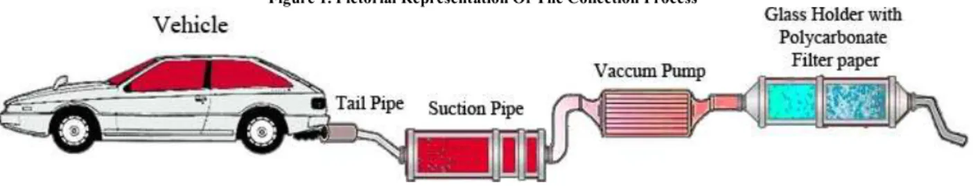
a vacuum pump for the suction of air7, which in turn was connected to a suction pipe through which the air flows resulting in an airflow of 6 ml/min in the monitor. For the particle collection, the vehicle engine was turned on and left at idle condition. The particulate phase of exhaust was collected on the polycarbonate filter paper (Fig.1) during the collection process. The total amount of particulate matter was determined by weighing the filter paper before and after sampling. The procedure was repeated several times until the desired quantity of diesel and petrol particulate matter was collected. The collected samples were scraped out from the filter paper and stored in airtight containers at room temperature and was used for further studies.
Figure 1. Pictorial Representation Of The Collection Process
Characterization of Exhaust Particles
Theaverage particle size, size distribution and morphology were examined by TEM at a voltage of 100 kV. The elemental analysis was performed using the Scanning Electron Microscopy with Energy-dispersive X-ray spectroscopy (SEM-EDX) technique.
Preparation of Cell Lines
The cell lines (HT29, VERO, HEP 2, A549 and MDA MB 231) were obtained from NCCS, Pune. All cultures were maintained in a Minimal Essential Medium (MEM), supplemented with 5% (v/v) Fetal Calf Serum (FCS), 1% (v/v) antibiotic (2 mM L-Glutamine, 100 U/ml Penicillin and 0.1 mg/ml Streptomycin, 0.02 mg/ml Amphotericin) and 7.5% sodium bi-carbonate. Cultured cells were kept at 37ºC in a humidified 5% CO2 incubator. Once the cells reached
confluency, they were sub-cultured. The culture medium was discarded from the flask and the cells were rinsed three times with sterile MEM. The confluent cell layers were enzymatically removed using TPVG [2% Trypsin,Phosphate Buffered Saline (PBS), 0.2% Ethylene Diamine Tetra Acetic acid (EDTA), 10% Glucose] solution and re-suspended in culture medium. Gentle passaging was done using a serological pipette.After passaging, the cells were split into 1:2 or 1:3 ratios for cytotoxicity studies. Cell viability was assessed by vital staining with trypan blue (0.4% [w/v]) and the cell numbers were determined using a light microscope. Preparation of Vehicle Exhaust Nanoparticles
The collected Diesel Exhaust Particles (DEP) and Petrol Exhaust Particles (PEP) was dissolved in Dimethyl Sulphoxide (DMSO) to obtain the stock concentration of 10 mg/ml (100 mg of DEP/PEP is dissolved in 10 ml of DMSO). The stock was prepared fresh and filtered through 0.45-mm filters before each assay. From this stock concentration, working concentrations of solution were prepared by dilution with serum-free culture medium. Working concentrations were prepared adding 0.5 ml of stock to 4.5 ml MEM to obtain the stock concentration of 1 mg/ml. From this working solution, a series of dilutions were prepared (500–1.953 mg/ml).
Cytotoxicity Tests
measure cell viability. It is a non-radioactive colorimetric assay to measure cell viability, proliferation or cytotoxicity. MTT a yellow tetrazolium salt, in metabolically active cells is able to get converted into a water-insoluble dark blue Formosan crystals by reductive cleavage of the tetrazolium ring8.The resultant Formosan crystals, then, can be dissolved and quantified by measuring the absorbance of the solution at 550 nm, and the resultant value is related to the viability of the cells.
To determine cell cytotoxicity/viability, the monolayer culture of HT29, VERO, HEP 2, A549and MDA MB 23 was plated at a density o 105 cells/ml/well in 24-well titre plates and incubated for 24–48 h at 37°C in 5% CO2 atmosphere.
After 24 and 48 h of incubation, the plates were microscopically examined for confluent monolayer, turbidity and toxicity. The medium in the wells was discarded. Fresh medium containing nanoparticles of different concentrations were added. Each dilution of the nanoparticles ranged from 1:1 to 1:64 were added to the respective wells of the 24-well titre plates. To the cell control wells, 1 ml MEM (w/o) FCS was added. The plates were incubated at 37º in 5% CO2 for
24 h and observed for cytotoxicity using an inverted microscope. After incubation, the medium from the wells was removed carefully for MTT assay. Each well was washed with MEM (w/o) FCS for 2–3 times, and then 200 µl of MTT (concentration of 5 mg/ml) was added and incubated for 6– 7 h in 5% CO2 incubator for cytotoxicity. After incubation,
1 ml of DMSO was added in each well and incubated for 45 s. The viability of cells was confirmed by the purple colour formation of formosan crystals after adding solubilising reagent, DMSO. The absorbance was read on a micro plate reader at 550 nm. Cell viability was defined as the ratio of absorbance of treated cells to untreated cells. The percentage of cell viability was calculated by,
% Cell viability = (Test absorbance/Control absorbance) × 100.
RESULTS
Particle Morphology (TEM)
The collected vehicle exhaust samples contained carbon aggregates consisting of tens to thousands of primary carbon particles and mineral particles. The PEP contained slightly larger size particles (Fig. 2) compared to the DEP (Fig. 3).Both samples contained particles of nano-size.
Figure 2. TEM Micrograph Of Petrol Sample Showing Carbon Aggregates Of Various Sizes
Figure 3. TEM Micrograph Of Diesel Sample Showing Carbon Aggregates Of Various Sizes
Elemental Analysis (SEM-EDX)
The elemental analysis for the two samples, PENPs and DENPs, was performed using the SEM-EDX technique. Both the samples contained carbon as the majority element. The percentage of carbon in PENPs was comparatively more than in DENPs. Besides carbon, the PENPs contained elements like aluminium, silica, lead, sulphur, calcium and iron in trace amounts (Fig. 4 and Table 1) compared to the DENPs, which
contained only carbon, oxygen and sulphur (Fig. 5 and Table 2).
Figure 4. X-Ray Microanalysis Spectra Of PENPs
Table 1. Elemental Composition of PENPs
Element Wt% At%
C K 88.77 92.24
O K 08.90 06.94
AlK 00.40 00.18
SiK 00.06 00.03
P K 00.01 00.01
S K 01.08 00.42
PbM 00.07 00.00
CaK 00.22 00.07
FeK 00.48 00.11
Figure 5. X-Ray Microanalysis Spectra Of DENPs
Table 2. Elemental Composition of DENPs
Element Wt% At%
C K 82.72 87.39
O K 14.53 11.52
S K 02.76 01.09
Cytotoxicity Studies
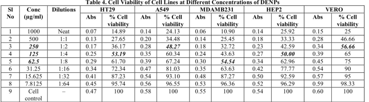
The PENPs and DENPs were screened for its cytotoxicity on HT29, A549, MDA MB 231, HEP 2 and VERO cell lines at various concentrations up to 1,000 µg. The cytotoxicity activity was carried out by using the MTT assay. Absorbance values that are lower than the control cells indicate a reduction in the rate of cell proliferation and hence, cell viability (Table 3&4).
Table 3. Cell Viability of Cell Lines at Different Concentrations of PENPs Sl
No
Conc (µg/ml)
Dilutions HT29 A549 MDAMB231 HEP2 VERO
Abs % Cell viability
Abs % Cell viability
Abs % Cell viability
Abs % Cell viability
Abs % Cell viability
1 1000 Neat 0.05 10.63 0.11 17.46 0.14 26.41 0.09 15.25 0.25 37.31 2 500 1:1 0.13 27.65 0.17 26.98 0.19 35.84 0.15 25.42 0.29 43.28 3 250 1:2 0.17 36.17 0.24 38.09 0.24 45.28 0.20 33.89 0.34 50.74
4 125 1:4 0.24 51.06 0.29 46.03 0.27 50.94 0.25 42.37 0.39 58.20
5 62.5 1:8 0.29 61.70 0.37 58.73 0.34 64.15 0.30 50.84 0.44 65.67
6 31.25 1:16 0.31 65.95 0.45 71.42 0.40 75.47 0.38 64.40 0.52 77.61 7 15.625 1:32 0.37 78.72 0.50 79.36 0.44 83.01 0.44 74.57 0.57 85.07 8 7.8125 1:64 0.43 91.48 0.59 93.65 0.52 98.11 0.52 88.13 0.64 95.52 9 Cell
control
Table 4. Cell Viability of Cell Lines at Different Concentrations of DENPs Sl
No
Conc (µg/ml)
Dilutions HT29 A549 MDAMB231 HEP2 VERO
Abs % Cell viability
Abs % Cell viability
Abs % Cell viability
Abs % Cell viability
Abs % Cell viability
1 1000 Neat 0.07 14.89 0.14 24.13 0.06 10.90 0.14 25.92 0.15 25 2 500 1:1 0.13 27.65 0.20 34.48 0.14 25.45 0.18 33.33 0.28 46.66 3 250 1:2 0.17 36.17 0.28 48.27 0.18 32.72 0.23 42.59 0.34 56.66
4 125 1:4 0.25 53.19 0.35 60.34 0.24 43.63 0.27 50.00 0.39 65
5 62.5 1:8 0.29 61.70 0.39 67.24 0.30 54.54 0.34 62.96 0.45 75
6 31.25 1:16 0.34 72.34 0.47 81.03 0.35 63.63 0.42 77.77 0.54 90 7 15.625 1:32 0.41 87.23 0.54 93.10 0.48 87.27 0.50 92.59 0.57 95 8 7.8125 1:64 0.45 95.74 0.56 96.55 0.53 96.36 0.52 96.29 0.59 98.33 9 Cell
control
– 0.47 100 0.58 100 0.55 100 0.54 100 0.60 100
The IC50 (Concentration at which growth of 50% of the cell
lines is inhibited) values for DENPs and PENPs in VERO (monkey kidney cell lines) and HT29 (colon cancer cell lines) were found to be 250 and 125 µg/ml, respectively. Both the nanoparticles exhibited same level of effect on the above two cell lines. Whereas in contrast to this, in A549 (lung cancer cell lines) and HEP2 (larynx cancer cell lines), the PENPs were found to be toxic at low concentrations of 62.5 µg in comparison to the DENPs which were toxic to A549 only at 250 µg and HEP2 at 125 µg. Studies on MDA MB231 (breast
cancer cell lines) indicated that DENPs were more toxic at low concentrations of 62.5 µg than PENPs, which were found to be toxic only at 125 µg. Hence, the studies indicate different levels of cytotoxicity of these two particles on five different cell lines.
The relative cell viability of various cell lines is explained by the following graph (Figs. 6 and 7). The graphical data shows that cell viability was significantly reduced in a dose-dependent manner after the cell lines were treated with the vehicle exhaust nanoparticles using the MTT assay.
Figure 6. Relative Cell Viability Of Various Cell Lines Treated With PENPs
Figure 7. Relative Cell Viability Of Various Cell Lines Treated With DENPs
The morphology of the treated cell lines were different (Fig 8B&8C) when compared to the normal cell lines represented(Fig.8A). The general cytotoxic changes that were observed included rounding up of cell, cell aggregation, cell
shrinkage, nuclear condensation and granule formation .
Figure.8. Morphological Characterization of Various Cell
A) Normal Cell Lines B) PEP treated Cell Lines C) DEP treated Cell Lines
DISCUSSION
The environmental problems in India are increasing rapidly. Industrial pollution, deforestation, soil erosion, rapid industrialization, urbanization and land degradation are all worsening problems. Over-exploitation of the country’s resources like land or water, and the urbanization process has resulted in the environmental degradation of resources. The population is mainly exposed to high air pollution concentrations, where motor vehicle emissions constitute the main source of fine and ultrafine particles, having a serious impact on our urban air quality and public health. Diesel and petrol particulate matters are the major source of air pollution in the urban areas.
Inhaled particulate air pollution with particle diameter >2.5µm contributes to respiratory diseases, cardiovascular morbidity and mortality. The DEPs and PEPs, which are the major contributors to PM 2.5 and ultrafine particles in cities, have been identified in a number of epidemiological studies to cause adverse health effects.
Accurately assessing the toxicity and safety of these vehicle exhaust nanoparticles to human health is of utmost importance. Toxicity data generated in this study can potentially be used to assess human risk exposure to these nanoparticles. Future studies should be focused on investigating the potential risk of these nanoparticles to human health at the microscopic cellular level by implementing appropriate in vivo toxicity method to reveal the general mechanisms of organ toxicity. More studies should be carried out in detail for organs, like the brain and kidneys.
In conclusion, the results of the present study indicate that these nanoparticles can be toxic to normal cell lines and to the cancerous cell lines at varying levels. Thus, the in vivo studies should be carried out to study in detail the vehicle exhaust particle-mediated toxicity.
ACKNOWLEDGMENT
One of the authors would like to acknowledge the financial support provided by DST, New Delhi under the INSPIRE FELLOWSHIP Scheme, Proc.No.8946/PD6/2010.
REFERENCES
1.Peters A,Veronesi B,Calderon-Garciduenas L,Gehr P,Chen L.C,Geiser M,Reed W, Rothen-Rutishauser B, Schurch S, Schulz. Translocation and potential neurological effects of fine and ultrafine particles a critical update. Part Fibre Toxicol. 2006; 3:13.
2.Bathmanabhan S,Madanayak S.N.S.Analysis and interpretation of particulate matter – PM10 , PM2.5 and PM1 emissions from the heterogeneous traffic near an urban roadway.Atmos Pollut. 2010; 1:184-194. 3.Sayes C.M, Wahi R,Kurian P.A,Liu Y,West J.L ,Ausman K.D,Warheit D.B et al.,. Correlating nanoscale titania structure with toxicity: a cytotoxicity and inflammatory response study with human dermal fibroblasts and human lung epithelial cells.Toxicol. Sci. 2006; 92:174-185.
4.Bakand S,Winder C,Khalil C,Hayes A.An experimental in vitro model for dynamic direct exposure of human cells to airborne contaminants.Toxicol Lett.2006a; 165:1-10.
5.Bakand S,Winder C,Khalil C ,Hayes A. A novel in vitro exposure technique for toxicity testing of selected volatile organic compounds.J. Environ. Monitor.2006b; 8:100-105.
6.Lestari F,Green A.R,Chattopadhyay G,Hayes A. An alternative method for fire smoke toxicity assessment using human lung cells. Fire Safe J. 2006; 41:605-615.
7.Kochbach A,Li. Y,Yttri K.E, Cassee F.R, Schwarze P.E, Namork E. Physicochemical characterization of combustion particles from vehicle exhaust and residential wood smoke.Particle Fibre Toxicol. 2006; 3:1. 8.Mosmann T. Rapid colorimetric assay for cellular growth and survival: application to proliferation and cytotoxic assay.J.Immunol Methods. 1993; 95:55-63.