Triclabendazole Sulfoxide Causes
Stage-Dependent Embryolethality in Zebrafish and
Mouse
In Vitro
Nuria Boix1, Elisabet Teixido1,2, Marta Vila-Cejudo3, Pedro Ortiz4, Elena Ibáñez3, Juan M. Llobet1, Marta Barenys1,5
*
1GRET-CERETOX, INSA-UB and Toxicology Unit, Pharmacology and Therapeutical Chemistry Department, Faculty of Pharmacy, University of Barcelona, Barcelona, Spain,2Grup de Mutagènesi, Departament de Genètica i de Microbiologia, Facultat de Biociències, Universitat Autònoma de Barcelona, Bellaterra, Spain,3Departament de Biologia Cellular, Fisiologia i Immunologia, Unitat de Biologia Cellular, Facultat de Biociències, Universitat Autònoma de Barcelona, Bellaterra, Spain,4Facultad de Ciencias Veterinarias, Universidad Nacional de Cajamarca, Cajamarca, Perú,5IUF-Leibniz Research Institute for Environmental Medicine, Düsseldorf, Germany
Abstract
Background
Fascioliasis and paragonimiasis are widespread foodborne trematode diseases, affecting millions of people in more than 75 countries. The treatment of choice for these parasitic dis-eases is based on triclabendazole, a benzimidazole derivative which has been suggested as a promising drug to treat pregnant women and children. However, at the moment, this drug is not approved for human use in most countries. Its potential adverse effects on em-bryonic development have been scarcely studied, and it has not been assigned a pregnan-cy category by the FDA. Thus, to help in the process of risk-benefit decision making upon triclabendazole treatment during pregnancy, a better characterization of its risks during ges-tation is needed.
Methodology
The zebrafish embryo test, a preimplantation and a postimplantation rodent whole embryo culture were used to investigate the potential embryotoxicity/teratogenicity of triclabenda-zole and its first metabolite triclabendatriclabenda-zole sulfoxide. Albendatriclabenda-zole and albendatriclabenda-zole sulfox-ide were included as positive controls.
Principal Findings
Triclabendazole was between 10 and 250 times less potent than albendazole in inducing dysmorphogenic effects in zebrafish or postimplantation rodent embryos, respectively. However, during the preimplantation period, both compounds, triclabendazole and tricla-bendazole sulfoxide, induced a dose-dependent embryolethal effect after only 24 h of
OPEN ACCESS
Citation:Boix N, Teixido E, Vila-Cejudo M, Ortiz P, Ibáñez E, Llobet JM, et al. (2015) Triclabendazole Sulfoxide Causes Stage-Dependent Embryolethality in Zebrafish and MouseIn Vitro. PLoS ONE 10(3): e0121308. doi:10.1371/journal.pone.0121308
Academic Editor:Peter James Hansen, University of Florida, UNITED STATES
Received:December 5, 2014
Accepted:January 30, 2015
Published:March 20, 2015
Copyright:© 2015 Boix et al. This is an open access article distributed under the terms of theCreative Commons Attribution License, which permits unrestricted use, distribution, and reproduction in any medium, provided the original author and source are credited.
Data Availability Statement:All relevant data are within the paper and its Supporting Information files.
exposure in rodent embryos and zebrafish (lowest observed adverse effect concentra-tions = 10μM).
Conclusions/Significance
In humans, after ingestion of the recommended doses of triclabendazole to treat fascioliasis and paragonimiasis (10 mg/kg), the main compound found in plasma is triclabendazole sulf-oxide (maximum concentration 38.6μM), while triclabendazole concentrations are
approxi-mately 30 times lower (1.16μM). From our results it can be concluded that triclabendazole,
at concentrations of the same order of magnitude as the clinically relevant ones, does not entail teratogenic potentialin vitroduring the organogenesis period, but its first metabolite
triclabendazole sulfoxide has a high embryotoxic capacityin vitroduring the preimplantation
stage.
Introduction
Fascioliasisandparagonimiasisare food-borne trematode diseases affecting millions of people worldwide [1]. They are acquired through ingestion of food contaminated with the larval stages of the parasites and they affect mainly the liver and the lung, respectively. The treatment of choice for these two parasitic diseases is based on triclabendazole (TCBZ) [2,3,4], a benzimid-azole derivative active against both mature and immature flukes. Although TCBZ is only ap-proved for veterinary use in most countries (except in Egypt, Venezuela and France where it is also registered for human use [5,6,7]), it is included in the World Health Organization model list of essential medicines for use in these two parasitic diseases [4], and it has even been sug-gested as a promising treatment forFasciola hepaticainfections occurring in pregnant women and children [8].
TCBZ is regarded as a safe compound during pregnancy, especially in comparison to other benzimidazoles used to treat parasitic diseases. Most of these other benzimidazoles are terato-genic in animals (reviewed in [9]) and have been classified within the Food and Drug Adminis-tration (FDA) pregnancy category C (animal reproduction studies have shown an adverse effect on the foetus and there are no adequate and well- controlled studies in humans, but po-tential benefits may warrant use of the drug in pregnant women despite popo-tential risks). How-ever, this alleged safety relies on a disproportioned difference of available information among the different benzimidazolic compounds. The developmental adverse effects of most benzimid-azole derivatives have been comprehensively studiedin vivo[10,11,12,13,14,15] andin vitro [16,17,18,19,20] while the adverse effects of developmental exposure to TCBZ have been scarcely described and extrapolation from the data obtained from the other benzimidazolic compounds is not possible since the mechanism of action of TCBZ seems to be different [21, 22]. There is a single original article available about developmental exposure to TCBZ which describes that it has no teratogenic or embryocidal effects in rats, but the study only covers a third part of the gestation period, leaving the initial and final developmental stages unstudied [23]. The WHO has compiled and published a monography on this drug [24] in which the studies on embryotoxicity and teratogenicity have been summarized. None of the studies of the embryotoxicity and teratogenicity section (2.2.5) covers TCBZ treatment during the whole pre-implantation period in any of the species tested (rats, rabbits, sheep and cattle). The reproduc-tion study included (secreproduc-tion 2.2.4), which is performed in rats, covers the preimplantareproduc-tion
data collection and analysis, decision to publish, or preparation of the manuscript.
period, but the higher dose administered is much lower than the therapeutical doses given to humans because the study was intended to evaluate TCBZ as a possible residue in food from veterinary administration and not as a product directly administered to humans. Moreover, in the revision of Hurtt [9] TCBZ was classified as fetotoxic in two species, being the rabbits more sensitive than rats, although the original data leading to this classification is not publicly avail-able [9]. Summarizing, there is no public information onin vitrostudies covering TCBZ treat-ment during the preimplantation period at human therapeutically relevant doses. Besides, concerning official classifications, TCBZ has not been assigned a pregnancy category by the FDA yet [25]. Thus, to help in the important process of risk-benefit decision making upon TCBZ treatment during pregnancy, a better characterization of TCBZ risks during develop-ment, and anin vivoreproduction study covering treatment during the preimplantation period at human relevant doses, are needed.
Previous studies with other benzimidazolic compounds like albendazole (ABZ; FDA preg-nancy category C) have shown that the parent compound exhibits more teratogenic potential than the first sulfoxide metabolitein vitro[16,17], while the teratogenic effectsin vivomight be mainly caused by the sulfoxide metabolite due to higher concentrations reached in plasma and the rapid metabolism of the parent compound [16,26,27]. Taking this into account, the aims of our study were: 1) to evaluate the developmental toxicity of TCBZ, 2) to study the rela-tionship between the developmental toxic potential of the parent compound and its first me-tabolite triclabendazole sulfoxide (TCBZSO), 3) to compare TCBZ and ABZ developmental toxic potential, as well as that of their sulfoxide metabolites.
To address the planned objectives the developmental effects of TCBZ and TCBZSO were tested separately using threein vitrotechniques: the postimplantation whole embryo culture (postWEC), the zebrafish embryo test (ZFET) and the preimplantation whole embryo culture (preWEC). These techniques have been widely used for the assessment of developmental toxic-ity [28,29,30,31,32]. They cover early developmental periods before and during the main pe-riod of organogenesis, and they allow testing in a whole organism a parent compound and its metabolites independently. ABZ and albendazole sufoxide (ABZSO) were used as positive con-trols because they are well known teratogenic compounds in animals and they have been stud-iedin vitrousing the postWEC and the ZFET [16,19,20], thus allowing the comparison of previous publications with the present study and with the results of TCBZ and TCBZSO. This strategy has been used before in the study of developmental effects of other benzimidazolic compounds [33], and avoids the bias introduced by the use of different protocols among labo-ratories when comparing effective concentrations.
Our findings for ABZ and ABZSO correlated with the previous published results, thus con-firming the suitability of the approach. TCBZ and TCBZSO developmental toxicity assessment revealed no teratogenic potential for none of them, but a strong embryotoxic potential of TCBZSO at relevantin vivoconcentrations, being this effect restricted to the preimplantation period.
Materials and Methods
Test substances
by the Organic Chemistry Laboratory from the University of Barcelona (SinteFarma UB). Both sulfoxide metabolites were analysed and proved to be free of parent compound residues.
Animals
Sprague Dawley rats (Harlan Interfauna Iberica; Barcelona, Spain) and B6CBAF1 (C57BI/ 6xCBA/J) mice (Charles River, Spain) were kept at a constant dark-light cycle of 12–12 hours (h) and maintained at temperature of 20°C ± 2°C and humidity of 50 ± 10%. Rats and mice were fed with 2014 Teklad Global 14% Protein Rodent Maintenance Diet (Harlan Interfauna Iberica, Barcelona, Spain). Both received tap waterad libitum. They were monitored daily for general health.
AdultDanio reriozebrafish (BCN Piscicultura Iberica; Terrassa, Spain) were kept in aquari-ums with a closed flow-through system in standardized water as specified in ISO 7346-1 and 7346-2 (ISO, 1996; 2 mM CaCl2.2H2O; 0.5 mM MgSO4.7H2O; 0.75 mM NaHCO3; 0.07 mM
KCl). Animals were maintained in an environmentally controlled room: temperature of 26 ± 1°C and constant dark-light cycle of 10–14 h respectively. Females and males were housed separately and fed twice a day with commercial flakes (Tetramin Flakes) and with brine shrimp to stimulate the egg production.
Ethics Statement
The postWEC and the ZFET studies were approved by the Ethic Committee for Animal Exper-imentation of the University of Barcelona. The preWEC study was approved by the Ethics Committee on Animal and Human Research of the Autonomous University of Barcelona. All protocols were accepted by the Department of Environment and Housing of theGeneralitat de Catalunyawith the following license numbers, postWEC: DAAM 7148, ZFET: DAAM 7971, preWEC: DAAM 6064, and all were adhered to theGeneralitat de CatalunyaDecree 214/1997 of 30th of July, which regulates the use of animals for experimental and for other scientific purposes.
Postimplantation whole embryo culture (postWEC)
Nulliparous female rats, which were checked to be in the oestrus phase of the oestrous cycle, were housed with adult males (1:1) for 4 h, considering 2 h of light and 2 h of darkness. Mating was confirmed by the presence of sperm in vaginal smear, and the following 24 h were consid-ered as gestational day 0 (GD 0).
Rat embryos were explanted and evaluated as described before [34] with some variations. Briefly, embryos were explanted at GD 9.5 under sterile conditions. Two embryos were incor-porated in flasks filled with 3.5 mL of culture medium which contained 20% of rat serum and 80% of serum mixture (Biochrom AG; Berlin, Germany). TCBZ, TCBZSO, ABZ and ABZSO were dissolved in dimethyl sulfoxide (DMSO; Sigma Aldrich Spain) and added to the culture medium to a final DMSO concentration of 0.1%. Culture flasks were oxygenated at the begin-ning of the culture with a gas mixture contaibegin-ning 12% O2and after 38 h with a gas mixture
con-taining 50% O2. Embryos were cultured at a temperature of 38°C, under rotation at a speed of
is the sum of the scores given to several embryonic structures depending on their differentia-tion stage, corresponding higher values with higher stages of differentiadifferentia-tion [35]. Furthermore, the percentage of dysmorphogenesis was calculated according to [36].
Zebrafish embryo test (ZFET)
The day before the test, adult male and female zebrafish were transferred (1:1) to breeding tanks (Aquaneering, San Diego, California). Artificial plants and marbles were used as spawn-ing substrate. Spawnspawn-ing and fertilization took place within 30 min after the onset of light in the morning. Eggs were collected and extensively cleaned with ISO-standard 7346 water diluted 1:5 using deionized water. Fertilized eggs were selected under a dissection stereomicroscope (Motic SMZ168, Motic China group, LTD., China). The eggs presenting overt anomalies like asymmetries, formation of vesicles or damaged membranes were discarded.
Exposure and evaluation of eggs was performed as described before [32] with slight varia-tions. At 2 hours post-fertilization (hpf) fertilized eggs were selected and transferred to 6-well plates (Greiner Bio-one, Germany). Ten embryos were randomly distributed into wells and filled with 5 mL of freshly prepared test solutions. TCBZ, TCBZSO, ABZ and ABZSO were dis-solved in DMSO and subsequently diluted in 0.3x Danieau’s buffer to a final DMSO concentra-tion of 0.05% (v/v). Embryos were incubated at 26 ± 1°C with a dark-light cycle of 10–14 h for 48 h. Embryolethality was determined at 8 hpf on the basis of egg coagulation, at 26 hpf by the absence of tail detachment or somites formation, and at 50 hpf by the absence of heartbeat. De-velopmental and teratogenic effects were assessed at 50 hpf by the total morphological score described in [32].
Preimplantation whole embryo culture (preWEC)
Female mice (6–12 weeks old) were superovulated by intraperitoneal injections of 5 IU of preg-nant mare’s serum gonadotropin (Intervet, Spain) and 5 IU of human chorionic gonadotropin (Farma-Lepori, Spain), 48 h apart, and mated with adult males (2:1). Embryos at the one-cell stage were obtained 24 h after the second injection by tearing the oviducts in Hepes-buffered Chatot Ziomek Bavister medium with 150 U/ml hyaluronidase (HCZB) [37] to dissociate the cumulus cells. Denuded embryos were then washed in HCZB and cultured for 96 h in drops of culture medium, potassium simplex optimized medium (KSOM) [38] under oil at 37°C and 5% CO2, with or without the compounds of study. TCBZ and TCBZSO dissolved in DMSO
were diluted in KSOM medium and the final DMSO concentration in the culture medium was
1μM. Control embryos were cultured in the presence of 1μM DMSO. Embryos were observed
under the stereomicroscope every 24 h to assess if they were progressing normally until the blastocyst stage or, otherwise, they were arrested in a previous developmental stage. In each ob-servation the number of embryos at the correct developmental stage (two-cells at 24 h, four-cells at 48 h, morula at 72 h and blastocyst at 96 h) were counted.
Working concentrations
For the postWEC experiments, the selected concentrations were 140μM, 278μM and
556μM for TCBZ and 267μM, 666μM and 932μM for TCBZSO. The tested concentrations
for ABZ analysis were 0.4μM, 1.1μM and 1.9μM and for ABZSO were 3.4μM, 9μM, 12μM,
14μM and 16μM.
The working concentrations for ZFET experiments were 0.1μM, 0.5μM, 1μM, 2.5μM and
5μM for TCBZ studies and 0.5μM, 1μM, 2.5μM, 5μM, 10μM and 50μM for TCBZSO. The
experiments with ABZ were performed including concentrations of 0.025μM, 0.05μM,
0.1μM, 0.3μM and 0.5μM and for ABZSO they included 1μM, 6μM, 12μM, 25μM and
50μM.
TCBZ and TCBZSO concentration ranges for the preWEC study were selected based on the results of the ZFET. The tested concentrations for this technique were 1μM, 3μM and 10μM for TCBZ studies and 3μM, 10μM, 30μM and 100μM for TCBZSO studies.
Statistics
For the postWEC and ZFET statistical analysis the StatGraphics program was used. The homo-geneity of variances of continuous variables was assessed by the Bartlett’s test. Statistical com-parisons of homogeneous parameters were made using a one-way analysis of variance, with the ANOVA and the Bonferroni’s test. Statistical comparisons of non-homogeneous parameters were made using the Kruskal-Wallis test. Categorical variables were analysed with the Fisher’s exact test. Quantal data obtained in the preWEC experiments were analysed with the Chi-squared test, using the GraphPad Instat program. In all statistical analyses, a probability of p< 0.05 was considered as statistically significant.
Results
Effects of TCBZ and TCBZSO in the rodent postimplantation whole
embryo culture
All embryos cultured in the postWEC study presented yolk sac circulation and heartbeat at the end of the culture, except one embryo exposed to ABZSO 14μM. Therefore, all cultured em-bryos except this one were selected for further evaluation.
TCBZ exposure for 48 h at 140μM did not produce any adverse effect in rodent GD 9.5–
11.5 embryos (Table 1). This high concentration, far above thein vivorelevant ones, was con-sidered the highest concentration at which embryos did not present adverse effects, and was se-lected as the no observed adverse effects concentration (NOAEC) for TCBZ in the postWEC. At higher concentrations (up to 556μM), TCBZ induced adverse effects in embryonic growth, assessed by crown-rump length measurement, and in differentiation shown by somites number and total morphological score, being the lowest observed adverse effect concentration
(LOAEC) 278μM. These concentrations also increased the percentage of embryos presenting at least one dysmorphic feature, reaching 100% at 556μM. In this group of embryos the num-ber of somites could not be determined due to widespread abnormalities (abbreviated in the table as n.c.: not possible to count). The most observed abnormalities were in the yolk sac, branchial bars, flexion, head and optic vesicles (S1 Table). Detailed descriptions of all abnor-malities assessed in the postWEC are described inS2 Table.
(Table 1). Besides, a dose-dependent significant increase in the percentage of abnormalities was observed. The main dysmorphogenic effects induced by TCBZSO were in the yolk sac, branchial bars, flexion, head and optic and otic vesicles (S1 Table).
To confirm the validity of these results, and thus the absence of teratogenic potential of TCBZ and TCBZSOin vitro, a pair of positive control compounds belonging to the same chemical class, ABZ and ABZSO, was used for comparison. ABZ and ABZSO decreased the so-mites number and the total morphological score and increased the percentage of dysmorpho-genesis dose-dependently (Table 1). The LOAECs were obtained atin vivorelevant
concentrations and were as low as 1.1 and 9μM respectively. All embryos incubated at the highest ABZ concentration (1.9μM) presented dysmorphogenesis, mainly in yolk sac, in bran-chial bars, head, heart and caudal part and presence of subcutaneous blisters (S1 Table). ABZSO exposure also produced subcutaneous blisters and a dose-dependent correlation for branchial bars, head and caudal part dysmorphogenesis. Other main abnormalities presented after ABZSO treatment were in the yolk sac, flexion, heart and optic and otic vesicles (S1 Table).
Summarizing the postWEC results, TCBZ and TCBZSO exposure between GD 9.5 and 11.5 did not induce adverse effects in rodent embryos until concentrations at least approximately seven times higher than thein vivorelevant ones, indicating that these compounds have a rath-er low trath-eratogenic potential atin vivorelevant concentrations.
Effects of TCBZ and TCBZSO in the zebrafish embryo test
Zebrafish embryos incubated with TCBZ at 1μM did not present any adverse effects. Higher TCBZ exposure produced an effect on the zebrafish embryonic differentiation
Table 1. Effects of TCBZ and TCBZSO in the rodent postimplantation whole embryo culture.
E Yolk sac circulation Crown-rump length Somites number Morphological score Dysmorphogenic embryos
Median mm±SD Mean±SD Mean±SD %
Control 76 3.0 3.7±0.6 26.8±1.9 39.1±2.6 2.6
140μM 8 3.0 3.5±0.2 26.9±1.1 39.0±1.5 0
TCBZ 278μM 8 1.0* 3.2±0.5 25.7±2.0* 35.1±5.9* 62.5*
556μM 8 1.0* 2.6±0.6* n.c. 21.0±3.0* 100*
267μM 9 3.0 3.4±0.4 26.6±1.6 37.9±2.8 0
TCBZSO 666μM 9 3.0* 3.1±0.7* 25.1±2.1* 30.8±7.1* 55.6*
932μM 8 1.0* 2.3±0.5* n.c. 20.3±2.7* 100*
0.4μM 8 3.0 3.5±0.1 25.8±1.9 38.9±0.6 0
ABZ 1.1μM 8 3.0 3.2±0.2 23.9±1.1* 30.8±2.1* 50*
1.9μM 8 1.0* 2.9±0.4* 21.9±4.5* 26.4±5.2* 100*
3.4μM 8 3.0 3.6±0.2 26.9±1.6 38.5±2.1 12.5
9μM 13 3.0 3.4±0.3 25.0±2.8 35.8±5.2 30.8*
ABZSO 12μM 10 3.0 3.6±0.2 25.5±2.0 38.6±1.2 40*
14μM 11 2.0* 3.4±0.2 22.9±1.6* 29.7±6.6* 72.7*
16μM 11 1.0* 3.1±0.6* 19.3±2.4* 21.8±6.7* 100*
Total number of embryos (E), yolk sac circulation, crown-rump length, number of somites, total morphological score and percentage of dysmorphogenesis obtained in each concentration group. SD: standard deviation, n.c.: not possible to count
*: p<0.05.
(LOAEC = 2.5μM), assessed by a significant decrease on the total morphological score (Table 2). It also produced a significant increase in the percentage of embryos presenting at least one dysmorphic feature (LOAEC = 2.5μM). All embryos incubated with TCBZ at the concentration of 5μM were dead at 50 hpf. The most observed abnormalities in zebrafish em-bryos exposed to TCBZ are detailed inS3 Table.
TCBZSO exposure also induced an embryolethal effect at 26 hpf, but starting at 10μM con-centrations (LOAEC = 10μM). None of the studied concentrations of TCBZSO decreased the total morphological score or induced any dysmorphogenesis on the treated embryos (Table 2).
Thus, TCBZSO NOAEC was established at 5μM.
To compare the embryotoxic potential of TCBZ and TCBZSO with previously studied com-pounds of the same family, ABZ and ABZSO were used as positive control comcom-pounds. ABZ and ABZSO produced a significant increase in the percentage of lethality in the higher concen-trations, reaching 100% at 0.5 and 50μM respectively. ABZ and ABZSO produced a significant effect in the embryonic differentiation expressed as a decrease in the total morphological score at the highest concentrations in which embryos were still alive, 0.3 and 25μM respectively. At this concentrations a significant increase in the percentage of embryos presenting
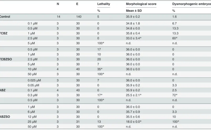
Table 2. Effects of TCBZ and TCBZSO in the zebrafish embryo test.
N E Lethality Morphological score Dysmorphogenic embryos
% Mean±SD %
Control 14 140 5 35.9±0.2 1.6
0.1μM 3 30 0 34.8±1.8 6.7
0.5μM 3 30 0 34.8±0.0 13.3
TCBZ 1μM 3 30 0 35.8±0.4 13.3
2.5μM 3 30 0 33.0±3.4* 60*
5μM 3 30 100* n.d. n.d.
0.5μM 3 30 17 36.0±0.0 0
1μM 3 30 10 36.0±0.0 0
TCBZSO 2.5μM 3 30 20 36.0±0.0 0
5μM 3 30 7 36.0±0.0 0
10μM 4 40 35* 36.0±0.0 0
50μM 3 30 100* n.d. n.d.
0.025μM 3 30 7 36.0±0.0 0
0.05μM 3 30 0 35.9±0.2 3.3
ABZ 0.1μM 4 40 0 35.9±0.2 2.5
0.3μM 3 30 17* 25.5±2.1* 72*
0.5μM 3 30 100* n.d. n.d.
1μM 3 30 0 36.0±0.0 0
6μM 3 30 0 35.7±0.9 3.3
ABZSO 12μM 3 30 0 35.5±0.6 10
25μM 3 31 13 18.0±0.0* 100*
50μM 3 30 100* n.d. n.d.
Number of independent experiments (N), total number of embryos (E), percentage of lethality, total morphological score and percentage of dysmorphogenesis obtained in each concentration group. SD: standard deviation, n.d.: not determined
*: p<0.05.
dysmorphogenesis, was also observed for both compounds, being of 100% in the case of ABZSO (S3 Table).
To sum up, the ZFET results showed that TCBZ exposure until 50 hpf produced a signifi-cant effect in embryonic differentiation and in lethality at concentrations 2.5 and 5μM, respec-tively. On the other hand, TCBZSO exposure did not produce any effects in embryonic growth or differentiation, but produced a significant embryolethal effect during the first 24 h of expo-sure at relevantin vivoconcentrations, starting at 10μM, and therefore rising a concern about the embryotoxic potential of TCBZSO during the first stages of development.
Effects of TCBZ and TCBZSO in the rodent preimplantation whole
embryo culture
To confirm if embryolethal effects of TCBZSO observed in zebrafish embryos were also rele-vant for mammalian embryos, a 96 h mouse embryo preimplantation culture, checking embry-onic development every 24 h, was performed. In this case, the analysis of TCBZ and TCBZSO was performed via blind studies, and ABZ and ABZSO were not used as positive controls due to the absence of previous studies analysing the effects of these compounds during the rodent preimplantation periodin vitro.
TCBZ exposure did not produce any significant effect in preimplantation embryos at 3μM (Table 3). Nonetheless, it produced a significant increase in the percentage of lethality of the highest studied concentration group (LOAEC = 10μM), which started after only 24 h of exposure.
TCBZSO did not induce adverse effects at 3μM. Starting at 10μM (LOAEC), TCBZSO pro-duced a significant concentration-dependent lethal effect, which was already evident during the first 24 h of exposure. At 30μM, which is a concentration still relevant after parasitosis treatment in humans, TCBZSO produced lethality at higher rates than 50% during the first 48 h of culture.
Consequently, the preWEC results confirmed a strong embryotoxic potential of TCBZSO also in mammalian embryosin vitro(For a graphical overview on the results of TCBZSO in the threein vitrotechniques used, seeFig. 1).
Table 3. Effects of TCBZ and TCBZSO in the rodent preimplantation whole embryo culture.
N E 2-cells 24 h 4-cells 48 h Morula 72 h Blastocyst 96 h
% % % %
Control 4 48 2.1 4.2 6.3 22.9
1μM 4 48 4.2 8.3 8.3 18.8
TCBZ 3μM 4 48 6.3 6.3 10.4 20.8
10μM 3 49 18.4* 24.5* 24.5* 36.7
3μM 3 48 4.2 14.6 16.7 31.3
TCBZSO 10μM 3 49 24.5* 30.6* 36.7* 55.1*
30μM 3 49 24.5* 79.6* 95.9* 98*
100μM 1 15 100* 100* 100* 100*
Number of independent experiments (N), total number of embryos (E), percentage of lethality in every developmental stage for each concentration group. *: p<0.05
Discussion
Fascioliasisandparagonimiasisare parasitic infections caused by flatworms or flukes. They are classified within the foodborne trematodiases group, which is included among the major ne-glected tropical diseases. Millions of people affected by these parasitoses are in need of effective and safe therapies, and among them women at childbearing age represent an important portion of population. Direct exclusion of all these women from massive treatment programs to avoid exposure during pregnancy would prevent their access to treatment for a large proportion of their reproductive lives. Thus, it is of uppermost importance to correctly characterize the risks
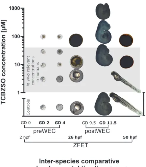
Fig 1. Graphical summary comparing TCBZSO results across species and developing time. Representative pictures of rodent embryos exposed to increasing concentrations of TCBZSO from GD 0 to GD 4 in the preWEC culture (LOAEClethality = 10μM) and from GD 9.5 to GD 11.5 in the postWEC culture
(LOAECdysmorphogenesis = 666μM). Zebrafish embryos were exposed to TCBZSO from 2 hpf to 50 hpf, a
developmental period comprising the stages covered by both rodent cultures. No dysmorphogenesis were observed (maximum concentration tested = 50μM), but TCBZSO was embryolethal during the first 24 h of
culture (LOAEClethality = 10μM). Pictures correspond to the developmental time points marked in bold in the
x-axis. PreWEC embryos pictures are 10 times magnified respect to postWEC and zebrafish embryos pictures. Scale bar: 4 mm for postWEC and zebrafish embryos; and 400μm for preWEC embryos. ZFET:
zebrafish embryo test (concentrations = 0, 5, 10 and 50μM); preWEC: preimplantation whole embryo
culture (concentrations = 0, 3, 10, 30 and 100μM); postWEC: postimplantation whole embryo culture
(concentrations = 0, 27, 267 and 666μM); GD: gestational day; hpf: hours post-fertilization.
associated with TCBZ treatment during pregnancy, and compile the maximum information on concentrations of the same order of magnitude as the clinically relevant ones, to identify if there are developmental periods where the risk associated with the exposure is lower than in others.
In vivo, TCBZ concentrations found in plasma after its administration are very low due to its rapid metabolism [22]. For this reason it is also important to study the effects of its main metabolite TCBZSO, which achieves higher concentrations in plasma. For example in rabbits, the TCBZSO maximum plasma concentration (Cmax) after a single 10 mg/kg TCBZ dose is
33μM (transformed toμM from [40]). Apart from the data coming from laboratory animals,
several studies have evaluated the relevant concentrations after TCBZ therapeutical adminis-tration to food producing animals and humans. In sheep, TCBZ doses of 10 mg/kg resulted in 37.26μM Cmaxof TCBZSO (calculated from [41]), and in cows after a 12 mg/kg dose, the Cmax
was 71.34μM (calculated from [39]). In humans, after a therapeutical dose of 10 mg/kg TCBZ, the TCBZ Cmaxwas 1.16μM ([42] and reviewed by [22]) and the TCBZSO Cmaxwas 26.6μM
(calculated from [43]) or between 15.8μM and 38.6μM depending on the concomitant food intake situation ([44] and reviewed by [22]).In vivorelevant concentrations of ABZ and ABZSO in food producing animals and in humans have recently been reviewed by Eckardt [16].
In our studies with rodent embryos exposed during the postimplantation period, the terato-genic potential of TCBZ was approximately 250 times lower than the potential of ABZ
(Table 4). Both sulfoxide metabolites had less teratogenic potential than their respective parent compounds, correlating with previous observations with other benzimidazolic derivatives [16, 17]. In this case, TCBZSO was 2.5 times less potent than its parent compound (Table 4). Even if it occurred at very different concentration ranges, the four tested compounds induced abnor-mal head and abnorabnor-mal branchial bars in rodent embryos exposed during organogenesis. These adverse effects are characteristic of developmental exposure to benzimidazoles, being the second effect also representative of other azolic derivatives like triazoles [45]. Besides these al-terations, ABZ and ABZSO exposed embryos exhibited very evident subcutaneous blisters in the facial laterals, abnormal caudal part and abnormal heart (S1 Table). Although TCBZ and TCBZSO have the ability of producing the same dysmorphogenesis as other compounds of their family, these effects occur at concentrations much higher than those achievedin vivoin
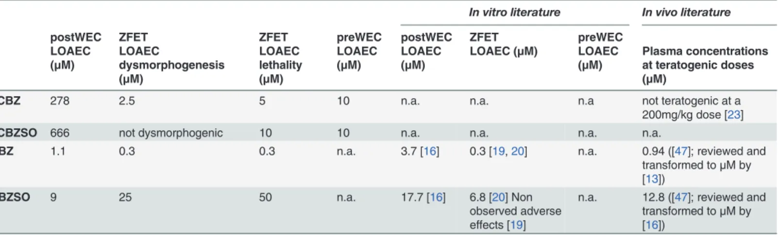
Table 4. Summary of LOAEC values of rodent and zebrafish embryos exposed to TCBZ, TCBZSO, ABZ and ABZSO during 48 h in comparison to the availablein vitroandin vivoliterature.
In vitro literature In vivo literature
postWEC ZFET ZFET preWEC postWEC ZFET preWEC
LOAEC (μM)
LOAEC
dysmorphogenesis (μM)
LOAEC lethality (μM)
LOAEC (μM)
LOAEC (μM)
LOAEC (μM) LOAEC (μM)
Plasma concentrations at teratogenic doses (μM)
TCBZ 278 2.5 5 10 n.a. n.a. n.a not teratogenic at a
200mg/kg dose [23]
TCBZSO 666 not dysmorphogenic 10 10 n.a. n.a. n.a. n.a.
ABZ 1.1 0.3 0.3 n.a. 3.7 [16] 0.3 [19,20] n.a. 0.94 ([47]; reviewed and
transformed toμM by
[13])
ABZSO 9 25 50 n.a. 17.7 [16] 6.8 [20] Non
observed adverse effects [19]
n.a. 12.8 ([47]; reviewed and transformed toμM by
[16])
LOAEC: lowest observed adverse effect concentration, n.a.: data not available.
humans after parasitosis treatment, indicating that neither TCBZ nor TCBZSO have teratogen-ic potential at the actual recommended therapeutteratogen-ical doses.
The results of the zebrafish embryo test reproduced those of the postWEC concerning dys-morphogenic potential. Again, ABZ was more potent than ABZSO at concentrations in good agreement with the published literature (0.3μM), and TCBZ was more potent than TCBZSO which was not dysmorphogenic at all (Table 4). However, TCBZSO concentrations causing embryolethality were five times lower than ABZSO concentrations killing the embryos or 2.5 times lower than concentrations producing malformations (Table 4). These results show no dysmorphogenic activity but point to a remarkable embryotoxic potential of TCBZSO. In our test, compound exposure started at 2 hpf, leaving the very first stages of development untreat-ed. As previous studies observed higher sensitivity to ABZ during the first stages of develop-ment, it cannot be excluded that TCBZSO has higher embryotoxicity when exposure starts at 0 hpf. After 48 h of exposure, the observed effects for all compounds in the zebrafish embryos were of general toxicity, as for example cardiac oedema or decreased pigmentation and there were no evident characteristic dysmorphogenesis for each one. Other studies working with zeb-rafish and azolic derivatives showed that the ZFET could correctly classify the potency of the compounds but could not reproduce thein vivoobserved specific dysmorphogenesis [45].
The differences in embryolethality observed in the ZFET and the postWEC TCBZSO results could be due to 1) inter-species differences in susceptibility to the compound or in compound availability, or to 2) different susceptibility of the early and middle developmental periods after exposure to TCBZSO, as the ZFET covers earlier developmental stages than the postWEC (Fig. 1).
To distinguish between these two options, another rodent culture experiment was per-formed with TCBZ and TCBZSO but in this case covering only the earliest period of develop-ment (from GD 0 to GD 4). And indeed, after only 24 h of exposure TCBZSO caused a significant increase in lethality at concentrations as low as 10μM, thus confirming the rele-vance of the high embryotoxic potential of TCBZSO in mammals. Previously published mecha-nistic effects of this compound like protein synthesis inhibition or microtubule inhibition in fluke vitelline cells [46] could be related to TCBZSO effects in preimplantation embryos, but further studies need to be done to elucidate the exact mechanisms by which TCBZSO exerts its embryotoxicity in mammals. Besides, studies exposing pregnant rodent dams during the pre-implantation period are required to confirm the stage-dependent embryotoxic potential of TCBZSOin vivo. On the other hand, TCBZ only increased lethality in preimplantation embry-os, at concentrations 10 times higher than thein vivorelevant ones.
From our results it can be concluded that TCBZ, at concentrations of the same order of magnitude as those achieved after intake of the recommended treatment doses forfascioliasis andparagonimiasis, does not entail relevant dysmorphogenic potentialin vitroduring the or-ganogenesis period, but its first metabolite TCBZSO has a high embryotoxic capacityin vitro during the preimplantation stage.
Supporting Information
S1 Table. Frequency (%) of dysmorphogenesis observed in postWEC experiments.
(DOCX)
S2 Table. Description of the abnormalities observed in the embryos cultured using the postWEC technique.
S3 Table. Frequency (%) of dysmorphogenesis observed in ZFET experiments.
(DOCX)
Author Contributions
Conceived and designed the experiments: NB PO EI JML MB. Performed the experiments: NB ET MVC MB. Analyzed the data: NB ET MVC EI JML. Wrote the paper: NB MB.
References
1. Hopkins DR. Homing in on helminths. Am J Trop Med Hyg. 1992; 46: 626–634. PMID:1535760 2. World Health Organization. Report of the WHO Expert Consultation on Foodborne Trematode
Infec-tions and Taeniasis/Cysticercosis. 2011. Available:http://www.who.int/neglected_diseases/ preventive_chemotherapy/WHO_HTM_NTD_PCT_2011.3.pdf. Accessed 25 November 2014. 3. Fairweather I. Triclabendazole progress report, 2005–2009: an advancement of learning? J Helminthol.
2009; 83: 139–150. doi:10.1017/S0022149X09321173PMID:19366485
4. Ofori-Adjei D, Dodoo ANO, Appiah-Danquah A, Couper M. A review of the safety of niclosamide, pyran-tel, triclabendazole and oxamniquine. Int J Risk Saf Med. 2008; 20: 113–122.
5. World Health Organization. Report of the WHO Informal Meeting on use of triclabendazole in fasciolia-sis control. 2007. Available:http://www.who.int/neglected_diseases/preventive_chemotherapy/WHO_ CDS_NTD_PCT_2007.1.pdf. Accessed 25 November 2014.
6. Ministerio de Salud y Desarrollo Social. Junta revisora de productos farmaceuticos, Boletín n.41 del Ministerio de Salud y Desarrollo Social. 2004. Available:www.caveme.org/documentos/Boletin%2041. pdf. Accessed 24 November 2014.
7. Agence Nationale de Sécurité du Médicament et des produits de santé. Available:http://agence-prd. ansm.sante.fr/php/ecodex/frames.php?specid=67258794&typedoc=R&ref=R0197475.htm. Accessed 24 November 2014.
8. Richard-Lenoble D, Chandenier J, Duong TH. Antiparasitic treatments in pregnant women and in chil-dren in 2003. Med trop (Mars). 2003; 63: 491–497. PMID:14763305
9. Hurtt ME, Cappon GD, Browning A. Proposal for a tiered approach to developmental toxicity testing for veterinary pharmaceutical products for food-producing animals. Food Chem Toxicol. 2003; 41: 611– 619. PMID:12659713
10. Teruel MT, Felipe AE, Solana HD, Sallovitz JM, Lanusse CE. Placental and fetal toxicity of albendazole sulphoxide in wistar rats. Vet Hum Toxicol. 2003; 45: 131–136. PMID:12776788
11. Cristòfol C, Navarro M, Franquelo C, Valladares JE, Carretero A, Ruberte J, et al. Disposition of Netobi-min and Albendazole, and its metabolites in the pregnant rat: Developmental Toxicity. Toxicol Appl Pharmacol. 1997; 144: 56–61. PMID:9169069
12. Mantovani A, Ricciardi C, Stazi AV, Macri C. Effects observed on gestational day 13 in rat embryos ex-posed to albendazole. Reprod Toxicol. 1995; 9: 265–273. PMID:7579911
13. Capece BP, Navarro M, Arcalis T, Castells G, Toribio L, Perez F, et al. Albendazole sulphoxide enantio-mers in pregnant rats embryo concentrations and developmental toxicity. Vet J. 2003; 165: 266–275. PMID:12672373
14. El-Makawy A, Radwan HA, Ghaly IS, El-Raouf AA. Genotoxical, teratological and biochemical effects of anthelmintic drug oxfendazole Maximum Residue Limit (MRL) in male and female mice. Reprod Nutr Dev. 2006; 46: 139–156. PMID:16597420
15. Yoshimura H. Effect of oral dosing vehicles on the developmental toxicity of flubendazole in rats. Reprod Toxicol. 2003; 17: 377–385. PMID:12849847
16. Eckardt K, Kaltehäuser J, Kilb C, Seiler A, Stahlmann R. Relative potency of albendazole and its sulfox-ide metabolite in twoin vitrotests for developmental toxicity: The rat whole embryo culture and the mouse embryonic stem cell test. Reprod Toxicol. 2012; 34: 378–384. doi:10.1016/j.reprotox.2012.05. 037PMID:22652462
17. Whittaker SG, Faustman EM. Effects of albendazole and albendazole sulfoxide on cultures of differenti-ating rodent embryonic cells. Toxicol Appl Pharmacol. 1991; 109: 73–84. PMID:2038752
18. Whittaker SG, Faustman EM. Effects of benzimidazole analogs on cultures of differentiating rodent em-bryonic cells. Toxicol Appl Pharmacol. 1992; 113: 144–151. PMID:1553749
20. Mattson A, Ullerås E, Patring J, Oskarsson A. Albendazole causes stage-dependent developmental toxicity and is deactivated by a mammalian metabolization system in a modified zebrafish embryotoxi-city test. Reprod Toxicol. 2012; 34: 31–42. doi:10.1016/j.reprotox.2012.02.007PMID:22414603 21. Köhler P. The biochemical basis of anthelmintic action and resistance. Int J Parasitol. 2001; 31: 336–
345. PMID:11400692
22. Keiser J, Engels D, Büscher G, Utzinger J. Triclabendazole for the treatment of fascioliasis and parago-nimiasis. Expert Opin Investig Drugs. 2005; 14: 1513–1526. PMID:16307491
23. Yoshimura H. Teratogenic evaluation of triclabendazole in rats. Toxicology. 1987; 43: 283–287. PMID: 3824395
24. World Health Organization. Toxicological evaluation of certain veterinary drug residues in food: Tricla-bendazole. 1993. Available:http://www.inchem.org/documents/jecfa/jecmono/v31je05.htm. Accessed 24 November 2014.
25. CDC, Centers for Disease Control and Prevention. Available:http://www.cdc.gov/dpdx/fascioliasis/tx. html. Accessed 30 September 2014.
26. Hennesy DR, Lacey E, Steel JW, Prichard RK. The kinetics of triclabendazole disposition in sheep. J Vet Pharmacol Ther. 1987; 10: 64–72. PMID:3586125
27. Mestorino N, Formentini EA, Lucas MF, Fernandez C, Modamio P, Hernández EM, et al. Pharmacoki-netic disposition of triclabendazole in cattle and sheep; discrimination of the order and the rate of the absorption process of its active metabolite triclabendazole sulfoxide. Vet Res Commun. 2008; 32: 21– 33. PMID:17457687
28. Piersma AH. Invittox Protocol No. 123: Embryotoxicity Testing in Post-Implantation Embryo Culture-Method of Piersma. 2002.
29. Flick B, Klug S. Whole embryo culture: an important tool in developmental toxicology today. Curr Pharm Des. 2006; 12: 1467–1488. PMID:16611129
30. Nagel R. DarT: The embryo test with the zebrafish Danio rerio: a general model in ecotoxicology and toxicology. ALTEX. 2002; 19: 37–48. PMID:12200112
31. Organization for Economic Cooperation and Development. Guideline for the testing of chemicals, Sec-tion 2. Test No. 236 Fish Embryo Acute Toxicity (FET) test. July 26, 2013. Available: http://www.oecd-ilibrary.org/environment/test-no-236-fish-embryo-acute-toxicity-fet-test_9789264203709-en. Accessed 2015 March 7.
32. Teixidó E, Gómez-Catalán J, Piqué E, Llobet JM. Assessment of developmental delay in the zebrafish embryo teratogenicity assay. Toxicol In Vitro. 2013; 27: 469–478. doi:10.1016/j.tiv.2012.07.010PMID: 22898132
33. Longo M, Zanoncelli S, Colombo PA, Harhay MO, Scandale I, Mackenzie C, et al. Effects of the benz-imidazole anthelmintic drug flubendazole on rat embryosin vitro. Reprod Toxicol. 2013; 36: 78–87. doi: 10.1016/j.reprotox.2012.12.004PMID:23287076
34. Barenys M, Flick B, Boix N, Almeida B, Joglar J, Klug S, et al. Effects of MDMA (ecstasy) and two of its metabolites on rat embryosin vitro. Reprod Toxicol. 2012; 34: 57–65. doi:10.1016/j.reprotox.2012.02. 001PMID:22391229
35. Klug S, Lewandowski C, Neubert D. Modification and standardization of the culture of early postimplan-tation embryos for toxicological studies. Arch Toxicol. 1985; 58: 84–88. PMID:4091661
36. Flick B, Talsness CE, Jäckh R, Buesen R, Klug S. Embryotoxic potential of Nmethyl-pyrrolidone (NMP) and three of its metabolites using the rat whole embryo culture system. Toxicol Appl Pharmacol. 2009; 237: 154–167. doi:10.1016/j.taap.2009.02.024PMID:19281833
37. Biggers J, McGuinnis LK, Raffin M. Amino acids and preimplantation development of the mouse in the protein-free KSOM. Biol Reprod. 2000; 63: 281–293. PMID:10859270
38. Chatot CL, Ziomek CA, Bavister BD, Lewis JL, Torres I. An improved culture medium supports develop-ment of random-bred 1-cell mouse embryos in vitro. J Reprod Fertil. 1989; 86: 679–688. PMID: 2760894
39. Imperiale F, Ortiz P, Cabrera M, Farias C, Sallovitz JM, Iezzi S, et al. Residual concentrations of the flu-kicidal compound triclabendazole in dairy cows’milk and cheese. Food Addit Contam Part A Chem Anal Control Expo Risk Assess. 2011; 28: 438–445. doi:10.1080/19440049.2010.551422PMID: 21337234
40. Alvarez-Bujidos ML, Ortiz AI, Negro A, Cubría JC, Ordóñez D. Pharmacokinetics of Triclabendazole in rabbits. Comp Biochem Physiol C. 1993; 106: 805–808. PMID:7905812
42. Bogan JA, Kinabo LD, Strong MB, Formica C, Galtier P, Alvinerie M. Pharmacokinetics of triclabenda-zole in cattle, sheep, goats, horses, ponies, donkeys, pigs and man. Proceedings of the 4thCongress of the European Association for veterinary pharmacology and toxicology, University of Veterinary Sci-ence, Budapest, Hungary. 1988;159–163.
43. El-Tantawy WH, Salem HF, Mohammed Safwat NA. Effect of Fascioliasis on the pharmacokinetic pa-rameters of triclabendazole in human subjects. Pharm World Sci. 2007; 29: 190–198. PMID:17265093 44. Lecaillon JB, Godbillon J, Campestrini J, Naquira C, Miranda L, Pacheco R, et al. Effect of food on the
bioavailability of triclabendazole in patients with fascioliasis. Br J Clin Pharmacol. 1998; 45: 601–604. PMID:9663817
45. de Jong E, Barenys M, Hermsen SA, Verhoef A, Ossendorp BC, Bessems JG, et al. Comparison of the mouse Embryonic Stem cell Test, the rat Whole Embryo Culture and the Zebrafish Embryotoxicity Test as alternative methods for developmental toxicity testing of six 1,2,4-triazoles. Toxicol Appl Pharmacol. 2011; 253: 103–111. doi:10.1016/j.taap.2011.03.014PMID:21443896
46. Stitt AW, Fairweather I. Fasciola hepatica: disruption of the vitelline cells in vitro by the sulphoxide me-tabolite of triclabendazole. Parasitol Res. 1996; 82: 333–339. PMID:8740549