Hemispatial Neglect during a Visual Search Task
Laura M. Jelsone-Swain*¤a
, David V. Smith¤b, Gordon C. Baylis¤c Department of Psychology, University of South Carolina, Columbia, South Carolina, United States of America
Abstract
Patients with hemispatial neglect exhibit a myriad of profound deficits. A hallmark of this syndrome is the patients’ absence of awareness of items located in their contralesional space. Many studies, however, have demonstrated that neglect patients exhibit some level of processing of these neglected items. It has been suggested that unconscious processing of neglected information may manifest as a fast denial. This theory of fast denial proposes that neglected stimuli are detected in the same way as non-neglected stimuli, but without overt awareness. We evaluated the fast denial theory by conducting two separate visual search task experiments, each differing by the duration of stimulus presentation. Specifically, in Experiment 1 each stimulus remained in the participants’ visual field until a response was made. In Experiment 2 each stimulus was presented for only a brief duration. We further evaluated the fast denial theory by comparing verbal to motor task responses in each experiment. Overall, our results from both experiments and tasks showed no evidence for the presence of implicit knowledge of neglected stimuli. Instead, patients with neglect responded the same when they neglected stimuli as when they correctly reported stimulus absence. These findings thus cast doubt on the concept of the fast denial theory and its consequent implications for non-conscious processing. Importantly, our study demonstrated that the only behavior affected was during conscious detection of ipsilesional stimuli. Specifically, patients were slower to detect stimuli in Experiment 1 compared to Experiment 2, suggesting a duration effect occurred during conscious processing of information. Additionally, reaction time and accuracy were similar when reporting verbally versus motorically. These results provide new insights into the perceptual deficits associated with neglect and further support other work that falsifies the fast denial account of non-conscious processing in hemispatial visual neglect.
Citation:Jelsone-Swain LM, Smith DV, Baylis GC (2012) The Effect of Stimulus Duration and Motor Response in Hemispatial Neglect during a Visual Search Task. PLoS ONE 7(5): e37369. doi:10.1371/journal.pone.0037369
Editor:Nicole Wenderoth, Katholieke Universiteit Leuven, Belgium
ReceivedDecember 31, 2011;AcceptedApril 21, 2012;PublishedMay 25, 2012
Copyright:ß2012 Jelsone-Swain et al. This is an open-access article distributed under the terms of the Creative Commons Attribution License, which permits unrestricted use, distribution, and reproduction in any medium, provided the original author and source are credited.
Funding:This work was supported by the National Institutes of Health (GCB: NS042047) and by intramural funding from the University of South Carolina. The funders had no role in study design, data collection and analysis, decision to publish, or preparation of the manuscript.
Competing Interests:The authors have declared that no competing interests exist.
* E-mail: [email protected]
¤a Current address: Department of Radiology, University of Michigan, Ann Arbor, Michigan, United States of America
¤b Current address: Department of Psychology and Neuroscience, Duke University, Durham, North Carolina, United States of America
¤c Current address: Administrative Council in Research and Research Foundation, University of Western Kentucky, Bowling Green, Kentucky, United States of America
Introduction
Hemispatial visual neglect is a syndrome of attention deficit that frequently occurs after unilateral brain damage, such as from stroke. Patients with neglect are unaware of, or unresponsive to, information in the side opposite their damage (referred to as the contralesional side) [1]. For example, they may shave only one side of their face or color only the ipsilesional side of a picture. Despite the obvious behavioral impairments of visual neglect, patients with this disorder often display marked anosognosia [2] (i.e., they appear to lack awareness of their inattention).
Although people with neglect may lack explicit awareness of information in their contralesional space, it is well established that unconscious processing of this visual field can take place. This phenomenon has been repeatedly demonstrated across different modalities and behavioral functions [3–14]. For example, one highly cited study described a patient with left-sided neglect who was shown two drawings of the same house, however the left side of one of these houses was in flames [10]. When asked to choose the house she would prefer to live in, she consistently chose the
non-burning house while claiming that both were identical. Even though such examples like this are common, the mechanistic underpinnings behind unconscious processing of neglected in-formation are still under debate.
Laeng and colleagues examined the fast denial theory in a single patient case study [16]. The results of this study did not support the fast denial theory because their patient was faster to neglect stimuli than to detect them. A major difference between the studies conducted by Mijovic´-Prelec [15] and Laeng [16] was that one required a verbal response and the other a motoric button-press. Therefore, interpretation of differences from the results between these two case studies is limited given that the measure of reaction time is based on different methods of reporting, and both studies examined behavior from only one person.
Motor and higher level visual systems are directly influenced by each other. For example, visual feedback is necessary to guide movements such as reaching. Mattingley et al. [17] demonstrated a relationship between motor planning and visual awareness in neglect patients. It was concluded that this relationship stems from functioning of the inferior parietal lobe rather than from the frontal lobe, as was previously thought. It can be inferred from this study that dual functioning of the inferior parietal lobe, an area commonly damaged in neglect patients [18–20], might result in altered visual search abilities when a motor response is required. Of course, lateralized motor responses by neglect patients during visual search tasks and exploration can be influenced by visual information [21,22,23]. However, it is not known how motor responses are affected when compared to verbal responses during visual search processes if non-lateralized motor movements are required.
The underlying purpose of our study was to examine the theory of fast denial in a group of patients with neglect. We tested this theory with two different manipulations. First, we aimed to compare verbal and motor responses during a visual search task. To the best of our knowledge, no study to date has directly examined neglect behavior between these two response types with a neglect patient group. In addition to addressing response modality, as this may have contributed to differences seen between previous studies [15,16], motor and verbal responses were compared to test for the possible interchangeability between these two methods in future studies.
Our second aim was to examine duration effects in a visual search task. Therefore we compared responses during two visual search tasks when the target either remained in the visual field or was displayed for only several hundred milliseconds. Many visual search tasks display target stimuli for an unlimited amount of time in the visual field. However, we often experience brief intervals of information under normal conditions, and this brief presentation of information may affect unconscious processing differently in patients with neglect.
We conducted two experiments to test our aims. The main difference between experiments was that each stimulus in Experiment 1 remained on the computer screen until a response was made and in Experiment 2 each stimulus was displayed briefly. Also, the paradigm in Experiment 1 was similar to that designed by Mijovic´-Prelec et al. [15] in attempt to induce fast-denial responses, whereas the template of the visual search task was simplified in Experiment 2. All other factors remained the same, and a verbal and motor task was included in each experiment. Verbal and motor responding was not included within the same task to eliminate a possible interaction effect [23]. It was hypothesized that reaction times to neglected stimuli would be longer in Experiment 1 given the unlimited amount of time to process each stimulus, assuming that unconscious processing is taking place. It was also hypothesized that reaction times during the motor modality would be longer due to the hand-motor coordination needed to press the target button, and because of the
additional component of having to associate the chosen response (yes or no) with the appropriate button.
Methods
Ethics Statement
The Institutional Review Board at the University of South Carolina approved all procedures, and all participants provided written informed consent prior to their participation in any of the assessments or experiments.
2.1. Participants
A total of 28 participants were recruited in our study. All participants were without any visual impairment that could have affected their performance during the experiment, including hemianopia. Participants enrolled in the study on a full voluntary basis, with no monetary compensation for their time. Patients with neglect and control patients with parietal damage without neglect were recruited from Health South Rehabilitation Center (Colum-bia, South Carolina). Healthy control participants were recruited from the general community.
2.1.1. Participants with Hemispatial Neglect. Twelve patients with hemispatial neglect resulting from stroke were recruited for this study. These patients were without speech or ipsilateral motor impairment that would have impacted their reaction time in this study. Seven patients completed Experiment 1, of which three completed both the verbal and the motor task, resulting in five completed data sets for the motor and verbal task each (Mage-motor= 65.4, Mage-verbal= 70.4). Eight patients with
neglect participated in Experiment 2 (Mage-motor= 63.8, M age-verbal= 69.8). Two patients completed both tasks, resulting in five
data sets per task. Three patients were only able to complete one block in the motor task. All patients performed the experiment in their hospital rooms, except for patient#8 who was recruited as an outpatient participant. All patients were assessed for neglect symptoms using the diagnosis criteria discussed below. See Table 1 for neglect patient demographic information.
2.1.2. Participants with Parietal Lesions Without Neglect. This control group included 11 stroke patients with lesions in their parietal lobe, without history or current symptoms of neglect. This group was chosen as a control because parietal injury often leads to hemispatial neglect [24]. In Experiment 1, seven patients total participated (Mage-motor= 58.6, M age-ver-bal= 65.75). Three of these participated in both the motor and
verbal task, resulting in five data sets per task. In Experiment 2, seven patients total participated (Mage-motor= 64.8, M age-ver-bal= 56.66). Two of these completed both motor and verbal tasks,
and one participant in each task was only able to complete one block. Overall there were five data sets created in the motor task and four in the verbal task in Experiment 2. Three patients had right parietal damage in each experiment and all were right handed. All patients performed the experiment while in their hospital rooms.
2.1.3. Healthy Controls. Five healthy volunteers without any history of brain damage or head trauma were recruited. Five people participated in each task in Experiment 1 (Mage= 64.6).
Five people participated in Experiment 2; three completed both tasks resulting in four participants per task (Mage-motor= 64.25,
Mage-verbal= 65.25). All participants were right handed. 2.2. Neglect Diagnosis Criteria
midline, held their index fingers upright in each visual field, and wiggled their finger one at a time at four different visual angles. Each patient was asked to keep their eyes fixated at centerline and to report what side of their peripheral space they detected movement. A patient was considered to have neglect when they missed more than 30% of the stimuli presented in the contrale-sional hemispace. 2) Line-bisection test: Patients were asked to bisect a nine-inch horizontal line (on paper). This paper was positioned central to the patient’s midline. At least a 10% deviation toward the ipsilesional side (from the middle at 0%) suggested visual neglect. 3) Clock-drawing test: Patients were asked to manually write the numbers inside a circle (60 diameter) that was described to them as a ‘‘clock-face’’. A spatial bias toward the ipsilesional side indicated possible neglect, such that the majority of numbers were located within this space of the paper. All patients who were considered to have visual neglect and participated in the current study met at least two of these three criteria. No control participant displayed any symptoms suggestive of neglect behavior.
2.3. Protocol and Materials
2.3.1. Stimuli and Apparatus. A Cornea TFT LCD flat-screen monitor (flat-screen size: 14611 inches) was used to display each visual search task. The experimenter controlled the search task from a Hewlett Packard laptop that was connected to the monitor. The monitor was placed central to the patient’s midline, approximately three feet from their body. The search tasks were programmed in E-Prime, version 1.1 (Psychology Software Tools, INC).
2.3.2. Experiment 1 Paradigm. The paradigm of Experi-ment 1 closely replicated that of Mijovic´-Prelec, et al. [17] (see Figure 1). Each block contained 72 pseudo-randomly assigned (without replacement) trials, in which one or two blocks were completed for each task. The target, a black ‘‘X’’ (563 cm), appeared a third of the time in the left and right visual fields each, and a third of the time it did not appear. When present, the target appeared equally in a fixed location in the center of any one of the four quadrants. This stimulus followed 50 ms after fixation (exclamation point, 461 cm).
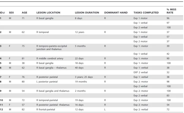
2.3.3. Experiment 2 Paradigm. There were only minor differences in the paradigm of Experiment 2 compared to Table 1.Demographic information, tasks performed, and percent of contralesional stimuli neglected in each task for all participants with visual neglect, in both Experiments 1 and 2.
ID# SEX AGE LESION LOCATION LESION DURATION DOMINANT HAND TASKS COMPLETED
% MISS RATE
1 M 71 R basal ganglia 8 days R Exp 1 motor 96
Exp 1 verbal 97
Exp 2 verbal 100
2 M 62 R temporal 12 years R Exp 1 motor 37
Exp 1 verbal 57
Exp 2 motor 37
3 F 75 R temporo-parieto-occipital junction and thalamus
5 months R Exp 1: motor 39
Exp 1 verbal 42
4 F 81 R middle cerebral artery 22 days R Exp 1: motor 90
5 M 38 R basal ganglia 18 days R Exp 1: motor 100
6 M 62 R basal ganglia – thalamus 40 days R Exp 1: verbal 38
EXP 2 verbal 32
7 F 76 R posterior parietal 3 years; 25 days R Exp 1: verbal 38
8 M 80 L posterior parietal 19 months R Exp 2: motor 98
Exp 2 verbal 100
9 M 54 R basal ganglia and thalamus 2 months R Exp 2: motor 100
Exp 2 verbal 83
10 M 72 R temporal-parietal 19 days R Exp 2: motor 100
11 F 57 R posterior parietal -thalamus 16 days R Exp 2: motor 54
12 M 82 R frontal-parietal 12 days L Exp 2: verbal 72
Age is in years, lesion duration is between the time of stroke and testing session, and dominant hand is self-reported. Lesion location is based on neurological medical report at time of admittance into Health-South Rehabilitation Center.
Patient 7 suffered from first stroke three years before but was readmitted to hospital 25 days prior to participation after suffering a second stroke. doi:10.1371/journal.pone.0037369.t001
Figure 1. Example of a single possible trial in Experiment 1 when a stimulus appeared in the upper right quadrant (the ipsilesional side for a patient with right hemisphere damage).
Experiment 1. First, the stimulus was only presented for a fixed duration. Second, the template design of the paradigm was simplified (see Figure 2) to help produce a pop-out effect. A circle fixation cue (3 cm in diameter) in the center of the screen prompted each trial. This circle flashed twice (50 ms duration each) and 50 ms later either the target (a 365 cm black X) would or would not appear (200 ms onset from trial initiation). The target appeared two-thirds of the time out of 72 trials (per block), and occurred equally in either the right or left visual field. All stimuli were presented pseudo randomly without replacement. Baylis et al [24] demonstrated that 400 ms was the average calibrated stimulus duration needed for patients with extinction, a disorder similar to neglect, to be capable of performing above chance in detecting flashing stimuli. Therefore we chose to display each stimulus for 300 milliseconds as the baseline for each participant during the practice session. This stimulus duration was checked to verify that detection of targets (in the ipsilesional field for neglect participants) was above chance in the neglect patient group and above 90% in the control groups. This duration time was too fast for neglect patient#12 and therefore adjusted to 700 ms.
2.3.4. Verbal Task. An E-Prime compatible microphone connected to a serial subject response box (Psychology Software Tools, INC.) was used. Reaction time for each trial was recorded as the time between stimulus onset and the participant’s response. Voice activation into the microphone terminated each trial. Each participant’s response was manually entered into the computer by the experimenter, who then started the next trial. Verbal responses were all unambiguous.
2.3.5. Motor Task. Two round plastic buttons (‘‘jelly-bean’’ switches), one red and one green (34 cm in diameter), were secured on top of a portable ‘‘lap desk’’, which sat comfortably on the participant’s lap. The jelly-bean buttons were placed vertically in front of the participant on their ipsilesional side to ensure no lateral confound was induced [25,26]. These buttons remained in-line with each participant’s ipsilesional arm, therefore not requiring the participant to make a lateral movement with their arm or hand. Both buttons were connected to the HP laptop by an E-Prime compatible serial response box (Psychology Software Tools, INC.). Reaction times were recorded as the time between stimulus onset and button press, and E-Prime recorded which
button was pressed for each trial. Trials were terminated when either button was pressed. Like in the verbal task, the experimenter controlled the onset of each new trial.
2.3.6. Procedure. Participants were told to respond to whether they saw the stimulus, that the stimulus was an X, and that it would either appear in the center of any of the four quadrants or not at all. Instructions were to verbally or motorically report the presence or absence of the stimulus in each trial, and to make their responses as quickly and accurately as possible. Each participant was given a 12-trial (equal number of all stimulus conditions) practice session before they began the experiment.
In the verbal task, participants held the microphone in a fixed position near their mouth throughout the session and were asked to speak either ‘‘yes’’ or ‘‘no’’ into the microphone for each trial. In the motor task, participants were instructed that the green button symbolized ‘‘yes’’ and that the red button symbolized ‘‘no.’’ Each participant was familiarized with the buttons to ensure they comprehended this instruction. The motor and verbal tasks were counterbalanced across participants. Button position (red vs. green in the upper position) was also counterbalanced across both participants and block sessions (for those who completed two blocks) in the motor task.
2.3.7. Statistical Analysis. It was our goal to compare motor and verbal responses in each experiment. Additionally, our goals were to measure neglect behavior to timed and untimed stimulus presentations during verbal and motor responses, and to assess if these measurements fit a fast denial model.
We used a linear mixed effects model to examine the effects of task (motor and verbal) and stimulus type (contralateral, ipsilateral, and absent stimuli conditions) on the outcome variable reaction time in each experiment. In the neglect patient group the contralateral stimulus condition was specific to targets presented in the contralesional space that were neglected and the ipsilateral condition was specific to correctly reporting stimulus presence in the ipsilesional space. Correctly responding to contralesional stimuli in the neglect group was not part of thea priorihypothesis, therefore was not included in the analyses. In the control groups, the contralateral stimulus condition was measured as the correct responses to stimulus presence. Therefore, a single statistical model combining all groups could not be performed. The left side was considered the healthy control group’s contralateral side given that all participants in this group were right handed.
The linear mixed effects model was conducted in Experiments 1 and 2 for each group, in order to account for correlated data across tasks that incorporated a mixture of independent and repeated subject samples. Task (verbal and motor) and stimulus type (contra, ipsi, and no) were included as the fixed effects measures. An interaction between task and stimulus was also included in the model but because no interactions were significant they will not be discussed further. The intercept was included as the random effect measure and stimulus type was also included as a repeated measures variable. Variance components heteroge-neous compound symmetry was chosen for the covariance structure and the restricted maximum likelihood estimation was used.
The account of a fast denial involves reacting to neglected and detected stimuli equally fast. According to this theory, this is because the neglected stimuli are detected in the same manner as the non-neglected stimuli, however this detection fails to reach conscious awareness. Because we were specifically interested in comparing reaction times to neglected and detected stimuli, we conducted paired sample t-tests for all stimulus condition comparisons within each experiment and task.
Figure 2. Example of a single possible trial in Experiment 2 when a stimulus appeared in the upper right quadrant (the ipsilesional side for a patient with right hemisphere damage).
Exploration of the raw reaction times (RT) of each participant in each condition indicated that RTs were positively skewed. Therefore, logarithmic (base 10) transformations of individual raw RTs in each condition per each participant were performed. All statistical tests measuring reaction time were analyzed using these transformed means. To allow for interpretation of the data, raw reaction times of the neglect group for each condition are presented at the individual level (see Tables 2 & 3). The raw data are shown using the trimmed means (top and bottom 2.5% removed), which is a robust measure of central tendency. All statistical results were derived from the statistical package SPSS, version 19.0.
Results
3.1. Accuracy: No Differences in Neglecting Between Motor and Verbal Tasks
A primary goal of our study was to examine if performance during a visual search task is affected by response modality or the duration of stimulus presentation. Looking specifically at responses made to stimuli presented in the contralesional field, patients with neglect consistently neglected these stimuli regardless of how they responded. The average miss rate in Experiment 1 for this group was 72.2% (SE = 14.1) and 54.3% (SE = 11.4) in the motor and verbal tasks, respectively. In Experiment 2, miss rate averages in the motor and verbal tasks were 77.4% (SE = 12.5) and 77.8% (SE = 13.5), respectively. Miss rate percentages of contralesional stimuli per neglect participant are presented in Table 1. All
Table 2.Trimmed means (top and bottom 2.5% removed) showing robust center of raw reaction times (ms) and standard errors (from non-trimmed raw data) for individual neglect patients and control groups in Experiment 1.
PARTICIPANTS MOTOR TASK VERBAL TASK
Neglect Patients CONTRA IPSI NO CONTRA IPSI NO
1
Mean 1769.135 1211.565 1535.190 1703.994 943.997 2091.050
SE 355.048 128.160 353.587 370.576 41.819 424.170
2
Mean 1343.537 1133.934 2337.262 925.856 793.566 1215.296
SE 156.924 121.897 329.152 106.248 23.875 103.122
3
Mean 3008.924 1753.527 3606.605 1785.208 1115.905 2502.639
SE 621.197 479.420 420.251 249.041 69.784 387.940
4
Mean 4913.120 2717.630 5591.963 NA NA NA
SE 384.152 260.881 535.240 NA NA NA
5
Mean 5277.773 1230.329 4395.593 NA NA NA
SE 695.645 34.885 487.075 NA NA NA
6
Mean NA NA NA 4494.778 1891.622 3697.188
SE NA NA NA 628.803 343.630 317.200
7
Mean NA NA NA 5803.852 4215.931 5956.571
SE NA NA NA 1272.814 720.356 689.287
GROUPS CONTRA IPSI NO CONTRA IPSI NO
Neglect Patients
Mean 3262.500 1609.400 3493.323 2942.737 1792.204 3092.549
SE 442.593 205.048 425.061 525.496 239.893 384.344
Parietal Controls
Mean 1087.605 978.351 1456.076 1064.739 894.559 1684.411
SE 73.558 48.104 135.852 72.992 57.474 183.062
Healthy Controls
Mean 742.707 753.757 863.273 670.270 661.677 713.022
SE 28.118 29.854 35.004 18.647 18.832 15.086
CONTRA (in the neglect group) = misses of contralesional targets; CONTRA (in the control groups) = hits of contralesional/lateral targets; IPSI = hits of ipsilesional/lateral targets; NO = correct response to target absence.
patients with neglect except for patient 10 correctly identified ipsilesional stimuli at least 80% of the time and correctly reported stimulus absence at least 90% of the time. Patient 10, who had severe neglect, missed stimuli in their ipsilesional field 62% of the time in Experiment 2 during the motor task. All control participants correctly responded with over 90% accuracy for all three stimuli conditions.
Non-parametric independent sample tests (Mann-Whitney) were performed to compare task and experiment neglect rates (of contralesional stimuli) after removal of repeated subjects. In order to conduct these analyses with independent samples, tasks
were compared including individuals in both experiments, and experiments were compared including individuals in both tasks. There was no difference in miss rates between task (U = 7, Z(11) =21.47,p= .14) or experiment (U = 9, Z(9) =2.25,p= .80). 3.2. Reaction Time
3.2.1. Participants with Neglect Were Equally Slow to Neglected and Absent Stimuli. Again our goals in this study were to test if hemispatial visual neglect is affected by methods of response and to further evaluate the fast denial theory. Therefore we examined the speed of reaction between tasks (verbal and Table 3.Trimmed means (top and bottom 2.5% removed) showing robust center of raw reaction times (ms) and standard errors (from non-trimmed raw data) for individual neglect patients and control groups in Experiment 2.
PARTICIPANTS MOTOR TASK VERBAL TASK
Neglect Patients CONTRA IPSI NO CONTRA IPSI NO
2
Mean 2934.592 980.015 3438.520 NA NA NA
SE 499.442 38.301 520.577 NA NA NA
8
Mean 3259.767 843.164 3007.937 1011.849 652.750 897.184
SE 433.989 168.764 581.518 229.861 140.402 284.673
9
Mean 1692.303 519.359 1567.687 4729.400 709.161 5198.750
SE 210.080 32.373 284.576 461.266 59.660 357.408
10
Mean 3979.310 993.357 6363.340 NA NA NA
SE 835.158 184.113 1293.320 NA NA NA
11
Mean 4843.680 1002.444 4621.804 NA NA NA
SE 760.965 84.061 784.236 NA NA NA
12
Mean NA NA NA 4739.556 1229.004 4372.722
SE NA NA NA 532.599 101.274 356.090
1
Mean NA NA NA 13033.170 816.956 8428.601
SE NA NA NA 2483.734 54.245 1467.167
6
Mean NA NA NA 7192.278 1336.133 8744.317
SE NA NA NA 954.701 154.672 811.444
GROUPS CONTRA IPSI NO CONTRA IPSI NO
Neglect Patients
Mean 3341.930 867.668 3799.858 6141.252 948.800 5528.314
SE 547.927 101.522 692.845 932.432 102.051 655.356
Parietal Controls
Mean 858.373 840.345 1848.351 680.813 640.078 1257.415
SE 52.516 44.438 284.024 42.896 44.190 121.804
Healthy Controls
Mean 666.188 675.952 922.055 673.226 645.031 989.698
SE 24.010 25.545 118.953 15.804 19.613 87.503
CONTRA (in the neglect group) = misses of contralesional targets; CONTRA (in the control groups) = hits of contralesional/lateral targets; IPSI = hits of ipsilesional/lateral targets; NO = correct response to target absence.
motor) and stimulus type (contralesional misses, ipsilesional hits, correct target absence responses) in both experiments using a linear mixed effects model. There was a significant effect of stimulus type in both Experiments 1 and 2, F(2, 8.58) = 38.49, p,.001, F(2, 8.40) = 27.15,p,.001, respectively. The motor task was slower in Experiment 1, F(1, 7.34) = 7.64, p= .03, but there was no task effect in Experiment 2,F(1, 7.88) = .73,p= .42.
Taking a closer look at the significant task effect in Experiment 1, it appears that this result may have been driven by the three subjects who participated in both tasks. Overall the group means are very similar between tasks (see Table 2), however participants 1, 2, and 3 were slower in the motor task. These participants contributed twice the marginal percentage in the mixed model compared to those who only participated in one task. Therefore a third linear mixed model was conducted to examine reaction times between the motor and verbal task after collapsing participants between experiments (removing repeated subjects who completed the same task; no subjects were repeated within the same group). This model yielded null results between tasks,F(1, 6.94) = .09,p= .78. Collapsing of participants in this analysis was not confounding as no differences were found between Experi-ments (see section 3.5).
To specifically assess the fast denial theory, paired sample t-tests were conducted for every a priori stimulus condition pair (e.g. contralesional misses versus ipsilesional hits) in each task and experiment. This analysis failed to support fast denials of neglected information. Instead, RTs of neglected and detected stimuli were significantly different in each task in both experiments, with patients being faster when detecting the ipsilesional stimulus. Additionally, reaction times between neglected and absent stimuli trials were not significantly different, indicating equal reaction times during these conditions. This finding was consistent across both tasks in each Experiment, and group means are depicted in Figure 3. Paired t-test statistical results are presented in Table 4.
3.2.2. Reaction Time for Control Participants. We tested for differences between stimuli and task type in both control groups using a linear mixed model. In the parietal-lesion patient group there was a significant main effect of stimulus type in both Experiments 1 and 2,F(2, 8.12) = 8.38,p= .01,F(2, 8.02) = 4.95, p = .04, respectively. Both experiments showed a null result for task type,F(1, 7.14) = .60,p= .47 andF(1, 5.70) = 1.27,p= .31.
Overall, results from the healthy control group were very similar to the parietal-lesion patient control group. In Experiment 1 there was a significant effect for stimulus type, F(2, 5.56) = 10.51,
p= .013, but not for task type, F(1, 4.86) = 3.82, p= .11. In Experiment 2 this group showed no significant effects of stimulus type,F(2, 6.71) = 4.16,p= .07 or task type,F(1, 4.99) = .02,p= .90. See Table 4 for paired t-test comparisons for both control groups.
3.3. Experiment 1 Compared to Experiment 2:
Participants With Neglect Respond Slower to Ipsilesional Stimuli in Experiment 1
To address the effects of stimulus duration in a visual search task we compared neglect patient reaction times in each stimulus condition between Experiments 1 and 2. Specifically, we were interested in comparing neglect patients’ responses to untimed versus timed stimuli. Because several participants completed both tasks within the same experiment, we analyzed between experi-ment effects by task. As in previous analyses, we conducted a linear mixed effects model to account for repeated subjects between experiments in the verbal condition. No differences were found between experiments,F(1, 5.54) = .12,p= .74, and again there was a stimulus effect,F(2, 6.38) = 16.57,p= .003.
Only one person was repeated between experiments in the motor task, therefore a linear mixed effects model could not be performed. This person was removed from the analysis and a repeated measures analysis of variance (ANOVA) test was conducted using experiment and stimulus type as the between and within subjects factors. There was no difference between experiments,F(1, 6) = .80,p= .40 but there was a significant main effect for stimulus,F(2, 12) = 50.97,p,.001.
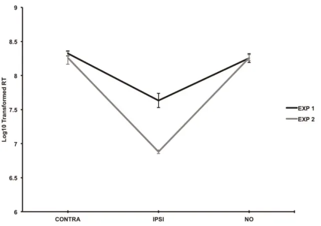
It was our a priori goal to examine duration effects for each stimulus condition (contra, ipsi, no) between Experiments 1 and 2 in the neglect patient group. Interestingly, results from this analysis showed that the only significant difference occurred between detected ipsilesional stimuli. Specifically, patients were significantly slower when responding to ipsilesional stimuli in Experiment 1 compared to Experiment 2 (see Figure 4). This core result was shown using independent sample t-tests after removal of repeated subjects, combining motor and verbal tasks in the same sample (t(7) = 3.85, p= .01). Also, when separating independent sample tests per motor and verbal task, the overall results of this analysis did not change in the motor task (t(6) = 2.92, p= .03). An individual test for the verbal task could not be conducted however because the sample size was too small. Reaction times were not significantly different between experiments when patients ne-glected contralesional stimuli or when they responded to absent stimuli trials (t(7) = .32,p= .76;t(7) =2.06,p= .96). Overall, these results consistently show a duration effect that was specific to detecting targets in the ipsilesional visual field.
Discussion
Our goal of this study was to investigate the effects of different response modalities and stimulus durations on hemispatial visual neglect behavior and unconscious processing. Specifically, we measured reaction time and accuracy when patients with neglect responded either motorically or verbally to timed and untimed stimuli presented in a visual search task. The theory of a fast denial account was assessed with these measurements. To the best of our knowledge, comparing verbal to non-lateralized motor responses in patients with visual neglect is a novel comparison.
4.1. Failed to Support the Fast Denial Theory of Unconscious Processing
4.1.1. Slow Responses to Neglected Stimuli. Overall, our results failed to support evidence for the behavior of unconscious processing based on the fast denial theory [15]. In fact, results from both experiments failed to show any evidence of unconscious processing when patients neglected stimuli in their contralesional visual field. Instead, the most reproducible finding across all four tasks was that all neglect patients responded to contralesional stimuli in the same manner as when the stimuli were truly absent, such that reaction times to neglected stimuli were the same as stimulus-absent trials.
pre-attention mechanism was initiated despite our effort to induce a pop-out effect (Experiment 2).
Attention is necessary at any level of information processing. Previous research has shown that facilitation of pre-attentive mechanisms during information processing in healthy individuals can occur, for example, by providing the context of a scene [27], grouping [28], and by providing a cue. Patients with neglect can actually increase their performance in detecting contralesional information with such facilitators [29–32], emphasizing that pre-attentive processing is important for contralesional information gathering (conscious or unconscious). It has been suggested that neglect results from specific attention impairments, such as the inability to orient to the contralesional space [30] or to disengage from the ipsilesional side [29].
It is very possible that our experiment design did not engage the necessary attention processes that would have resulted in overt unconscious processing behavior. It is also possible that the severity of neglect shared by patients within this group may have contributed to our findings. All patients met two out of the three criteria for assessing clinical symptoms of neglect, when in many cases only one of the assessments is needed to diagnose neglect disorder. It would therefore be expected that stronger pre-attentive cues would be needed to facilitate contralesional information gathering by patients with severe neglect, even at the subconscious level. Future studies should therefore attempt to disentangle the effects of pre-attentive mechanisms and unconscious processing in patients with varying degrees of hemispatial neglect.
4.1.2. Importance of Ipsilesional Information Processing. Most experiments conducted on unilateral neglect have focused on the properties and mechanisms of when patients
neglect information in their contralesional space. However, few studies have been interested in the behavior that occurs when patients are consciously aware of information in their ipsilesional space, even though it has been shown that ipsilesional information can influence neglect behavior [3]. An important finding from our study is that patients with neglect were significantly slower to respond to ipsilesional targets in Experiment 1 compared to Experiment 2. Again, the main difference between these experiments was that the stimulus remained on the computer screen in Experiment 1 but was presented for only a brief duration in Experiment 2.
Slower responses to ipsilesional targets by patients with neglect in Experiment 1 suggest that the only processing affected by stimulus duration in this study was during conscious detection of ipsilesional information. Thus, differences between reaction times of neglected and detected stimuli were not just a factor of neglect-specific behavior, but also a factor of when patients consciously detected a stimulus. This challenges the claim by Mijovic´-Prelec et al. [15] ‘‘…that the denied targets and the detected targets were processed in the same way…’’ (p. 157). In other words, our results from Experiments 1 and 2 suggest that different processes are in fact taking place when stimuli reaches awareness; and are influenced by whether or not the stimulus remains in the visual field.
4.2. General Consistency Across Motor and Verbal Tasks. Overall, reaction times for the response conditions (contra, ipsi, no) between the verbal and motor tasks did not differ in Experiment 2, but were slower in the motor task in Experiment 1. After collapsing participants across experiments however, no differences were found between tasks. It could be
Figure 3. Bar graphs showing equally slow responses to neglected and absent stimuli trials in the neglect group compared to control groups for each task in both experiments.Mean reaction times (based on the log10 transformed raw data) and standard errors for each group in each condition are shown. A: Experiment 1 motor task. B: Experiment 1 verbal task. C: Experiment 2 motor task. D. Experiment 2 verbal task. CONTRA (in the neglect group) = misses of contralesional targets; CONTRA (in the control groups) = hits of contralesional/lateral targets; IPSI = hits of ipsilesional/lateral targets; NO = correct responses to target absence.
suggested that priming a motoric system (without a lateral movement confound) may resemble the verbal system in patients with neglect. Even though this finding was against our hypothesis, the implications of this finding may be of invaluable importance to future studies. Specifically, it may be possible to use these two methods interchangeably when implementing a visual search task with this patient group. This could, therefore, increase the accessibility of patients with neglect who may be unable to make a verbal response (a common disability post stroke).
Still under debate is the prevalence of hemispatial visual neglect among patients with left hemisphere lesions. It is the typical
consensus that neglect most commonly occurs after damage to the right hemisphere [1,33,34], although it could also occur after left-sided damage [35–37]. Prevalence rates of right-left-sided neglect may be higher than what is reported, possibly influenced by language impairments commonly seen after damage to the left hemisphere. For example, the presence of aphasia may mask the expression or diagnosis of neglect in patients with left hemispheric injury.
Integration of motor-based responses into diagnostic or experimental paradigms may help alleviate some of the method-ological issues regarding evaluations of neglect patients with comorbid language impairment. It may also lead to better Table 4.Paired t-test results for logarithmic base 10 transformed reaction times in each condition-pair across both experiments in each group.
Tasks Neglect Patients Parietal Patient Controls Healthy Controls
t-value p-value t-value p-value t-value p-value
Experiment 1 Verbal
Contra vs. Ipsi 4.650 .010** 1.821 .143 .523 .629
Contra vs. No 2.949 .396 22.917 .043* 21.747 .156
Ipsi vs. No 28.510 .001** 22.995 .040* 21.944 .124
Mean St. Dev Mean St. Dev Mean St. Dev
Contra 7.697 .709 6.873 .394 6.583 .135
Ipsi 7.238 .590 6.739 .308 6.479 .169
No 7.773 .592 7.272 .408 6.548 .185
Experiment 1 Motor
Contra vs. Ipsi 3.029 .039* 1.704 .164 2.865 .436
Contra vs. No 2.936 .402 23.231 .032* 23.299 .030*
Ipsi vs. No 25.282 .006** 22.711 .053 22.688 .055
Mean St. Dev Mean St. Dev Mean St. Dev
Contra 7.720 .504 6.942 .279 6.583 .135
Ipsi 7.239 .308 6.852 .166 6.593 .150
No 7.833 .425 7.154 .395 6.761 .090
Experiment 2 Verbal
Contra vs. Ipsi 4.324 .012** 1.233 .285 2.112 .125
Contra vs. No 0.781 .478 21.531 .201 21.820 .166
Ipsi vs. No 23.990 .016** 21.552 .196 21.951 .146
Mean St. Dev Mean St. Dev Mean St. Dev
Contra 8.224 .875 6.442 .407 6.500 .182
Ipsi 6.780 .304 6.413 .390 6.451 .153
No 8.155 .913 6.838 .691 6.770 .445
Experiment 2 Motor
Contra vs. Ipsi 12.071 ,.001** .499 .644 2.269 .805
Contra vs. No 2.389 .717 23.921 .017* 22.240 .111
Ipsi vs. No 210.377 ,.001** 23.739 .020* 22.092 .128
Mean St. Dev Mean St. Dev Mean St. Dev
Contra 7.819 .365 6.701 .293 6.488 .087
Ipsi 6.720 .274 6.685 .296 6.450 .070
No 7.860 .460 7.224 .527 6.761 .309
CONTRA (in the neglect group) = misses of contralesional targets; CONTRA (in the control groups) = hits of contralesional/lateral targets; IPSI = hits of ipsilesional/lateral targets; NO = correct response to target absence.
**Indicates p-values corrected for multiple comparisons (Bonferroni, p,.016).
estimates of prevalence rates among left hemisphere/right-sided visual neglect. It is important however for future studies to further test motor and verbal responding methods and results before these assumptions can be validated.
4.3. Conclusions
Although our study did not elicit overt unconscious processing behavior, it cannot be definitively concluded that this type of processing did not take place. Behavioral evidence of unconscious processing has converged with neuroimaging results showing cortical activation to neglected information [34]. In one example using fMRI, a patient with comorbid visual extinction and neglect showed amygdala activity to unconsciously perceived fearful faces [38]. In a more recent study, Vuilleumier et al. [39] used fMRI to examine right and left retinotopic cortical activation in the occipital lobe when neglect patients viewed checkerboard images displayed in both hemifields. In this study, significant bi-lateral cortical activation occurred despite the failure to acknowledge these images in the neglected visual field. Therefore, future studies could benefit from the incorporation of neuroimaging techniques, which may better parse the characteristics and mechanisms behind unconscious processing in hemispatial neglect.
Collectively, our study failed to support the fast denial theory. Patients with neglect reported the same to neglected and absent stimuli, yet were affected by stimulus duration when detecting targets in the ipsilesional field. Additionally, our results suggest that patients with neglect sometimes behave similarly to visual information when they respond verbally or motorically (without
a lateral confound). This finding may be invaluable for accessing patients with verbal impairment either for diagnostic or experi-mental purposes. Using motor and verbal responses interchange-ably may also lead to better estimates of hemispatial neglect prevalence rates of those with left hemisphere damage. Given that this is a novel comparison between non-lateralized motor and verbal responding in patients with neglect, more studies need to be conducted to test this theory. Future studies also need to disentangle the effects of pre-attentive mechanisms and other factors that could affect unconscious processing along a spectrum of symptom severity. Last, incorporation of neuroimaging techniques in future studies could provide valuable information behind the mechanistic underpinnings of unconscious processing in hemispatial visual neglect.
Acknowledgments
Our appreciation is given to Chris Rorden for his time spent reviewing this manuscript and for his insightful comments and overall feedback. We also acknowledge Charlotte Laird for assisting in data collection and Iman Choucair for helping with data organization. We thank the staff at Health South Rehabilitation Center (Columbia, South Carolina) for their helpfulness, and importantly we thank all the participants who gave their time during the study.
Author Contributions
Conceived and designed the experiments: LMJ GCB. Performed the experiments: LMJ DVS. Analyzed the data: LMJ. Wrote the paper: LMJ. Funded project: GCB.
Figure 4. Graph showing reaction time difference in the neglect patient group between Experiments 1 and 2 when ipsilesional stimuli were detected.Data is taken from independent samples collapsed across the motor and verbal tasks. Mean reaction times (based on the log10 transformed raw data) and standard errors are shown. CONTRA = misses of contralesional targets; IPSI = hits of ipsilesional targets; NO = correct responses to target absence.
References
1. Bartolomeo P, Chokron S (2002) Orienting of attention in left unilateral neglect. Neuroscience and Biobehavioral Reviews 26: 217–234.
2. Bisiach E, Vallar G, Perani D, Papagno C, Berti A (1986) Unawareness of disease following lesions of the right hemisphere: anosognosia for hemiplegia and anosognosia for hemianopia. Neuropsychologia 24: 471–482.
3. Baylis GC, Simon-Dack SL, Greene K, Jelsone L, Rorden C (2004) The effect of ipsilesional cues on line-bisection errors: the importance of predictive value. Neuropsychologia 42: 175–182.
4. Berti A, Oxbury S, Oxbury J, Affanni P, Umilta C, et al. (1999) Somatosensory extinction for meaningful objects in a patient with right hemispheric stroke. Neuropsychologia 37: 333–343.
5. Cappelletti M, Cipolotti L (2006) Unconscious processing of Arabic numerals in unilateral neglect. Neuropsychologia 44: 1999–2006.
6. Driver J, Mattingley JB (1998) Parietal neglect and visual awareness. Nat Neurosci 1: 17–22.
7. Kanne SM (2002) The role of semantic, orthographic, and phonological prime information in unilateral visual neglect. Cogn Neuropsychol 19: 245–261. 8. Ladavas E, Shallice T, Zanella MT (1997) Preserved semantic access in neglect
dyslexia. Neuropsychologia 35: 257–270.
9. Maravita A (1997) Implicit processing of somatosensory stimuli disclosed by a perceptual after-effect. Neuroreport 8: 1671–1674.
10. Marshall JC, Halligan PW (1988) Blindsight and insight in visuo-spatial neglect. Nature 336: 766–767.
11. Rafal R, Ward R, Danziger S (2006) Selection for action and selection for awareness: evidence from hemispatial neglect. Brain Res 1080: 2–8. 12. Sackur J, Naccache L, Pradat-Diehl P, Azouvi P, Mazevet D, et al. (2008)
Semantic processing of neglected numbers. Cortex 44: 673–682.
13. Volpe BT, Ledoux JE, Gazzaniga MS (1979) Information processing of visual stimuli in an ‘‘extinguished’’ field. Nature 282: 722–724.
14. Vuilleumier P, Schwartz S, Husain M, Clarke K, Driver J (2001) Implicit processing and learning of visual stimuli in parietal extinction and neglect. Cortex 37: 741–744.
15. Mijovic´-Prelec D, Shin LM, Chabris CF, Kosslyn SM (1994) When does ‘‘no’’ really mean ‘‘yes’’? A case study in unilateral visual neglect. Neuropsychologia 32: 151–158.
16. Laeng B, Brennen T, Espeseth T (2002) Fast responses to neglected targets in visual search reflect pre-attentive processes: an exploration of response times in visual neglect. Neuropsychologia 40: 1622–1636.
17. Mattingley JB, Husain M, Rorden C, Kennard C, Driver J (1998) Motor role of human inferior parietal lobe revealed in unilateral neglect patients. Nature 392: 179–182.
18. Karnath HO, Rennig J, Johannsen L, Rorden C (2011) The anatomy underlying acute versus chronic spatial neglect: a longitudinal study. Brain 134: 903–912.
19. Karnath HO, Rorden C (2011) The anatomy of spatial neglect. Neuropsycho-logia.
20. Corbetta M, Shulman GL (2011) Spatial neglect and attention networks. Annu Rev Neurosci 34: 569–599.
21. Broeren J, Samuelsson H, Stibrant-Sunnerhagen K, Blomstrand C, Rydmark M (2007) Neglect assessment as an application of virtual reality. Acta Neurol Scand 116: 157–163.
22. Danckert J, Ferber S (2006) Revisiting unilateral neglect. Neuropsychologia 44: 987–1006.
23. Gore CL, Rodriguez PD, Baylis GC (2001) Deficits of motor intention following parietal lesions. Behavioral Neurology 13: 29–37.
24. Baylis GC, Gore CL, Rodriguez PD, Shisler RJ (2001) Visual extinction, the importance of binding dorsal and ventral visual pathways.Visual Cognition8: 359–379.
25. Bisiach E, Berti A, Vallar G (1985) Anological and logical disorders underlying unitaleral neglect of space. In: Posner MI, Marin OSM, eds. Attention and Performance. Hillsdale, NJ: Lawrence Erlbaum. pp 239–244.
26. Eskes GA, Butler B (2006) Using limb movements to improve spatial neglect: the role of functional electrical stimulation. Restor Neurol Neurosci 24: 385–398. 27. Palmer SE (1975) The effects of contextual scenes on the identification of
objects.Memory and Cognition3: 519–526.
28. Baylis GC, Driver J (1993) Visual attention and objects: evidence for hierarchical coding of location. Journal of Experimental Psychology Human Perception and Performance 19: 451–470.
29. Di Pellegrino G (1995) Clock-drawing in a case of left visuo-spatial neglect: a deficit of disengagement? Neuropsychologia 33: 353–358.
30. Posner MI, Walker JA, Friedrich FJ, Rafal RD (1984) Effects of parietal injury on covert orienting of attention. J Neurosci 4: 1863–1874.
31. Rastelli F, Funes MJ, Lupianez J, Duret C, Bartolomeo P (2008) Left visual neglect: is the disengage deficit space – or object-based? Exp Brain Res 187: 439–446.
32. Robertson IH (2001) Do we need the ‘‘lateral’’ in unilateral neglect? Spatially nonselective attention deficits in unilateral neglect and their implications for rehabilitation. Neuroimage 14: S85–90.
33. Karnath HO, Fruhmann Berger M, Kuker W, Rorden C (2004) The anatomy of spatial neglect based on voxelwise statistical analysis: a study of 140 patients. Cereb Cortex 14: 1164–1172.
34. Driver J, Vuilleumier P (2001) Perceptual awareness and its loss in unilateral neglect and extinction. Cognition 79: 39–88.
35. Cubelli R, Beschin N, Della Sala S (2011) Ipsilateral neglect for non-verbal stimuli following left brain damage. Cortex 47: 899–901.
36. Snow JC, Miranda RR, Humphreys GW (2011) Impaired visual sensitivity within the ipsilesional hemifield following parietal lobe damage. Cortex 1–14. 37. Suchan J, Karnath HO (2011) Spatial orienting by left hemisphere language
areas: a relict from the past? Brain 134: 3059–3070.
38. Vuilleumier P, Armony JL, Clarke K, Husain M, Driver J, et al. (2002) Neural response to emotional faces with and without awareness: event-related fMRI in a parietal patient with visual extinction and spatial neglect. Neuropsychologia 40: 2156–2166.