Submitted16 December 2015
Accepted 14 March 2016
Published5 April 2016
Corresponding author
Olga Panagiotopoulou, [email protected]
Academic editor
Andrew Farke
Additional Information and Declarations can be found on page 15
DOI10.7717/peerj.1895
Copyright
2016 Panagiotopoulou et al.
Distributed under
Creative Commons CC-BY 4.0
OPEN ACCESS
Architecture of the sperm whale forehead
facilitates ramming combat
Olga Panagiotopoulou1, Panagiotis Spyridis2,3, Hyab Mehari Abraha1,
David R. Carrier4and Todd C. Pataky5
1Faculty of Medicine and Biomedical Sciences, Moving Morphology & Functional Mechanics Laboratory, School of Biomedical Sciences, The University of Queensland, Brisbane, Australia
2Polytropos Ltd, London, United Kingdom
3Department of Civil Engineering and Natural Hazards, University of Natural Resources and Life Sciences, Vienna, Austria
4Department of Biology, University of Utah, Salt Lake City, Utah, United States of America
5Institute for Fiber Engineering, Department of Bioengineering, Shinshu University, Ueda, Nagano, Japan
ABSTRACT
functions, our results are consistent with the hypothesis that the structure also evolved to function as a massive battering ram during male-male competition.
SubjectsAnimal Behavior, Evolutionary Studies, Marine Biology, Zoology
Keywords Sperm whale, Spermaceti junk, Ramming impact, Finite element analysis, Probabilistic simulation, Connective tissue partitions
INTRODUCTION
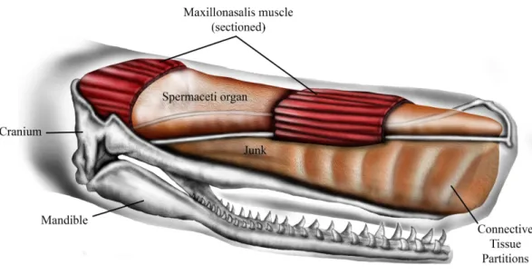
The sperm whale (Physeter macrocephalus L.) is unique in having a massively expanded forehead that is highly sexually dimorphic, being much larger and extending up to a meter and a half beyond the anterior tip of the jaws in mature males (Berzin, 1972;Cranford, 1999). Internally the forehead is composed of two large oil-filled sacs, stacked one on top of the other, known as the dorsal spermaceti organ (or spermaceti case) and the junk (Fig. 1). These sacs extend for one-third of the total length of the whale and can constitute more than one-quarter of the whale’s mass (Berzin, 1972;Clarke, 1978). The oil contained in the upper sac (spermaceti organ) was a primary target of the whaling industry of the early 19th century. At the same time, the forehead of sperm whales was considered by whalers to be a battering ram that the whales sometimes used to attack and sink oak whaling ships of up to 238 tons (Chase, 1821;Starbuck, 1878;Philbrick, 2000).
The lower sac (the junk) is derived from the odontocete melon (Heyning & Mead, 1990) and is organized into sections by transverse partitions of connective tissue that contain waxy oil (Clarke, 1978) (Fig. 1). The connective tissue partitions embedded in the junk are widest about 10–25% of the length from the anterior end and the sections are narrow ventrally and broad dorsally (Clarke, 1978). The partitions become thinner progressively towards the posterior aspect of the junk until they are totally replaced by a mixture of oil and wax. The oil and connective tissue partitions of the junk are enclosed in a fibrous connective tissue case which sits in a trough formed by the upper jaw (Clarke, 1978).
The function of the spermaceti organ and the junk in adding directionality and amplitude to sonar clicks is relatively well studied and accepted (Møhl et al., 2000;Møhl, 2001;Møhl et al., 2003a;Møhl et al., 2003b;Huggenberger, André & Oelschläger, 2014). Previous studies have also suggested that the unique structural configuration of the sperm whale forehead is functionally related to acoustic sexual selection (Cranford, 1999), acoustic prey debilitation (Norris & Møhl, 1983), communication (Madsen, Wahlberg & Møhl, 2002) and buoyancy control (Clarke, 1970). Although all of these functional hypotheses are plausible, they cannot explain how the forehead of sperm whales may function as a battering ram capable of sinking ships that are four to five times the mass of the whale.
The ramming hypothesis was originally proposed by whalers following the sinking of at least 2 whaling ships,the Essex in 1821 andthe Ann Alexander in 1851 (Chase, 1821;
Figure 1 Schematic representation of sperm whale head structure. Image courtesy of Ali Nabavizadeh.
to use their foreheads as battering rams in male–male contests over harems of females (Carrier, Deban & Otterstrom, 2002).
The ramming hypothesis remains highly controversial because (1) the structures that generate sound, the distal sac and monkey lips of the right nasal passage, are located at the rostral end of spermaceti organ (Fig. 1) and are therefore assumed to be in harm’s way in a ramming event (Huggenberger, André & Oelschläger, 2014), and (2) ramming episodes have not been observed by scientists who study the behavior of sperm whales. Although the monkey lips do reside at the front end of the spermaceti organ, these structures are located well above and to the right of the rostral end of the junk (Huggenberger, André & Oelschläger, 2014), and it is the junk, not the spermaceti organ, that has been suggested to function as a battering ram during aggressive encounters (Chase, 1821;Carrier, Deban & Otterstrom, 2002). As far as we know, the scientific literature does not include observations of sperm whale ramming behavior, yet there is one documented observation of male-male ramming that we report here (Supplemental Information 1). On January 30, 1997, a wildlife pilot, while flying over the Gulf of California, watched two mature males swim directly toward each other, from an initial observed distance of approximately 6.4 km, at an estimated average swimming speed of 17 km/h and collide forehead-to-forehead. Shortly before impact both whales, which had been swimming at the surface, ‘‘shallow dove’’ so that the impact occurred below the surface of the water. This ramming event occurred a few miles north of a group of approximately of 50 females. This observation plus reports of ramming attacks on 19th century whaling ships suggest that sperm whales may indeed engage in ramming contests. If these ramming contests generally occur at a shallow depth, they may be much more common than whale biologists realize because a human observer would have to be located well above the surface of the water to watch it happen.
mammals, body size sexual dimorphism is generally greatest in polygynous species in which males compete through fighting and the threat of fighting (Clutton-Brock & Harvey, 1977;
Parker, 1983;Andersson, 1994). Additionally, because sexual dimorphism is often greatest in those characters that enhance a male’s capacity to dominate other males (Clutton-Brock & Harvey, 1977;Hamilton, 1979;Clutton-Brock, Albon & Harvey, 1980;Parker, 1983;Jarman, 1983;Andersson, 1994), the observation that head length is the most dimorphic anatomical feature (Nishiwaki, Ohsumi & Maeda, 1963) is consistent with the head being a weapon important to male-male competition.
This paper addresses the battering-ram hypothesis using finite element analysis and probabilistic simulation. Our main objective was to determine if the connective tissue partitions of the junk have potential to reduce stress in the bones of the skull during ramming impact. We predict that the vertically oriented connective tissue partitions within the junk can dissipate load through tension during posteriorly directed compressive loading of the forehead. Bone stress reduction would be particularly important on the anterior aspect of the skull (i.e., upper jaw) that would otherwise be most vulnerable to potential tissue damage.
MATERIALS AND METHODS
Finite element analysis (FEA) is a numerical technique well entrenched in comparative biomechanics as a tool to assess the mechanical architecture of anatomical tissues and to better comprehend the complex interaction of their form–function relationships. Nevertheless, FEA accuracy is dependent on a variety of factors and its reproducibility is often obscured in scientific publications due to both public unavailability of the underlying models and the lack of standard reporting guidelines (Erdemir et al., 2012). To mitigate these problems we here describe our methods in accordance with biomechanical FEA reporting guidelines (Erdemir et al., 2012) and we also make all raw data and FE models available for public use (Panagiotopoulou et al., 2016).
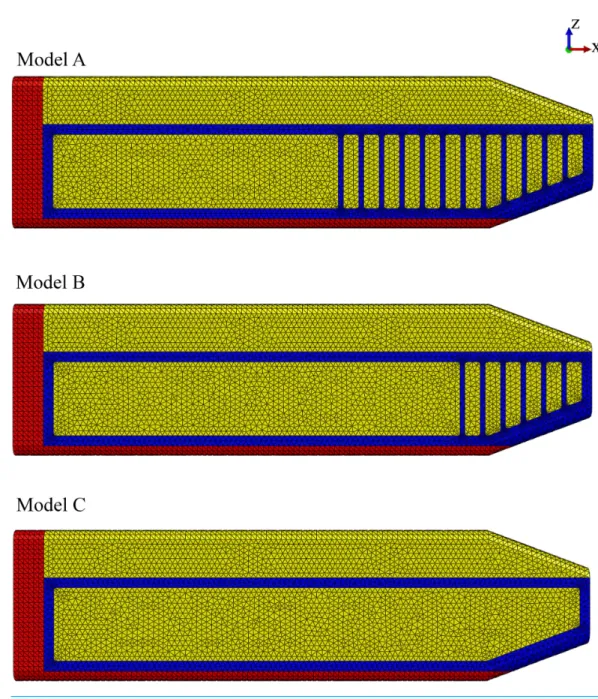
Model identification. Our study utilized three FE models to study the effect of the connective tissue partitions on the reduction of bone stresses in quasi-static loading of the sperm whale head (Fig. 2).
Model name.Sperm Whale Head Model A Generic Base consisted of twelve connective tissue partitions embedded in the spermaceti tissue of junk.
Sperm Whale Head Model B Half Partitions had reduced number (six) of connective tissue partitions.
Sperm Whale Head Model C No Partitions had no connective tissue partitions. Model keywords.Sperm whale skull, quasi-static impact.
Version.0.1 (Panagiotopoulou et al., 2016)
Physiological domain.No segmental motion, evenly distributed anterior surface loading, small deformations of hard and soft tissue.
Mechanical domain.All models were static and linear elastic.
Figure 2 Finite element models.Young’s moduli for the connective tissue partitions (blue), spermaceti organ (yellow) and skull (red) were, 2 GPa, 1 GPa and, 14.8 GPa, respectively. Models A, B and C have twelve, six and zero connective tissue partitions, respectively.
Demographics.Adult male sperm whale (Physeter macrocephalus L.). State of represented organism.in vitro.
Disease state.Healthy.
Spatial scale.Within a volume of (length 5.3 m×height 1.6 m×width 0.1 m). Time scale.Not applicable (quasi-static analysis).
Figure 3 Loads and constraints assigned to all FE models.A force of 764 kN was applied to the anterior surface of the head (1). Motion was constrained at the posterior surface (2) in all directions.
Primary highlight.To elucidate the possible mechanically protective role of the vertical connective tissue partitions within the sperm whale skull.
Secondary highlight.Not applicable.
Primary limitation. Linear isotropic and homogeneous materials.
Due to lack of experimental data on the elasticity of the sperm whale head tissues, anisotropy and heterogeneity, as well as environmental and time dependencies could not be modeled in this study. Thus, isotropy, homogeneity and linear elasticity were assumed and the material properties assigned to each tissue were the closest estimations based on published values of tissues similar to those of interest (Rho, Ashman & Turner, 1993;Shahar et al., 2007). A biologically unrealistic consequence of this assumption was that the dorsal horizontal components of connective tissue partitions provided resistance to compression in the model. To this end we assigned a Young’s modulus (E) value of 14.8 GPa and Poisson (v) value of 0.1 for the skull;E=2 GPa andv=0.2 for the connective tissue partitions;
andE=1 GPa andv=0.49 for the oil/wax mixture enclosed within the spermaceti organ
and the junk (Fig. 2) (Rho, Ashman & Turner, 1993;Shahar et al., 2007). Nevertheless, our study was comparative and such an assumption likely created a constant error across all models. Additionally, uncertainties due to material variations had been handled through numerical statistic elaboration of the models. Lastly, the basic mechanism of skull-stress reduction we described was independent of model realism.
Secondary limitation. Simplifications in the model geometry (see below), static simulation. Reference to publications.Clarke (1970)andClarke (1978); no explicit mechanical model described.
Model structure
of 20.56◦(collinear with the model’s anterior part of the connective tissue) to the impact
force. We also varied the application point of the impact force, applying the load to the spermaceti organ, and the superior, middle and inferior aspects of the junk respectively for all models (Fig. S1). The magnitude of the applied force per surface area is shown in Table S1. We calculated the applied force by assuming that each of the two colliding whales were traveling at an intermediate speed of 6.26 ms−1(Aoki et al., 2007) had masses of
39,000 kg, and decelerated over a distance of 1 m upon impact. The deceleration distance was based on the length of the junk that extended beyond the tip of the skull. Boundary conditions included no-displacement constraints on all external nodes on the posterior surface of the skull.
Primary output variables.von Mises stress.
Source of anatomy. To test our hypothesis, we developed FE models based partially on previously published structural properties and schematic configurations of male sperm whale adult cadavers. Due to the inaccessibility of sperm whale cadaveric species, the report byClarke (1978)was the most detailed hitherto available and encompassed skeletal and soft tissues such as the connective tissue partitions, and the oil cases of the spermaceti organ and the junk. To calculate the dimensions of the various structural components of the model, we scaled the anatomical elements shown in Fig. 1 ofClarke (1978)to a total spermaceti organ length of 5 m (Clarke, 1970). For modelling purposes and due to the unclear description of the individual connective tissue partitions thickness, we assumed a universal thickness of 0.05 m and 0.150 m for all connective tissue partitions and the junk compartments between the connective tissue partitions respectively (Fig. 2). In order to ensure that the modeling simplification with regards to the thickness of the connective tissue partitions did not influence our overall conclusions and were biologically sound, we conducted a sensitivity analysis and compared the von Mises skull stresses of Model A, against a modification with connective partitions thickness reported byClarke (1978)and listed inTable S2.
Model A, representative of the sperm whale head, consisted of the upper sack or spermaceti organ; the lower sack or junk; the connective tissue partitions with a universal thickness of 0.05 m and 0.150 m and their subsequent connective tissue case enclosed in the junk and the upper jaw (Fig. 2). Model A2 was identical to Model A but the connective tissue partitions were assigned different thickness (Table S2).
We compared Model A against two modified models (Models B and C) to assess the mechanical function of the spermaceti organ (Fig. 2). Model B had fewer connective tissue partitions than Model A. Model C lacked the connective tissue partitions altogether (Fig. 2).
The skin and the blubber were discarded from the modeling process due to their negligible thickness and stiffness.
case; 93,482 for the junk; and 55,664 for the connective tissue partitions). Model C had 227,925 elements in total (28,137 for the upper jaw; 65,519 for the spermaceti case; 134,269 for the junk. The nominal element size was 50 mm (0.05 m), and the actual elements sizes across the model varied approximately from 15 to 85 mm.
Reference configuration.The Abaqus defaultx (cranial-caudal),y (medial-lateral),z
(vertical) coordinate system was used.
Simulation structure
Name of simulation software.Abaqus/CAE (Simulia-Dassault Systemes). Version of simulation software.6.12
Solution strategy.Abaqus/Standard implicit direct static solver. Minimum and maximum increments set to 1.000E–05 and 1 respectively.
Numerical algorithms.Full Newton default iterations.
Convergence criteria.Default convergence tolerances of the simulation software were used. We interpreted stress differences amongst our models using a Monte Carlo simulation. A total of 1,000 Monte Carlo iterations were run for each of the three models, varying the three materials’ stiffness values randomly with a standard deviation of 10%, and von Mises stress distributions were stored for each iteration (Supplemental Information). This resulted in a population of 1,000 random individuals which represented the population of interest, under an assumption of 10% error in each of the material parameters. The latter is an essential approach in cases when the assigned material properties are based on generalized published values and not on experimental analysis of the tissues of interest.
For each population pair (i.e., Model B vs. Model A, Model C vs. Model B and Model C vs. Model A), the following statistic was calculated for each element:
zi=
(si)B−(si)A 1
2[(σi)A+(σi)B]
whereiindexes the elements, A and B represent models,si represents mean elemental
von Mises stress andσirepresents elemental standard deviation. Since the input variance
nonlinearly maps to elemental variance, thezdistribution is a non-trivial function of mean group differences.
Next, the ‘‘significance’’ of thez distribution was assessed using paired non-parametric permutation tests (one for each model pair). A total of 10,000 label permutations were applied to each model pair, yielding a non-parametric distribution of thez statistic at each element. The 99th percentile of that distribution was taken as the ‘‘significance’’ threshold. In other words, if an element’sz value survived that threshold, it would suggest that 99% of all randomly labelled individuals would yield azvalue less than that observed in the original labellings, and thus that threshold-surviving elements represented true population differences at alpha =0.01 under the assumption of 10% true population
material parameter variance.
Validation.Validation of the FE models against experimentalex vivodata was not feasible due to size and accessibility constraints. Nevertheless our study is comparative and conclusions are fundamentally mechanical rather than empirical.
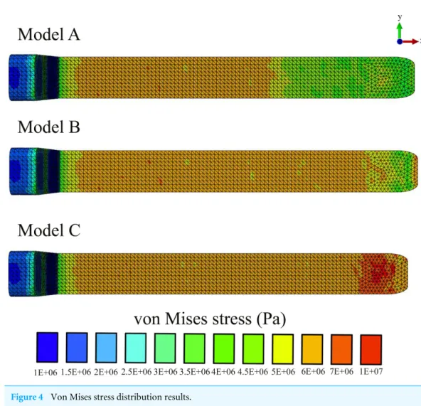
Figure 4 Von Mises stress distribution results.
RESULTS
In all our FE models the highest concentration of von Mises stresses occurred in the most anterior aspect of the skull (Figs. 4and5andTable 1). The anterior connective tissue partitions within the junk were subjected to higher tensile loading than the posterior portions (Fig. 6). Tension in the connective tissue partitions redistributed compressive stresses across the skull (Models A and B) and the absence of the partitions (Model C) raised anterior skull stresses (Figs. 4and5andTable 1).
A reduced number of partitions (Model B) did reduce stresses in the anterior skull, but stress reduction was not as effective as Model A (Figs. 4and5andTable 1). The skull stress difference distributions resulting from Monte Carlo simulations suggest that our main finding regarding the load-redistribution functionality of the connective tissue is insensitive to relatively large changes in both material parameters (Fig. 7) and, indirectly, load magnitudes.
Figure 5 Region definitions (blue vertical bars).
Table 1 Maximum (Max.), mean and minimum (Min.) percentage increase of the regional (Fig. 5) von Mises stress values (Pa) between Models A and C and Models B and C.
% increase
1 2 3 4 5
Max. Mean Min. Max. Mean Min. Max. Mean Min. Max. Mean Min. Max. Mean Min.
Model A– Model C
42.9 45.7 3.6 25 24.1 9.4 −8.8 6.1 60.8 −6.5 −0.9 −27.3 1.4 −0.3 −62.8
Model B– Model C
10.1 15.5 12.8 −7.3 4.1 35.7 0.6 0.8 −6.9 −6.7 −1.4 −27.4 1.4 −0.2 −59.6
Figure 6 Maximum principal stress distributions across the connective tissue partitions.Positive and negative stresses indicate areas of tension and compression respectively.
increased and the connective tissue partitions underwent increased tension. From the top of the anterior junk to the bottom, variations in the angle of the applied force on the junk increased skull stresses on the posterior aspect of the skull in all models, yet the model without partitions (Model C) showed the highest stress concentration across the whole skull and thus the least resistance to bending (Fig. S6).
Figure 7 Probabilistic FEA simulation.Zstatistic distributions depicting mean elemental von Mises stress differences divided by elemental standard deviation under an assumed population material stiffness variance of 10%. Data are thresholded at alpha=0.01.
(with no connective tissue partitions). This raises confidence that the model simplifications on the partition design for Models A and B did not compromise the biological results of our study.
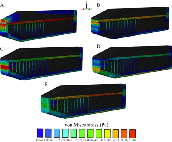
Figure 8 Von Mises stress distribution results following variations in the topological application of the impact force.(A) Impact force on the spermaceti organ; (B) Impact force on the superior aspect of the spermaceti junk; (C) Impact force on the mid spermaceti junk; (D) Impact force on the inferior aspect of the spermaceti junk; (E) Impact force on the entire anterior aspect of the spermaceti junk. Warm (red) and cold (blue) colors show higher and lower von Mises stresses respectively.
(Supplemental Information 2and3). This suggested that whilst the stiffness between Models A and B was quite similar, Model C was substantially different. To ensure that the comparisons between the models reflected a true effect of architectural design rather than solely being due to the changes in the system stiffness, we varied the stiffness of Models B and C by changing the Young’s modulus of the connective tissue until we obtained similar reaction force for all models (Fig. S8). Comparisons of von Mises stress between Models A, B, and C with equivalent stiffnesses further supported our findings that Model C increases stress on the anterior aspect of the skull (Fig. S8).
DISCUSSION
upon impact, the oil between the partitions is displayed vertically and laterally, placing the connective tissue partitions into tension (Fig. 6). This connective tissue tension allows the total compressive bone load to be shared over a greater volume (Figs. 4–7). While our static simulations do not quantify dynamic effects of the connective partitions including energy dissipation, our results suggest that connective partition tension would in fact dissipate energy in dynamic impacts because dynamic loading, like the current quasi-static loading, would be distributed over a broader region of the skull. Additionally, during a dynamic impact soft tissues within the skull would displace, and the connective tissue partitions would limit this displacement. The connective tissue might therefore protect both bone and soft tissue from injury. Absence of the partitions increased stresses by 45%, concentrated on the most anterior aspect of the skull, making the skull more prone to tissue failure (Table 1andFigs. 4–7).
From an engineering perspective one could argue that the mechanical effect of the connective tissue partitions in minimizing skull stresses might be due to variations in the system stiffness rather than the actual architecture of the junk. Our stiffness sensitivity analysis, however, confirmed that the connective tissue partitions are insensitive to system stiffness. Thus, the architecture and anatomical configuration of the connective tissue partitions assist in reducing impact stresses, particularly on the anterior aspect of the skull.
Our modeling indicates that an impact load applied to the spermaceti organ generates lower skull stresses than the impact loads on the junk. Based on this finding and the fact that the rostral end of spermaceti organ often extends anteriorly beyond the junk, it would be expected for sperm whales to use the spermaceti organ during head-butting incidents. However, scarring on the forehead of adult male sperm whales is largely confined to the rostral end of the junk, rather than the spermaceti organ (Carrier, Deban & Otterstrom, 2002,Fig. 7), suggesting that sperm whales avoid contact with the spermaceti organ and use the junk in ramming events. Behaviorally, confining impact to the junk makes sense given the lack of structural reinforcement of the spermaceti organ and the presumably delicate sound generator of the sonar system housed in the anterior spermaceti organ that could potentially be injured in a ramming event (Huggenberger, André & Oelschläger, 2014). In addition, whilst impacts on the spermaceti organ appear more effective in reducing skulls stresses than impacts on the junk in our static model, the former cause an increased concentration of compressive stresses on the spermaceti organ and also on the connective tissue case. Because connective tissue cannot offer resistance to compressive forces, compressive forces applied to the case would tend to cause its collapse resulting in (1) an ineffective ram applied to the opponent whale and (2) secondary impact of the junk. Additionally, compressive collapse of the connective tissue case would not only endanger the sonar apparatus but would ultimately greatly increase skull stresses.
The connective tissue partitions of the junk are acquired traits that likely facilitate a variety of functions. In addition to echolocation (Madsen, Wahlberg & Møhl, 2002), the partitions may play an important role in the dissipation of stresses during ramming combat to protect the skull and brain. This ‘‘mechanical advantage’’ is a trait that is likely related to selection on male-male aggressive behavior. Such developmentally non-independent morphological features of the junk are an example of how a derived structure, such as the connective tissue partitions, facilitates evolutionary modifications while maintaining functional integrity (Wagner & Altenberg, 1996).
Our results are not directly relevant to the behavioral strategies behind ramming impacts; however, our findings are consistent with the hypothesis proposed in 1821 by Owen Chase (Chase, 1821). Following the sinking of theEssexwhaling ship, Owen Chase hypothesized that sperm whales not only use their immense and elaborately complex foreheads as battering rams when fighting, but also that ‘‘the whale’s head is admirably designed for this mode of attack.’’ The prevalence of head-butting in sperm whales is not well documented. However, ramming is a basal behavior for Bovidae (Farke, 2008;Alvarez, 1990) and Cetacea (Carrier, Deban & Otterstrom, 2002), including humpback whales (Baker & Herman, 1984), bottle-nosed whales (Gowans & Rendell, 1999), narwhales (Silverman & Dunbar, 1980), long-finned pilot whales (Reilly & Shane, 1986) and killer whales (Goley & Straley, 1994). Based on these reports, it has previously been hypothesized that the spermaceti organ of male sperm whales may function as a weapon and is more developed in males due to sexual selection (Carrier, Deban & Otterstrom, 2002). If this is true, then males may be exposed to increased stresses during head-butting ramming and as such necessitate additional support via a dramatically increased and more structurally robust melon.
Our study illustrates how structural engineering principles and probabilistic simulation can be used to address hypotheses of mechanical function in biological systems that are too big or inaccessible to be studied directly. We anticipate our study will stimulate future research aimed at unraveling the mechanical function of the head during aggressive head-butting and ramming in other species such as theHippopotamus(Kingdon, 1979) in which head-butting aggressive behavior is common but remains unsimulated.
ACKNOWLEDGEMENTS
We thank Sandra Lanham for sharing with us her report on the sperm whale ramming incident that occurred near Mildriff Island at the Gulf of California, on the 30th of January 1997. We are also grateful to Dr. Ali Nabavizadeh (University of Chicago) for creatingFig. 1. Our most sincere thanks go to our editor and the two reviewers for the very constructive comments and suggestions during the review process that improved the quality of our work.
ADDITIONAL INFORMATION AND DECLARATIONS
Funding
Competing Interests
Panagiotis Spyridis is an employee of Polytropos Ltd, London.
Author Contributions
• Olga Panagiotopoulou conceived and designed the experiments, performed the experiments, analyzed the data, wrote the paper, prepared figures and/or tables, reviewed drafts of the paper.
• Panagiotis Spyridis conceived and designed the experiments, performed the experiments, analyzed the data, reviewed drafts of the paper.
• Hyab Mehari Abraha analyzed the data, reviewed drafts of the paper.
• David R. Carrier conceived and designed the experiments, reviewed drafts of the paper.
• Todd C. Pataky performed the experiments, analyzed the data, contributed reagents/
materials/analysis tools, reviewed drafts of the paper.
Data Availability
The following information was supplied regarding data availability: Dryad DOI:10.5061/dryad.81rp6.
Supplemental Information
Supplemental information for this article can be found online athttp://dx.doi.org/10.7717/ peerj.1895#supplemental-information.
REFERENCES
Alvarez F. 1990.Horns and fighting in male Spanish ibex,Capra pyrenaica.Journal of
Mammalogy71:608–616DOI 10.2307/1381801.
Andersson M. 1994. Sexual selection. Princeton: Princeton University Press.
Aoki K, Amano M, Sugiyama N, Muramoto H, Suzuki M, Yoshioka M, Mori K, Tokuda D, Miyazaki N. 2007. Measurement of swimming speed in sperm whales. In:
Underwater technology and workshop on scientific use of submarine cables and related technologies. Piscataway: IEEE, 467–471.
Baker CS, Herman LM. 1984.Aggressive behavior between humpback whales
(Megaptera novaeangliae) wintering in Hawaiian waters.Canadian Journal of Zoology
62:1922–1937DOI 10.1139/z84-282.
Berzin AA. 1972.The sperm whale. Jerusalem: Israel Program for Scientific Translations. Carrier RC, Deban SM, Otterstrom J. 2002.The face that sank the Essex: potential
function of the spermaceti organ in aggression.Journal of Experimental Biology
205:1755–1763.
Chase O. 1821.Shipwreck of the whale-ship Essex. New York: Gilley.
Clarke MR. 1970.Function of the spermaceti organ of the sperm whale.Nature
228:873–874DOI 10.1038/228873a0.
Clarke MR. 1978.Structure and proportions of the spermaceti organ in the sperm whale.
Clutton-Brock TH, Albon SD, Harvey PH. 1980.Antlers, body size and breeding group size in the Cervidae.Nature285:565–566DOI 10.1038/285565a0.
Clutton-Brock TH, Harvey PH. 1977.Primate ecology and social organization.Journal of
Zoology London183:1–39DOI 10.1111/j.1469-7998.1977.tb04171.x.
Cranford TW. 1999.The sperm whale’s nose: sexual selection on a grand scale?Marine
Mammal Science15:1133–1157DOI 10.1111/j.1748-7692.1999.tb00882.x.
Erdemir A, Guess TM, Halloran J, Tadepalli SC, Morrison TM. 2012.Considerations for reporting finite element analysis studies in biomechanics.Journal of Biomechanics
45:625–633DOI 10.1016/j.jbiomech.2011.11.038.
Farke AA. 2008.Frontal sinuses and head-butting in goats: a finite element analysis.
Journal of Experimental Biology211:3085–3094DOI 10.1242/jeb.019042.
Goley PD, Straley JM. 1994.Attack on gray whales (Eschrichtius robustus) in Monterey Bay, California by killer whales (Orcinus orca) previously identified in Glacier Bay, Alaska.Canadian Journal of Zoology72:1528–1530DOI 10.1139/z94-202.
Gowans S, Rendell L. 1999.Head-butting in northern bottlenose whales (Hyperodon am-pullatus): a possible function for big heads.Marine Mammal Science15:1342–1350 DOI 10.1111/j.1748-7692.1999.tb00896.x.
Hamilton WD. 1979. Wingless and fighting males in fig wasps and other insects. In: Blum MS, Blum NA, eds.Sexual selection and reproductive competition in insects. New York: Academic Press, 167–220.
Heyning JE, Mead JG. 1990. Evolution of the nasal anatomy of cetaceans. In: Thomas J, Kastelein R, eds.Sensory abilities of cetaceans. New York: Plenum Press, 67–79. Huggenberger S, André M, Oelschläger HHA. 2014.The nose of the sperm whale:
overviews of functional design, structural homologies and evolution.Journal of
Marine Biological Association of the United Kingdom1–24
DOI 10.1017/S0025315414001118.
Jarman PJ. 1983.Mating system and sexual dimorphism in large, terrestrial, mammalian herbivores.Biology Reviews58:485–520DOI 10.1111/j.1469-185X.1983.tb00398.x. Kingdon J. 1979.East African Mammals. Vol. III. London: Academic Press.
Madsen PT, Wahlberg M, Møhl B. 2002.Male sperm whale (Physeter macrocephalus) acoustics in a high-latitude habitat: implications for echolocation and communica-tion.Behavioural Ecology and Sociobiology53:31–41DOI 10.1007/s00265-002-0548-1. Møhl B. 2001.Sound transmission in the nose of the sperm whalePhyseter catodon.
A post mortem study.Journal of Comparative Physiology. A, Sensory, Neural, and
Behavioral Physiology187:335–340DOI 10.1007/s003590100205.
Møhl B, Madsen PT, Wahlberg M, Au WWL, Nachtigall PE, Ridgway SH. 2003a.Sound transmission in the spermaceti complex of a recently expired sperm whale calf.
Acoustics Research Letters Online4:19–24DOI 10.1121/1.1538390.
Møhl B, Wahlberg M, Madsen PT, Miller LA, Surlykke A. 2000.Sperm whale clicks: directionality and source level revisited.Journal of the Acoustical Society of America
107:638–648DOI 10.1121/1.428329.
Nishiwaki M, Ohsumi S, Maeda Y. 1963.Change of form in the sperm whale accompa-nied with growth.Scientific Reports of the Whales Research Institute Tokyo17:1–17. Norris KS, Møhl B. 1983.Can odontocetes debilitate prey with sound?American
Naturalist 122:85–104.
Panagiotopoulou O, Spyridis P, Mehari Abraha H, Carrier DR, Pataky T. 2016.
Architecture of the sperm whale forehead facilitates ramming combat. Dryad Digital Repository.Available athttp:// dx.doi.org/ 10.5061/ dryad.81rp6.
Parker GA. 1983.Arms races in evolution—an ESS to the opponent independent costs game.Journal of Theoretical Biology101:619–648
DOI 10.1016/0022-5193(83)90019-X.
Philbrick N. 2000.In the heart of the sea. Harmondsworth: Penguin Books Ltd. Reilly SB, Shane SH. 1986. Pilot whale. In: Haley D, ed.Marine Mammals of Eastern
North Pacific and Arctic Waters. Seattle: Pacific Search Press, 133–139.
Rho JY, Ashman RB, Turner CH. 1993.Young’s modulus of trabecular and cortical bone material: ultrasonic and microtensile measurements.Journal of Biomechanics
26:111–119DOI 10.1016/0021-9290(93)90042-D.
Sawtell CC. 1962.The Ship Ann Alexander of New Bedord, 1805-1851. Mystic: Marine Historical Association.
Shahar R, Zaslansky P, Barak M, Friesem AA, Currey JD, Weiner S. 2007.Anisotropic Poisson’s ratio and compression modulus of cortical bone determined by speckle interferometry.Journal of Biomechanics40:252–264
DOI 10.1016/j.jbiomech.2006.01.021.
Silverman HB, Dunbar MJ. 1980.Aggressive tusk use by the narwhal (Monodon
monoceros L).Nature284:57–58DOI 10.1038/284057a0.
Starbuck A. 1878.History of the American whale fishery, from its earliest inception to the
year 1876. New York: Argosy-Antiquarian Ltd.
Wagner GP, Altenberg L. 1996.Complex adaptations and the evolution of evolvability.
Evolution50:967–976DOI 10.2307/2410639.