Corresponding author: Dra. Marilia Farignoli Romeiro. e-mail: [email protected]
Received 11 January 2016 Accepted 26 May 2016
Evaluation and optimization of SYBR Green real-time
reverse transcription polymerase chain reaction
as a tool for diagnosis of the
Flavivirus
genus in Brazil
Marilia Farignoli Romeiro
[1], William Marciel de Souza
[1], Aline Lavado Tolardo
[1],
Luiz Carlos Vieira
[1], Tatiana Elias Colombo
[2], Victor Hugo Aquino
[3],
Maurício Lacerda Nogueira
[2], and Luiz Tadeu Moraes Figueiredo
[1][1]. Centro de Pesquisa em Virologia, Faculdade de Medicina de Ribeirão Preto, Universidade de São Paulo, Ribeirão Preto, São Paulo, Brasil. [2]. Laboratório de Pesquisa em Virologia, Faculdade de Medicina de São José do Rio Preto, São José do Rio Preto, São Paulo, Brasil. [3]. Laboratório de Virologia, Departamento de Análises Clínicas, Toxicológicas e Bromatológicas, Faculdade de Ciências Farmacêuticas de Ribeirão Preto,
Universidade de São Paulo, Ribeirão Preto, São Paulo, Brasil.
Abstract
Introduction: The genus Flavivirus includes several pathogenic species that cause severe illness in humans. Therefore, a rapid and accurate molecular method for diagnosis and surveillance of these viruses would be of great importance. Here, we evaluate and optimize a quantitative real-time reverse transcription polymerase chain reaction (RT-PCR) method for the diagnosis of the
Flavivirus genus. Methods: We evaluated different commercial kits that use the SYBR Green system for real-time RT-PCR with a primer set that ampliies a fragment of the NS5 lavivirus gene. The speciicity and sensitivity of the assay were tested using twelve laviviruses and ribonucleic acid (RNA) transcribed from the yellow fever virus. Additionally, this assay was evaluated using the sera of 410 patients from different regions of Brazil with acute febrile illness and a negative diagnosis for the dengue
virus. Results: The real-time RT-PCR ampliied all laviviruses tested at a melting temperature of 79.92 to 83.49°C. A detection
limit of 100 copies per ml was determined for this assay. Surprisingly, we detected dengue virus in 4.1% (17/410) of samples from patients with febrile illness and a supposedly negative dengue infection diagnosis. The viral load in patients ranged from
2.1×10⁷to 3.4×10³copies per ml. Conclusions: The real-time RT-PCR method may be very useful for preliminary diagnoses in
screenings, outbreaks, and other surveillance studies. Moreover, this assay can be easily applied to monitor viral activity and to measure viral load in pathogenesis studies.
Keywords: Flavivirus. Arbovirus. Real-time RT-PCR. Diagnosis.
Major Article
INTRODUCTION
Members of the Flavivirus genus, the Flaviviridae family,
are single-stranded, positive-sense ribonucleic acid (RNA) viruses with a genome approximately 11 kilobases in length that encodes three structural and seven non-structural proteins(1).
Most laviviruses are maintained in nature via complex cycles that include arthropod vectors and vertebrate hosts. Currently, 57 Flavivirus species have been described worldwide; humans
serve as the end hosts for most laviviruses(2). Many of these
viruses pose signiicant public health problems, including dengue virus (DENV), Zika virus (ZIKV), yellow fever virus (YFV), West Nile virus (WNV), and Saint Louis encephalitis virus (SLEV)(3) (4) (5).
Human lavivirus infection can lead to diseases ranging
from mild fever to encephalitis and hemorrhagic fevers(1) (2).
Therefore, for many lavivirus diseases, early diagnosis is essential for patient management and adoption of preventive measures(2) (6).
The diagnosis of flavivirus infections is commonly performed using serological tests such as enzyme-linked immunoabsorbent assays (ELISAs) for immunoglobulin M (IgM)-capture, which are commercially available for a number of viruses(1) (2) (7). The diagnosis of lavivirus infection can also be conducted by viral isolation in cell cultures including in C6/36
Aedes albopictus cells; however, this method is time-consuming and expensive and runs the risk of contamination. Alternative assays for the diagnosis or detection of lavivirus infection, such as low cytometry and microarrays, have been reported; however, these assays are not used routinely because of their high costs and complexity(8) (9) (10). Thus, the principal molecular
method for the diagnosis and detection of Flavivirus species
developed for the main speciic virus or multiplex causing human lavivirus diseases(11) (12) (13). Here, we describe the evaluation
and optimization of a broad speciicity SYBR Green I real-time RT-PCR method for the universal detection of laviviruses.
METHODS
Real-time RT-PCR
To evaluate the real-time RT-PCR method for the universal detection of flaviviruses previously described by Moureau
et al.(11),we tested the following commercial kits: SuperScript
III Platinum SYBR Green One-Step (Invitrogen, USA), SYBR Green PCR Master Mix (Applied Biosystems, USA), SYBR Select Master Mix (Applied Biosystems, USA), Power SYBR Green PCR Master Mix (Applied Biosystems, USA), and KAPA SYBR FAST Universal 2X quantitative polymerase chain reaction (qPCR) Master Mix (Kapa Biosystems, USA). All of the kits tested were used according to the manufacturer’s
recommendations. The assays were performed in a StepOnePlusTM
Real-Time PCR System (Applied Biosystems, USA).
Flaviviruses, RNA extraction, and cDNA synthesis
The laviviruses used for evaluation of the real-time RT-PCR
were propagated in C6/36 Aedes albopictus cells cultured in
Leibovitz’s-15 medium supplemented with 10% heat-inactivated fetal bovine serum, 50mg/mL of gentamicin, and 2mg/mL of amphotericin B (Vitrocell, Brazil). The infected cells were incubated for 5 to 10 days at 28°C. Next, viral RNA was extracted from the laviviruses using the QIAamp viral RNA extraction kit (Qiagen, Germany) and converted to double-stranded complementary DNA (cDNA) using Superscript III reverse transcriptase (Invitrogen, USA), according to the manufacturer’s instructions.
Standard curve, limit of detection, and
speciicity of real-time RT-PCR
To enable quantitation using real-time RT-PCR, we introduced a 272bp fragment of the NS5 gene ofthe YFV 17D vaccine strain into the pET28a vector and then transformed
it into Escherichia coli DH5α cells (Invitrogen, USA),
according to the manufacturer’s instructions.The insertion was subsequently, conirmed by PCR and plasmid extraction using the QIAquick PCR Puriication Kit (Qiagen, Germany).
In vitro transcription was performed using the MEGAscript T7
Transcription kit (Invitrogen, USA), quantiied with a Qubit 2.0 luorometer (Invitrogen, USA) and stored at -70ºC. The number of RNA copies/ml was determined using the following formula: RNA concentration (g per ml)/the number of nucleotides in the
transcript × 340×6.022×1023.
The standard curve for quantitation of RNA laviviruses in real-time RT-PCR was created with ive decimal serial dilutions of transcribed RNA, as described above. The real-time RT-PCR limit of detection for laviviruses was determined in
triplicate with serial decimal dilutions of in vitro transcribed
RNA (2.29×10¹¹ to 2.29×10⁻¹ RNA copies per ml). Additional
arboviruses, such as the Venezuelan equine encephalitis virus, Western equine encephalitis virus, Eastern equine encephalitis virus, Mayaro virus, Aura virus, and Mucambo virus were also tested to conirm the speciicity of the assay.
Clinical samples
To evaluate the clinical applicability of Flavivirus real-time RT-PCR, we tested a total of 410 serum samples from patients with acute febrile illnessesand negative diagnosesfor dengue
virususing conventional RT-PCR(14) or microarrays for arbovirus
and robovirus (unpublished data), as described in Table 1. Of
these sera samples, 88 were obtained during the dengue outbreak of 2011 to 2013 inthe City of São Jose do Rio Preto in São Paulo, Brazil, 55 were obtained during the dengue outbreak of 2006 to 2008 in Ribeirao Preto in São Paulo and 167 were obtained from patients with suspected dengue infection in 2011 in Sinop in Mato Grosso, Brazil. RNA was extracted from the samples and subjected to real-time RT-PCR.
Ethical considerations
This study was approved by the Human Research Ethics Committee of the School of Medicine of the University of São Paulo, Brazil (Process nr. 2013/164.277).
Nucleotide sequencing
The gene products ampliied using our real-time RT-PCR were conirmed by nucleotide sequencing. The amplicons were treated with ExoSAP-IT (Affymetrix, USA) to remove unused
deoxynucleotide triphosphates (dNTPs) and the puriied DNA
was sequenced using primers PF1S and PF2Rbis and the BigDye Terminator v3.1 Ready Reaction Cycle Sequencing Mixture (Applied Biosystems, USA) following the manufacturer’s protocol. The reaction was performed with an ABI Prism 3100 Sequencer Genetic Analyzer (Applied Biosystems, USA). The nucleotide sequences were analyzed using the Basic Local Alignment Search Tool for viral identiication.
RESULTS
To evaluate SYBR Green I real-time RT-PCR for the universal detection of laviviruses, several commercial kits were tested. The KAPA SYBR FAST Universal 2X qPCR Master Mix kit exhibited the maximal levels of sensitivity and speciicity for RNA from several laviviruses as well as in vitro transcribed RNAs. Evaluation
of the other four commercial real-time RT-PCR kits for Flavivirus
detection yielded different results; however, all kits had low or no ampliication. Non-speciic ampliication and primer-dimers
were also observed (Figure 1). Of the commercial kits evaluated
using Flavivirus stocks, only SuperScript III Platinum SYBR
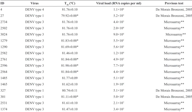
TABLE 1 -Positive samples identiied in this study using our real-time RT-PCR for Flavivirus.
ID Virus TM (ºC) Viral load (RNA copies per ml) Previous test
4 DENV type 4 81.76±0.10 1.1×10⁶ De Morais Bronzoni, 2005
27 DENV type 1 79.92±0.00* 5.2×103 De Morais Bronzoni, 2005
2734 DENV type 3 81.76±0.10 4.4×10³ Microaarray**
2502 DENV type 3 81.76±0.10 2.0×10⁴ Microaarray**
3024 DENV type 3 81.76±0.10 9.0×10⁴ Microaarray**
1279 DENV type 3 81.83±0.00* 5.5×103 Microaarray**
1290 DENV type 3 81.69±0.00* 5.6×103 Microaarray**
2582 DENV type 3 81.46±0.10 1.2×10⁴ Microaarray**
2761 DENV type 3 81.84±0.00* 4.9×103 Microaarray**
2596 DENV type 3 81.98±0.00* 7.7×103 Microaarray**
2564 DENV type 3 81.84±0.00* 4.4×104 Microaarray**
1485 DENV type 3 81.77±0.09 5.1×10³ Microaarray**
1465 DENV type 3 81.62±0.10 1.9×10⁴ Microaarray**
327 DENV type 1 80.74±0.11 5.1×10³ De Morais Bronzoni, 2005
301 DENV type 1 81.11±0.00* 5.0×103 De Morais Bronzoni, 2005
2531 DENV type 3 81.61±0.10 2.1×10⁷ Microaarray**
1374 DENV type 3 81.47±0.10 3.4×10³ Microaarray**
RT-PCR: real-time reverse transcription polymerase chain reaction; ID: identiication;TM: temperature melting; RNA: ribonucleic acid; DENV: dengue virus.
*Standard deviation < 0.001. **Unpublished.
The was ampliication conditions consisted of: 95°C for 10 minutes for Taq polymerase activation and double-stranded cDNA denaturation, followed by 45 cycles of 95°C for 15 seconds for denaturation and 60°C for 1 minute for primer
annealing. The ampliication the genomes of different Flavivirus
species was detected at the expected temperatures ranging
from 79.92 to 83.49°C (Table 2 and Figure 2). As expected,
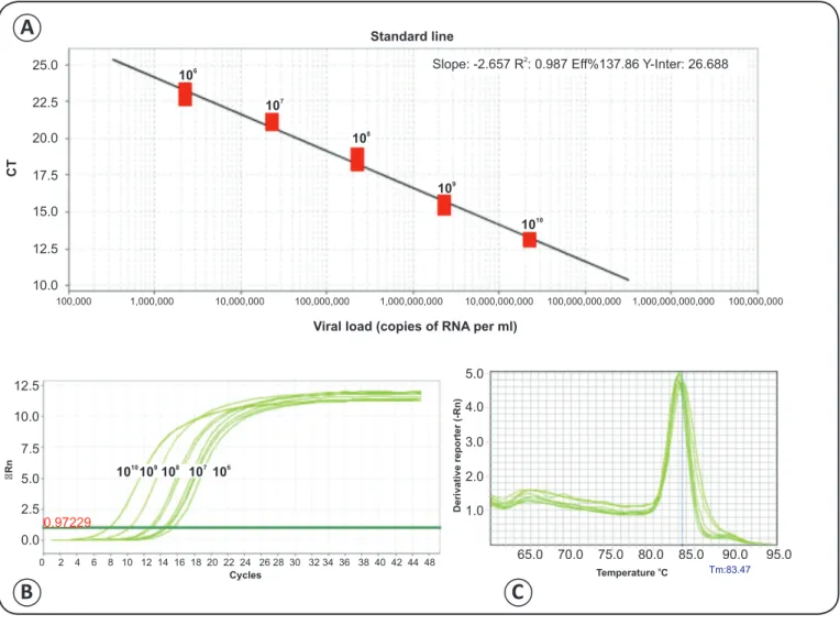
ampliication of the genomes of other RNA arboviruses was not observed. To quantify viral RNA, a standard curve of serially
diluted transcribed RNA was generated by plotting the CT value
against the number of copies of RNA per ml (Figure 3). The
limit of detection was at least 100 copies of RNA per ml. In our evaluation of real-time RT-PCR for the detection
of laviviruses, we detected Flavivirus viral genomes in 4.1%
(17/410) of samples from patients with febrile illness anda
negative diagnosis for dengue virus (Table 1). The amplicons
were sequenced and compared with sequences deposited in GenBank, showing that the patients were infected with dengue
virus types 1, 3, and 4 with viral loads ranging from 3.4×10³ to
2.1×10⁷ copies of RNA per ml (Table 1).
DISCUSSION
Flaviviruses are an important public health concern world wide(5) (6) (15). Therefore, a fast, eficient, and sensitive method for Flavivirus diagnosis is essential for the management of infected patients(2) (16) (17). In this study, we report the evaluation of
real-time RT-PCR for the universal detection of laviviruses as well as adaptations that allow for the measurement of viral load(11).
The use of real-time RT-PCR for the diagnosis of Flavivirus
infection has many advantages compared with virus isolation, serological assays, and conventional RT-PCR. Virus isolation
is considered to be the gold standard for the diagnosis of many
Flavivirus infections(18). However, this method has low sensitivity and requires several days for completion. Serological tests can cross-react between laviviruses or have false negative results(18).
Compared with conventional RT-PCR, real-time RT-PCR has several advantages such as rapidity, low risk of false positive results, high sensitivity, speciicity, and the possibility of viral quantiication(12) (19) (20). In fact, a previously described real-time
RT-PCR method(11) was able to detect a broad range of laviviruses
found in Brazil(3) (21) (22). This assay ampliied the genomes of 12
mosquito-borne lavivirusesthat are among the principal arboviruses responsible for human disease in Brazil (Table 2)(3) (23). The genomes
100.0 80
7.5
5.0
2.5
0.0
40
30
20
10
0
5
4
3
2
1
0 70 60 50 40 30 20 10
8.0 7.0
6.0 5.0
4.0
3.0
2.0
1.0
12.0
10.0
8.0
6.0
4.0
2.0
35,000.0
30,000.0
25,000.0
20,000.0
15,000.0
10,000.0
5,000.0
0
0 2 4 6 8 10 12 14 16 18 20 22 24 26 28 30 32 34 36 38 40 42
0 2 4 6 8 10 12 14 16 18 20 22 24 26 28 30 32 34 36 38 40 42
0 2 4 6 8 10 12 14 16 18 20 22 24 26 28 30 32 34 36 38 40 42 0 2 4 6 8 10 12 14 16 18 20 22 24 26 28 30 32 34 36 38 40 42 80.0
60.0
40.0
20.0
65.0 70.0 75.0 80.0 85.0 90.0 95.0
65.0 70.0 75.0 80.0 85.0 90.0 95.0
65.0 70.0 75.0 80.0 85.0 90.0 95.0
65.0 70.0 75.0 80.0 85.0 90.0 95.0 Tm:80.81
Tm:80.51
Tm:81.26
Tm:72.75
Derivative reporter (-Rn)
Derivative reporter (-Rn)
Derivative reporter (-Rn)
Derivative reporter (-Rn)
Temperature Co
Temperature Co
Temperature Co
Temperature Co
Cycles
Cycles
Cycles Cycles
∆
Rn
∆
Rn
∆
Rn
∆
Rn
FIGURE 1 - Melting peaks and ampliication curves of real-time RT-PCR for mosquito-borne Flavivirus using four different kits. A). Ampliication curve using Power SYBR Green PCR Master Mix (Applied Biosystems, Foster City, CA, USA). B). Melting peaks for real-time RT-PCR using Power SYBR
Green PCR Master Mix. C). Ampliication curve using SYBR® Green PCR Master Mix (Applied Biosystems). D). Melting peaks for real-time RT-PCR using SYBR® Green PCR Master Mix. E). Ampliication curve using SYBR® Select Master Mix (Applied Biosystems). F). Melting peaks for real-time RT-PCR using SYBR® Select Master Mix. G). Ampliication curve using SuperScript III Platinum SYBR® Green One-Step (Invitrogen, Carlsbad, CA, USA). H). Melting peaks for SuperScript III Platinum SYBR® Green One-Step. RT-PCR: real-time reverse transcription polymerase chain reaction.
A
B
C
D
E
F
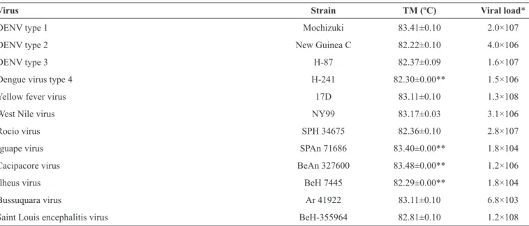
TABLE 2 -Flavivirus strains detected by real-time RT-PCR.
Virus Strain TM (ºC) Viral load*
DENV type 1 Mochizuki 83.41±0.10 2.0×10⁷ DENV type 2 New Guinea C 82.22±0.10 4.0×10⁶
DENV type 3 H-87 82.37±0.09 1.6×10⁷
Dengue virus type 4 H-241 82.30±0.00** 1.5×10⁶ Yellow fever virus 17D 83.11±0.10 1.3×10⁸ West Nile virus NY99 83.17±0.03 3.1×10⁶ Rocio virus SPH 34675 82.36±0.10 2.8×10⁷ Iguape virus SPAn 71686 83.40±0.00** 1.8×10⁴ Cacipacore virus BeAn 327600 83.48±0.00** 1.2×10⁶ Ilheus virus BeH 7445 82.29±0.00** 1.8×10⁴ Bussuquara virus Ar 41922 83.11±0.10 6.8×10³ Saint Louis encephalitis virus BeH-355964 82.81±0.10 1.2×10⁸
RT-PCR: real-time reverse transcription polymerase chain reaction; TM: temperature melting; DENV: dengue virus. RNA: ribonucleic acid.*RNA copies per
ml. **Standard deviation < 0.001.
Rn
12.5
5.0
65.0 70.0 75.0 80.0 85.0 90.0 95.0 4.0
3.0
2.0
1.0
Cycles Amplification curve
Melting Curve
10.0
7.5
5.0
2.5
0.0
0 2 4 6 8 10 12 14 16 18 20 22 24 26 28 30 32 34 36 38 40 42 44 46 Derivative reporter (-Rn)
Tm:83.49
Temperature Co
FIGURE 2 - Melting peaks and amplification curves of real-time RT-PCR for mosquito-borne Flavivirus. A). Amplification curve obtained
for the laviviruses described in Table 2. B). Melting peaks of real-time RT-PCR obtained for the mosquito-borne laviviruses described in Table 2.
RT-PCR: real-time reverse transcription polymerase chain reaction.
A
B
The high sensitivity of this real-time RT-PCR method for
Flavivirus detection was demonstrated using in vitro transcribed RNA showing that 100 copies of viral RNA per ml could be detected. The results from patients with indications of dengue infection presenting a viral load of 1.5×10⁴ to 9.9×10⁵ copies
per ml demonstrate the high sensitivity of our detection limit(27).
Other Flavivirus real-time RT-PCRs, such as those for DENV,
JEV (Japanese Encephalitis virus), WNV, or Usutu virus,
showed similar sensitivity (20 to 1,000 copies of viral RNA per ml); however, these assays do not exhibit as broad a range of Flavivirus detection as our assay(28) (29) (30) (31).
To evaluate real-time RT-PCR, the sera of patients with a negative diagnosis for the dengue virus were tested as described in the methods section. Our results indicated that 4.1% (17) of
samples were Flavivirus positive; surprisingly, these positive
samples were identified as dengue infection by nucleotide sequencing. However, although our assay exhibits a broad range of
Flavivirus detection, this assay does not allow for the discrimination of viral species by melting temperature. Nevertheless, the
Flavivirus amplicon of approximately 272bp obtained with this
test allows for distinguishing and identifying different viruses
by nucleotide sequencing, as performed in our assay(11). This
ability further enhances the high sensitivity observed using in
vitro transcribed RNA, suggesting that the real-time RT-PCR for Flavivirus is more sensitive than the traditional RT-PCR assay(14).
25.0
Standard line
106
1010109108 10 107 6
107
108
109
1010
CT
Slope: -2.657 R : 0.987 Eff%137.86 Y-Inter: 26.6882
22.5
20.0
17.5
15.0
12.5
12.5 5.0
4.0
3.0
2.0
1.0
65.0 70.0 75.0 80.0 85.0 90.0 95.0 10.0
7.5
5.0
2.5
0.97229
0.0 10.0
Viral load (copies of RNA per ml)
0 2 4 6 8 10 12 14 16 18 20 22 24 26 28 30 32 34 36 38 40 42 44 48
Cycles
Rn
Derivative reporter (-Rn)
Tm:83.47
Temperature Co
100,000 1,000,000 10,000,000 100,000,000 1,000,000,000 10,000,000,000 100,000,000,000 1,000,000,000,000 100,000,000
FIGURE 3 - Standard line of real-time RT-PCR with Flavivirus transcribed RNA. A). Standard line with serial decimal dilutions of transcribed Flavivirus
RNA. B). Ampliication curve obtained using serial decimal dilutions of transcribed Flavivirus RNA. C). Melting peaks for real-time RT-PCR using serial decimal dilutions of transcribed Flavivirus RNA.CT: threshold cycle; ∆Rn: normalized reporter; RT-PCR: real-time reverse transcription polymerase
chain reaction; RNA: ribonucleic acid.
A
C
B
REFERENCES
1. Gubler DJ, Kuno G, Markoff L. Flaviviridae. In: Knipe DM,
Howley HP, Grifin DE, editor. Fields virology, Philadelphia: Lippincott William & Wilkins; 2007. p. 1153-1253.
2. Go YY, Balasuriya UB, Lee CK. Zoonotic encephalitides caused by arboviruses: transmission and epidemiology of alphaviruses and laviviruses. Clin Exp Vaccine Res 2014; 3:58-77.
3. Figueiredo LT. The Brazilian laviviruses. Microbes Infect 2000;
2:1643-1549.
4. Le Flohic G, Porphyre V, Barbazan P, Gonzalez JP. Review of climate, landscape, and viral genetics as drivers of the Japanese encephalitis virus ecology. PLoS Negl Trop Dis 2013; 7:e2208.
5. Guzman MG, Harris E. Dengue. Lancet 2015; 385:453-465.
6. Mackenzie JS, Gubler DJ, Petersen LR. Emerging laviviruses: the spread and resurgence of Japanese encephalitis, West Nile and dengue viruses. Nat Med 2004; 10:S98-109.
7. Cleton N, Koopmans M, Reimerink J, Godeke GJ, Reusken C. Come ly with me: review of clinically important arboviruses for global travelers. J Clin Virol 2012; 55:191-203.
we improved the assay to enable quantiication of the viral genome, thus it can be easily implemented for monitoring viral activity and measuring viral load in pathogenesis studies.
Acknowledgments
We wish to thank Profª Roberta Vieira de Morais Bronzoni of Universidade Federal de Mato Grosso for supplying us with patient samples from Mato Grosso State, Brazil.
Conlict of interest
The authors declare that there is no conlict of interest.
Financial Support
Fundação de Amparo à Pesquisa do Estado de São Paulo (Grant nr.
8. Woda M, Mathew A. Fluorescently labeled dengue viruses as probes to identify antigen-speciic memory B cells by multiparametric low cytometry. J Immunol Methods 2015; 416:167-177.
9. Kao CL, Wu MC, Chiu YH, Lin JL, Wu YC, Yueh YY, et al. Flow cytometry compared with indirect immunoluorescence for rapid detection of dengue virus type 1 after ampliication in tissue culture. J Clin Microbiol 2001; 39:3672-3677.
10. Cleton NB, Godeke GJ, Reimerink J, Beersma MF, Doorn HR, Franco L, et al. Spot the difference-development of a syndrome based protein microarray for speciic serological detection of multiple lavivirus infections in travelers. PLoS Negl Trop Dis 2015; 9:e0003580.
11. Moureau G, Temmam S, Gonzalez JP, Charrel RN, Grard G, de Lamballerie X. A real-time RT-PCR method for the universal detection and identiication of laviviruses. Vector Borne Zoonotic
Dis 2007; 7:467-477.
12. Chao DY, Davis BS, Chang GJ. Development of multiplex real-time reverse transcriptase PCR assays for detecting eight medically important laviviruses in mosquitoes. J Clin Microbiol 2007; 45:584-589.
13. Johnson N, Wakeley PR, Mansield KL, McCracken F, Haxton B, Phipps LP, et al. Assessment of a novel real-time pan-lavivirus RT-polymerase chain reaction. Vector Borne Zoonotic Dis 2010;
10:665-671.
14. de Morais Bronzoni RV, Baleotti FG, Ribeiro Nogueira RM, Nunes M, Moraes Figueiredo LT. Duplex reverse transcription-PCR followed by nested PCR assays for detection and identiication of Brazilian alphaviruses and laviviruses. J Clin Microbiol 2005;
43:696-702.
15. Monath TP, Vasconcelos PF. Yellow Fever. J Clin Virol 2015;
64:160-173.
16. Ishikawa T, Yamanaka A, Konishi E. A review of successful lavivirus vaccines and the problems with those laviviruses for which vaccines are not yet available. Vaccine 2014; 32:1326-1337.
17. Maia FG, Chavez JH, de Souza WM, Romeiro MF, de Castro-Jorge LA, da Fonseca BA, et al. Infection with Saint Louis encephalitis virus in the city of Ribeirao Preto, Brazil: report of one case.
Int J Infect Dis 2014; 26:96-97.
18. Maeda A, Maeda J. Review of diagnostic plaque reduction neutralization tests for lavivirus infection. Vet J 2013; 195:33-40.
19. Shu PY, Huang JH. Current advances in dengue diagnosis. Clin Diagn Lab Immunol 2004; 11:642-650.
20. Chien LJ, Liao TL, Shu PY, Huang JH, Gubler DJ, Chang GJ. Development of real-time reverse transcriptase PCR assays to detect and serotype dengue viruses. J Clin Microbiol 2006; 44: 1295-1304.
21. Batista WC, Tavares GS, Vieira DS, Honda ER, Pereira SS, Tada MS. Notiication of the irst isolation of Cacipacore virus in a human in the State of Rondonia, Brazil. Rev Soc Bras Med Trop 2011;44: 528-530.
22. Vieira MA, Romano AP, Borba AS, Silva EV, Chiang JO, Eulalio KD, et al. West Nile virus encephalitis: the irst human case recorded in Brazil. Am J Trop Med Hyg 2015; 93:377-379.
23. Huang YJ, Higgs S, Horne KM, Vanlandingham DL. Flavivirus-mosquito interactions. Viruses 2014; 6:4703-4730.
24. Vazquez A, Sanchez-Seco MP, Palacios G, Molero F, Reyes N, Ruiz S, et al. Novel laviviruses detected in different species of mosquitoes in Spain. Vector Borne Zoonotic Dis 2012; 12:223-229.
25. Roiz D, Vazquez A, Seco MP, Tenorio A, Rizzoli A. Detection of novel insect lavivirus sequences integrated in Aedes albopictus
(Diptera: Culicidae) in Northern Italy. Virol J 2009; 6:93.
26. Figueiredo LT, Batista WC, Kashima S, Nassar ES. Identiication of Brazilian laviviruses by a simpliied reverse transcription-polymerase chain reaction method using Flavivirus universal primers. Am J Trop Med Hyg 1998; 59:357-362.
27. Pal T, Dutta SK, Mandal S, Saha B, Tripathi A. Differential clinical symptoms among acute phase Indian patients revealed signiicant association with dengue viral load and serum IFN-gamma level. J Clin Virol 2014; 61:365-370.
28. Mohammed H, Linnen JM, Munoz-Jordan JL, Tomashek K, Foster G, Broulik AS, et al. Dengue virus in blood donations, Puerto Rico, 2005. Transfusion 2008; 48:1348-1354.
29. Nikolay B, Weidmann M, Dupressoir A, Faye O, Boye CS, Diallo M, et al. Development of a Usutu virus speciic real-time reverse transcription PCR assay based on sequenced strains from Africa and Europe. J Virol Methods 2014; 197:51-54.
30. Faye O, Faye O, Diallo D, Diallo M, Weidmann M, Sall AA. Quantitative real-time PCR detection of Zika virus and evaluation with ield-caught mosquitoes. Virol J 2013; 10:311.