DOI: 10.2298/AVB1301101Z UDK 578.826+636.52/.58:613.36-002
INCLUSION BODY HEPATITIS (IBH) OUTBREAK ASSOCIATED WITH FOWL ADENOVIRUS TYPE 8b IN BROILERS
ZADRAVEC M*, SLAVEC BRIGITA*, KRAPE@ U*, KAJÁN GL**, RA^NIK J*, JUNTES POLONA***, JUR[I^ CIZERL RAHELA****, BENKÕ MÁRIA** and ZORMAN ROJS OLGA*
*Institute for Poultry Health, Veterinary Faculty of Ljubljana, Slovenia
**Veterinary Medical Research Institute, Hungarian Academy of Sciences, Budapest, Hungary ***Institute of Pathology, Forensic and Administrative Veterinary Medicine,
Veterinary Faculty of Ljubljana, Slovenia
****Veterinary practice Perutnina Ptuj, Veterinarstvo d.o.o., Ptuj, Slovenia
(Received 21thJuly 2012
The causative agent of inclusion body hepatitis (IBH) was identified as fowl adenovirus (FAdV) type 8b, a member of the Fowl adenovirus E species, based on PCR results of adenoviral polymerase and the hexon gene in an outbreak of acute mortality that affected a broiler flock of 12,000 animals. In two waves of elevated mortality rate, a total of 264 chickens were found dead. Affected birds showed ruffled feathers, depression, watery droppings and limping. The most common pathological lesions seen on necropsy were pale, swollen and friable livers. On histological examination, acute hepatitis characterized by necrosis of hepatocytes, with large basophilic intranuclear inclusion bodies, were observed. In addition, infectious bursal disease virus and infectious bronchitis virus were detected in the same flock.
Key words: broilers, FAdV8b, IBH outbreak, Slovenia
INTRODUCTION
Fowl adenoviruses (FAdV) are a very heterogeneous group of viruses. Twelve types (formerly serotypes), named FAdV-1 to 8a, and FAdV-8b to 11, are classified into five different species (A-E) (Benköet al.,2005). They are believed to be ubiquitous in poultry farms (McFerran and Smyth 2000). Not all FAdV are considered to be pathogenic for chickens but every type has already been recovered from naturally occurring cases of inclusion body hepatitis (IBH) (Gomis et al.,2006; McConnell and Fitzgerald, 2008).
weeks (Howellet al.,1970; Macphersonet al.,1974; McFerranet al.,1976; Baar and Scott, 1988; Ojki}et al.,2008b). The severity of the disease may also depend on some other predisposing factors that enhance the pathogenic potential of FAdV infection, such as a poor environment and management (Toroet al.,2000; Ojki} et al., 2008b). It has been proven that the initial involvement of immunosuppressive agents, including infectious bursal disease virus (IBDV) and chicken anemia virus (CAV) or some mycotoxins, such as aflatoxins, is needed for IBH onset (Fadlyet al.,1975; Rosenberget al.,1975; Toroet al.,2000; Toroet al., 2002; Shivachandraet al.,2003).
On the other hand, different FAdV types, called highly pathogenic FAdVs, have been considered to be the primary pathogen in IBH. Among them, FAdV-8 and 9 are the most commonly detected ones (Christensen and Saifuddin 1989; Saifuddinet al.,1992; Gomiset al.,2006; Ojki}et al.,2008a; Ojki}et al.,2008b) and seem to predominate in some areas (Gomiset al.,2006; Ojki}et al.,2008a).
The present paper is the first report of IBH with detailed clinical and laboratory findings in an outbreak in commercial broilers in Slovenia.
MATERIAL AND METHODS
Case history
Acute mortality was observed in a broiler flock of 12,000 animals. The birds were housed as day-old chickens and were vaccinated against Newcastle disease (ND) on day 6 (Pestikal La Sota SPF, Veterina, CRO), IBD on day 13 (Nobilis Gumboro 228E, Intervet/Schering-Plough, USA) and infectious bronchitis on day 16 (Nobilis IB 4-91, Intervet/Schering-Plough, USA). At the age of 17 days, 146 chickens (1.22%) were found dead at random throughout the house. The flock was treated with amoxicillin (Paracilin, Intervet/Schering-Plough, USA) for 5 days. In the six days following the first onset, 118 chickens (0.98%) died. Affected birds showed ruffled feathers, depression, watery droppings and limping.
Fourteen broiler chickens that died at the age of 23 days were submitted to the Institute of Poultry Health, Veterinary Faculty (VF), University of Ljubljana on the day of death.
The overall production results in the affected flock were comparable to those obtained in other broiler flocks. The mortality was 2.90%. Nonetheless, a higher feed conversion rate (1.94 kg compared to a predicted 1.88 kg) was obtained and the average body weight at the age of 34 days was higher than expected; 1.70 kg compared to 1.50 kg planned.
Gross and histopathological examinations
Pathologic examinations were performed on all birds submitted. Tissue blocks of the liver, kidney, heart and spleen were fixed in 10% buffered formalin, embedded in paraffin, sectioned at 4mm and stained with hematoxylin and eosin
Virological examinations
For molecular investigations, DNA and RNA were extracted from material taken at necropsy and frozen until investigation. Portions of liver and spleen were separately homogenized in phosphate buffered saline (PBS), prepared as a 10–20% w/v suspension. DNA was extracted by using a QIAamp®DNA Mini Kit (Qiagen, Hilden, Germany) according to the manufacturer’s instructions. RNA was extracted from the trachea and cloacal swab by using QIAamp Viral RNA Mini Kit (Qiagen, Hilden, Germany) according to the manufacturer’s instructions. Before RNA extraction, 2 mL of PBS was added to each swab and vortexed vigorously.
Detection of FAdV
Attempts to detect adenoviruses in the liver and spleen tissue were made by using nested polymerase chain reaction (PCR) with degenerate, consensus primers targeting the viral DNA dependent DNA polymerase gene, as described by Wellehanet al.(2004). In addition, more specific, degenerate primers hexon A and hexon B that amplify a region of the hexon gene were used for establishing FAdV serotype, as reported previously (Meulemanset al.,2001).
Detection of CAV
For detection of CAV in the liver and spleen tissue, primers that target the highly conserved gene encoding VP2 were used, as described by Notebornet al. (1992).
Detection of IBDV
For detection of IBDV from cloacal swabs, a reverse transcription (RT)-PCR method was applied, as described previously (Barli~-Maganjaet al.,2002), using primers specific to the genome fragment that codes for the hypervariable region of VP2 (Caoet al.,1998).
Detection of infectious bronchitis virus (IBV)
The presence of the IBV genome in tracheal and cloacal swabs was tested by RT-PCR. Primer pair CK2/CK4 targeting the variable region of S1 gene was used, as described by Keeleret al. (1998).
PCR product analysis and typing
Kimura two parameters model (Kimura, 1980). The Fitch program was used by the Fitch–Margoliash method with global rearrangements for phylogenetic tree reconstruction. The trees were visualized using Mega (Tamuraet al., 2007).
Isolation of FAdV
For adenovirus isolation, 8-day-old SPF embryonated chicken eggs (Lohman, Cuxhaven, Germany) were used. Liver homogenate was prepared and inoculated into the egg yolk, as described previously (Cowen, 1988). Briefly, liver tissue was homogenized. Ten percent liver solution was made adding the minimum essential medium and penicillin streptomycin solution and centrifuged at 1500×g for 10 minutes. A total of 0.1 mL of the supernatant was used for each inoculation. Inoculated eggs were observed by candling daily. All dead embryos were necropsied. Livers were taken for histopathology and molecular examinations for FAdV detection, as described above.
Bacteriological examinations
Routine bacteriology was performed on liver samples. Samples were cultured aerobically at 37oC on 5% sheep's blood and Drigalski agar plates. Cultures were considered negative if no growth was detected after a 48-hour incubation period.
Serological examinations
For serological investigation, 20 blood samples were taken on the 36thday of age. Antibodies against IBV, CAV and IBDV were tested by enzyme-linked immunosorbent assays (ELISAs) (IDEXX, Westbrook, ME), according to the manufacturer's instructions.
RESULTS AND DISCUSSION
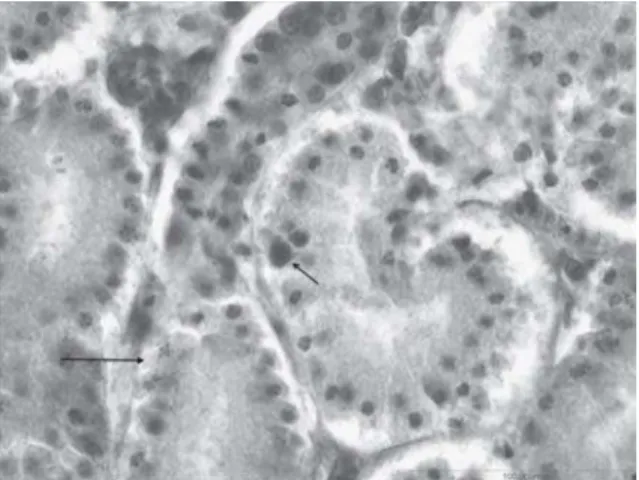
Necropsy and histopathology results revealed pathological changes characteristic of IBH (Howell et al., 1970; Macpherson et al., 1974; McFerran 2000). The predominating gross lesions were pale, swollen and friable livers, kidneys with subcapsular petechial hemorrhages and pale myocardium. Occasionally, mild tracheitis and catarrhal enteritis were noticed. All examined birds were in good body condition. Microscopic examination revealed acute hepatitis, with randomly distributed multifocal areas of acute necrosis, as well as numerous disseminated hepatocytes with large basophilic intranuclear inclusion bodies scattered among necrotic hepatocytes (Figure1). Multiple subcapsular hemorrhages, multifocal groups of hepatocytes with lipid degeneration, and cholestasis were also present. Similar large intranuclear basophilic inclusion bodies as in the liver and karyorrhexis were found in the red pulp cells of the spleen and in the tubular cells of the kidneys but were less frequent than in the liver (Figure 2).
determined partial hexon gene sequences from the liver and spleen samples were found to be 100% identical on the nucleotide level. According to the phylogenetic tree, the newly detected virus could be classified as FAdV type 8b, a member of the Fowl adenovirus E species (Figure 3). Based on literature data, FAdV-E type 8b is one of the most common causative agents involved in IBH (Christensen and Saifuddin, 1989; Ojki} et al., 2008a; Ojki} et al., 2008b). The nucleotide sequences described in the present paper were submitted to GenBank and assigned accession numbers JF766220 for DNA polymerase gene and JF766221 for hexon gene, respectively.
Figure 1. Hepatocytes with large basophilic intranuclear inclusion bodies (arrows) (400×)
The infection was also confirmed by virus isolation. All embryos inoculated with liver homogenate of affected chickens died by day 5 post inoculation. On histological examination, acute hepatitis with distortion of the liver plates, multiple necrotic areas and disseminated individual necrotic hepatocytes was diagnosed. Variation of the nuclear size was evident in the hepatocytes of affected embryos, but no inclusion bodies were found. The presence of FAdV-8b was detected by PCR from the liver tissue of the embryos.
also been described (Christensen and Saifuddin 1989; Gomiset al.,2006; Ojki}et al.,2008a; Ojki}et al.,2008b).
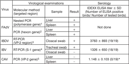
In our case, in addition to FAdV-8b, the presence of IBDV but not CAV was detected. RT-PCR performed to test IBDV in cloacal swabs resulted in a product of appropriate size of approximately 630-bp. However, direct sequencing of the PCR product from the VP2 gene failed to give unambiguous results. A concurrent infection with vaccine and field strains of IBDV apparently occurred, resulting in heterogeneous PCR products that could not be sequenced without prior molecular cloning. Serological testing at the age of 36 days of age revealed an antibody response to IBDV (Table 1) originating from vaccination or/and from field infection. Since Zorman Rojset al. (2011) obtained significantly lower antibody titers (from 102 to 518) detected by the same (IDEXX) ELISA system in non-infected broilers vaccinated with intermediate plus vaccine, field infection is most likely in the present case. Infection with pathogenic strains of IBDV, including some less attenuated vaccine strains, has well-known immunosuppressive effects in chickens and might induce the development of IBH (Fadlyet al.,1975; Rosenberget al.,1975; Toroet al.,2000).
Table 1. Summary of virological and serological results in IBH affected broiler flock
Virus
Virological examinations Serology
Molecular method
(targeted region) Sample Result
IDEXX ELISA titer ± SD (Number of ELISA positive birds/ Number of tested birds)
FAdV
Nested PCR (polymerase gene)1
Liver +
Not done
Spleen +
PCR (hexon gene)2 Liver +
Spleen +
IBDV RT-PCR(VP-2 region)3 Cloacal swab + 3783 ± 893 (19/19) IBV RT-PCR (S-1 gene)4 Tracheal swab + 1326 ± 650 (18/19)
Cloacal swab –
CAV PCR (VP-2 gene)5 Liver – 1.148 ± 0.103 (0/19)*
Spleen –
In addition, the presence of IBV in the trachea was confirmed. Further molecular investigation showed that strain QX was involved (accession number GU 564331). Tracheitis, found at necropsy in some of the submitted chickens, might have been caused by IBV infection. Presumably it did not have a significant influence on the course of IBH in our case. Interestingly, Ojki}et al.,(2008b) found that co-infections with other viruses (IBDV, IBV and reovirus) were more frequent in FAdV infections not related to IBH.
(2008) demonstrated by experimental infection that FAdV-4 caused a depletion of B- and T-lymphocytes in lymphoid organs in SPF chickens. The question of whether immunosuppressive interactions of FAdVs with the host organism are enough to cause clinical manifestation of different diseases, remains unanswered.
Address for correspondence: Marko Zadravec
Institute for Poultry Health Veterinary Faculty of Ljubljana Gerbi~eva 60
1000 Ljubljana, Slovenia
E-mail: marko.zadravecªvf.uni-lj.si
REFERENCES
1.Baar DA, Scott P, 1988, Adenovirus and IBH, In: Proc. 2ndAsian/Pacific Poultry Health Conf[Proc
112]. University of Sydney, Sydney, Australia, 323-6.
2.Barli~-Maganja D, Zorman Rojs O, Grom J, 2002, Detection of infectious bursal disease virus in different lymphoid organs by single-step reverse transcription polymerase chain reaction and microplate hybridization assay,J Vet Diagn Invest, 14, 243-6.
3.Benkö M, Harrach B, Both GW, Russell WC, Adair BM, Adam E et al., 2005, Family Adenoviridae, In:
Fauquet CM, Mayo MA, Maniloff J, Desselberger U and Ball LA (Eds.): Virus Taxonomy. VIIIth Report of the International Committee in Taxonomy of Viruses, Elsevir, New York, 213-28. 4.Cao YC, Yeung WS, Bi YZ, Leung FC Lim BL, 1998, Molecular characterization of seven Chinese
isolates of infectious bursal disease virus: classical, very virulent, and variant strains,Avian Dis,
42, 340-51.
5.Christensen NH, Saifuddin MD, 1989, A primary epidemic of inclusion body hepatitis in broilers,
Avian Dis, 33, 622-30.
6.Cowen BS, 1988, Chicken embryo propagation of type 1 avian adenoviruses,Avian Dis, 32, 347-52. 7.Fadly AM, Winterfield RW, Olander HJ, 1976, Role of Bursa of Fabricius in the pathogenicity of
inclusion body hepatitis and infectious bursal disease viruses,Avian Dis, 20, 467-77.
8.Felsenstein J, 1989, PHYLIP - Phylogeny Inference Package (Version 3.2), Cladistics 5, 164-6.
9.Gomis S, Goodhope R, Ojki} D, Willson P, 2006, Inclusion body hepatitis as a primary disease in broilers in Saskatchewan, Canada,Avian Dis, 50, 550-5.
10.Hall TA, 1999, BioEdit: a user-friendly biological sequence alignment editor and analysis program
for Windows 95/98/NT,Nucl Acids Symp Ser, 41, 95-8.
11.Howell J, MacDonald DW, Christian RG, 1970, Inclusion body hepatitis in chickens,Can Vet J, 11, 99-101.
12.Keeler CL Jr, Reed KL, Nix WA, Gelb J Jr, 1998, Serotype identification of avian infectious bronchitis virus by RT-PCR of the peplomer (S-1) gene,Avian Dis, 42, 275-84.
13.Kimura MA, 1980, Simple method for estimating evolutionary rates of base substitutions through
comparative studies of nucleotide sequences,J Mol Evol, 16, 111-20.
14. Macpherson I, McDougall JS, Laursen-Jones AP, 1974, Inclusion body hepatitis in a broiler integration,Vet Rec, 95, 286-9.
15.McConnell Adair B, Fitzgerald SD, 2008, Group I adenovirus infections, In: Saif YM (Ed.): Diseases of poultry, 12thed. Wiley-Blackwell, Iowa, 252-66.
16.McFerran JB, Smyth JA, 2000, Avian adenoviruses,Rev Sci Tech, 19, 589-606.
18. Meulemans G, Boschmans M, van den Berg TP, Decaesstecker M, 2001, Polymerase chain
reaction combined with restriction enzyme analysis for detection and differentiation of fowl adenoviruses,Avian Pathol, 30, 655-60.
19. Mobyleªpasteur, Available at: http://mobyle.pasteur.fr, Last modified March 3, 2011, Accessed April 20, 2011.
20.Noteborn MHM, Verschueren CAJ, van Roozelaar DJ, Veldkamo S, van der Eb AJ, Boer GF, 1992,
Detection of chicken anemia virus by DNA hybridization and polymerase chain reaction,Avian Pathol, 21, 107-18.
21.Ojki} D, Krell PK, Tuboly T, Nagy E, 2008a, Characterization of fowl adenoviruses isolated in Ontario and Quebec, Canada,Can J Vet Res, 72, 236-41.
22.Ojki} D, Martin E, Swinton J, Vaillancourt JP, Boulianne M, Gomis S, 2008b, Genotyping of Canadian
isolates of fowl adenoviruses,Avian Pathol, 37, 95-100.
23.Rosenberg JK, Klopp S, Krauss WC, 1975, The role of the infectious bursal agent and several avian adenoviruses in the hemorrhagic-aplastic-anemia syndrome and ganfrenous dermatitis,Avian Dis, 19, 717-29.
24.Saifuddin MD, Wilks CR, Murray A, 1992, Characterisation of avian adenoviruses associated with
inclusion body hepatitis,N Z Vet J, 40, 52-4.
25.Schonewille E, Singh A, Göbel TW, Gerner W, Saalmüller A, Hess M, 2008, Fowl adenoviruses (FAdV) serotype 4 causes depletion of B and T cells in lymphoid organs in specific pathogen-free chickens following experimental infection,Vet Immunol Immunopath, 121, 130-9.
26.Shivachandra SB, Sah RL, Singh SD, Kataria JM and Manimaran K, 2003, Immunosuppression in
broiler chicks fed aflatoxin and inoculated with fowl adenovirus serotype-4 (FAV-4) associated with hydropericardium syndrome,Vet Res Comm, 27, 39-51.
27.Tamura K, Dudley J, Nei M, Kumar S, 2007, MEGA4: Molecular Evolutionary Genetics Analysis (MEGA) software version 4.0,Mol Biol Evol, 24, 1596-9.
28.Thompson J, Higgins D and Gibson T,1994, CLUSTAL W: improving the sensitivity of progressive
multiple sequence alignment through sequence weighting, position-specific gap penalties and weight matrix choice,Nucl Acids Res, 22, 4673-80.
29.Toro H, Gonzales C, Cerda L, Hess M, Reyes E, Geisse C, 2000, Chicken anemia virus and fowl adenoviruses: association to induce the inclusion body hepatitis/hydropericardium syndrome,
Avian Dis, 44, 51-8.
30.Toro H, Gonzales C, Cerda L, Morales MA, Donner P, Salamero M, 2002, Prevention of inclusion body hepatitis/hydropericardium syndrome in progeny chickens by vaccination of breeders with fowl adenovirus and chicken anemia virus, Avian Dis, 46, 547-54.
31.Wellehan JFX, Johnson AJ, Harrach B, Benkö M, Pessier AP, Johnson CM et al., 2004, Detection and analysis of six lizard adenoviruses by consensus primer PCR provides further evidence of a reptilian origin for the Atadenoviruses,J Virol, 78, 13366-9.
32.Zorman Rojs O, Krape` U, Slavec B, Jur{i~-Cizerl R, Poljanec T, 2011, Field efficacy of different
POJAVA HEPATITISA SA INKLUZIJAMA (IBH) ZAJEDNO SA ADENOVIRUSOM @IVINE TIPA 8b KOD BROJLERA
ZADRAVEC M, SLAVEC BRIGITA, KRAPE@ U, KAJÁN GL, RA^NIK J,
JUNTES POLONA, JUR[I^ CIZERL RAHELA, BENKÕ MÁRIA i ZORMAN ROJS OLGA
SADR@AJ