85
Pakistan Veterinary Journal
ISSN: 0253-8318 (PRINT), 2074-7764 (ONLINE)Accessible at: www.pvj.com.pk
Avian Adenoviruses Infections with Special Attention to Inclusion Body Hepatitis/
Hydropericardium Syndrome and Egg Drop Syndrome
Hafez Mohamed Hafez*
Institute of Poultry Diseases, Faculty of Veterinary Medicine, Free University Berlin, Königsweg, 14163 Berlin, Germany
*Corresponding author: hafez@vetmed.fu-berlin.de
A R T I C L E H I S T O R Y A B S T R A C T Received:
Revised: Accepted:
October 30, 2010 December 14, 2010 December 15, 2010 Key words:
Avian Adenoviruses infections
Egg drop syndrome Hydropericardium syndrome
Inclusion body hepatitis
The first avian adenovirus (AAV) associated with clinical disease was isolated from an outbreak of respiratory disease in quail in 1950 (Olson, 1950). Since that time, AAVs have been found in all types and breeds of chickens and from a variety of other avian species. The infections may be asymptomatic or associated with several clinical and pathological conditions. Vertical transmission via the egg is the most common way of transmission. Also horizontal transmission through faeces, contaminated egg trays, crates and trucks play a role in the infection route. Studies have demonstrated the presence of antibodies in healthy poultry, and viruses have been isolated from normal birds. Avian adenoviruses in chickens are the etiological agents of 2 diseases known as inclusion body hepatitis (IBH) and hydropericardium syndrome (HP). In some cases each condition is observed separately, however, recently the 2 conditions have frequently been observed as a single entity; therefore, the name hepatitis hydropericardium has been widely used to describe the pathologic condition. The syndrome is an acute disease of young chickens associated with anemia, haemorrhagic disorders, hydropericardium and high mortality.
Egg-Drop-Syndrome (EDS) is caused also by an adenovirus. The disease is characterised by a severe drop in egg production as well as the production of shell-less, thin-shelled, discoloured or misshapen eggs in apparently healthy birds. Ducks and geese are the natural host of the EDS virus. It was first described in chickens in the 1970s and spread to several countries world wide. The birds usually do not show any other signs of disease, and mortality is not expected. There is no specific treatment of the AAV infections. Active immunization by vaccination using an inactivated is wide spread.
©2011 PVJ. All rights reserved To Cite This Article: Hafez HM, 2011. Avian adenoviruses infections with special attention to inclusion body hepatitis/hydropericardium syndrome and egg drop syndrome. Pak Vet J, 31(2): 85-92.
Etiology
The avian adenoviruses are non-enveloped DNA viruses. Adenoviruses are resistant to many several disinfectants and are relatively tolerant to heat and pH changes. Iodophor and aldehyde disinfectants seem to be effective if they are allowed to have contact to the virus for longer time. Composting infected chicken carcasses for 20 days completely inactivates the virus (Senne et al., 1994).
In the past the family Adenoviridae was divided by host range and antibody reactivity into two genera: the genus Mastadenovirus for viruses isolated from mammalian and the genus Aviadenovirus for viruses isolated from birds. Recently, the International Committee
formerly designated as group III avian adenoviruses, includes the Egg drop syndrome virus (EDS) (Table 1).
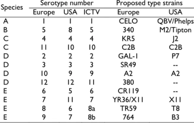
According to Jensen and Villegas (2005) the nomenclature used for the serotypes of avian adenoviruses from chicken has created some confusion as different systems have been used in Europe and the U.S.; however, a revised nomenclature system has been published (Benkő
et al., 2005) that, if adopted, will clarify matters (Table 2).
Table 1: Classification of adenoviruses from birds(Smyth and
McNulty, 2008)
Genus Species Serotype
Fowl adenovirus A FAdV-1
Fowl adenovirus B FAdV-5
FAdV-4 Fowl adenovirus C
FAdV-10 FAdV-2 FAdV-3 FAdV-9 Fowl adenovirus D
FAdV-11 FAdV-6 FAdV-7 FAdV-8a Fowl adenovirus E
FAdV-8b GoAdV-1 GoAdV-2 Goose adenovirus
GoAdV-3
(Duck adenovirus B) DAdV-2
(Pigeon adenovirus B) PiAdV
TAdV-1 Aviadenovirus
(Turkey adenovirus B)
TAdV-2
Siadenovirus Turkey adenovirus A TAdV-3
Atadenovirus Duck adenovirus A DAdV-1
Table 2: Classification of fowl adenoviruses (Jensen and
Villegas, 2005)
Serotype number Proposed type strains
Species
Europe USA ICTV Europe USA
A 1 1 1 CELO QBV/Phelps
B 5 8 5 340 M2/Tipton
C 4 4 4 KR5 J2
C 11 10 10 C2B C2B
D 2 2 2 GAL-1 P7
D 3 3 3 SR49 --
D 10 9 9 A2 A2
D 12 12 11 380 --
E 6 5 6 CR119 --
E 7 11 7 YR36/X11 X11
E 8 6 8a TR59 T8
E 9 7 8b 764 B3
Inclusion body hepatitis (IBH)/Hydropericardium syndrome (HPS)
IBH was first described in 1963 in the USA (Helmboldt and Frazier, 1963). Then after, the disease has been reported in many countries worldwide. It is a sporadic disease condition caused by several serotypes of fowl adenoviruses (Fitzgerald, 2008; Smyth and McNulty, 2008).
In 1988, a new broiler disease was reported from Angara Goth near Karachi in Pakistan and called as Angara Disease. The clinical signs and the course of the disease were similar to IBH. The main pathological findings were the accumulation of a clear, straw coloured fluid in the pericardial sac, therefore the disease was called Hydropericardium Syndrome “HPS” (Jaffery,
1988). The disease has subsequently been recorded in Iraq (Abdul-Aziz and Al-Attar, 1991), India (Gowda and Satyanarayana, 1994), Mexico, Ecuador, Peru, Chile (Voss et al., 1996; Toro et al., 1999), South and Central America (Shane, 1996), Slovakia (Jantosovic et al., 1991), Russia (Borisov et al., 1997) and Japan (Abe et al., 1998). An adenovirus was detected and later isolated (Rabbani and Naeem, 1996; Voss et al., 1996; Mazaheri et al.,
1998; Singh et al., 2002). The inclusion body hepatitis/hydropericardium syndrome (IBH/HP) has been reported to occur in both broilers and layers (Cowen, 1992). It seems that immunosuppression, prior to or concurrently with a FAdV infections, is necessary to develop IBH. Infectious bursal disease virus (IBDV), chicken anaemia virus (CAV) and mycotoxins are known to increase the pathogenicity of FAV infections (Rosenberger et al., 1975; Fadly et al., 1976; Bülow et al.,
1986; Toro et al., 2000; Shivachandra et al., 2003). However, several cases of IBH occurred without obvious influence of infectious immunosuppression (Reece et al.,
1986; Christensen and Saifuddin, 1989). On the other hand, Zavala et al. (2002) infected 1-day-old grandparent meat-type chickens carrying maternal antibodies against FAV with a field isolate of FAV associated with inclusion body hepatitis in broilers, avian leukosis virus subgroup J ALV-J, or both FAV and ALV-J and they found no significant differences in the dually infected birds in comparison with chickens that received a monovalent challenge with either FAV or ALV-J.
The infection is transmitted by vertical and horizontal means. Vertical transmission is reported as an important feature of fowl adenovirus (FAV) to spread from parent birds to progenies (McFerran and Adair, 1977; McCracken and Adair, 1993). Infected breeder shed virus to their progeny for three to six weeks until development of immunity occurs (Toro et al., 2001; Mazaheri et al., 2003). There is evidence that adenovirus infections can become latent and that periods of stress, such as the onset of egg production, will reactivate viral shedding (Girshick
et al., 1980). Fadly et al. (1980) reported that there is evidence that adenovirus infection can remain latent and undetected for at least one generation in a specific-pathogen-free flock.
The bird-to-bird transmission of the virus in a flock occurs horizontally by the oral-faecal route and further spread take place by mechanical means and by contamination with infected faeces. Commercial hatching eggs may be a mechanism of spread of AAV from one country to another (Cook, 1974; Ahmad et al., 1992; Akhtar et al., 1992; Akhtar, 1995; Adair and Smyth, 2008a).
Under field condition the disease is characterized by sudden onset of mortality in chickens < 6 weeks old and as young as 4 days of age. Mortality normally ranges from 2-40 percent, especially when birds are < 3 weeks of age. However, there have been outbreaks in which mortality has reached 80 % depending on the pathogenicity of the virus, immune status of the chicks and concurrent secondary infections. Mortality generally peaks within three to four days and ceases within 9-14 days. Clinically the birds showed lethargy, huddling with ruffled feathers, inappetence and yellow, mucoid droppings may be seen. The infection can be accompanied with bad feed conversion and a reduced weight gain (Anjum et al., 1989; Cowen, 1992).
Gross lesions include an enlarged pale and friable liver sometimes with necrotic foci, also ecchymotic haemorrhages may be present in the liver and less consistently ecchymotic haemorrhage can be observed in leg and breast muscles (Howell et al., 1970, Macpherson
et al., 1974; McFerran and Adair, 1977). The heart can be flabby with a mild hydropericardium. In case HPS a straw-coloured transudate is present in the pericardial sac (Anjum et al., 1989). In addition, nephritis, enlarged spleens and thymus atrophy could be observed in most dead birds. Histopathological lesions include necrotic focal lesions and some of the livers had basophilic intranuclear inclusion bodies. Haemorrhages under the epicardium with multifocal necrosis in the myocard are the major findings in the hearts and lymphoid depletion of spleen, thymus and bursa of Fabricius could be observed (Ahmad et al., 1989; Gowda and Satyanarayana, 1994). The liver showed histological changes, such as small multifocal areas of coagulative necrosis, mononuclear cell infiltration and the presence of basophilic inclusion bodies in the hepatocytes surrounded by a clear halo or filling the entire enlarged nucleus (Kumar et al., 1997; Nakamura et al., 1999).
Egg drop syndrome (EDS)
EDS is a disease characterized by a drastic drop in egg production as well as the production of abnormal eggs in apparently healthy chickens and quails. The disease was firstly described in 1976 by Van Eck et al. (1976) in The Netherlands. Thereafter, the disease was observed in several countries around the world including France (Picault, 1978), Great Britain (Baxendale, 1978), Northern Ireland (McFerran et al., 1978), Belgium (Meulemans et al., 1979), Hungary (Zsák and Bartha, 1979), Israel (Malkinson and Weisman 1980), Australia (Firth et al., 1981), Japan (Yamaguchi et al., 1981), Singapore (Singh and Chew-Lim, 1981), Taiwan (Lu, et al., 1985), South Africa (Bragg, et al., 1991), India (Kumar et al., 1992), China (Zhu and Wang, 1994), Bolivia (Bishop and Cardozo, 1996). The antibody to the virus was demonstrated from chickens in Denmark (Badstue and Smidt, 1978), Brazil, (Hwang et al., 1980); Mexico (Rosales et al., 1980), Nigeria (Nawathe and Abegunde, 1980), USA (Calnek, 1978), Germany (Kaleta et al., 1980) and New Zealand (Howell, 1982).
The initial outbreak in chickens was probably caused by a contaminated vaccine grown in duck embryo fibroblasts, since antibodies to EDS virus have been detected in the sera of duck, geese, and herring gulls prior
to 1975 (Bartha et al., 1982) i.e. before the diseases was recognized in hens (McFerran, 1979). In addition, the EDS outbreaks were observed in the quail flocks reared together with infected chickens and resulted in the fall of the egg production and in the increase of number of soft-shelled eggs (Das and Pradhan, 1992).
In spite of the fact that the disease outbreaks were mostly recorded in laying hens only and some time in quails, EDSV or the antibodies against the virus have been detected in ducks and geese (Schlör, 1980), pheasants, guinea fowls (Zanella et al., 1980), pigeon (Durojaiye et al., 1992) and in wild birds (Malkinson and Weisman, 1980).
In 2001, EDSV showed to cause a severe acute respiratory disease of the young goslings in Hungary. The disease affected goslings between 4 and 20 days of age. The symptoms included anorexia, depression, sneezing, coughing, dyspnoea, and rales (Ivanics et al., 2001). Recently, Biđin et al. (2007) reported on a naturally occurring EDS in turkey breeder flocks in Croatia, which were accompanied with a significant decrease in both egg quality and production.
The disease caused is by duck adenovirus a member of genus Atadenovirus. The virus has haemagglutination activity and has its reservoir in ducks and geese. The complete nucleotide sequence data revealed that it is an intermediate virus between mammalian and avian adenoviruses (Hess et al., 1997).
EDS virus transmits vertically from hens to chicks and also horizontally from chicken to chicken (Cook and Darbyshire, 1980 and 1981; Darbyshire and Peters, 1980). Contaminated eggs as well as egg trays or faeces seem to be the main sources for virus spread (Smyth and Adair, 1988). However, some outbreaks have been attributed to contact with wild birds or water contaminated by feces from wild birds.
Smyth et al. (1988) carried out an investigation on the pathogenesis of EDS in laying hen. After experimental infection viral antigen and intranuclear inclusion bodies were detected in the surface epithelium of the nasal cavity of conventional hens 2 to 6 days p.i. Low levels of viral antigen were detected in lymphoid tissue throughout the body 2 to 5 days p.i. and inflammatory lesions and viral antigen were observed in the infundibulum 3 to 5 days p.i.. Viral replication was first detected in the pouch shell gland (PSG) 8 days p.i.. Viral antigen was never detected in the surface epithelium of the alimentary tract.
many outbreaks to levels lower than half the pre-disease level. Subsequently, flocks gradually resume production to levels that could be considered normal for birds. Outbreaks lasted approximately 3 to 8 weeks (Van Eck, 1976; McFerran et al., 1978).
Although signs of EDS are quite characteristic, diagnosis must not be made on the clinical picture alone but should be confirmed by laboratory tests, since several infectious and non infectious causes can cause drop in egg production and might impair the external as well as internal egg quality. Examples are infectious bronchitis (IB), Newcastle disease (ND), avian encephalomyelitis (AE), fowl pox, infectious laryngotracheitis (ILT), avian metapneumovirus (AmPV), Mycoplasma gallisepticum (MG), and Mycoplasma synoviae (MS). Among the parasites, nematodes (Ascaridia, Capillaria, and Heterakis) may be responsible for egg drops. In addition several non-infectious factors such as stocking density, management and quality of feed and water are involved in egg production failures and should be considered (Meulemans, 1993; Gupta, 2008; Feberwee et al., 2009)
At necropsy there is no specific lesion, but a slight atrophy of ovary and oviduct can be observed. Histopathological changes can be seen in the oviduct and uterus (shell gland). There may be severe degeneration and desquamation of the epithelial cells, atrophy of the uterine glands and infiltration of heterophils, lymphocytes, and plasmacytes. Intranuclear inclusion bodies may be found in the epithelial cells of the uterus, isthmus, and vaginal gland region (Adair and Smyth, 2008b; Smyth and McNulty, 2008).
Diagnosis of Adenovirus infections
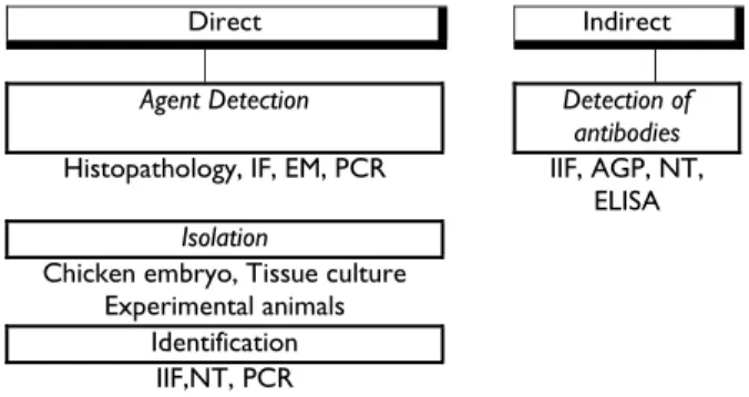
The diagnosis of poultry diseases in less developed regions of the World is generally based upon case history, clinical signs and post-mortem examination as important steps toward disease diagnosis, but it should not be the final step. In most cases clinical signs and lesions of many diseases are similar and laboratory tests are required to identify the specific cause (Fig. 1).
Direct Indirect
Agent Detection Detection of
antibodies
Histopathology, IF, EM, PCR IIF, AGP, NT,
ELISA
Isolation
Chicken embryo, Tissue culture Experimental animals
Identification
IIF,NT, PCR
Fig. 1: Laboratory diagnosis of poultry diseases
The laboratory diagnosis can be applied to direct detection as well as for isolation and identification of the causative agent or indirectly to detect antibodies (Hafez and Hess, 1999). The diagnosis of adenovirus infections in poultry is in most cases based on histological investigations and detection of intranuclear inclusion bodies in hepatocytes or on detection of the antigen or virus particles using Immunofluorescence test or electron
microscopy. In the last few years several molecular biological tools such as PCR, Real-time PCR and REA were developed allow the detection of the Virus – DNA as well as the further identification and typing of adenoviruses (Erny et al., 1991; Raue and Hess, 1998; Hess et al., 1999; Hess, 2000; Raue et al., 2002; Lüschow
et al., 2007; Steer et al., 2009).
However the isolation of the aviadenoviruses using chicken embryo liver (CEL) cell culture and chicken embryo fibroblast cell culture with further identification and determination of the pathogenicity seems to be very important, since the pathogenicity of the isolates within the same serotype can be widely differ. The cross neutralization tests and /or molecular biological tools are necessary to serotype the isolated virus and to determine a new serotype (McCracken and Adair, 1993; Kumar et al., 2003; Lüschow et al., 2007; Steer et al., 2009).
EDSV can be isolated in embryonated duck or goose eggs, and in cell cultures. Susceptible cell lines include duck and chick embryo liver, duck kidney, and fibroblast cells. Also isolation has been made in chicken embryo kidney or liver cells (Adair et al., 1979). The virus may be isolated directly from the reproductive tract of affected hens. Alternatively, abnormal eggs may be fed to naive hens; virus isolation is attempted from the shell gland of these hens when they produce abnormal eggs (McFerran and Adair, 2003).
The most common serologic test is the immunodiffusion test that detects the group specific antigen. This test is not sensitive enough. A group specific ELISA and IIF tests are more sensitive. The serum neutralization test has been used to detect serotype-specific antibody but is labour intensive and expensive. In general the interpretation of serologic tests is difficult because antibodies against AAVs can be found in both healthy and diseased birds (McFerran and Adair, 2003).
In addition for the detection of antibodies against EDSV haemagglutination inhibition using fowl RBC can also use. Dhinakar Raj et al. (2007) developed immunofiltration (flow through) test to detect the presence of antibodies to egg drop syndrome 76 (EDS) virus in chicken sera and compared it with HI and ELISA. In total, the immunofiltration test could detect EDS antibodies with a sensitivity and specificity of 90.14 and 92.86%, respectively as compared to the HI test. Compared to ELISA, the sensitivity and specificity of the developed immunofiltration assay was 79.45 and 94.58%, respectively. The disadvantage of this test is the qualitative detection of antibodies in the serum, which may not be highly informative on all occasions and this test can be used as a preliminary test before confirmation can be done by another more sophisticated laboratory based assay.
Control of Adenovirus infections of poultry
Control of IBH/HPS
pressure the disease has been brought under control by formalin-inactivated vaccines prepared from liver homogenates from infected birds or by inactivated cell culture – derived (Shane, 1996, Kumar et al., 1997; Kataria et al., 1997; Balamurugan and Kataria, 2004).
Roy et al. (1999) compared two inactivated vaccines prepared from the liver of experimentally infected chickens. One was prepared without adjuvants described by (Afzal and Ahmed, 1990). For the 2nd one, liquid paraffin was used as adjuvants after chloroform extraction and inactivation with formalin. The oil emulsion vaccine applied subcutaneously at 10th day of age provided 100% cent protection up to seven weeks of age against challenge. The inactivated vaccine without adjuvant was less efficacious. In a similar study by Afzal and Ahmed (1990), owing to the poor performance of a similar vaccine, they suggested that birds should be vaccinated twice at 10 and 21 days of age. In the field trial, the oil emulsion vaccine was highly effective; there were no deaths due to hydropericardium syndrome and birds were sold to the market at eight weeks.
Balamurugan and Kataria (2004) review the experiences of several authors using the vaccines to control HPS in poultry. In two field trials, involving 570 000 birds on 128 farms, the overall mortality ranged from 0.77 to 3.8% in vaccinated and from 11.11 to 30% in unvaccinated birds (Ahmad et al., 1990). In another trial, the mortality in vaccinated birds was 0.52% compared to 5.34% in unvaccinated birds kept on the same premises. Vaccination was also effective when carried out in the face of an outbreak; mortality in the vaccinated infected birds being 2.33% compared with 10.27% in unvaccinated infected birds (Afzal and Ahmad 1990). Inactivated chicken liver cell culture and embryonated egg-propagated vaccine used subcutaneously at 103.5 LD50/dose/bird provided protection against challenge with 1 ml of a 20% liver homogenate at a biological titre of 2× 10⁵ LD50/0.5 ml (Naeem et al., 1995a, b). Shane (1996) evaluated five inactivated vaccines used in Mexico. Complete protection, with an absence of histological changes in chicks challenged with 103.5LD50 of the DCV-94 adenovirus strain, was observed. Icochea et al. (2001) evaluated the efficacy of three inactivated vaccines against IBH/HPS in Peru in two different experiments and concluded that the protective effect of a commercial oil-adjuvant cell culture IBH vaccine was superior to the autogenous vaccines and that the mortality rates were not dose-dependent. As most cases of IBH are the result of vertical transmission, vaccines have been proved to be highly successful at controlling IBH by preventing vertical transmission and inducing maternal immunity (Toro et al., 2002; Alvarado et al., 2007). Toro et al. (2002) reported that effective protection of the progeny of chickens against IBH-HPS could be achieved by dual vaccination of breeders with FAV-4 and CAV.
Alvarado et al. (2007) isolated pathogenic adenovirus, identified as Stanford strain and characterized as European serotype 9. The level of protection against IBH was evaluated in two broiler-breeder progenies from AAV 8/11– vaccinated grandparent flocks and a commercial broiler flock by challenge at 1 or 7 days of age with the AAV 8 and 11 serotypes and/or the Stanford strain. The broiler-breeder progenies and the commercial
broiler flock exhibited protection against IBH after challenge. They conclude that broiler-breeder progenies from 30- to 50-wk-old grandparents vaccinated with the AAV 8/11 vaccine were adequately protected against challenge with the AAV 8 and 11 serotypes and the Stanford strain (serotype 9).
Control of EDS
Beside biosecurity, vaccination with an inactivated vaccine prior to lay is mostly important to prevent egg production losses and reduced egg shell quality in commercial layer and breeder flocks. Initial vaccination occurs between 14 and 16 weeks of age. However, vaccination cannot completely inhibit virus excretion in feces, but decrease virus shedding (Heffels et al., 1982).
References
Abdul-Aziz TA and MA Al-Attar, 1991. New syndrome in Iraqi chicks. Vet Rec, 129: 272.
Abe T, K Nakamura, H Tojo, M Mase, T Shibahara, S Yamaguchi and N Yuasa, 1998. Histology, immunohistochemistry and ultra-structure of hydropericardium syndrome in adult broiler breeders and broiler chicks.Avian Dis, 42: 606-612.
Adair BM and JA Smyth, 2008a. Group I Adenovirus infections. In: Diseases of Poultry, 12th Ed, Saif YM, Fadly AM, Glisson JR, McDougald LR, Nolan LK and Swayne DE, eds. Iowa State University Press, Iowa, USA, pp: 252-266.
Adair BM and JA Smyth, 2008b. Egg drop syndrome. In: Diseases of Poultry, 12th Ed, Saif YM, Fadly AM, Glisson JR, McDougald LR, Nolan LK and Swayne DE, eds. Iowa State University Press, Iowa, USA, pp: 267-276.
Adair BM, JB McFerran, TJ Connor, MS McNulty and ER McKillop, 1979. Biological and Physical properties of a virus (strain127) associated with egg drop syndrome 1976. Avian Pathol, 8: 249-264. Afzal M and I Ahmad, 1990. Efficacy of an inactivated
vaccine against hydropericardium syndrome in broilers. Vet Rec, 126: 59-60.
Ahmad I, M Afzal, MI Malik, Z Hussain and W Hanif, 1989. Studies on the disease pattern and etiology of hydropericardium syndrome (Angara disease) in broiler chickens in Pakistan. Pak J Agric Res, 10: 195-199.
Ahmad I, MI Malik, K Iqbal, K Ahmed and S Naz, 1990. Efficacy of formalinized liver-organ-vaccine against Angara diseases in broilers. Vet Arhiv, 60: 131-138. Ahmad K, I Ahmad, MA Muneer and M. Ajmal, 1992.
Experimental transmission of Angara disease in broiler fowls. Stud Res Vet Med, 1: 53-55.
Akhtar S, 1995. Lateral spread of the aetiologic agent(s) of hydropericardium syndrome in broiler chickens. Vet Rec, 136: 118-120.
Akhtar S, S Zahid and MI Khan, 1992. Risk factors associated with hydropericardium syndrome in broiler flocks. Vet Rec, 131: 481-484.
with an avian adenovirus isolate associated with inclusion body hepatitis. Avian Dis, 51: 27-32.
Anjum AD, MA Sabri and Z Iqbal, 1989. Hydropericarditis syndrome in broiler chickens in Pakistan. Vet Rec, 124: 247-248.
Badstue, P B and B Smidt, 1978. Egg drop syndrome 76 in Danish poultry. Nord Vet Med, 30: 498-505.
Balamurugan V and JM Kataria, 2004. The hydropericardium syndrome in poultry - a current scenario. Vet Res Commun, 28: 127-148.
Bartha A, J Meszaros and J Tanyi, 1982. Antibodies againsr EDS-76 avian adenovirus in bird species before 1975. Avian Pathol, 11: 511-513.
Baxendale W, 1978. Egg drop syndrome 76. Vet Rec, 102: 285-286.
Benkő M, B Harrach, WC Russell, BM Adair, É Ádám, JC De Jong, M Hess, M Johnson, A Kajon, AH Kidd, HD Lehmkuhl, QG Li, V Mautner, P Pring-Akerblom and G Wadell, 2005. Family Adenoviridae. In: Virus Taxonomy. Classification and Nomenclature of Viruses. The 8th Report of the International Committee on Taxonomy of Viruses. (Fauquet CM, Mayo MA, J Maniloff, U Desselberger, LA Ball; eds.). Elsevier/Academic Press, London, pp: 213-228. Biđin Z, I Lojkić, M Mikec and B Pokrić, 2007. Naturally Occurring Egg Drop Syndrome Infection in Turkeys. Acta Vet Brno, 76: 415-421.
Bishop SC and P Cardozo, 1996. Egg Drop syndrome '76 in Bolivia. Trop Anim Health Prod, 28: 199-206. Borisov VV, AV Borisov and AA Gusev, 1997.
Hydropericardium syndrome in chickens in Russia. Proc 10th Int Cong World Vet Poult Assoc (Budapest, Hungary), pp: 258.
Bragg RR, DM Allwright and L Coetzee, 1991. Isolation and identification of adenovirus 127, the causative agent of egg drop syndrome (EDS), from commercial laying hens in South Africa. Onderstepoort J Vet Res, 58: 309-310.
Bülow von V, R Rudolph and B Fuchs, 1986. Folgen der Doppeltinfektion von Küken mit Adenovirus oder Reovirus und dem Erreger der infektiösen Anämie. J Vet Med B, 33: 717-726.
Butcher GD, JP Jacob and FB Mather, 1999. Common Poultry Diseases. http://edis.ifas.ufl.edu/ps044
Calnek BW, 1978. Hemagglutination-inhibition antibodies against an adenovirus (virus 127) in white Pekin ducks in the United States. Avian Dis, 22: 798-801.
Christensen NH and MD Saifuddin, 1989. A primary epidemic of inclusion body hepatitis in broilers. Avian Dis, 33: 622-630.
Cook JKA, and JH Darbyshire, 1980. Epidemiological studies with egg drop syndrome-1976 (EDS-76) virus. Avian Pathol, 9: 437-443.
Cook JKA, and JH Darbyshire, 1981. Longitudinal studies on the egg drop syndrome 1976 (EDS 76) in the fowl following experimental infection at 1-day old. Avian Pathol, 10: 449-459.
Cook JKA, 1974. Spread of an avian adenovirus (CELOvirus) to uninoculated fowls. Res Vet Sci, 16: 156-161.
Cowen B, 1992. Inclusion body hepatitis-anemia and hydropericardium syndromes: aetiology and control. World's Poult Sci J, 48: 247-254.
Darbyshire, JH, and RW Peters, 1980. Studies on EDS-76 virus infection in laying chickens. Avian Pathol, 9: 277-290.
Das BB and HK Pradhan, 1992. Outbreaks of egg drop syndrome due to EDS-76 virus in quail (Coturnix coturnix japonica). Vet Rec, 131: 264-265
Dhinakar R, G, V Thiagarajan and K Nachimuthu, 2007. Detection of antibodies to egg drop syndrome virus in chicken serum using a field-based immunofiltration (flow-through) test. Avian Dis, 51: 788-790.
Durojaiye OA, AS Ahmed and DF Adene, 1992. Egg drop syndrome ‘76 in poultry and other avian species in Nigeria. Rev Elev Méd Vét Pay Trop, 44: 37-38. Erny KM, DA Barr and KJ Fahey, 1991. Molecular
characterization of highly virulent fowl adenoviruses associated with outbreaks of inclusion body hepatitis. Avian Path, 20: 597-606.
Fadly AM, RW Winterfield and HJ Olander, 1976. Role of the bursa of Fabricius in the pathogenicity of inclusion body hepatitis and infectious bursal disease viruses. Avian Dis, 20: 467-472.
Fadly, AM, BJ Riegle, K Nazerian and EA Stephens, 1980. Some observations on an adenovirus isolated from specific pathogen-free chickens. Poult Sci, 59: 21-27.
Feberwee A, JJ de Wit and WL Landman, 2009. Induction of eggshell apex abnormalities by Mycoplasma synoviae: field and experimental studies. Avian Pathol, 38: 77-85.
Firth GA, MJ Halland and JB McFerran, 1981. Isolation of haemagglutinating adeno-like virus related to virus 127from an Australian poultry flock with an egg drop syndrome. Aust Vet J, 57: 239-242.
Fitzgerald SV, 2008. Adenovirus Infections. In: Diseases of Poultry, 12th Ed, Saif YM, AM Fadly, JR Glisson, LR McDougald, LK Nolan and DE Swayne, eds. Iowa State University Press, Iowa, USA, pp: 251-266.
Girshick T, CK Crary and RE Luginbuhl, 1980. Serologic detection of adenovirus infections in specific-pathogen-free chickens. Avian Dis, 24: 527-531. Gowda RNS and ML Satyanarayana, 1994.
Hydropericardium syndrome in poultry. Indian J Vet Pathol, 18: 159-161.
Gupta L, 2008. Maintaining Egg Shell Quality. http://www.thepoultrysite.com/articles/979/maintaini ng-egg-shell-quality
Hafez HM and M Hess, 1999. Modern techniques in diagnosis of poultry diseases. Archiv für Geflügelkunde, 63: 237-245.
Heffels U, SED Khalaf and EF Kaleta, 1982. Studies on the persistence and excretion of egg drop syndrome 1976 virus in chickens. Avian Pathol, 11: 441-452. Helmboldt CF and MN Frazier, 1963. Avian hepatic
inclusion bodies of unknown significance. Avian Dis, 7: 446-450.
Hess M, 2000. Detection and differentiation of avian adenoviruses: a review. Avian Path, 29: 195-206. Hess M, H Blocker and P Brandt, 1997. The complete
an intermediate between mastadenoviruses and aviadenoviruses. Virology, 238: 145-156.
Hess M, R Raue and HM Hafez, 1999. PCR for specific detection of haemorrhagic enteritis virus of turkeys, an avian adenovirus. J Virol Methods, 81: 199-203. Howell J, 1982. Egg drop syndrome in Ross Brown hens:
An interim report. Surveillance,9: 10-11.
Howell J, DW McDonald and RG Christian, 1970. Inclusion body hepatitis in chickens. Can Vet J, 11: 99-101.
Hwang MH, JM Lamas, O Hipolito and EN Silva, 1980. Egg drop syndrome 1976 a serological survey in Brazil. Proc 6th Eur Poult Conf, Hamburg, Germany, pp: 371-378.
Icochea E, M Alba, L Fiory and A Ramirez, 2001. Eficacia de tres vacunas inactivadas contra la hepatitis a corpusculos de inclusion y sindrome del hidropericardion en el peru. http://www. visionveterinaria.com/articulos/09.html.
Ivanics E, V Palya, R Glávits, Á Dán, V Pálfi, T Révész and M Benkö, 2001. The role of egg drop syndrome virus in acute respiratory disease of goslings. Avian Pathol, 30: 201-208.
Jaffery MS, 1988. A treatise on Angara disease (hydropericardium pulmonary oedema – hepatoneph- ritis syndrome). Pak Vet Med Assoc, pp: 1-33. Jantosovic J, J Konard, J Saly, I Skardova, J Kusev and K
Beninghausova, 1991. Hydropericardium syndrome in chicks. Veterinastvi, 41: 261-263.
Jensen L and P Villegas, 2005. Inclusion body hepatitis: control in breeder and broiler chickens. AviaTech- Eric Technical Information for the Broiler Industry, 2: 1-6.
Kaleta EF, SED Khalaf and O. Siegman, 1980. Antibodies to egg drop syndrome 76 virus in wild birds in possible conjunction with egg-shell problems. Avian Pathol, 9: 587-590.
Kataria JM, KC Verma, SJ Jadhao, JN Deepak and RL Shah, 1997. Efficacy of an inactivated oil emulsified vaccine against inclusion body hepatitis-hydropericardium syndrome (Litchi disease) in chicken prepared from cell culture propagated fowl adenovirus. Indian J Comp Microbiol Immunol Infect Dis, 18: 38-42.
Kumar R, R Chandra and SK Shukla, 2003. Isolation of etiological agent of hydropericardium syndrome in chicken embryo liver cell culture and its serological characterization. Indian J Exp Biol, 41: 821-826. Kumar R, R Chandra, SK Shukla, DK Agrawal and M
Kumar, 1997. Hydropericardium syndrome in India: a preliminary study on causative agent and control of disease by inactivated autogenous vaccines. Trop Anim Health Prod, 29: 158-164.
Kumar R, GC Mohanty, KC Verma and Ram-Kumar, 1992. Epizootiological studies on egg drop syndrome in poultry. Indian J Animal Sci, 62: 497-501.
Lu Y S, DF Lin, HJ Tasi, YL Lee, SY Chui, C. Lee, and ST Huang,1985. Outbreaks of egg drop syndromen1976 in Taiwan and isolation of the etiological agent. J Chinese Soc Vet Sci, 11: 157-165. Lüschow D, C Prusas, M Lierz, H Gerlach, D Soike and
HM Hafez, 2007. Adenovirus of psittacine birds: investigations on isolation and development of a
real-time polymerase chain reaction for specific detection. Avian Path, 36: 487-494.
Macpherson I, JS McDougall and AP Laursen-Jones, 1974. Inclusion body hepatitis in a broiler integration. Vet Rec, 95: 286-289.
Malkinson M and Y Weisman, 1980. Serological survey for the prevalence of antibodies to egg drop syndrome 1976 virus in domesticated and wild birds in Israel. Avian Pathol, 7: 483-490.
Mazaheri A, C Prusas, M Voss and M Hess, 1998. Some strains of serotype 4 fowl adenoviruses cause inclusion body hepatitis and hydropericardium syndrome in chickens. Avian Path, 27: 269-276. Mazaheri A, C Prusas, M Vossand M Hess, 2003. Vertical
transmission of fowl Adenovirus serotype 4 investigated in specified pathogen-free birds after experimental infection. Archiv für Geflügelkunde, 67: 6-10.
McCracken JB and BM Adair, 1993. Avian adenoviruses. In: Virus infections of birds; JB McFerran and MS McNulty (eds), Elsevier Science Publishers B.V., Amsterdam, pp: 123-144.
McFerran JB and BM Adair, 1977. Avian adenoviruses - A review. Avian Pathol, 6: 189-217.
McFerran JB and BM Adair, 2003. Egg Drop Syndrome. In: Poultry Diseases, Saif YM, HJ Barnes, JR Glisson, AM Fadly, LM McDougald, and DE Swayne (eds). 11th Ed, Iowa State Press, Ames, Iowa, USA, pp: 227-237.
McFerran JB, 1979. Egg drop syndrome. Vet Quart, 1: 176-180.
McFerran JB, RM McCracken, ER McKillop, R Eileen, MS McNulty and DS Collins, 1978. Studies on a depressed egg production syndrome in Northern Ireland. Avian Pathol, 7: 35-47.
Meulemans G, 1993: Aetiology and diagnosis of drops in egg production. In: Virus infections of birds; McFerran JB and MS McNulty (eds), Elsevier Science Publishers BV, Amsterdam, pp: 555-567. Meulemans G, D Dekegel, J Peeters, E Van Meirhaeghe
and P Halen, 1979. Isolation of an adenolike virus from laying chickens affected by egg drop syndrome 1976. Vlaams Diergeneeskd Tijdschr, 2: 151-157. Naeem K, M Rabbani, M Hussain and AH Cheema,
1995b. Development of cell culture vaccine against HPS in poultry. Pak Vet J, 15: 150-151.
Naeem K, T Niazi, SA Malik and AH Cheema, 1995a. Immunosuppressive potential and pathogenicity of an avian adenovirus isolate involved in hydro-pericardium syndrome in broilers. Avian Dis, 39: 723-728.
Nakamura K, M Mase, S Yamaguchi, T Shiobahara and N Yuasa, 1999. Pathologic study of specific pathogen free chicks and hens inoculated with adenovirus isolated from hydropericardium syndrome. Avian Dis, 43: 414-423.
Nawathe DR and A Abegunde, 1980. Egg drop syndrome 76 in Nigeria: Serological evidence in commercial farms. Vet Rec, 107: 466-467.
Picault JP, 1978. Chutes de ponte associees a la production d`oeufs sans coquille fragile: Proprietes de l`agent infectious isole au cours de la maladie. L`Aviculteur, 379: 57-60.
Rabbani M and K Naeem, 1996. In vitro and in vivo evaluation of avian adenovirus isolates from outbreaks of hydropericardium syndrome. Proc Int Symp Adenovirus and Reovirus Infections in Poultry, Rauischholzhausen, Germany, pp: 26-31.
Raue R and M Hess, 1998. Hexon based PCRs combined with restriction enzyme analysis for rapid detection and differentiation of fowl adenoviruses and egg drop syndrome virus. J Virol Methods, 73: 211-217. Raue R, HM Hafez and M Hess, 2002. A fiber gene-based
polymerase chain reaction for specific detection of pigeon adenovirus. Avian Path, 31: 95-99.
Reece RL, DC Grix and DA Barr, 1986. An unusual case of inclusion body hepatitis in a cockerel. Avian Dis, 30: 224-227.
Rosales G, A Antillon and C Morales, 1980. Reporte en Mexico sobre la presencia de anticuerpos contra el adenovirus causante del syndrome de la baja en postura (CEPABC-14) en parvadas de gallinas domesticas. Proc 29th West Poultry Dis Conf, pp: 192-196.
Rosenberger JK, S Klopp, RF Eckroade and WC Krauss, 1975. The role of the infectious bursal disease agent and several avian adenoviruses in the haemorrhagic-aplastic anemia syndrome and gangrenous dermatitis. Avian Dis, 19: 717-729.
Roy P, M Koteeswaran and R Manickam, 1999. Efficacy of an inactivated oil emulsion vaccine against hydropericardium syndrome in broilers. Vet Rec,
145: 458-459.
Schlör GM, 1980. Frequency of antibody to adenovirus 127 in domestic ducks and wild waterfowl. Avian Dis, 24: 91-98.
Senne DA, B Panigrahy and R Morgan, 1994. Effect of composting poultry carcasses on survival of exotic avian viruses: highly pathogenic avian influenza (HPAI) virus and adenovirus of egg drop syndrome-76. Avian Dis, 38: 733-737.
Shane SM, 1996. Hydropericardium-hepatitis syndrome, the current world situation. Zootecnica Int, 18: 20-27. Shivachandra SB, RL Sah, SD Singh, JM Kataria, and K
Manimaran, 2003. Immunosuppression in broiler chicks fed aflatoxin and inoculated with fowl adenovirus serotype-4 (FAV-4) associated with hydropericardium syndrome. Vet Res Comm, 27: 39-51.
Singh A, MS Oberoi, GS Grewal, HM Hafez and M Hess, 2002. The use of PCR combined with restriction enzyme analysis to characterize fowl adenovirus field isolated from Northern Indian. Vet Res Comm, 26: 577-585.
Singh KY and M Chew-Lim, 1981. Breeder farm egg drop syndrome 1976 (EDS76) in Singapore. Singapore Vet J, 5: 8-13.
Smyth JA and BM Adair, 1988. Lateral transmission of egg drop syndrome-76 virus by the egg. Avian Path, 17: 193-200.
Smyth JA and MS McNulty, 2008. Adenoviridae. In: Poultry Diseases. 6th Ed, Pattison M, PF McMullin,
JM Bradbury and D Alexander (eds), Butterworth Heinemann-Elsevier, pp: 367-381.
Smyth JA, MA Plattern and JB McFerran, 1988. A study of the pathogenesis of egg drop syndrome in laying hens. Avian Pathol, 17: 653-666.
Steer PA, NC Kirkpatrick, D O'Rourke and AH Noormohammad, 2009. Classification of fowl adenovirus serotypes by use of High-Resolution Melting-Curve Analysis of the hexon gene region. J Clin Microbiol, 47: 311-321.
Toro H, C Gonzalez, L Cerda, M Hess, E Reyes and C Geissea, 2000. Chicken anemia virus and fowl adenoviruses: Association to induce the inclusion body hepatitis/hydropericardium syndrome. Avian Dis, 44: 54-58.
Toro H, C Gonzalez, L Cerda, MA Morales, P Dooner and M Salamero, 2002. Prevention of inclusion body hepatitis/hydropericardium syndrome in progeny chickens by vaccination of breeders with fowl adenovirus and chicken anemia virus. Avian Dis, 46: 547-554.
Toro H, C Prusas, R Raue, L Cerda, C Geisse, C González and M Hess, 1999. Characterization of fowl adenoviruses from outbreaks of inclusion body hepatitis/hydropericardium syndrome in Chile. Avian Dis, 43: 262-270.
Toro H, O González, C Escobar, L Cerda, MA Morales and C González, 2001. Vertical induction of the inclusion body hepatitis/hydropericardium syndrome with fowl adenovirus and chicken anemia virus. Avian Dis, 45: 215-222.
Van Eck JHH, FG Davelaar, TAM Van den Heuvel-Plesman, N Van Kol, B Kouwnhoven and FHM Guldie, 1976. Dropped egg production, soft shelled and shell-less eggs associated with appearance of precipitins to adenovirus in flocks of laying fowl. Avian Pathol, 5: 261-272.
Voss M, E Vielitz, M Hess, CH Prusas and A Mazaheri, 1996. Aetiological aspects of hepatitis and HPS caused by pathogenic adenoviruses in different countries. Proc Int Symp Adenovirus and Reovirus Infections in Poultry, Rauischholzhausen, Germany, pp: 75-78.
Yamaguchi S, H Imada, H Kawamura, T Taniguchi, H Saio and K Shimamatsu, 1981. Outbreaks of egg drop syndrome 1976 in Japan and its etiological agent. Avian Dis, 25: 628-641.
Zanella A, A Di Donato, A Nigrelli and G Poli, 1980. Egg drop syndrome (EDS´76), ethiopatogenesis, epidemiology, immunology and control of the disease. Clin Vet, 103: 459-469.
Zavala G, L Dufour-Zavala, P Villegas, J El-Attrache, DA Hilt and MW Jackwood, 2002. Lack of interaction between Avian Leukosis virus subgroup J and Fowl Adenovirus (FAV) in FAV-Antibody-Positive chickens. Avian Dis, 46: 979-984.
Zhu GQ and YK Wang, 1994. Study on egg drop syndrome 1976 (EDS-76) and its control. J Jiangsu Agric Coll, 15: 5-13.