RESUMO.- [Adenovirus aviário Ggrupo I como um agen-te causal da síndrome da hepatiagen-te por corpúsculo de in-clusão/hidropericárdio (IBH/HPS) em lotes de frangos de corte.]Lotes comerciais de frangos de uma granja locali-zada no Estado de São Paulo, Brasil, apresentavam diarreia, depressão, aumento de mortalidade e baixo ganho de peso. Após o exame post-mortem, sinais clássicos da síndrome de hepatite por corpúsculo de inclusão/hidropericárdio (IBH/HPS) foram observados incluindo hepatomegalia com aspecto amarelado pálido e líquido de coloração amarelo palha no saco pericárdio. Além disso, as alterações macros-cópicas foram também observadas nos rins, pâncreas, timo, intestinos e vesícula biliar. Amostras destes órgãos foram
analisadas pela técnica de PCR para detectar o adenovírus aviário do grupo I através do gene Hexon. Os resultados fo-ram positivos para ambos os lotes (A e B) utilizando-se a técnica de PCR. As lesões macroscópicas associadas à detec-ção do adenovírus aviário do grupo I pela técnica de PCR em vários destes órgãos acometidos permitiu a identifica -ção da síndrome de hepatite/hidropericárdio em frangos no Brasil. Ao nosso conhecimento, este é a primeira descrição da síndrome de hepatite/hidropericárdio causado por ade-novírus aviário do grupo I, no Brasil. Estes achados podem contribuir com a epidemiologia mundial do adenovírus me-diando a síndrome de hepatite/hidropericárdio.
TERMOS DE INDEXAÇÃO: Adenovírus aviário, síndrome hepatite por corpúsculo de inclusão/hidropericárdio, IBH/HPS, galinha.
INTRODUCTION
Fowl Adenovirus (FAdVs) expression appears to be ubi-quitous in domesticated fowl and is often isolated from asymptomatic chickens (McFerran & Adair 2003, Wang
Fowl adenovirus Group I as a causal agent of inclusion body
hepatitis/hydropericardium syndrome (IBH/HPS) outbreak
in brazilian broiler flocks
1Elena Mettifogo2, Luis F.N. Nuñez2, Silvana H. Santander Parra2, Claudete S. Astolfi-Ferreira2 and Antonio J. Piantino Ferreira2*
ABSTRACT.- Mettifogo E., Nuñez L.F.N., Santander Parra S.H., Astolfi-Ferreira C.S. & Ferrei -ra A.J.P. 2014. Fowl adenovirus Group I as a causal agent of inclusion body hepatitis/
hydropericardium syndrome (IBH/HPS) outbreak in brazilian broiler flocks. Pesqui-sa Veterinária Brasileira 34(8):733-737. Departamento de Patologia, Faculdade de Medicina Veterinária e Zootecnia, Universidade de São Paulo, Av. Prof. Dr. Orlando Marques de Paiva 87, São Paulo, SP 05508-270, Brazil. E-mail: ajpferr@usp.br
Commercial broiler flocks from a farm located in the State of São Paulo, Brazil, pre -sented diarrhea, depression, increased mortality and poor weight gain. Upon post-mor-tem examination, classical signs of Inclusion Body Hepatitis/Hydropericardium Syndrome (IBH/HPS) were observed, including enlarged pale yellow-colored livers and straw-colo-red liquid in the pericardial sac. In addition, gross lesions were also observed in the kid-neys, pancreas, thymus, intestines and gallbladder. Samples of these organs were analyzed by PCR for the detection of the hexon gene of the Fowl Adenovirus (FAdVs) Group I. The results were positive for both flocks (A and B) assayed by PCR. The macroscopic lesions associated with the detection of FAdV Group I by PCR in several of these affected organs allowed for the identification of IBH/HPS. In fact, this is the first report in Brazil of IBH/HPS in broilers, which identifies FAdVs group I as a causal agent of the disease. These findin -gs may contribute to the worldwide epidemiology of the adenovirus-mediated hepatitis/ hydropericardium syndrome.
INDEX TERMS: Fowl adenovirus, hepatitis, hydropericardium, IBH/HPS syndrome, chicken.
1 Received on November 27, 2013. Accepted for publication on June 23, 2014.
et al. 2011). However, the hepatitis syndrome characteri-zed by corpuscle inclusion (Inclusion Body Hepatitis-IBH) has attracted the attention of the poultry industry and the scientific community since the emergence of IBH in the 1960s and subsequent significant economic losses. This disease was originally described by Hemboldt & Frazier in the USA (1963) in seven-week-old chickens that exhibited necrotizing hepatitis and the presence of inclusion bodies in hepatocytes. In 1987, a new presentation of the disease emerged that was called hydropericardium-hepatitis syn-drome (HPS) or Angara disease. HPS was associated with the same group of viruses, and it devastated the poultry industry of Pakistan (Cowen et al. 1978, Hess et al. 1999, Asthana et al. 2013). The syndrome and its various mani-festations have been reported in several countries in Nor-th and SouNor-th America, Europe, Asia and Oceania, causing considerable economic losses (Mendelson et al. 1995, Toro et al. 1999, Ono et al. 2003, Rahul et al. 2005, Gomis et al. 2006, Manarolla et al. 2009, Mase et al. 2009, Alemnesh et al. 2010, Choi et al. 2012). Adenoviruses belong to the fa-mily Adenoviridae. The genus Mastadenovirus contains ade-noviruses that infect mammals, whereas the genus Aviade-novirus has been isolated from birds. Avian adenoviruses are further subdivided into three serological groups (I - III). The Fowl Adenoviruses (FAdVs) belong to Group I, which comprises five species (A to E) and 12 serotypes (1 to 12) and share a common group antigen with viruses isolated from chickens, geese, ducks and turkeys (Alemnesh et al. 2010, Senties-Cue et al. 2010).
In Brazil, the first detection of Fowl adenovirus group 1 (chicken) was demonstrated by antibody detection with immunodiffusion tests for the presence of Avian adenovirus group 1. Although Fowl adenovirus group 1 was present in 78.2% of serum collected, its expression was not related to any disease (Romero et al. 1989). Recently, another survey demonstrated that turkeys remain free of avian adenovirus group 2 (Moura-Alvarez et al. 2013), and the first molecu -lar detection of FAdV-1 in enteric content related to enteric diseases was performed by our research group (Mettifogo et al. 2014).
This paper describes the first report of Inclusion Body Hepatitis/Hydropericardium Syndrome (IBH/HPS) in Bra-zilian broiler chickens flocks, identifying avian adenovirus group 1 as a causative agent of the disease. Furthermore, this paper describes the differential diagnosis of IBH/HBS
and other viral infections by PCR and observations of clini-cal signs and pathologiclini-cal lesions.
MATERIALS AND METHODS
Description of clinical history
A commercial poultry company’s flock of broilers had a conti -nuous history of malabsorption syndrome, with poor weight gain, high feed conversion rates and diarrhea. The onset of these signs began the fourth week of bird life, and signs were more
pronoun-ced in flocks that were housed during months with higher tem
-peratures. The farm had 21 flocks, with a population of 22,000 to 41,000 birds per flock and a density of 14.6 to 15.5 birds/m2. Im-munization procedures were routinely performed in the hatchery against Infectious Bronchitis Virus (IBV-Bio Bronk-Vet H120), In-fectious Bursal Disease Virus (IBDV-Gumbor-Vet), Marek’s disea-se (HVT-Bio Mark-Vet L) and fowl pox virus (FPV-Bouba Suave). All vaccines were obtained from the Bio Vet Laboratory, Vargem Grande Paulista, SP, Brazil. According to veterinarian reports, some attempts at antibiotic treatments were performed at the onset of
signs, including bacitracin at 300 ppm/ton of feed for five days as a
preventive treatment. This procedure was repeated 10 days later,
no significant improvement was observed.
On 13 March, 2011, two flocks (A and B) were chosen for
analysis based on case history, clinical signs, necropsy and
labora-tory analysis. In the two flocks, the broilers were 41 days old and
were at the end of their life cycle. The main clinical signs presented
by the broilers were severe diarrhea, prostration, ruffled feathers,
depigmentation of the leg skin, severe dermatitis, increased mor-tality after the fourth week of life, high feed conversion (1: 2.10) and low weight gain. Poultry litter was very wet as a result of
se-vere diarrhea. In flock “A”, the mortality rate reached 5%, and the final weight loss was 37%; in flock “B”, mortality was 2.4%, and
the weight gain was 45%, which was lower than expected.
Virus detection by PCR technique
Ten chickens (five birds/flock) were sacrificed for postmor -tem examination. Fragments of the organs with or without gross
lesions were collected and frozen at – 20˚C for PCR to detect the
Fowl Adenovirus (FAdVs) Group I hexon gene, and for differential diagnosis for Infectious Bronchitis virus (IBV), avian reovirus, In-fectious Bursal Diseases virus (IBDV), and Chicken Anemia virus (CAV) according to the authors in Table 1. All samples were tested
in duplicate, since nuclei acid extraction until the final PCR process.
Serology
Additionally, serum samples (five birds/flock “A” and “B”) were
also collected to determine the presence of antibodies against In-fectious Bronchitis Virus (IBV), InIn-fectious Bursal Disease virus
Table 1. Primers sets, nucleotide sequences, amplicon sizes and the corresponding references that were used to screen for the viruses
Virus detected Primers Nucleotide sequence (5’ - 3’) Amplicon size Reference
(bp)
Avian Adenovirus Hexon A CAA RTT CAG RCA GAC GT 897 Meulemans et al. group I Hexon B TAG TGA TGM CGS GAC ATC AT 2001 Infectious Bronchitis UTR 41+ ATG TCT ATC GCC AGG GAA ATG TC 179 Cavanagh et al. Virus UTR 31 GGG CGT CCA AGT GCT GTA CCC 2002 UTR 11 GCT CTA ACT CTA TAC TAG CCT A
Avian Reovirus S4-F13 GTG CGT GTT GGA GTT TCC CG 1120 Pantin-Jackwood et al.
S4-R1133 TAC GCC ATC CTA GCT GGA 2008
Infectious Bursal VP2F ACC ATA AAC GCC GTG ACC 631 Yamaguchi et al. Diseases Virus VP2R CCG TGG ATC GTC ACT GCT A 1996 Chicken Anemia CAV4a GAC TGT AAG ATG GCA AGA CGA GCT C 675 Todd et al.
Table 3. ELISA results for Chicken Anemia Virus, Reovirus, Infectious Bronchitis Virus and Infectious Bursal Disease Virus and the average antibody titers of positive samples
from the A and B flocks
Agent Flock A Flock B
+/n PC NC Positive +/n PC NC Positive samples‡GMT samples‡GMT
CAV 5/10 0.229 0.826 0.321 2/10 0,229 0.826 0.230↑
Reovirus 8/10 0.164 0.059 0.292↑ 6/10 0.164 0.059 0.109 IBV 5/10 0.377 0.053 0.224↑ 3/10 0.377 0.053 0.150 IBDV 10/10 0.318 0.051 0.303 10/10 0.318 0.051 0.332↑
CAV = Chicken Anemia Virus, IBV = Infectious Bronchitis Virus, IBDV = Infec-tious Bursal Disease Virus, +/n = positive samples/total of samples, PC = positive control, NC = negative control, ‡ Average titer of positive samples.
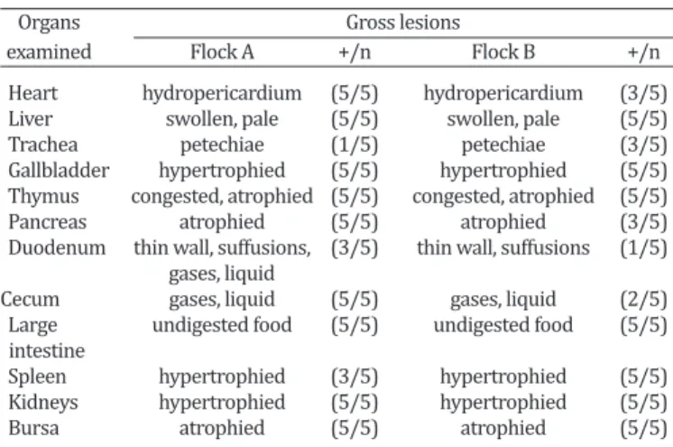
Table 2. Gross lesions observed in organs during post-mortem
examination of five broilers from each flock
Organs Gross lesions
examined Flock A +/n Flock B +/n
Heart hydropericardium (5/5) hydropericardium (3/5) Liver swollen, pale (5/5) swollen, pale (5/5) Trachea petechiae (1/5) petechiae (3/5) Gallbladder hypertrophied (5/5) hypertrophied (5/5) Thymus congested, atrophied (5/5) congested, atrophied (5/5) Pancreas atrophied (5/5) atrophied (3/5) Duodenum thin wall, suffusions, (3/5) thin wall, suffusions (1/5) gases, liquid
Cecum gases, liquid (5/5) gases, liquid (2/5) Large undigested food (5/5) undigested food (5/5) intestine
Spleen hypertrophied (3/5) hypertrophied (5/5) Kidneys hypertrophied (5/5) hypertrophied (5/5) Bursa atrophied (5/5) atrophied (5/5) +/n = positive samples/number total of samples.
(IBDV), Reovirus and Chicken Anemia Virus (CAV), but not for Fowl Adenovirus group 1, by competitive enzyme-linked immu-nosorbent assay (ELISA, Idexx Laboratories, Inc., Westbrook, MN, USA). All sera were screened in duplicate.
RESULTS
During post-mortem examinations, the classical gross le-sions of hepatitis and hydropericardium were mainly ob-served in flock “A”, which presented enlarged, pale yellow --colored livers and straw--colored liquid in the pericardial sac (Fig.1 and 2). Approximately 7 ml of liquid could be extracted from the pericardial sac of a bird from flock “A”. Moreover, more birds had organs presenting lesions in flock “A” than in flock “B”. In addition, the intensity of the macroscopic lesions was also more pronounced in flock “A”, particularly in the heart, pancreas and duodenum (Table 2). Our results reflect an impairment of the birds from flock “A” and atrophy of the lymphoid organs (thymus and bur -sa) in the two flocks. ELISA tests performed on both flocks were positive for reovirus and IBV in the flock “A”; CAV and IBDV in the flock “B” (Table 3). According to IBDV and IBV antibodies titer values were consistent with vaccinated bir-ds and did not represent active infection.
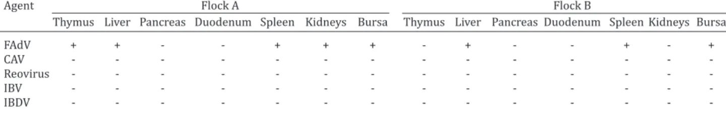
The results of PCR performed on samples of each pool of organs (thymus, liver, pancreas, duodenum, spleen, kid-neys and bursa of Fabricius) for the detection of FAdVs group I, CAV, reovirus, IBV and IBDV are shown in Table 4.
The macroscopic lesions of the hydropericardium and pale-swollen liver that were observed in the broilers, toge-ther with the PCR results, indicate that the clinical signs ob-served were predominantly due to an adenovirus infection. In addition to these signs, gross lesions were also present in other organs, and PCR analysis revealed FAdVs group I ex-pression in the thymus, pancreas, kidneys, and duodenum.
DISCUSSION
Inclusion body hepatitis (IBH) and Hydropericardium syn-drome (HPS) have been described as entities characterized by hepatic (swelling, discoloration, ecchymosis or petechial hemorrhages) and renal dysfunction (kidney inflammation, distension of the tubules and degenerative changes). These entities are characterized by basophilic inclusion bodies or eosinophilic hepatocytes and have been associated mainly with the group of adenoviruses belonging to serotypes 4, 8, 11 and 12 (McFerran & Adair 2003, Alemnesh et al. 2010). Although classical lesions in the heart and liver are charac-teristic of HPS, FAdVs have also been reported to cause le-sions in other organs such as the pancreas, proventriculus, and lymphoid organs and in the respiratory system.
Dama-Fig.1. Broiler chicken with hydropericardium and hepatitis. The liver is pale, enlarged and discolored.
ge to these organs caused by adenoviruses has been descri-bed previously, both alone and in combination with other viruses (Cowen et al. 1978).
In this report, atrophy of lymphoid organs (thymus and bursa) was observed in both flocks (A and B) and was as -sociated with both FAdVs group I and CAV. Similar patho-logical lesions in lymphoid organs have been described in broilers with CAV. These lesions include thymic, splenic and bursal atrophy, aplastic bone marrow and anemia, which may vary in severity depending on the presence of other pathogens (Adair 2000). However, the increased intensity of gross lesions in flock “A”, which had lower antibody ti -ters and was negative for CAV as assessed by PCR, suggests that the detected serotype of FAdV is capable of induc-ing the macroscopic lesions observed in these lymphoid organs.
Based on the available information on the vaccination programs performed at the hatchery against IBV and IBVD and the results obtained here, the lower antibody titers against the diseases observed in flocks A and B indicated that these antibodies were induced by vaccination and not by field virus infection. By contrast, the antibody titers against reovirus indicate a field virus infection because the flocks were not vaccinated against this disease. However, PCR analysis revealed that the organs were negative for reovirus. Although reovirus has been reported to cause in-juries similar to those caused by FAdV (Ni & Kemp 1995), this virus causes tenosynovitis and consequently was not considered to be one of the possible causative agents of the signs observed in the chickens in this report.
CAV antibodies were higher in flock B, in which the signs and gross lesions were less intense than those obser-ved in flock A. Furthermore, the PCR results indicated that this flock was negative for CAV. It has long been believed that adenoviral IBH results only when birds are also infec-ted with IBDV, CAV, or other immunocompromising agents (McFerran & Adair 2003). In the 1970s, researchers were able to reproduce the disease using isolates from infected birds presenting lesions in the lymphoid tissue (McCracken et al. 1976, Grimes et al. 1977). Outbreaks of IBH in whi-ch no immunosuppressive pathogens were detected have been reported in Northern Ireland, Australia, Korea and New Zealand - countries that have experienced epidemic outbreaks of IBH in the absence of IBDV (Reece et al. 1986, Christensen & Saifuddin 1989, Choi et al. 2012). Experi-ments performed by Toro and colleagues could not confirm the hypothesis that the association of CAV and FAdVs group I is necessary for the induction of IBH/HP syndrome (Toro
et al. 2001). Those authors followed the premise that cer-tain isolates, such as FAdV-4, can reproduce the syndrome by themselves, but other strains appear to require the pre-sence of an immunosuppressive agent.
According to our results, the hepatitis and hydropericar-dium observed in the broilers were caused mainly by FAdV Group I. The presence of CAV according to ELISA and its ac-companying immunosuppressive effects most likely caused a synergistic effect with FAdV that may have accentuated the general signs. To our knowledge, this is the first report of FAdV infection causing hepatitis and hydropericardium syndrome in Brazil. Further investigation is needed to veri-fy the spread of this virus in Brazilian poultry flocks.
CONCLUSION
This is the first report of Inclusion body hepatitis/hydrope -ricardium syndrome (IBH/HPS) caused by Fowl Adenovi-rus group I in Brazil. Indeed, this description may contribu-te to the worldwide epidemiology of adenovirus-mediacontribu-ted hepatitis/hydropericardium syndrome.
Acknowledgments.- This study was sponsored by FAPESP (Fundação de Amparo à Pesquisa do Estado de São Paulo), grant #06/59332-9, and
CNPq (Conselho Nacional de Desenvolvimento Científico e Tecnológico).
REFERENCES
Adair C.W. 2000. Immunopathogenesis of chicken anemia virus infection. Dev. Comp. Immunol. 24:247-255.
Alemnesh W., Hair-Bejo M., Aini I. & Omar A.R. 2010. Pathogenicity of fowl
adenovirus in specific pathogen free chicken embryos. J. Comp. Pathol.
1:1-7.
Asthana M., Chandra R. & Kumar R. 2013. Hydropericardium syndrome: current state and future developments. Arch. Virol. 158:921-931. Cavanagh D., Mawditt D., Welchman B., Britton P. & Gough R.E. 2002.
Co-ronaviruses from Pheasants (Phasianuscolchicus) are genetically closely related to coronaviruses of domestic fowl (infectious bronchitis vírus) and turkeys. Avian Pathol. 31:81-93.
Choi K.S., Kye S.J., Kim J.Y., Jeon W.J., Lee E.K., Park K.Y. & Sung H.W. 2012. Epidemiological investigation of outbreaks of fowl adenovirus infection in commercial chickens in Korea. Poult. Sci. 91:2502-2506.
Christensen N.H. & Saifuddin M.D. 1989. A primary epidemic of inclusion body hepatitis in broilers. Avian Dis. 33:622-630.
Cowen B., Mitchell G.B. & Calnek B.W. 1978. An adenovirus survey of
poul-try flocks during the growing and laying periods. Avian Dis. 22:115-121.
Gomis S., Goodhope A.R., Ojkic A.D. & Willson P. 2006. Inclusion body he-patitis as a primary disease in broilers in Saskatchewan, Canada. Avian Dis. 50:550-555.
Grimes T.M., King D.J., Kleven S.H. & Fletcher O.J. 1977. Involvement of a type-8 avian adenovirus in the etiology of inclusion body hepatitis. Avian Dis. 21:26-38.
Table 4. PCR results for the detection of Fowl Adenovirus (FAdV), Chicken Anemia Virus (CAV), Reovirus, Infectious Bronchitis Virus (IBV), and Infectious Bursal Disease Virus (IBDV) from samples of several organs of chickens from
flocks A and B
Agent Flock A Flock B
Thymus Liver Pancreas Duodenum Spleen Kidneys Bursa Thymus Liver Pancreas Duodenum Spleen Kidneys Bursa
FAdV + + - - + + + - + - - + - +
CAV - - -
-Reovirus - - -
-IBV - - -
-Helmbolt C.F. & Frazier M.N. 1963. Avian hepatic inclusion bodies of
un-known significance. Avian Dis. 7:446-450.
Hess M., Raue R. & Prusas C. 1999. Epidemiological studies on fowl adeno-viruses isolated from cases of infectious hydropericardium. Avian Dis. 28:433-439.
Manarolla G., Pisoni G., Moroni P., Gallazzi D., Sironi G. & Rampin T. 2009.
Adenoviral gizzard erosions in Italian chicken flocks. Vet. Rec. 164:754-756.
Mase M., Mitake H., Inoue T. & Imada T. 2009. Identification of group I-III
avian adenovirus by PCR coupled with direct sequencing of the hexon gene. J. Vet. Med. Sci. 71:1239-1242.
McCracken R.M., McFerran J.B., Evans R.T., Connor T.J. 1976. Experimen-tal studies on the a etiology of inclusion body hepatitis. Avian Pathol. 5:325-339.
McFerran J.B. & Adair B.M. 2003. Group I Adenovirus Infections, p.214-227. In: Saif Y.M., Fadly A.M., Glisson J.R., McDougald L.R., Nolan L.K. & Swayne D.E. (Eds), Diseases of Poultry. 12th ed. Blackwell Publishing, Iowa.
Mettifogo E., Nuñez L.F., Chacón J.L., Santander Parra S.H., Astolfi-Ferreira
C.S., Jerez J.A., Jones R.C. & Piantino Ferreira A.J. 2014. Emergence of en-teric viruses in production chickens is a concern for avian health.
Scien-tific World Journal. ID 450423. http://dx.doi: 10.1155/2014/450423.
Meulemans G., Boschmans M., Van den Berg T.P. & Decaesstecker M. 2001. Polymerase chain reaction combined with restriction enzyme analysis for detection and differentiation of fowl adenoviruses. Avian Pathol. 30:655-660.
Mendelson C., Nothelfer H.B. & Monreal G. 1995. Identification and charac
-terization of an avian adenovirus isolated from a “spiking mortality syn
-drome” field outbreak in broilers on the Delmarva. Pennsilvania, USA.
Avian Pathol. 24:693-706.
Moura-Alvarez J., Chacon J.V., Scanavini L.S., Nuñez L.F., Astolfi-Ferreira
C.S., Jones R.C. & Piantino Ferreira A.J. 2013. Enteric viruses in Brazilian
turkey flocks: single and multiple virus infection frequency according to
age and clinical signs of intestinal disease. Poult. Sci. 92:945-955. Ni Y. & Kemp M.C. 1995. A comparative study of avian reovirus
pathoge-nicity: virus spread and replication and induction of lesions. Avian Dis. 39:554-566.
Ono M., Okuda Y., Yazawa S., Shibata I., Sato S. & Okada K. 2003. Outbreaks of adenoviral gizzard erosion in slaughtered broiler chickens in Japan. Vet. Rec. 153:775-779.
Pantin-Jackwood M.J., Day J.D., Jackwood M.W. & Spackman E. 2008. En-teric viruses detected by molecular methods in commercial chicken and
turkey flocks in the United States between 2005 and 2006. Avian Dis. 52:235-244.
Rahul S., Kataria J.M., Senthilkumar N., Dhama K., Sylvester S.A. & Uma R. 2005. Association of fowl adenovirus serotype 12 with hydropericar-dium syndrome of poultry in India. Acta Virol. 49:139-143.
Reece R.L., Grix D.C. & Barr D.A. 1986. An unusual case of inclusion body hepatitis in a cockerel. Avian Dis. 30:224-227.
Romero C.H., Brentano L., Rowe C.A., Wentz I., Flores R.S. & Rodrigues J.C. 1989. Ocorrência de anticorpos para vírus aviários em frangos de corte em região de intensa produção avícola. Pesq. Vet. Bras. 9:1-7.
Senties-Cue C.G., Wills R.W., Stayer P.A., Burleson M.A. & Magee D.L. 2010. Epidemiology and Effect of Production Parameters of an Outbreak of In-clusion Body Hepatitis in Broilers. Avian Dis. 54:74-78.
Todd D., Mawhinney K.A. & McNulty M.S. 1992. Detection and differen-tiation of chicken anemia virus isolates by using the polymerase chain reaction. J. Clin. Microbiol. 30:1661-1666.
Toro H., González O., Escobar C., Cerda L., Morales M.A. & Gonzalez C. 2001. Vertical induction of the inclusion body hepatitis/hydropericardium syndrome with fowl adenovirus and chicken anemia virus. Avian Dis. 45:215-222.
Toro H., Prusas C., Raue R., Cerda L., Geisse C., González C. & Hess M. 1999. Characterization of fowl adenoviruses from outbreaks of inclu-sion body hepatitis/hydropericardium syndrome in Chile. Avian Dis. 43:262-270.
Wang J., Zhu L., Zhu J., Sun H. & Zhu G. 2011. Molecular characterization and phylogenetic analyses of an avian adeno-associated virus originat-ing from a chicken in China. Arch. Virol. 77:71-77.
Yamaguchi T., Ogawa M., Inoshima Y., Miyoshi M., Fukushi H. & Hirai K.