Epizootics in the Indo-Pacific
Meir Sussman1,2*, Bette L. Willis1, Steven Victor3, David G. Bourne2
1ARC Centre of Excellence for Coral Reef Studies, School of Marine and Tropical Biology, James Cook University, Townsville, Australia,2Australian Institute of Marine Science (AIMS), PMB3, Townsville MC, Townsville, Australia,3Palau International Coral Reef Center (PICRC), Koror, Republic of Palau
Abstract
Background:White Syndrome (WS), a general term for scleractinian coral diseases with acute signs of advancing tissue
lesions often resulting in total colony mortality, has been reported from numerous locations throughout the Indo-Pacific, constituting a growing threat to coral reef ecosystems.
Methodology/Principal Findings: Bacterial isolates were obtained from corals displaying disease signs at three WS
outbreak sites: Nikko Bay in the Republic of Palau, Nelly Bay in the central Great Barrier Reef (GBR) and Majuro Atoll in the Republic of the Marshall Islands, and used in laboratory-based infection trials to satisfy Henle-Koch’s postulates, Evan’s rules and Hill’s criteria for establishing causality. Infected colonies produced similar signs to those observed in the field following exposure to bacterial concentrations of 16106cells ml21. Phylogenetic 16S rRNA gene analysis demonstrated that all six
pathogens identified in this study were members of thec-ProteobacteriafamilyVibrionacae, each with greater than 98%
sequence identity with the previously characterized coral bleaching pathogenVibrio coralliilyticus. Screening for proteolytic activity of more than 150 coral derived bacterial isolates by a biochemical assay and specific primers for aVibriofamily zinc-metalloprotease demonstrated a significant association between the presence of isolates capable of proteolytic activity and observed disease signs.
Conclusion/Significance:This is the first study to provide evidence for the involvement of a unique taxonomic group of
bacterial pathogens in the aetiology of Indo-Pacific coral diseases affecting multiple coral species at multiple locations. Results from this study strongly suggest the need for further investigation of bacterial proteolytic enzymes as possible virulence factors involved inVibrioassociated acute coral infections.
Citation:Sussman M, Willis BL, Victor S, Bourne DG (2008) Coral Pathogens Identified for White Syndrome (WS) Epizootics in the Indo-Pacific. PLoS ONE 3(6): e2393. doi:10.1371/journal.pone.0002393
Editor:Niyaz Ahmed, Centre for DNA Fingerprinting and Diagnostics, India
ReceivedMarch 4, 2008;AcceptedApril 2, 2008;PublishedJune 18, 2008
Copyright:ß2008 Sussman et al. This is an open-access article distributed under the terms of the Creative Commons Attribution License, which permits unrestricted use, distribution, and reproduction in any medium, provided the original author and source are credited.
Funding:This study was funded by an ARC Discovery grant and a James Cook University CRIG grant to BL Willis. The authors wish to thank CD Harvell, Chair of the World Bank Coral Disease Working Group, L Raymundo, University of Guam, and A Hooten from the IOC-GEF/World Bank Coral Reef Targeted Research and Capacity Building Program for contributing funding and support for travel and field work in Palau. Work on the research vessel Lady Basten and in the PC2 laboratory at Cape Ferguson was funded by the Australian Institute of Marine Science.
Competing Interests:The authors have declared that no competing interests exist.
* E-mail: dsmb007@jcu.edu.au
Introduction
Reports on coral disease continue to rise [1] with currently 29 reported syndromes in the Caribbean [2] and 7 syndromes reported from the Indo-Pacific [3]. However, the causes for coral disease and the methods by which to investigate them are still heavily debated [4–6]. Most efforts are directed towards traditional surveillance [7], with comparatively less research directed towards developing strategies for active engagement in coral reef health management, disease prevention and cure [8– 10]. Unfortunately, a lack of knowledge of coral disease causative agents propels this debate to a stand still. To date, only 5 bacterial species and one fungal agent have been determined as causative agents for coral infectious diseases [11–17], and currently no diagnostic tools or management efforts are able to validate these findings at a level required for active intervention. [18–19].
The study of disease in complex environmental settings is often difficult. Modern studies have cast a shadow on traditional
presence once detected. Nevertheless, the benefits from isolating and culturing pathogens are still many, especially when precise disease identification for health control purposes is needed [31].
The study of epidemiology has revolutionized many concepts associated with disease studies [32] including some of the terminology used in infectious disease classifications. Traditional distinctions between primary vs. secondary, exogenous vs. endogenous and opportunistic agents [33–34] are being replaced by schemes classifying the genes involved in infectivity (the ability to physically infect a host [35]) and virulence (the severity of disease outcome inflicted by infection [36]). Modern studies have demonstrated that host, pathogen and environment form a constantly evolving disease equilibrium [37] contributing to a growing list of newly emerging infectious diseases [38]. The hierarchy of causation has been translated into causal models and complex outbreaks are now considered as multi-factorial, comprised of an often-unknown range of component causes [32], which need to be explored both independently and in conjunction with other causes. Nevertheless it remains a paradox, that despite the growing complexity in our understanding of disease causation, it is often expected that emerging infectious outbreaks be successfully curtailed before causation is fully established [39], shifting the focus from cure of individuals to disease-prevention in entire populations.
The aims of this study were therefore twofold: firstly, to identify possible causative agents for white syndromes widespread throughout the Indo-Pacific by combining both traditional microbial tools such as culturing with biochemical and molecular methods, and secondly, to investigate the aetiology of WS in order to recommend the development of novel diagnostic tools that could be implemented and validated in an active coral reef health management plan targeted ‘‘to protect against disease in the framework of the concept of ecosystem management’’ [40].
Since 2003, a variety of white syndromes have been reported from numerous locations throughout the Indo-Pacific and under various names [3,41–44]. Willis et al. [3] suggested the use of a common term: white syndrome (WS), for Indo-Pacific scleracti-nian coral diseases displaying acute tissue loss exposing white skeleton in the absence of other disease signs or established causation. Three independent WS outbreaks were chosen for this 3-year study (2003–2006) in order to determine whether WS is one disease or possibly many, and whether a standard disease investigation protocol could be developed that could be used in future monitoring and management efforts (for a short video clip of a WS outbreak in the Republic of the Marshall Islands see Movie S1 in Supporting Information).
Results
Higher bacterial counts on WS corals
Densities of cultivable bacteria (measured as CFU’s ml21g21 wet weight) associated with corals sampled from each of the three Indo-Pacific outbreak sites examined in this study were signifi-cantly higher on corals displaying disease signs than on those lacking disease signs (Fig. 1A–C). Mean CFU’s from Pachyseris speciosasamples collected from Nikko Bay Palau (Fig. 1A) plated on Figure 1. Bacterial density on corals sampled from the field: A.
Mean CFU’s ml21g21 from crushed Pachyseris speciosa fragments
sampled in Nikko Bay Palau. B.Mean CFU’s ml21
g21
from crushed
Montipora aequituberculata fragments sampled in Nelly Bay GBR C.
Mean CFU’s ml21
g21
from crushed Acropora cytheria fragments sampled in Majuro Atoll the Marshall Islands. &–Bacterial isolates
streaked on TBCS agar.%-Bacterial isolates streaked on MA.Control– samples from coral fragments lacking disease signs. Healthy–Coral tissue lacking disease signs sampled from fragments displaying signs of disease. Interface–Coral tissue sampled at the border between exposed skeleton and healthy tissue.Skeleton–Exposed skeleton in areas of tissue lesions. CFU’s ml21g21are presented in a logarithmic
scale. Bars = Standard Errors.
a general heterotrophic Marine Agar (MA) were,20 times higher
for diseased corals (Mean 1.5060.426106CFU’s ml21g21) than for corresponding samples lacking disease signs (Mean 8.060.56104 CFU’s ml21g21). A ,200 fold difference was
observed when the same samples were plated on TCBS agar selective for members of the family Vibrionacae (Mean 4.4261.846105 and mean 2.060.16103 CFU’s ml21g21, respectively), suggesting higherVibriodensities on diseased corals. Cultivable bacterial densities were also found to be significantly higher on Montipora aequituberculata fragments (Nelly Bay GBR) displaying visual WS disease lesions, compared to coral fragments lacking lesions. Diseased fragments sampled from the interface (I) between lesions and healthy tissue (Fig 1B), gave rise to,7 times
more Vibrio CFU’s counts (Mean 4.9261.536102 CFU’s ml21g21) than the corresponding healthy fragments (H) from the same corals (Mean 6.861.36101CFU’s ml21g21). Fragments sampled from exposed coral skeleton (S) gave rise to,50 times
more CFU’s (Mean 3.4260.776103 CFU’s ml21g21) than healthy fragments (H) from the corresponding corals. Fragments sampled from Acropora cytherea corals (Marshall Islands) similarly had a significantly higher mean CFU’s counts on TCBS for samples derived from the lesion interface (I) and skeleton (S) compared directly against healthy looking fragments (H) of the corresponding corals (Fig. 1C), suggesting an association between Vibrio densities and disease lesions within a coral colony. Laboratory exposure trials were subsequently designed to test for isolate infectivity and to satisfy Hill’s criterion 4 [22], namely that disease signs follow a ‘‘time sequence’’ with cause (bacterial presence) preceding effect (disease lesions).
Inoculation Experiment I: Exposed colonies display disease signs
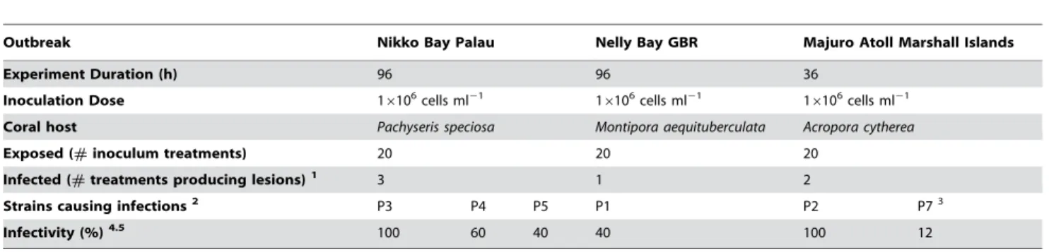
Bacterial strains isolated from corals displaying disease signs at each of the three outbreak sites (10 isolates from TCBS medium plates and 10 isolates from MA medium plates per site) were screened in infection trials with results from all inoculations presented in table 1. All fiveP. speciosafragments (Nikko Bay Palau) inoculated with isolate P3 (16106bacteria ml21) developed disease signs following exposure for 96 h, while treatments with isolates P4 and P5 demonstrated lower infectivity (Fig. S1). Coral fragments in control treatments (n = 17) including treatments with 7 other TCBS derived isolates and 10 isolates from MA plates remained unaffected for the duration of the experiment. Healthy fragments
ofM. aequituberculata (Nelly Bay GBR) were only infected by one strain (P1) of the 20 strains tested, with 40% of fragments displaying disease signs after a 96 h exposure to P1. 100% and 12% of healthy A. cytherea fragments (Majuro Atoll Marshall Islands) exposed to strains P2 and P7, respectively, displayed disease signs after 36 h. A repeat of the experiment with strain P7 resulted in no further positive results and therefore the strain was eliminated as a possible putative pathogen. Results from inoculation experiment I satisfied Hill’s criterion 4 [22] of ‘‘time sequence’’ (cause precedes effect) by demonstrating successful infectivity following putative pathogen inoculations.
Inoculation experiment II: Fulfilling Henle-Koch’s postulates
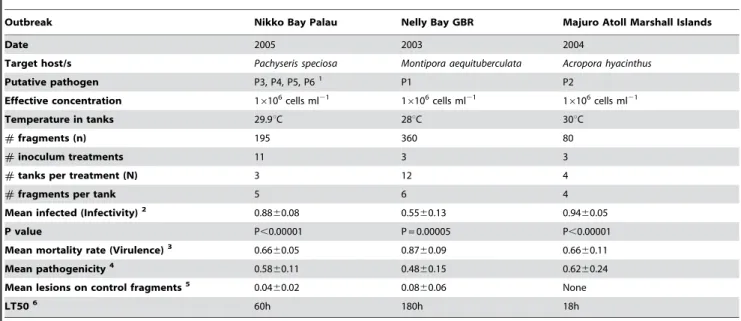
Results from three replicated experimental inoculation trials conducted to fulfil Henle-Koch’s’ postulates and determine the virulence of putative pathogens by causing mortality to infected corals are presented in table 2. Healthy colony fragments exposed to putative pathogens P1–P6 (16106cells ml21) displayed signs of disease similar to those observed in the field in all experiments (Fig. 2A–F). Exposure ofM. aequituberculatato putative pathogen P1 resulted in lesions covered by a sulphurous deposit, which matched disease signs in the field (Fig 2A–B). Exposure of P. speciosa to putative pathogens P4 and P6 began by producing linear lesions resembling field observed lesions (Fig. 2C–D), while P. speciosa fragments exposed to P3 and P5 resulted in the development of larger lesions similar to a second, more common type of lesion observed at the site (Fig. 2E–F). Coral fragments inoculated with control strains (non-pathogenic) and un-inoculated control frag-ments did not develop signs of WS lesions (Fig. 3IA–B, 3IIA–B) in contrast to lesion signs and mortality observed in all treatments with putative pathogens (Fig 3IC–D, 3IIC–J). Bacterial isolates from infected fragments retrieved at the conclusion of the experimental exposure, demonstrated 100% 16S rRNA gene sequence identity to inoculated strains. Recovery of inoculated strains from infected fragments fulfilled Henle-Koch’s postulates for all 6 proposed agents examined in this study.
The proportion of exposed fragments per tank that became infected (infectivity) varied among the experiments, with 88% of fragments exposed to P3–P6, 55% of fragments exposed to P1 and 94% of fragments exposed to strain P2 becoming infected. Pathogenicity (proportion of exposed fragments that died) measured 58%, 48% and 62%, and mortality rate, or virulence
Table 1.Inoculation experiment I
Outbreak Nikko Bay Palau Nelly Bay GBR Majuro Atoll Marshall Islands
Experiment Duration (h) 96 96 36
Inoculation Dose 16106cells ml21 16106cells ml21 16106cells ml21
Coral host Pachyseris speciosa Montipora aequituberculata Acropora cytherea
Exposed (#inoculum treatments) 20 20 20
Infected (#treatments producing lesions)1 3 1 2
Strains causing infections2 P3 P4 P5 P1 P2 P73
Infectivity (%)4.5 100 60 40 40 100 12
1The number of pure cultures in each experiment causing visible disease signs (lesions) on experimental fragments. 2Bacterial isolates causing disease signs were named Pathogen 1–7 (P1–P7).
3Inoculation experiment I was repeated for both isolates from the Marshall Islands (P2, P7) that demonstrated infectivity before ruling out isolate P7 as a possible
putative pathogen.
4Infectivity represents the percent of fragments (#exposed/#infected) within each of the treatments displaying visible disease signs (lesions) 5No fragments in control treatments were infected
(proportion of infected fragments that died) equalled 66%, 87% and 66% for putative pathogens from Palau, Magnetic Island and the Marshall Islands, respectively. Similarly, the times needed for 50% of the fragments to experience mortality (LT50) were 60h, 180h and 18h, respectively.
Aetiology of WS: Adhesion of pathogens to coral tissue Putative pathogen P1 (Nelly Bay GBR) demonstrated an 87% reduction in mean seawater CFU’s (Fig 4A) within the first 12 h following inoculation into aquaria with fragments of M. aequitu-berculata (from mean 2.4860.376105 cells ml21 at t 0 to mean 3.1760.676104 cells ml21 at t 12). In comparison, only a 6% reduction was observed when the same corals were inoculated with control bacterial isolate MF1 (from mean 9.6061.816105 cells ml21at t 0 to mean 9.0761.016105cells ml21at t 12). CFU’s from un-inoculated control aquaria averaged 3.060.696102cells ml21 after 12 h. After 36 h, mean CFU counts from aquaria seawater treated with P1 dropped even further to 0.6% of the original inoculation concentration (Men 1.4160.276103 cells
ml21), which was similar to the density of cells in control tanks (Mean 2.2660.496103cells ml21). In contrast, putative pathogens that were inoculated into sterile seawater without corals main-tained a constant density of viable counts in suspension throughout the experiment (Fig. 4A), eliminating the possibility that bacteria died from the seawater itself or may have settled on the sides or bottom of aquaria. Vibrio density in aquaria containing M. aequituberculata fragments, which were inoculated with non-pathogen MF1 remained unchanged after 36 h, with mean 1.0460.156106cell ml21(100%) retrieved on TCBS agar plates. CFU counts of crushed coral samples (CFU ml21g21 wet weight) from aquaria inoculated with P1 reached a mean of 1.6060.786105ml21g21 after 12 h (Fig 4B). In comparison, fragments from aquaria inoculated with control bacteria (MF1), or un-inoculated controls, resulted in CFU counts that were 94% and 97% lower after 12 h (Mean 9.0862.826103ml21g21and mean 4.1262.456103ml21g21, respectively). Table S1 summarizes the data from adhesion experiments conducted with putative patho-gens and controls isolated from the three infection sites examined in this study.
Loss ofSymbiodinium followed by tissue lesions
Detailed photographs taken ofA. hyacinthus fragments infected experimentally with P2 (Fig 5A–C) revealed 2 distinct disease-phases. An initial loss ofSymbiodinium, visible as tissue paling was observed after 9–12 h of exposure (Fig 5B–C) followed by developing tissue lesions. Similar patterns of paling were also observed whenP. speciosafragments were exposed to P3 (Fig 5D). Paling and loss ofSymbiodinium commenced in coenosarc tissue (tissue between polyps) in distinct linear patterns starting 12 h post inoculation and corresponding with the peak in viable CFU counts retrieved from coral tissue. These early signs of disease then developed into lesions that resembled those observed in the field (Fig 5E–F), suggesting that disease progression was consistent (Hill’s [22] criterion 2) and followed measurable steps (Evans’ Rule F [21]). For a 24h time lapse video clip ofA. hyacinthusinoculated with pathogen P2, see Supporting Information Movie S2.
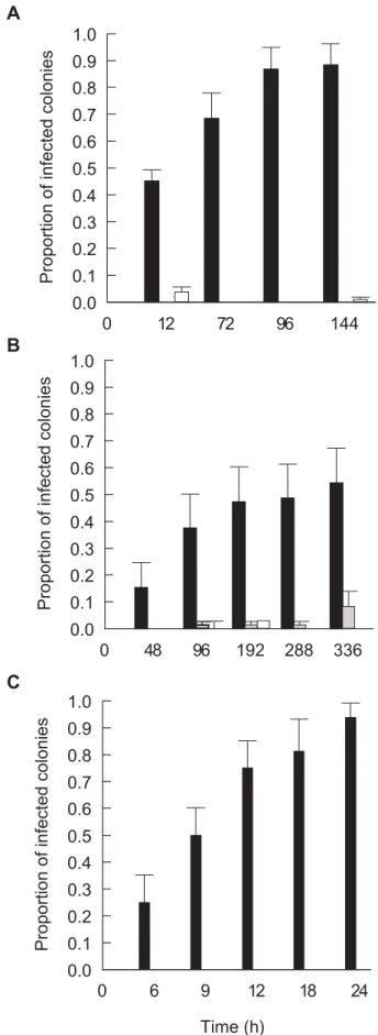
In all experimental treatments inoculated with putative pathogens P1–P6, the proportion of fragments displaying acute disease signs (lesions) increased with time to between 55% and 94% of fragments per tank (Fig 6A–C) conforming with Evans’ rules D and E [19], namely that disease occurs, temporally, following specific incubation times and that the number of new cases and the severity of outcome should correlate positively with time. The proportion of P. speciosa fragments from Palau, M. aequituberculata fragments from Nelly Bay and A. hyacinthus fragments from the Marshall Islands displaying acute disease signs increased consistently and significantly within the first 96 hours (Fig. 6A–6B) and 12 hours (Fig. 6C) of the start of inoculation experiment II, at each site, respectively, resembling standard infection curves [32]. In contrast, 0–8% of fragments in inoculated and un-inoculated control treatments developed disease signs (Fig 6A–C).
Isolates associated with disease signs are proteolytically active
Isolated bacteria (152 strains) recovered from both diseased and healthy corals were screened for proteolytic activity using the asocasein assay and specific PCR primers targeting the zinc-binding site of aVibriofamily zinc-metalloprotease. A total of 48% of strains (n = 33 strains) retrieved from diseasedP. speciosain the field (Nikko Bay Palau) demonstrated high ($3U) or medium (1-3U) proteolytic activity compared with 30% strains (n = 23 strains) demonstrating high or medium activity that were retrieved from Figure 2. WS signs observed in the laboratory and in the field:
A.Montipora aequituberculataexposed to pathogen P1 in laboratory inoculation experiment. B. M. aequituberculata with WS signs in the field (Nelly Bay GBR).C.Pachyseris speciosaexposed to pathogen P6 in laboratory inoculation experiment.D.P. speciosawith WS signs in the field (Nikko Bay Palau). E. P. speciosa exposed to pathogen P3 in laboratory inoculation experiment. F.P. speciosawith WS signs in the field (Nikko Bay Palau).
non-diseased colony fragments sampled in the field (Table 3). This difference, however, was not found to be statistically significant (Pearson’sx2= 1.825, DF = 1, p = 0.177). In contrast, 11 positive PCR bands and derived partial sequences of the Vibrio zinc-metalloprotease gene were obtained from DNA of isolates retrieved from diseasedP. speciosasampled in the field compared with only 1 partial sequence from a non-diseased colony fragment. This difference was found to be significant by testing for Pearson’s chi-square (x2= 6.763, DF = 1, p = 0.0093).
Similar results were obtained by screening field isolates from Nelly Bay GBR (Table S2). Bacteria demonstrating high and medium proteolytic activity by the asocasein assay made up 70% of all isolates retrieved from coral skeletons (S) exposed by WS disease at Nelly Bay GBR and 57% of all isolates from the lesion interfaces (I), compared with only 24% of all isolates obtained from healthy (H) tissue fragments on diseased colonies, demonstrating a significant difference in proteolytic activity between isolates associated with disease signs (I+S) and healthy (H) tissue (Pearson’s x2= 6.446, DF = 1, p = 0.011). A significant difference was also obtained for the same 38 isolates when screened by the molecular method using PCR primers (Pearson’s x2= 12.518, DF = 1, p,0.0001). Finally, screenings by the molecular method per-formed on DNA extracted from 56 isolates retrieved from both infected and non-infected fragments at the conclusion of inoculation experiment II in Palau (Table S3), demonstrated that results obtained by screening field isolates were consistent with screening laboratory derived isolates (Pearson’s x2= 6.725, DF = 1, p = 0.010). Thus, in both field and laboratory infections, the presence of aVibriofamily zinc-metalloprotease was associated with disease signs conforming to Evans’ rules B, C and G [21], suggesting that bacterial proteolytic activity may cause or contribute to observed WS lesions.
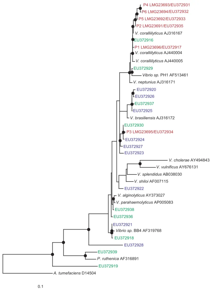
Pathogens identified by this study form a taxonomic cluster
Based on near complete 16S rRNA gene sequence comparisons, the six pathogens clustered in a tight taxonomic group and were found to share between 98–99% sequence identities with the previously characterized coral-bleaching pathogenVibrio coralliily-ticus [14]. All isolates which tested positive for the zinc-metalloprotease zinc-binding site and exhibited high proteolytic activity (when screened by the asocasein assay) were used to construct a maximum likelihood phylogenetic tree based on their 16S rRNA gene (Fig. 7). Our findings demonstrate that more isolates possess the genetic capacity to become proteolytically active than the six coral pathogens identified in this study, suggesting that successful infections require the expression of additional virulence genes, but also that other non-pathogens might be indirectly involved in enhancing infections.
Discussion
This study reports the successful isolation and identification of bacterial infectious agents implicated in a group of widespread Indo-Pacific coral diseases that affect numerous species at various geographical locations. Six coral pathogens were identified with close 16S rRNA gene phylogenetic affiliation with the previously identified coral pathogen V. coralliilyticus [14]. Vibrio pathogens have been previously demonstrated to cause fish, eel, shrimp and Figure 3. Inoculation experiment II: I A–B.Montipora
aequituber-culatacoral fragments in un-inoculated control treatment (t = 0h and t = 150h).I C–D.M. aequituberculatacoral fragments exposed to 16106 cells ml21of culture P1 (t = 0h and t = 150h).II A–B.Pachyseris speciosa
coral fragments in un-inoculated control treatment (t = 0h and t = 150h).
II C–D.P. speciosa coral fragments exposed to 16106cells ml21of culture P3 (t = 0h and t = 150h). II E–F. P. speciosa coral fragments exposed to 16106cells ml21of culture P4 (t = 0h and t = 150h).II G–H. P. speciosacoral fragments exposed to 16106cells ml21of culture P5
(t = 0h and t = 150h). II I–J. P. speciosacoral fragments exposed to 16106cells ml21of culture P6 (t = 0h and t = 150h).
human mortalities [45–49]. Seasonal bleaching of the coralOculina patagonicain the Mediterranean Sea has been shown to be caused byV. shiloi[5,11,50–51] andV. coralliilyticushas been identified as
the aetiological agent of Pocillopora damicornis bleaching in the Indian Ocean [14,52–53]. Other coral diseases in the Caribbean, such as White Band Disease type II, Yellow Blotch/Band and
Table 2.Inoculation experiment II
Outbreak Nikko Bay Palau Nelly Bay GBR Majuro Atoll Marshall Islands
Date 2005 2003 2004
Target host/s Pachyseris speciosa Montipora aequituberculata Acropora hyacinthus
Putative pathogen P3, P4, P5, P61 P1 P2
Effective concentration 16106cells ml21 16106cells ml21 16106cells ml21
Temperature in tanks 29.9uC 28uC 30uC
#fragments (n) 195 360 80
#inoculum treatments 11 3 3
#tanks per treatment (N) 3 12 4
#fragments per tank 5 6 4
Mean infected (Infectivity)2 0.88
60.08 0.5560.13 0.9460.05
P value P,0.00001 P = 0.00005 P,0.00001
Mean mortality rate (Virulence)3 0.66
60.05 0.8760.09 0.6660.11
Mean pathogenicity4 0.58
60.11 0.4860.15 0.6260.24
Mean lesions on control fragments5 0.0460.02 0.0860.06 None
LT506 60h 180h 18h
1Data for separate inoculation experiments with isolates P3–P6 was pooled together. 2Mean infectivity was calculated as mean#infected /#exposed
6SE.
3Mean mortality rate, or virulence, was calculated as mean#dead /#infected6SE. 4Mean pathogenicity was calculated as mean#dead/#exposed6SE.
5Mean lesions on control fragments were calculated as mean#lesions /#controls
6SE.
6LT50 is the time needed to cause 50% mortality of the exposed fragments.
doi:10.1371/journal.pone.0002393.t002
Figure 4. Adhesion experiment: A.CFU’s counts (ml21) from SW samples.
N-16106cells ml21of culture P1 inoculated into SW without corals.e -16106cells ml21of culture from control isolate MF1 inoculated into SW without corals.&-16106cells ml21of culture P1 inoculated into SW with Montipora aequituberculatacoral fragments.,-16106cells ml21of culture MF1 inoculated into SW withM. aequituberculatacoral fragments.m-SW
withM. aequituberculatacoral fragments without inoculation.B.CFU’s counts (ml21g21) from crushed coral fragments.
N-16106cells ml21of culture P1 inoculated into SW withM. aequituberculatacoral fragments.,- 16106cells ml21of culture MF1 inoculated into SW withM. aequituberculatacoral
fragments.&-M. aequituberculatacoral fragments without inoculation. Time represents hours (h) following exposure.CFU’s ml21
g21
are presented in a logarithmic scale. Bars = Standard errors.
Dark Spots Disease, were found to be associated with elevated Vibrio spp. prevalence [4,54–56], suggesting the involvement of Vibriostrains in numerous coral diseases including coral bleaching [5,57–59].
This study has found high prevalence of Vibrio spp. to be associated with WS signs in all diseased colonies sampled from three WS outbreaks. An association between bacterial presence and disease signs does not provide proof that bacteria actually cause the disease. However, such an association already fulfils requirements put forth by Evans’ rule A [21], namely that the ‘‘prevalence of the disease should be significantly higher in those exposed to the putative cause than in cases controls not so exposed’’. It also complies with the Read [60] definition of virulence, which highlights an agent’s contribution to reduction in host fitness caused by exploitation.
When bacterial strains were inoculated into tanks with healthy coral fragments, only putative pathogens adhered to coral tissue and a consistent peak in cultivable Vibrioabundance, 500–1000 fold greater than was found for control strains, was observed on fragments exposed to putative pathogens 12 h post inoculation. This experiment confirmed Hill’s criterion of ‘‘time sequence’’ [22], suggesting that following exposure and prior to the development of visual disease signs (lesions), putative pathogens were able to migrate towards the coral fragments, adhere to coral tissue and survive initial contact in a viable state. In contrast, control strains were unable to perform this transmission, suggesting that motility towards corals and adhesion may be regarded as traits involved in pathogenicity. Initial visual signs of tissue paling and lesions were observed following a peak in cultivable bacterial abundance for all six putative pathogens, demonstrating a common aetiology of adhesion followed by disease progression. Despite this peak in cultivable Vibrio abundance 12 h post exposure, less than 1% of the original inoculation was retrieved by plating coral fragments, potentially
indicating that Vibrio cells entered a viable but non-culturable (VBNC) state [61], or alternatively, died.
The colonization of target hosts by Vibriopathogens has been studied in detail, particularly the ability of Vibrios to adhere to mucus found either inside the gastro- internal track or externally on fish or corals [62–63]. Denkin and Nelson [64] have demonstrated that the transcription of zinc-metalloprotease by the fish pathogenV.anguillarumis regulated by mucus and can only occur after adhesion is completed. This duality inVibriofunction is often referred to as the ‘‘transmission-virulence trade-off’’ [65] and highlights the fact that the ultimate goal ofVibriopathogenicity is not to kill a host, or to complete a necessary biological life-cycle within it, but to re-enter the environment in larger numbers and initiate a new cycle of infections [66]. It explains why Vibrio pathogens are commonly found in environmental reservoirs [26], or transmitting through the water column, like pathogen P6 isolated in this study from seawater above infected corals at Nikko Bay Palau.
This study has demonstrated that 55%–94% of coral fragments exposed to pathogens cultured from diseased corals at their respective field sites become infected and that 66%–87% of those infected die, compared with significantly lower infection and mortality for fragments exposed to control bacterial strains (0–8%). These results conform to Evans’ rule G [21], requiring that experimental reproduction of the disease should occur in higher incidence in those exposed to the putative cause than in those not so exposed. However, a proportion of exposed fragments did not develop disease signs, demonstrating that the probability of becoming infected may not be equal among healthy colony fragments collected from the field, and that other host related factors potentially contribute to successful infections. Such unknown factors can be explored in future inoculation trials.
The presence ofVibrio spp. on both healthy and diseased corals has led to the conclusion by some authors [67–68] that Vibrio
Figure 5. Disease progression: A.Acropora hyacinthusfragmennt inoculated with 16106cells ml21of culture P2 (t = 0h).B.Loss ofSymbiodinium fromA. hyacinthusinoculated with 16106cells ml21of culture P2 (t = 12h).C.Polyp and surrounding tissue-loss ofSymbiodiniumfromA. hyacinthus inoculated with 16106cells ml21of culture P2 (t = 12h).D.Loss ofSymbiodiniumcells fromPachyseris speciosainoculated with 16106cells ml21of culture P3 (t = 12h).E.Tissue lesions onP. speciosainoculated with 16106cells ml21of culture P3 (t = 24h).F.Exposed skeleton onP. speciosa inoculated with 16106cells ml21of culture P3 (t = 60h).
infections of corals may be opportunistic in nature. This assumption fits well into models of disease occurring in environmental settings, where multiple factors, such as host density [69] and temperature [70] have been shown to influence the probability of successful infections. Combinations of virulent and a-virulent Vibrio strains are found readily in environmental samples [71] with non-clinicalV. cholerastrains found to be capable of causing infections despite lacking the cholera toxin gene [72]. ManyVibrios specialize in multiple host attachment and detach-ment [73–75], suggesting a broad scope for potential coral infections by Vibrios including possible host shifts due to fish depletion from coral reefs [76]. Amaro and Biosca [46] have demonstrated that Vibrio vulnificus biotype 2 is both a primary pathogen for eels and an opportunistic pathogen for humans, indicating that the identification of opportunistic pathogens requires rigorous testing. Nevertheless, none of the claims to defineVibriocoral infections as opportunistic have so far provided conclusive evidence to show that suspects (identified by molecular screening methods) found on healthy corals are in fact pathogenic (whether opportunistic or not), or that only compromised hosts become infected. In addition, not all coral mortalities are caused by infectious agents, but rather by exposure to extreme conditions, such as pesticides or high nutrient levels [77–78], which may result in indirect shifts in microbial abundance. Infectious outbreaks can be distinguished from non-infectious ones by plotting infection
Time (h)
0
6
9
12
18
24
Proportion of infected colonies
0.0
0.1
0.2
0.3
0.4
0.5
0.6
0.7
0.8
0.9
1.0
0
12
72
96
144
Proportion of infected colonies
0.0
0.1
0.2
0.3
0.4
0.5
0.6
0.7
0.8
0.9
1.0
0
48
96
192
288
336
Proportion of infected colonies
0.0
0.1
0.2
0.3
0.4
0.5
0.6
0.7
0.8
0.9
1.0
B
C
A
Figure 6. Disease transmission: A. Mean proportion of infected
Pachyseris speciosa coral fragments displaying WS signs following exposure to cultures of P3–P6 in comparison to proportions in
inoculated and un-inoculated control treatments.B.Mean proportion of infectedMontipora aequituberculatacoral fragments displaying WS signs following exposure to culture of P1 in comparison to proportions in inoculated and un-inoculated control treatments.C.Mean propor-tion of infectedAcropora cythereacoral fragments displaying WS signs following exposure to culture P2 in comparison to proportions in inoculated and un-inoculated control treatments.&-Coral fragments inoculated with 16106cells ml21of putative pathogen cultures.& -Coral fragments inoculated with 16106 cells ml21 culture of non-pathogen isolates. %-Coral fragments without inoculation. Time represents hours (h) following exposure. Bars = Standard errors. doi:10.1371/journal.pone.0002393.g006
Table 3.Proteolytic activity of bacterial isolates (Nikko Bay Palau)
Bacterial isolates retrieved from fieldPachyseris speciosa1 Total
Diseased colonies
Non-diseased colonies
+ve PCR product2 11 1 12
2ve PCR product2 22 22 44
Total 33 23 56
High proteolytic activity3 6 4 10
Medium proteolytic activity4 10 3 13
No proteolytic activity5 17 16 33
Total 33 23 56
1Isolates retrieved from diseased and non-diseasedPachyseris speciosacolonies
sampled in Nikko Bay Palau .
2Specific amplification of
Vibriozinc-metalloprotease active zinc binding site.
3High proteolytic activity.3U measured by the asocasein assay. 4Medium proteolytic activity 1-3U measured by the ascasein assay. 5No proteolytic activity
0.1
P6 LMG23694/EU372932
P5 LMG23692/EU372933
P2 LMG23691/EU372935
V. coralliilyticus AJ316167
EU372916
P1 LMG23696/EU372917
V. coralliilyticus AJ440004
V. coralliilyticus AJ440005
EU372929
Vibrio sp. PH1 AF513461
V. neptunius AJ316171
EU372920
EU372926
EU372937
EU372925
V. brasiliensis AJ316172
EU372930
P3 LMG23695/EU372934
EU372924
EU372927
EU372923
V. cholerae AY494843
V. vulnificus AY676131
V. splendidus AB038030
V. shiloi AF007115
EU372922
V. alginolyticus AY373027
V. parahaemolyticus AP005083
EU372938
EU372936
EU372921
Vibrio sp. BB4 AF319768
EU372918
EU372928
EU372939
P. ruthenica AF316891
EU372919
A. tumefaciens D14504
P4 LMG23693/EU372931
Figure 7. Phylogenetic tree of proteolitically-active isolates:Evolutionary distance maximum likelihood analysis based on 16S rRNA gene sequences of isolates obtained by this study. Coral pathogens are marked in red. Reference strains are marked in black. Isolates that demonstrated high proteolytic activity (asocasein assay) and tested positive for a zinc-metalloprotease gene are presented in blue (Palau isolates) and in green (Nelly Bay GBR isolates). Nodes represent bootstrap values$50% based on 1000 re-samplings. Scale bar corresponds to 10% estimated sequence divergence.
curves [32] to demonstrate a bell-shape increase and decrease in incidence rate with time.
This study did not find evidence for the presence of coral pathogens on healthy corals in the field, nor evidence that exposed fragments might be successfully infected due to stress other than the direct exposure to the pathogens themselves. Control treatments in all inoculations remained healthy, including a proportion of those exposed to pathogens. Further studies are recommended to determine the prevalence of pathogens in field samples by developing diagnostic tools to target specific virulence genes in large scale screening efforts. These studies could then determine the proportion of exposed corals in the field that develop acute disease signs and should become an integral part of establishing acute vs. chronic disease prevalence in environmental studies.
This is the first study to diagnose proteolytic activity as a possible component of the aetiology of WS through the screening of more than 150 isolates from both diseased and non-diseased corals. Zinc-metalloproteases have been characterized as virulence factors in manyVibriofamily pathogens, such asV. cholera[79],V. vulnificus[80], V. harveyi[81] andV. anguillarum [82].Vibrio zinc-metalloproteases are involved in cleavage of connective tissue [83], para-cellular perturbation [84], swarming and adhesion to mucus [85] and detachment [86]. The coral bleaching pathogensV. shiloi andV. coralliilyticushave been previously shown to harbour a zinc-metalloprotease [53,87] along with other toxins that cause photosynthetic 5 inhibition of coral Symbiodinium [88]. Serratia marcescens, the aetiological agent of acroporid serratiosis (coral White Pox disease [13]), resulting in acute tissue lesions, also possesses a virulent zinc-metalloprotease capable of connective tissue degradation [89]. However, it has been shown that both clinical and non-clinical strains possess zinc-metalloprotease genes [90], suggesting that it may not be the only virulence factor to cause successful infections. This study provided similar results, underlining the need to search for additional virulence factors in future studies.
Recent studies by Ainsworth et al. [91] did not detect bacteria associated with WS lesions of diseased corals sampled at Heron Island on the GBR, using direct microscopic techniques. In contrast, samples of WS corals obtained from Heron Island in this study for screening purposes demonstrated an abundance ofVibriospp. isolates on WS lesions, including proposed putative pathogens that are proteolytically active and possess a zinc-metalloprotease gene. These contradicting findings underline the importance of ‘comparative validation’ [92] in disease research and the need for standardized protocols for disease detection using better diagnostic tools.
Further histopathological studies by Ainsworth et al. [68, 91] utilizing commercial labelling kits have found that coral fragments displaying WS signs test positive for DNA fragmentation. These observations led to the hypothesis that WS is potentially the result of coral programmed cell death. However, further proof is needed in order establish whether DNA fragmentation (or apoptosis) in corals is cause or effect. The induction of apoptosis by bacterial pathogens (Salmonella Sp., E. coli, Shigella sp., C. difficile, L. monocytogenes, C. parvum and others) has been previously demon-strated by many studies [93-97], suggesting a possible link between bacterial infections and apoptosis. This link can be tested in future pathogen-exposure trials and used to design novel diagnostic protocols for WS, which would target bacterial enzymes causing DNA fragmentation.
In summary, this study demonstrated consistent results in applying cost effective culturing techniques combined with biochemical and molecular tools towards successful pathogen isolation, coral disease investigation and sample screening. Future
research should be conducted to explore the virulence components of all six pathogens identified in this study and to test the contribution of multiple factors (pathogen, environment and host related) to the aetiology of WS. Enhanced monitoring and management of WS outbreaks will not only benefit coral health, but would also further validate results obtained in this study.
Materials and Methods
Isolation and growth of bacteria from coral samples For inoculation experiment I, ten fragments (2–10 g wet weight) from corals displaying WS disease signs and ten fragments (2–10 g wet weight) from corals lacking WS disease signs were collected from depths between 3–15 m at each of the following locations: 1) Nelly Bay fringing reef (S19 109E 146 529) at Magnetic Island in the central section of the Great Barrier Reef (GBR) in September 2003; 2) Majuro Atoll the Republic of the Marshall Islands (N 9 009E 168 009) in August 2004; and 3) Nikko Bay, an enclosed bay among rock islands in the Republic of Palau (N 7 309E 134 309) in February 2005. WS mainly affected plate colonies of Pachyseris speciosa in Palau, tabular species ofAcropora (A. cytherea, A. hyacinthus and A. clathrata) in the Marshall Islands and plate colonies of Montipora aequituberculata at Nelly Bay GBR. At each site, samples were transported from the reef to the laboratory in sterile containers.
For calculating the abundance of bacteria associated with diseased and non-diseased fragments, the following sub-samples were obtained at each site: healthy tissue from coral fragments with no disease (CON, n = 3); tissue adjacent to lesions on coral fragments with WS disease signs (INF, n = 3); healthy tissue on coral fragments displaying disease signs (H, n = 3); lesion interface on coral fragments displaying disease signs (I, n = 3); and exposed coral skeleton on coral fragments displaying disease signs (S, n = 3). Samples were crushed and diluted with 10 ml of 0.22mm filtered seawater (Millipore, USA), and then vortexed for 3 min at maximum speed before being left to settle for 3 min [26]. Supernatant (100mL) was streaked on agar plates containing a general heterotrophic bacterial medium (Marine Agar: 1.8% Marine Broth, Difco-2216, USA 0.9% NaCl, 1.8% Agar Bacto, Difco-214010, USA) and thiosulfate citrate bile salts sucrose (TCBS) agar, aVibrionaceaselective growth medium (Difco, USA). Plating was conducted in triplicates of serial dilutions (161021– 161026) followed by incubation overnight at 30uC. Cultivable strains were quantified by counting colony forming units (CFU’s) and the density of bacteria associated with corals was determined as mean CFU’s per 1 ml of crush derived from 1 g (wet weight) of coral tissue (CFU’s ml21g21). Single CFU’s were picked from both Marine Agar (MA) and TCBS plates and transferred to fresh MA plates for further analyses.
Single isolates were grown in 250ml sterile flasks containing sterile marine broth (MB) media incubated at 30uC for 18h (i.e. to end of the logarithmic phase) with constant shaking (150rpm). Cell density in pure cultures was determined by plating triplicates of serial dilutions on MA and by measuring absorbance (595nm) in sterile microtitre well plates (n = 6).
Additional bacterial isolates from fragments displaying signs of ongoing tissue loss in association with WS and from healthy fragments (controls) were retrieved for screening purposes from corals at Heron Island, GBR (March 2004) and at Dip Reef GBR (November 2004).
manufacturer’s instructions. The 16S rRNA gene was amplified by using universal primers 27F and 1492R. [98]. In addition, primers
HA-F (59 –CATGAGGTCAGCCACGGTTTTACTGAGCAG)
and HA-R (59
–CGCGCGGTTAAACACGCCACTC-GAATGGTGAAC (Invitrogen, NZ) targeting a,225 bp region
including the zinc binding site of Vibrio-family zinc-metallopro-teasses [99] were used to screen all bacterial genomic DNA. PCR reactions (50mL) were run on an Eppendorf Mastercycler with the reaction mix consisting of 10 pmol of each primer, 5mL of 10xPCR buffer with 15 mM MgCl2, 50 nmol dNTP, 10 ng
template DNA and 1U Taq (iTaq, Intron Biotechnology, Korea). DDW (Milli-Q, millipore) was added to the volume of 50mL. Cycling conditions consisted of: 1) 27F/1492R-a 5 min denatur-ation step at 94uC followed by 30 cycles of 1 min at 94uC, 1 min at 52uC and 1 min at 72uC and concluded by a 7 min extension step at 72uC; and 2) HA-F/HA-R-a 5 min denaturation step at 94uC followed by 30 cycles of 20 sec at 94uC, 20 sec at 55uC and 1 min at 72uC, concluded by a 5 min extension step at 72uC. Amplified bands of the correct size were confirmed on a 1% ethidium bromide stained TAE agarose gel and amplified gene products were sequenced at MACROGEN Inc. (Seoul, Korea) on an ABI PRISM 3730XL analyzer (96 capillary-Applied Biosys-tems, CA, USA) using the ABI PRISM BigDyeTM Terminator Cycle Sequencing Kit. Retrieved gene sequences were aligned for closest matches using BLAST [100]. In total, 152 partial sequences were retrieved from coral fragments displaying WS signs and from controls sampled at the three-infection sites.
Phylogenetic analyses
Sequences were checked for chimera formation with the CHECK_CHIMERA software of the Ribosomal Database Project [101]. Sequence data were aligned to the most similar sequence using the BLAST database algorithm [100], and then further analysed with the ARB software package [102]. Tree topologies were evaluated by reconstructing phylogenies using maximum likelihood evolutionary distance analysis (Phylip Distance Method with Jukes and Cantor model) of aligned near full-length sequences (.1200 bp). Regions of ambiguous sequence (N) were removed from the analysis. Bootstrap values were obtained for branching patterns using the Phylip software package (version 3.65 [103]) and values $50% were included for main nodes of the tree.
Infection experiments
Infection experiments were run as incurred matrices in 2 consecutive stages, described below as inoculation experiments I and II.
Inoculation experiment I: Testing for infectivity of bacterial isolates
To screen bacteria for infectivity (the ability to initiate visual disease signs (lesions) regardless of their severity [35]), 20 isolates retrieved from coral samples at each of the three sites (10 most abundant isolates on both MA and TCBS plates from both healthy and diseased colonies at each site) were grown to end logarithmic phase in MB (as described above) and inoculated individually into 7L sterile aerated tanks (final inoculum concentration = 16106 cells ml21) containing 4–6 healthy fragments of corals collected from sites without disease signs (i.e. healthy fragments ofPachyseris speciosafrom a healthy Palau site, healthyAcropora cythereafragments from a healthy Marshall Islands site, and healthy Montipora aequituberculata fragments from a healthy Nelly Bay site). Prior to bacterial inoculation, coral fragments were acclimatized for 5 days
to allow recovery from handling and fragmentation following a protocol by Kushmaro et al. [50]. Each of the 20 culture inoculations was tested in two tanks [n = 168–252 fragments per site, N = 21 inoculation treatments including 1 negative control treatment]. The negative control tanks contained coral fragments with no bacteria added. Seawater in the tanks was replaced every 48 h and tanks were observed and photographed for 140 h in order to detect developing disease signs. At the end of each experiment, infectivity was calculated as the proportion of exposed fragments per tank that became infected. Both infected and non-infected fragments were crushed and individual CFU’s were picked and transferred to fresh MA plates for further analyses and DNA extraction, as previously described. Bacterial strains causing disease signs in this experiment were given the simplified names: P1–P7 (P1–fromM. aequituberculatain Nelly Bay GBR; P2 and P7 from A. cytherea in Majuro Atoll the Republic of the Marshall Islands; and P3–P6 fromP. speciosain Nikko Bay Palau) and were inoculated as pure cultures in the following experiments under these names. Bacterial strain P6 was isolated from seawater above diseased P. speciosa colonies at Nikko Bay Palau. It caused infections in a separate experiment and was therefore added to the list of putative pathogens. Strain P7 from the Marshall Island caused partial disease signs on only oneA. cythereafragment (out of four fragments) when inoculated into two tanks (n = 4 fragments per tank). Inoculation experiment I was repeated using this isolate (n = 12 fragments in each of 3 tanks) and it was removed from the putative pathogen list after failing to cause infections.
Inoculation experiment II: Replicated exposure trial to fulfil Henle-Koch’s postulates and test for virulence
To fulfil Henle-Koch’s postulates, a large multi-replicated exposure trial using successful putative pathogens that initiated disease signs in inoculation experiment I were grown as pure cultures and inoculated (final inoculum concentration = 16106 cells ml21) into multiple tanks with colony fragments (of P. speciosa, M. aequituberculata A. hyacinthus) collected from non-disease sites and acclimatized for 5 days. The number of fragments allocated to inoculation tanks at each site (n) was between 80–360, distributed as 4–6 fragments per tank, and the number of tanks per inoculation/ control treatments (N) was between 4 and 12. At each site, 4 negative controls were run including: 1 treatment comprising tanks to which no bacteria were added, 2 treatments comprising tanks to which control bacterial strains were added at identical concentrations, and 1 treatment comprising tanks to which sterile bacterial media was added (1 ml MB per 1 L seawater) as potential ‘‘growth enhancer’’ for putative pathogens that might be already present on experimen-tal coral fragments. Tanks were maintained temperatures identical to those measured at infection sites. Fragments in each tank were observed and photographed for the entire length of the experiment and developing disease signs were recorded. The experiments were terminated following mortality in infection tanks. Case mortality rate, or virulence (the proportion of infected fragments in each tank that died [36]) and the mean proportion of infected fragments per tank were calculated. Pathogenicity (the proportion of exposed fragments that died) was calculated following the formula of Thomas and Elkinton [35]: Pathogenicity = infectivity6virulence (where pathogenicity =#dying /#exposed, infectivity =#infected /# exposed and virulence =#dying /#infected). Finally, LT50 (the estimated time it takes to kill 50% of the infected fragments) was calculated as a temporal measure of virulence.
Complete alignment (100%) of the 16SrRNA gene sequences retrieved from bacteria re-isolated from infected fragments and the 16S rRNA gene sequences of inoculated bacteria (P1–P6) allowed the fulfilment of Henle-Koch’s postulates [20], namely, that:
1. An organism found only on infected corals could be isolated, taxonomically identified, and grown in pure culture.
2. The isolated organism reproduced disease signs when inocu-lated onto healthy corals.
3. An isolate retrieved from coral fragments that developed disease signs in inoculation experiments is demonstrated to be identical (by analysis of 16SrRNA partial gene sequences) with the organism used for inoculations.
Other rules and criteria for supporting causality used by this study
Results of experiments and screenings conducted in this study were used to evaluate compliance with Evans’ rules [21] and Hill’s criteria [22], defined as alternative requirements for establishing disease causation. Both Evans’ rules and Hill’s criteria are listed in the Supporting Information section (see Materials and Methods S1).
Adhesion of bacterial isolates to corals
To further test the physical ability of putative pathogens to migrate towards coral hosts, to adhere and to survive the initial contact with the coral host, before initial signs of infection are observed, mean bacterial CFU’s were quantified from random sub samples of tank seawater (N = 4 seawater sub-samples per treatment, each taken from a different tank) following inoculation with the six coral pathogens (P1–P6) identified in Inoculation Experiment I (final inoculum concentration = 16106 cells ml21). One ml of tank seawater was collected at inoculation time (t = 0 h), 1 h post inoculation (t = 1 h) and then at 12 h intervals (t = 12 h, t = 24 h, t = 36 h) from four infection tanks. 100mL aliquots from each sample were spread in triplicates on agar plates containing Marine Agar and TCBS, as described previously. Mean CFU’s ml21g21wet weight were determined from three crushed coral samples per treatment at corresponding times [63]. In addition, control bacterial strains were also tested to determine if they adhered to coral fragments following inoculation into tanks. Finally, both putative pathogens and control bacteria were inoculated into four seawater tanks (per bacterial treatment) lacking coral fragments to test their ability to survive and remain suspended in the water column for the experiment’s duration. Seawater samples were collected from these tanks and plated in triplicate as described previously to determine mean bacterial density in seawater (CFU’s ml21).
The asocasein proteolytic Assay
The proteolytic activities of supernatants derived from 152 isolated bacterial strains retrieved by this study were tested by the asocasein assay based on a protocol by Windle and Kelleher [104]. Briefly, 1 ml of bacterial cultures grown to end logarithmic phase were centrifuged at 10,000g (Eppendorf 5415D centrifuge) for 5 min. Supernatant was removed and filtered through a 0.22mm filter (Millipore, USA). 100mL supernatant was incubated for 30min at 30uC with 5g ml21
of asocasein as substrate (Sigma, USA) dissolved in Tris-Hcl (50 mM pH 8) containing 0.04% NaN3(wt vol21). The reaction was terminated by adding 10% (wt
vol21) of trichloroacetic acid (TCA) to a final concentration of 6.7% (wt vol21) and incubating samples for 1 min. Samples were then centrifuged for 3 min at 10,000g and transferred to 700mL of 525 mM NaOH. Absorbance of six replicates from triplicate
culture samples was measured in 96 microtitre well plates at 450 nm using a Wallac spectrophotometer (Perkin Elmer, USA). Blank controls were prepared from supernatant derived fromE.coli cultures boiled at 100uC for 10 min, treated with 5mg ml21
asocasein and directly thereafter by TCA. Protease activity was calculated as proteolytic units, when 1U = 10006(OD450
CFU21)6109[64]. Isolates were divided into 3 groups based on their proteolytic activity: High activity (.3U), medium activity (1-3U) and no activity (,1U).
Statistical analysis
Means and Standard Errors (SE) for bacterial colony forming unit (CFU) counts and for the proportion of infected colonies were compared among treatments using One-Way ANOVA (Statistica, StatSoft, Inc. USA). Colony forming unit (CFU) counts are presented in this study using logarithmic scales.
The association between categorical values related to bacterial isolates retrieved independently from diseased and non-diseased corals and demonstrating positive or negative proteolytic activity was estimated using 262 contingency tables (Pearson Chi-square). Significant results were determined whena#0.05.
Coral pathogens
Six coral pathogen strains that were identified by this study were submitted to the public collection of BCCM/LMG at the Gehnt University, Belgium and are available for acquisition under the following accession numbers: LMG23691-isolate P2 from a WS infected Acropora cytherea in Majuro Atoll the Republic of the Martshall Islands, LMG23692-isolate P5 from a WS infected Pachyseris speciosain Nikko Bay Palau, LMG 23693-isolate P4 from a WS infectedP. speciosain Nikko Bay Palau, LMG 23694-isolate P6 from seawater above a WS infectedP. speciosa in Nikko Bay Palau, LMG 23695-isolate P3 from a WS infectedP. speciosa in Nikko Bay Palau and LMG 23696-isolate P1 from a WS infected Montipora. aequituberculatain Nelly Bay the GBR. 16S rRNA gene sequences of all coral pathogens identified by this study were submitted to Genbank (www.ncbi.nih.nlm.gov/Genbank/) under the following accession numbers: P1 (LMG23696)-EU372917, P2
(LMG23691)-EU372935, P3 (LMG23695)-EU372934, P4
(LMG23693)-EU372931, P5 (LMG23692)-EU372933, P6
(LMG23692)-EU372932. 16S rRNA gene sequences retrieved from isolates demonstrating positive results for proteolytic activity in both screening tests conducted by this study (asocasein assay and PCR amplification) were submitted to Genbank under the following accession numbers: EU372918-EU372930, EU372936-EU372939, and are presented in Fig. 7.
Movie S2
Acropora hyacinthusfragments were inoculated with 16106 cells ml21of pathogen P2 (aquarium situated on the left hand side of the screen). Time lapse photography (every 20 seconds) was carried out for 36 h. Control treatment with no inoculation appears on the right hand side of the screen.
Supporting Information
Materials and Methods S1 Evans’ Rules and Hill’s Criteria
Found at: doi:10.1371/journal.pone.0002393.s001 (0.03 MB DOC)
Figure S1 Inoculation experiment I, Palau: A–B. Pachyseris
inoculated with 16106cells ml21of culture P4 (t = 0h and t = 96h). G–H.P.speciosacoral fragments inoculated with 16106cells ml21 of culture P5 (t = 0h and t = 96h).
Found at: doi:10.1371/journal.pone.0002393.s002 (1.19 MB TIF)
Table S1 Adhesion Experiment
Found at: doi:10.1371/journal.pone.0002393.s003 (0.03 MB DOC)
Table S2 Proteolytic activity of bacterial isolates (Nelly Bay
GBR)
Found at: doi:10.1371/journal.pone.0002393.s004 (0.04 MB DOC)
Table S3 Proteolytic activity of bacterial isolates (Palau)
Found at: doi:10.1371/journal.pone.0002393.s005 (0.03 MB DOC)
Movie S1 WS outbreak in Majuro Atoll the Republic of the
Marshall Islands (August 2004)
Found at: doi:10.1371/journal.pone.0002393.s006 (10.34 MB AVI)
Movie S2 Time lapse inoculation experiment of Acropora
hyacinthusfragments infected with 16106 cells ml21 of pathogen P2 for 36 hours (aquarium situated on the left hand side of the screen). Control treatment with no inoculation appears on the right hand side of the screen.
Found at: doi:10.1371/journal.pone.0002393.s007 (6.74 MB AVI)
Acknowledgments
The authors would like to thank S Anthony, Dr. W Dunlap, Dr. D Jacobson, E Matson, C Page, L Peplow, Prof. L Raymundo, members of the Palau International Coral Reef Center, the College of the Marshall Islands and the crew of the AIMS RSV Lady Basten for their assistance in laboratory work and sample collection.
Author Contributions
Conceived and designed the experiments: BW DB MS. Performed the experiments: MS SV. Analyzed the data: DB MS. Contributed reagents/ materials/analysis tools: BW DB. Wrote the paper: BW DB MS. Other: Performed sample collection: SV MS.
References
1. Harvell CD, Mitchell CE, Ward JR, Altizer S, Dobson AP, et al. (2002) Climate warming and disease risks for terrestrial and marine biota. Science 296: 2158–2162.
2. Weil E (2004) Coral reef diseases in the wider Caribbean. In: Rosenberg E, Loya Y, eds. Coral Health and Disease. Berlin: Springer-Verlag. pp 35–68. 3. Willis B, Page C, Dinsdale E (2004) Coral disease on the Great Barrier Reef. In:
Rosenberg E, Loya Y, eds. Coral Health and Disease. Berlin: Springer Verlag. pp 69–104.
4. Cervino JM, Hayes RL, Polson SW, Polson SC, Goreau TJ, et al. (2004) Relationship ofVibriospecies infection and elevated temperatures to yellow blotch/band disease in Caribbean corals. Appl Environ Microbiol 70: 6855–6864.
5. Rosenberg E, Falkovitz L (2004) TheVibrio shiloi/Oculina patagonica model system of coral bleaching. Annu Rev Microbiol 58: 143–59 Review. 6. Lesser MP, Bythell JC, Gates RD, Johnstone RW, Hoegh-Guldberg O (2007)
Are infectious diseases really killing corals? Alternative interpretation of the experimental and ecological data. J Exp Mar Biol Ecol 346: 36–44. 7. Morens DM, Gregory K, Folkers K, Fauci AS (2004) The challenge of
emerging and re-emerging infectious diseases. Nature 430: 242–249. 8. Efrony R, Loya Y, Bacharach E, Rosenberg E (2007) Phage therapy of coral
disease. Coral Reefs 26: 7–13.
9. Palumbi SR (2005) Germ theory for ailing corals. Nature 434: 713–714. 10. Pandolfi JM, Jackson JBC, Baron N, Bradbury RH, Guzman HM, et al. (2005)
Are U.S. Coral Reefs on the Slippery Slope to Slime? Science 307: 1725–1726. 11. Kushmaro A, Loya Y, Fine M, Rosenberg E (1996) Bacterial infection and
coral bleaching. Nature 380: 396.
12. Geiser DM, Taylor JW, Ritchie KB, Smith GW (1998) Cause of sea fan death in the West Indies. Nature 394: 137–138.
13. Patterson KL, Porter GW, Ritchie KB, Polson SW, Mueller E, et al. (2002) The etiology of white pox, a lethal disease of the Caribbean elkhorn coral, Acropora Palmate. Proc Natl Acad Sci USA. 99: 8725–8730.
14. Ben-Haim Y, Thompson FL, Thompson CC, Cnockaert MC, Hoste B, et al. (2003a)Vibrio coralliilyticus sp. nov., a temperature-dependent pathogen of the coralPocillopora damicornis. Int J Syst Evol Microbiol. 53: 309–15.
15. Denner EBM, Smith GW, Busse HJ, Schumann P, Narzt T, et al. (2003) Aurantimonas coralicidagen. nov., sp. nov., the causative agent of white plague type II on Caribbean scleractinian corals. Int J Syst Evol Microbiol 53: 1115–1122.
16. Barash Y, Sulam R, Loya Y, Rosenberg E (2005) Bacterial Strain BA-3 and a filterable factor cause a white plague-like disease in corals from the Eilat coral reef. Aquat Microb Ecol 40: 183–189.
17. Thompson FL, Barash Y, Swabe T, Sharon G, Swings J, et al. (2006) Thalassomonas loyana sp.Nov., a causative agent of the white plague-like disease of corals on the Eilat coral reef. Int J Sys Evol Microbiol 56: 365–368. 18. Hiney MP (1997) How to test a test: Methods of field validation for
non-culture- based detection techniques. Bull Eur Assoc Fish Pathol 17: 245–250. 19. Hiney MP, Smith PR (1998) Validation of Polymerase Chain Reaction–based techniques for proxy detection of bacterial fish pathogens: Framework, problems and possible solutions for environmental applications. Aquaculture 162: 41–68.
20. Koch R (1891) U¨ ber bakteriologische Forschung Verhandlungen des X internationalen medicinischen Congresses, Berlin 1890, 1, 35, august Hirschwald Berlin.
21. Evans AS (1976) Causation and disease: The Henle-Koch postulates revisited. Yale J Biol Med 49: 175–195.
22. Hill AB (1965) The environment and disease: association or causation? Proc R Soc Med 58: 295–300.
23. Amann RI, Ludwig W, Schleifer KH (1995) Phylogenetic identification and in situ detection of individual microbial cells without cultivation. Microbiol Rev 59: 143–69.
24. Snieszko SF (1974) The effects of environmental stress on outbreaks of infectious diseases of fishes. Journal of Fisheries Biology 6: 197–208. 25. Bourne DG (2005a) Microbial assessment of a disease outbreak on coral from
Magnetic Island (Great Barrier Reef, Australia). Coral Reefs 24: 304–312. 26. Sussman M, Loya Y, Fine M, Rosenberg E (2003) The marine fireworm
Hermodice carunculata is a winter reservoir and spring-summer vector for the coral-bleaching pathogenVibrio shiloi. Environ Microbiol 5: 250–255. 27. Edge SE, Morgan MB, Gleason DF, Snell TW (2005) Development of a coral
cDNA array to examine gene expression profiles inMonastraea faveolataexposed to environmental stress. Mar Pollut Bull 51: 507–523.
28. DeLong EF (2005) Microbial community geonomics in the ocean. Nature Rev Microbiol 3: 459–469.
29. Edwards RA, Rohwer F (2005) Viral metagenomics Nature Rev Microbiol 3: 504–510.
30. Yokuchi H, Fukuoka Y, Mukoyama D, Calugay R, Takeyama H, et al. (2006) Whole-metagenome amplification of a microbial community associated with scleractinian coral by multiple displacement amplification using phi 29 polymerase. Environ Microbiol 8: 1155–1163.
31. World Organization for Animal Health (OIE–2006) Manual of Diagnostic Tests for Aquatic Animals. http://www.oie.int/eng/normes/fmanual/ A_00004.htm
32. Thrusfield M (2005) Veterinary Epidemiology 3rd
Edition, Blackwell Science, Oxford, UK. .
33. Dubos R (1965) Man Adapting.Yale University Press, New Haven. . 34. Stainer RY, Doudoroff M, Adelberg EA (1971) General microbiology.
Macmillan, London, UK. .
35. Thomas SR, Elkinton JS (2004) Pathogenicity and virulence. J Invert Pathol 85: 146–151.
36. Day T (2002) On the evolution of virulence and the relationship between various measures of mortality. Proc R Soc Lond B 269: 1317–1323. 37. Osawa RO, Iyoda S, Nakayama SI, Wada A, Yamai S, et al. (2000) Genotypic
variations of Shiga toxin-converting phages from enterohaemorrhagic Esche-richia coliO157: H7 isolates. Molecular Epidemiology 49: 565–574. 38. Cohen ML (2000) Changing patterns of infectious diseases. Nature 406:
762–767.
39. Wilesmith JW (1993) BSE: Epidemiological approaches, trials and tribulations. Preventive Veterinary Medicine 18: 33–42.
40. FAO (1995) The challenge of sustainable forest management: what future for world’s forests. Rome: FAO. pp 128.
41. Jones RJ, Bowyer J, Hoegh-Guldberg O, Blackall LL (2004) Dynamics of a temperature-related coral disease outbreak. Mar Ecol Prog Ser 281: 63–77. 42. Aeby GS (2006) Outbreak of coral disease in the Northwestern Hawaiian
Islands. Coral Reefs 24: 481.
43. Roff G, Hoegh-Guldberg O, Fine M (2006) Intra-colonial response to Acroporid ‘‘white syndrome’’ lesions in tabular Acropora spp. (Scleractinia) Coral Reefs 25: 255–264.