RESEARCH ARTICLE
The Potential of Dark Septate Endophytes to
Form Root Symbioses with Ectomycorrhizal
and Ericoid Mycorrhizal Middle European
Forest Plants
Tereza Lukešová1,2, Petr Kohout1,2,3, TomášVětrovský4, Martin Vohník1,2 *
1Department of Plant Experimental Biology, Faculty of Science, Charles University in Prague, Prague, Czech Republic,2Department of Mycorrhizal Symbioses, Institute of Botany ASCR, Průhonice, Czech
Republic,3Department of Botany, Institute of Ecology and Earth Sciences, University of Tartu, Tartu, Estonia,4Laboratory of Environmental Microbiology, Institute of Microbiology ASCR, Prague, Czech Republic
*vohnik@ibot.cas.cz
Abstract
The unresolved ecophysiological significance of Dark Septate Endophytes (DSE) may be in part due to existence of morphologically indistinguishable cryptic species in the most common Phialocephala fortiniis. l.—Acephala applanataspecies complex (PAC). We inoculated three middle European forest plants (European blueberry, Norway spruce and silver birch) with 16 strains of eight PAC cryptic species and other DSE and ectomycorrhizal/ericoid mycorrhizal fungi and focused on intraradical structures possibly representing interfaces for plant-fungus nutrient transfer and on host growth response. The PAC speciesAcephala applanata simulta-neously formed structures resembling ericoid mycorrhiza (ErM) and DSE microsclerotia in blueberry.A.macrosclerotiorum, a close relative to PAC, formed ectomycorrhizae with spruce but not with birch, and structures resembling ErM in blueberry.Phialocephala glacialis, another close relative to PAC, formed structures resembling ErM in blueberry. In blueberry, six PAC strains significantly decreased dry shoot biomass compared to ErM control. In birch, oneA.macrosclerotiorumstrain increased root biomass and the other shoot biomass in com-parison with non-inoculated control. The dual mycorrhizal ability ofA.macrosclerotiorum sug-gested that it may form mycorrhizal links between Ericaceae and Pinaceae. However, we were unable to detect this species in Ericaceae roots growing in a forest with presence ofA. macrosclerotiorumectomycorrhizae. Nevertheless, the diversity of Ericaceae mycobionts was high (380 OTUs) with individual sites often dominated by hitherto unreported helotialean and chaetothyrialean/verrucarialean species; in contrast, typical ErM fungi were either absent or low in abundance. Some DSE apparently have a potential to form mycorrhizae with typical middle European forest plants. However, exceptA.applanata, the tested representatives of all hitherto described PAC cryptic species formed typical DSE colonization without specific structures necessary for mycorrhizal nutrient transport.A.macrosclerotiorumforms ectomy-corrhiza with conifers but not with broadleaves and probably does not form common mycor-rhizal networks between conifers with Ericaceae.
PLOS ONE | DOI:10.1371/journal.pone.0124752 April 23, 2015 1 / 25
a11111
OPEN ACCESS
Citation:Lukešová T, Kohout P, Větrovský T, Vohník M (2015) The Potential of Dark Septate Endophytes to Form Root Symbioses with Ectomycorrhizal and Ericoid Mycorrhizal Middle European Forest Plants. PLoS ONE 10(4): e0124752. doi:10.1371/journal. pone.0124752
Academic Editor:Raffaella Balestrini, Institute for Sustainable Plant Protection, C.N.R., ITALY
Received:September 13, 2014
Accepted:March 8, 2015
Published:April 23, 2015
Copyright:© 2015 Lukešová et al. This is an open access article distributed under the terms of the
Creative Commons Attribution License, which permits unrestricted use, distribution, and reproduction in any medium, provided the original author and source are credited.
Data Availability Statement:All relevant data are within the paper and its Supporting Information files.
Introduction
Fungal endophytes are defined as mycobionts which live inside living plant tissues, lack localized interfaces or specialized hyphae for nutrient transfer, their development is not synchronized with plant development and the plant does not nutritionally benefit from the symbiosis [1]. One of the most studied groups of fungal root endophytes, the so-called Dark Septate Endophytes (DSE), are a polyphyletic aggregate of fungi belonging to Class 4 of non-clavicipitaceous endophytes [2] which is broadly defined by the endophytic life strategy and presence of intraradical dark septate hyphae. The arguably most studied group of DSE is thePhialocephala fortiniis. l.—Acephala applanataspecies complex (PAC) [3] which comprises fungi that were formerly regarded as a single species,Phialocephala fortiniiWang & Wilcox (Helotiales, Ascomycota) [4]. However, it has been recently shown that this taxon is composed of at least 21 reproductively isolated line-ages representing putative cryptic PAC species, out of which 8 were formally described [5]. Endophytes related toP.fortiniis. l. show low to any host preference [6,7], they are common in-habitants of both mycorrhizal and non-mycorrhizal roots and frequently live as endophytes in roots of conifers, ericaceous plants and orchids in both the Northern and Southern Hemispheres [8–10]. Although ubiquitous on dry land, PAC species seem to be less frequent to absent in roots of aquatic plants [11] which are probably colonized by other guilds of fungal endophytes [12]. De-spite their frequent occurrence in terrestrial roots [6] and long coexistence with plants [13], the DSE influence on their hosts is still under debate. Results of numerous re-synthesis experiments are inconsistent—some report positive or neutral effects on plant growth [14–17] while others indicate mostly negative outcomes [18–20]. A recent meta-analysis of 18 research articles identi-fied only positive influence of inoculation with DSE [21]. In contrast, another meta-analysis sug-gested negative to neutral effects of inoculation with non-clavicipitaceous root fungal endophytes (including DSE) on host plant biomass and nitrogen content [22].
Because most DSE are thought to lack specialized interfaces for nutrient transfer with their plant partners, the cause of the reported positive influence of inoculation with DSE re-mains unclear. Possible explanations include mineralization of the substrate that can be then exploited by the host roots [23], similarly to non-symbiotic saprotrophic fungi which may be even more effective in supplying nutrients to plants than mycorrhizal fungi [24]; production of phytohormones or analogous substances [25]; breaking down complex carbohydrates and providing simple sugars to the host plants, especially at the seedling stage [26]; reduction of disease intensity of some fungal pathogens, either active or passive [27]; or reduction of in-traspecific competition between adult trees and their offspring [20]. However, many of these possible explanations remain at the level of hypotheses which need to be thoroughly tested under realistic experimental conditions. On the other hand, the number of structural studies investigating DSE associations in plant roots is considerably low [28].
DSE colonization is characterized by formation of microsleclerotia—aggregations of irregu-larly lobed hyphae [29] and dark septate hyphae growing inter- and intracellulary in the host root. In contrast, only a few DSE species were reported to form intraradical structures resembling those formed in mycorrhizal symbioses. For example,P.fortiniis. s. formed loose intracellular hy-phal loops morphologically resembling ericoid mycorrhizae in aRhododendroncultivar [30], Het-eroconium chaetospira(Grove) M. B. Ellis formed similar structures inRhododendron obtusum var.kaempferi(Planch.) Wilson [31] andAcephala macrosclerotiorumMünzenberger & Bubner formed a Hartig net and a hyphal mantle in axenic culture with Scots pine (Pinus sylvestrisL.) [32]. Interestingly, some DSE seem to be able to simultaneously form structures resembling eri-coid mycorrhizae and DSE colonization in the same ericaceous root [33]. However, functional as-pects of these intraradical hyphal structures, i.e., nutrient transfer and/or plant growth response to colonization, are only rarely investigated [16,34] which is problematic especially in the case of Competing Interests:The authors have declared
Ericaceae mycobionts, because ericaceous rhizodermis can be colonized by a wide range of non-mycorrhizal fungi, including typical basidiomycetous saprobes [35].
Mycorrhizal plants may be interconnected by common mycorrhizal networks (CMNs) which may influence their establishment, diversity, competition and community dynamics [36]. Existence of interconnecting mycelia has been investigated for arbuscular mycorrhizal, ectomy-corrhizal and ericoid myectomy-corrhizal plants and their respective myectomy-corrhizal fungi [37–40] but the role of fungal root endophytes remains unknown. A model mycorrhizal/DSE candidate poten-tially linking ectomycorrhizal conifers with ericoid mycorrhizal understorey might be ectomy-corrhiza-formingA.macrosclerotiorumprovided it forms ericoid mycorrhizae in Ericaceae at localities with the presence of its ectomycorrhizal morphotypePinirhiza sclerotia[32]. However, to our knowledge, the ErM potential ofA.macrosclerotiorumhas not yet been investigated.
In this study we took an advantage of our culture collection of DSE strains which includes all hitherto described cryptic PAC species, namelyAcephala applanata,Phialocephala euro-paea,Phialocephala fortiniis. s.,Phialocephala helvetica,Phialocephala letzii,Phialocephala subalpina,Phialocephala turiciensisandPhialocephala uotolensis(authorities for all these spe-cies are Grünig & Sieber), together with other related DSE fungi (A.macrosclerotiorum, Phia-locephala glacialisGrünig & Sieber), and tested the ability of different strains of these species to form specialized mycorrhizal structures in potentially ectomycorrhizal hosts (coniferous Norway spruce and broadleaved silver birch) and in a potentially ericoid mycorrhizal host (European blueberry). We described their respective colonization patterns in the roots of the host plants and compared these results with typical ectomycorrhizal and ericoid mycorrhizal fungi. The influence of inoculation with selected strains on plant growth underin vitro condi-tions was tested for blueberry and birch. Additionally, we investigated whetherA. macroscler-otiorumis present in ericaceous hair roots in a natural Scots pine forest with occurrence of the ectomycorrhizal morphotypeP.sclerotia, using both mycobiont isolation into pure culture and tag-encoded pyrosequencing, to tackle the possible mycelial/mycorrhizal links between ectomycorrhizal pines and their ericoid mycorrhizal understory.
Materials and Methods
Plant material
Norway spruce [Picea abies(L.) Karst.; spruce in the following text] was chosen as a model plant because it is arguably the most common forest conifer species in Central Europe. Both non-mycorrhizal and ectomycorrhizal spruce roots regularly host PAC [3,9]. European blue-berry (Vaccinium myrtillusL.; blueberry) is arguably the most common forest ericaceous spe-cies in Central Europe which frequently co-occurs with conifers [41]. Silver birch (Betula pendulaRoth; birch) is a common broadleaf species occurring across whole Central Europe and was included to explore the suggested specificity ofA.macrosclerotiorumfor conifers [32].
Fungal material
We tested 10 different DSE species, each being represented by two different strains; 8 species belonged to PAC (Acephala applanata,Phialocephala europaea,Phialocephala fortiniis. s., Phialocephala helvetica,Phialocephala letzii,Phialocephala subalpina,Phialocephala turiciensis, Phialocephala uotolensis) and 2 species were related to but outside PAC (Acephala macrosclero-tiorum,Phialocephala glacialis). One strain ofPaxillus involutus(Batsch) Fr. was used as a pos-itive ectomycorrhizal control and two strains ofRhizoscyphus ericae(Read) Zhuang & Korf as a positive ericoid mycorrhizal control (Table 1).
Root Symbiotic Potential of Dark Septate Endophytes
Experiment 1:
In vitro
inoculation of spruce and blueberry in an agar
substrate
Blueberry and spruce seeds were surface sterilized in 30% H2O2for 12 and 21 minutes,
re-spectively and then rinsed twice in autoclaved deionized water. Spruce seeds were placed on the MMN medium [42] containing (NH4)2HPO40.25 g, KH2PO40.5 g, MgSO4.7H2O 0.15 g,
CaCl2.2H2O 0.05 g, NaCl 0.025 g, FeEDTA 0.02 g, glucose 10.0 g, malt extract 3 g, thiamine
100μg, agar 7.5 g, deionized water 1000 ml and germinated in Petri dishes (diam. 9 cm) at room temperature in the dark for 2 weeks. Blueberry seeds were germinated on water agar (agar 10 g, deionized water 1000 ml) in Petri dishes (diam. 9 cm) at room temperature in the dark for 2 weeks. All fungal strains listed inTable 1exceptP.involutuswere used for inocula-tion; they were pre-cultivated on MMN at room temperature in the dark.
Square Petri dishes (12 × 12 cm) were filled with the MMN medium without glucose and malt extract and left to cool down. Two thirds of the solidified media were then removed leav-ing approximately 40 ml of the media in the lower part of each dish. The solidified medium was then covered by an autoclaved cellulose foil and each dish was inoculated with 9 agar plugs dissected from the pre-cultivated fungal cultures (see above).
One spruce and two blueberry seedlings were inserted into each dish with their roots placed on the surface of the medium covered by the foil. To prevent desiccation the roots were covered by a moistened autoclaved filter paper. Folded autoclaved filter paper was inserted in between the lid and the bottom of the dish to enable gas exchange and the dish was then sealed with an
Table 1. Fungal strains investigated in this study.
Species/putative ecology Strain Genbank no. Source/country Reference
Acephala applanata/DSE (PAC) AAP-1 EF093158 Picea abiesroot tip/Czech Republic [9]
AAP-2 n. a. Picea abiesroot tip/Czech Republic unpubl.
Acephala macrosclerotiorum/DSE (outside PAC) AMA-1 EU882732 Pinus sylvestrisroot tip/Germany [32]
AMA-11 n. a. Pinus sylvestrisroot tip/Czech Republic unpubl.
Paxillus involutus/ectomycorrhizal PIN5 n.a. Fruit body/Czech republic unpubl.
Phialocephala europaea/DSE (PAC) PF-EU-1 JN091538 Picea abiesroot/Switzerland [5]
PF-EU-2 JN091540 Picea abiesroot/Switzerland [5]
Phialocephala fortiniis. s./DSE (PAC) PFO-F EF446149 Vaccinium myrtillushair root/Czech Republic [9]
PFO-9 n.a. Pinus sylvestris/Czech Republic unpubl.
Phialocephala glacialis/DSE (outside PAC) PF-GL-1 EU434843 Vaccinium myrtillusroot/Switzerland [70]
PF-GL-2 EU434842 Picea abiesneedle/Switzerland [70]
Phialocephala helvetica/DSE (PAC) PF-HE-1 JN091541 Picea abiesroot/Switzerland [5]
PF-HE-2 JN091543 Picea abiesroot/Switzerland [5]
Phialocephala letzii/DSE (PAC) PF-LE-1 JN091534 Picea abiesroot/Switzerland [5]
PF-LE-2 JN091536 Picea abiesroot/Switzerland [5]
Phialocephala subalpina/DSE (PAC) PF-SU-1 JN091551 Vaccinium myrtillusroot/Switzerland [5]
PF-SU-2 JN091553 Picea abiesroot/Switzerland [5]
Phialocephala turiciensis/DSE (PAC) PFO-2 EF093162 Picea abiesroot tip/Czech Republic [9]
PFO-6 EF093157 Picea abiesroot tip/Czech Republic [9]
Phialocephala uotolensis/DSE (PAC) PF-UO-1 JN091547 Picea abiesroot/Switzerland [5]
PF-UO-2 JN091548 Picea abiesroot/Switzerland [5]
Rhizoscyphus ericae/ericoid mycorrhizal RER-1 AJ319078 Calluna vulgarishair root/UK [82]
RER-6 AF081437 Calluna vulgarisroot/UK [82]
DSE = dark septate endophyte; PAC = belonging to thePhialocephala fortiniis. l.—Acephala applanataspecies complex; n. a. = data not available.
air permeable foil. To shade the roots the bottom part of each dish was wrapped with an alumi-num foil. The dishes were stored vertically in a growth chamber (16 hours of light at 20C, one tungsten lamp and relative humidity 80%; Fitotron, SANYO, UK). There were three dishes per each inoculation treatment.
One dish per treatment was harvested after 3 months, the other two dishes one month later. Roots were washed in tap water and prepared for screening as follows. Blueberry roots were placed in 10% KOH, autoclaved for 10 minutes, rinsed in tap water, acidified in 3% HCl and stained in a solution of trypan blue in lactoglycerol (1: 1: 3 v.v. lactic acid: glycerol: deionized water, 0.05% solution) and then de-stained in lactoglycerol. An upright Olympus BX-60 micro-scope with differential interference contrast at high magnifications (400× and 1000×) was used to observe inter- and intracellular hyphal structures. Intact spruce roots removed from the dishes were observed under a dissecting microscope, ectomycorrhizal and non-colonized root tips were counted and the total root length was measured. One or two colonized root tips per each strain were cross-sectioned using a razor blade. Cross-sections were then observed with the upright microscope at high magnifications (400× and 1000×).
Experiment 2:
In vitro
inoculation of blueberry in a peat-based substrate
This experiment was set up to further investigate the influence of DSE on blueberry. However, due to low biomass and high mortality of blueberry seedlings in the previous experiment we decided to use different cultivation design, similar to that used by Vohníket al. [35]. In brief, both blueberry seedlings and fungal inoculum were pre-cultivated as described above. 10 ml of autoclaved MMN without sugars was poured into 50 ml sterile plastic tubes and left to solid-ify. One agar plug (diam. 0.5 cm) was dissected from a margin of an actively growing colony of the selected fungal strains (Table 1, the same strains as in Experiment 1) and transferred to the surface of the medium in each tube. Tubes for control plants were inoculated with agar plugs without fungal mycelium. The tubes were incubated in the dark at room temperature and the growth of the mycelium was checked periodically. After two months, 10 ml of twice autoclaved peat (pH before and after autoclaving 3.9 and 4.0, respectively) were added to the tubes which were then incubated in the dark at room temperature for another two weeks to enable fungal colonization of the peat. Single two-months-old sterile blueberry seedling were then inserted into the tubes which were consequently closed by lids, sealed with an air permeable film and incubated in the growth chamber under the same regime as above. The seedlings were har-vested after 3.5 months. Their roots were separated from shoots, gently washed under running tap water, dried with towel paper and weighted. The roots were then stained and de-stained as above. The percentage of colonized rhizodermal cells was measured using the upright micro-scope at 400× magnification as a proportion of 500 randomly screened rhizodermal cells per root system. The shoots were dried (90 min at 65°C) and weighted. Each treatment including non-inoculated control had 6 tubes (= replications).
Experiment 3:
In vitro
inoculation of birch in a peat-agar medium
In this experiment we tested whetherA.macrosclerotiorumwas able to form ectomycorrhizae in roots of a broadleaved tree underin vitroconditions, similarly to Münzenbergeret al. [32]. Birch seeds were surface sterilized in 30% H2O2for 12 minutes and rinsed twice in autoclaved
tap water. They were germinated in Petri dishes (diam. 9 cm) filled with MMN without glucose and malt extract at room temperature in the dark for three weeks, the seedlings were then culti-vated for another four weeks in the growth chamber under the regime described above. Four fungal strains were selected for this experiment: two strains ofA.macrosclerotiorum(AMA-1 and AMA-11), one strain ofA.applanata(AAP-1) as a representative of PAC and one strain of
Root Symbiotic Potential of Dark Septate Endophytes
P.involutus(PIN-5) as a representative of ectomycorrhizal fungi (Table 1). The experimental scheme was similar to Experiment 2: agar plugs with fungal mycelium were inserted into falcon tubes with 10 ml of solid MMN without glucose and malt extract, cultivated for one month in the dark and covered with 10 ml of a twice autoclaved peat-vermiculite substrate (volume ratio 1: 5: 6 peat: vermiculite: water). One birch seedling was then planted into each tube which was then closed, sealed and incubated in the growth chamber under the same regime as described above. Each treatment including control had 6 tubes (= replications).
Three days prior the harvest CO2concentration was measured using an HP 6850 gas
chro-matograph (Agilent, USA) equipped with a 0.53 mm × 15 m HP-Plot Q column and a 0.53 mm ×15 m HP-Plot Molecular Sieve 5A column, and a thermal conductivity detector, using helium as a carrier gas.
The seedlings were harvested after 6 months. Their roots were separated from shoots, gently washed with running tap water, surface dried with towel paper, weighted, stained with trypan blue in lactoglycerol, then de-stained in lactoglycerol (see above). The de-stained roots were screened using the upright microscope at high magnifications (400× and 1000×) and presence of microsclerotia and intraradical hyphae was counted in 80 microscopic fields per each root system under 400× magnification [11]. Colonization was not quantified in positive control (plants inoculated withP.involutus) but presence of Hartig net was checked to see whether the cultivation conditions were favorable for formation of ectomycorrhizal symbiosis. Shoots were dried (90 min at 65°C) and weighted. To check for contaminations and viability of the inoculat-ed fungi four samples of the peat substrate were aseptically taken from each of the microcosms and placed on MMN media in Petri dishes (diam. 9 cm). The dishes were kept for one month in the dark at 20°C and periodically screened for signs of fungal growth.
Statistical analyses
The data obtained in Experiments 2 and 3 were analyzed using the STATISTICA 12 software (StatSoft Inc., USA). As they did not meet the ANOVA criteria for normal distribution and homo-geneity of variances the non-parametric Kruskal-Wallis test followed by a multiple-comparison z-value test were used. Correlation between colonization and root fresh weight and shoot dry weight for inoculated plants in Experiment 2 was tested using Spearman R correlation.
Culture dependent screening of DSE-like fungi in Ericaceae roots from a
pine forest
Fourμl of isolated DNA were further amplified using the fungal specific primer pair ITS1F/ ITS4. The PCR products were purified and sequenced at Macrogen Europe Laboratory (Macro-gen Inc., South Korea). The obtained sequences were screened in Finch TV v1.4.0 (geospiza. com/finchtv), edited when needed and subjected to BLAST searches (megablast/blastn algo-rithms) in GenBank [43] to find out whether they comprise sequences ofA.macrosclerotiorum. For species identification, at least three closest matches preferably derived from cultured fungi with scientific names were considered and their taxonomic position was further checked with Blast Tree View (NJ, max. seq. difference 0.75).
Culture independent screening of fungal root symbionts in Ericaceae
roots from the pine forest
Fifteen samples were collected at three different experimental sites [CS1 (as above), CS2 (N 50°52.719´, E 14°22.806´) and CS3 (N 50°52.387´, E 14°26.725´)] in NPČeskéŠvýcarsko in September 2011 in accordance with the sampling permit (see above). The root samples were taken by a soil corer (diam. 5 cm x 10 cm) from patches overgrown by blueberry shrubs. The samples were then washed with tap water, Ericaceae and pine roots were separated and the pine roots were observed under a dissecting microscope for presence of theP.sclerotia mor-photype. WhenP.sclerotiawas present, neighboring Ericaceae roots were surface sterilized in 30 s in 30% H2O2, rinsed twice in autoclaved deionised water, surface-dried with a paper
towel and placed in sterile Eppendorf tubes in a freezer at -20°C. In this way we selected five samples from CS1, three from CS2 and two from CS3.
The roots were weighted and samples weighing between 50 and 100μg were prepared. The roots were then homogenized in liquid nitrogen. DNA was extracted using the DNeasy Plant Mini kit (QIAGEN, Germany) following manufacturer´s instructions. Isolated DNA was 10x diluted in dd H2O and amplified in 4 independent PCR reactions using primers ITS1F and
ITS4. Each PCR mix consisted of 16.75μl ddH2O, 2.5μl 10x Taq PCR buffer with KCl (Thermo Scientific, USA), 2μl MgCl2(25 mM), 0.5μl dNTP mixture (2 mM each), 0.5μl of each primer (10μM), 1μl BSA (20 mg/ml; Sigma, USA), 1U Taq DNA polymerase (Thermo Scientific, USA) and 2μl of diluted DNA template. Vapo.protect Mastercycler (Eppendorf, Germany) was used with following parameters: 94°C for 4 min, then 24 cycles at 94°C for 30 s, 55°C for 30 s, and 72°C for 90 s followed by 72°C for 10 min. To increase concentration of DNA the PCR product was purified using a QIAquick PCR purification kit (QIAGEN, Ger-many). To clear the sample from primers from the first PCR a Zymoclean GEL DNA Recovery kit (ZYMO RESEARCH, USA) was used.
After purification PCR with tagged primers was performed. PCR mix consisted of 31μl ddH2O, 10μl PCR buffer with MgCl2(5x Phusion HF buffer containing 7.5 mM MgCl2; Thermo
Scientific, USA), 1.5μl DMSO (dimethyl sulfoxide; Sigma, USA), 1μl dNTP mixture (2 mM each), 0.5μl tag-ITS 1 and tag-ITS 4 primers (10μM), 0.2μl Phusion polymerasis (2000 U/ml, BioLabs New England, USA) and 1μl of template. The cycler settings were 94°C for 5 min, then 20 cycles at 94°C for 30 s, 55°C for 30 s, and 72°C for 90 s followed by 72°C for 10 min. The re-sulting PCR products were purified using the QIAquick PCR purification kit and the Zymoclean GEL DNA Recovery kit. Purified products were subsequently equimolarly mixed and pyrose-quenced in GATC Biotech (Konstanz, Germany) using the Roche GS FLX+ platform.
In total, the tag-encoded pyrosequencing yielded 33999 raw sequences. The data were fil-tered and trimmed using the pipeline SEED ver. 1.2.1 [44]. All sequences with mismatches in tags were removed from the dataset. A pyrosequencing noise reduction was performed using the PyroNoise algorithm translation in Mothur ver. 1.28.0 [45]. Chimeric sequences were de-tected using Uchime implementation in USEARCH ver. 7.0.1090 [46] and deleted. A total of
Root Symbiotic Potential of Dark Septate Endophytes
22653 sequences were retained after the removal of low-quality sequences (mean<25),
se-quences shorter than 480 bases and potentially chimeric sese-quences. This dataset was trimmed to the 480 bp sequence length and the ITS1 region was extracted using ITSx [47]. This yielded 22634 extracted ITS1 sequences which were clustered to OTUs using UPARSE implementa-tion in USEARCH ver. 7.0.1090 [48] with 97% similarity threshold (794 chimeric sequences were excluded during this step). The consensus sequence from each OTU was constructed from a MAFFT alignment [49] based on the most abundant nucleotide at each position.
For sample comparison and diversity analyses the dataset was randomly resampled at the same sampling depth of 325 sequences. Diversity indices were computed in SEED ver. 1.2.1. OTUs identification was performed by BLASTn against local ITS database derived from NCBI GenBank on 10th March 2014. Each OTU was assigned at the taxonomic level of order (or nearest lower or higher level when order was not specified) by comparing the BLASTn best hits with the taxonomic information derived from the NCBI taxonomy server. Principal compo-nent analysis (PCA) was performed using PAST ver. 2.17c taking into account the number of sequences obtained for each OTU [50] and custom edited.
The taxonomic positions of OTU 1, OTU 4 and all OTUs related to theRhizoscyphus eri-caeaggregate were further checked by BLAST Tree View using BLAST pairwise alignments (NJ, max. seq. difference 0.75). The closest BlastN matches to OTU1 and OTU4 sequences were downloaded from NCBI Genbank. Sequence alignments were obtained using MAFFT 6 (http://mafft.cbrc.jp/alignment/software) [51]. The final dataset with OTU1 sequences had 105 sequences and 166 characters and with OTU4 sequence had 111 sequences and 171 characters. Maximum likelihood (ML) analyses were performed in PhyML 3.0 [52], using Kimura-2 parameter model and bootstrap support was obtained using 100 replicates. Evolu-tionary models were determined using MEGA 5.05 [53].
Results
Here we aimed at testing the central hypothesis that the variability in the PAC relationship with their host plants, i.e., the type of the relationship (mycorrhizal vs. endophytic vs. parasitic) and the resulting host plant growth response (positive vs. neutral vs. negative) is primarily cryptic species-dependent. The finding thatA.macrosclerotiorumwas able to form both ecto-mycorrhiza with spruce and ericoid ecto-mycorrhiza with blueberryin vitro, as well as the fact that it fromed ectomycorrhizae with pine in the screened forest, led us to hypothesize that this DSE species might mediate mycorrhizal interactions between conifers and their ericaceous under-growth. We therefore tested the main prerequisite for this hypothesis, i.e., whetherA. macro-sclerotiorumoccurred in neighboring conifer and Ericaceae roots under natural conditions. This additionally yielded the first comprehensive information on the spectra of mycobionts in-habiting hair roots of Ericaceae in central Europe based on tag-encoded pyrosequencing.
Experiment 1:
In vitro
inoculation of spruce and blueberry in an agar
substrate
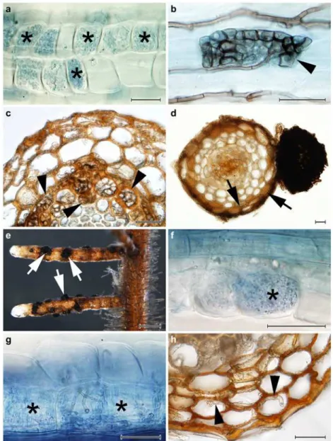
the tested PAC speciesA.macrosclerotiorumnever formed intracellular microsclerotia in spruce roots. On the other hand, intracellular hyphal coils resembling ErM (Fig 1f) together with intracellular microsclerotia typical for DSE were observed in blueberry roots colonized by A.macrosclerotiorum. A colonization pattern resembling ErM was formed also byP.glacialis Fig 1. The colonization patterns observed in Norway spruce (Picea abies) and European blueberry (Vaccinium myrtillus) roots in Experiment 1. 1a)Typical ericoid mycorrhizal colonization formed by
Rhizoscyphus ericaein blueberry roots (asterisks); stained with trypan blue, observed with DIC, bar = 25μm.
1b)An intracellular microsclerotium formed byPhialocephala helveticain a blueberry root (arrowhead); stained with trypan blue, observed with DIC, bar = 25μm.1c) Intracellular microsclerotia formed byP.
helveticain the vascular cylinder of a spruce root (arrowheads); observed with DIC, bar = 25μm.1d)A Hartig
net formed within the spruce root cortex (arrows) and an extraradical sclerotium formed on the spruce root surface (asterisk) byAcephala macrosclerotiorum; observed with DIC, bar = 25μm.1e)Spruce root tips colonized byA.macrosclerotiorumwith extraradical superficial sclerotia formed on the root surface (arrows); bar = 0.5 mm.1f)Intracellular hyphal loops morphologically resembling ericoid mycorrhizae (asterisks) formed byA.macrosclerotiorumin blueberry roots; stained with trypan blue, observed with DIC, bar = 25μm. 1g)Loose intracellular hyphal loops which may morphologically resemble ericoid mycorrhiza (asterisks) formed byPhialocephala glacialisin blueberry roots; stained with trypan blue, bar = 25μm.1h)Intracellular
colonization of spruce root cortex byP.glacialis(arrowheads); bar = 25μm.
doi:10.1371/journal.pone.0124752.g001
Root Symbiotic Potential of Dark Septate Endophytes
(Fig 1g) which however did not form EcM structures in spruce but colonized its roots intracel-lulary, including microsclerotia (Fig 1h) (Table 2).
Experiment 2:
In vitro
inoculation of blueberry in a peat-based substrate
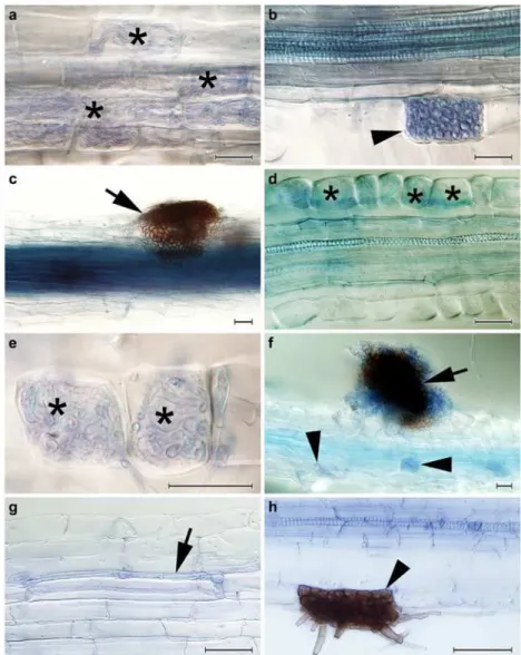
All of the inoculated blueberry seedlings possessed intraradical hyphal colonization similar to that found in Experiment 1 (Fig 2). Typical ErM structures were regularly formed in the roots of the plants inoculated withR.ericae. Intracellular hyphal coils resembling ErM colonization were formed byA.applanata(Fig 2a) which also formed intracellular microsclerotia and extraradical superficial sclerotia (Fig2band2c, respectively),A.macrosclerotiorum(Fig2dand2e) which also formed extraradical superficial sclerotia (Fig 2f) and to some extent also byP.glacialis. All DSE strains exceptA.macrosclerotiorumformed intracellular melanised or hyaline microsclero-tia. In contrast to Experiment 1 melanised sclerotia were formed on the root surface of the blue-berry seedlings inoculated withA.applanataAAP-1 and both strains ofA.macrosclerotiorum (see above;Table 2). Control plants did not possess any fungal colonization.
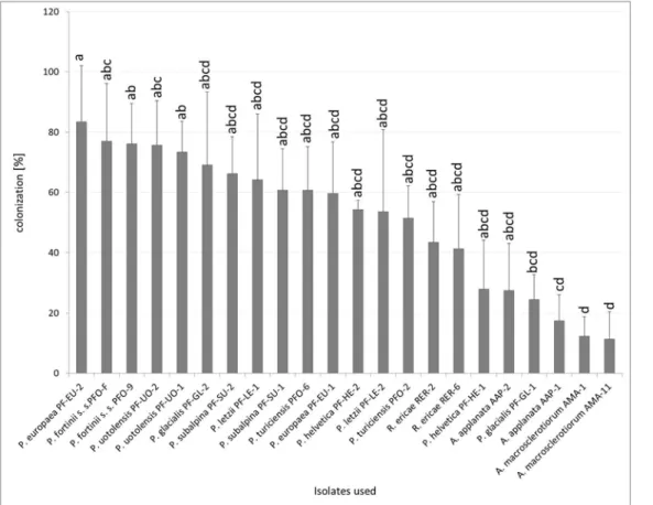
The colonization rates varied among the tested strains (Fig 3). Some DSE strains that formed ErM-like structures in the roots—A.macrosclerotiorum,A.applanataAAP-1 andP. glacialisPF-GL-1—had significantly lower colonization rates thanP.europaeaPF-EU-2. Both strains ofA.macrosclerotiorumhad significantly lower colonization rates than both strains ofP.fortiniis. s. andP.uotolensisbut no other differences between this ErM forming fungus and PAC were found. The percentage of colonized root cells byA.applanataAAP-1 was sig-nificantly lower thanP.fortiniis. s. PFO-9 andP.uotolensisPF-UO-1 but no other statistical-ly significant differences within PAC were found. Apparent trends of intraspecific differences were observed especially inP.glacialisandP.helveticaalthough these were statistically non-significant (Fig 3).
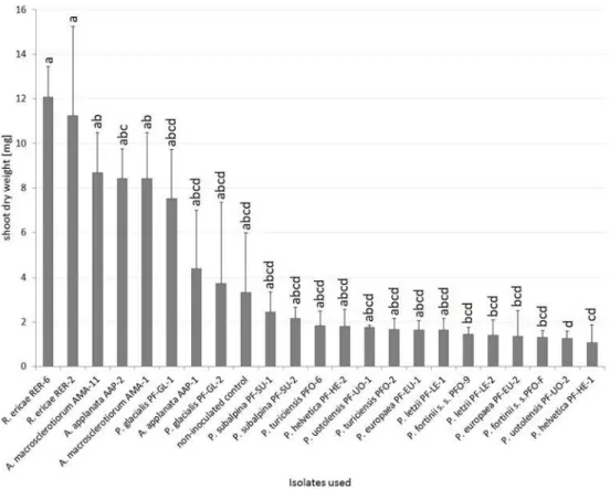
With respect to the influence of the inoculation on blueberry, six PAC strains (bothP. fortiniis. s. strains,P.europaeaPF-EU-2,P.helveticaPF-HE-1,P.letziiPF-LE-2 andP. uotolensisPF-UO-2) significantly decreased its dry shoot biomass when compared to posi-tive mycorrhizal control inoculated withR.ericae. Blueberries inoculated withA.applanata AAP-2 had significantly higher dry shoot biomass than those inoculated withP.uotolensis PF-UO-2 but no other differences within PAC were found. Blueberries inoculated with bothA.macrosclerotiorumstrains showed higher dry shoot biomass than two PAC strains (P.helveticaPF-HE-1andP.uotolensisPF-UO-2). The highest (although statistically non-significant) growth differences between conspecific strains were observed inA.applanata (AAP-1 vs. AAP-2) andP.glacialis(PF-GL-1 vs. PF-GL-2) (Fig 4).
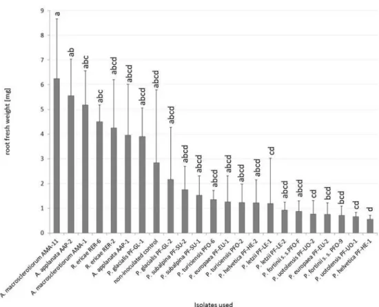
With respect to the influence of the inoculation on blueberry fresh root weight, the inocu-lated plants did not differ from the non-inocuinocu-lated control (Fig 5). Blueberries inoculated withR.ericaeRER-6 had significantly higher fresh root biomass than those inoculated with P.helveticaPF-HE-1 but no other statistically significant differences from mycorrrhizal con-trol were detected. Blueberries inoculated withA.applanataAAP-2 had significantly higher fresh root biomass thanP.helveticaPF-HE-1,P.letziiPF-LE-1 and both strains ofP. uotolen-sisbut no other statistically significant differences within PAC were found. Blueberries inocu-lated withA.macrosclerotiorumAMA-11 had higher fresh root biomass than six PAC strains (P.europaeaPF-EU-2,P.fortiniis. s. PFO-9,P.helveticaPF-HE-1,P.letziiPF-LE-1 and both strains ofP.uotolensis).A.macrosclerotiorumAMA-1 differed significantly only formP. hel-veticaPF-HE-1. The most prominent intraspecific variation was found inP.subalpinaandP. glacialisalthough it was not statistically significant (Fig 5).
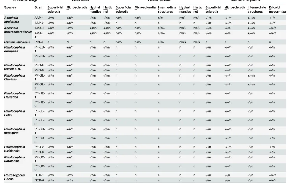
Table 2. Fungal structures observed in the roots of Norway spruce (Picea abies), silver birch(Betula pendula)and European blueberry (Vaccinium myrtillus) colonized by the tested fungal strains.
Inoculated fungi Picea abies Betula pendula Vaccinium myrtillus
Species Strain Superficial sclerotia
Microsclerotia Hyphal mantles
Hartig net
Superficial sclerotia Microsclerotia Intermediate structures Hyphal mantles Hartig net
Superficial sclerotia Microsclerotia Intermediate structures Ericoid mycorrhiza Acephala applanata
AAP-1 -/n/n +/n/n -/n/n -/n/n n/n/+ n/n/+ n/n/+ n/n/- n/n/- -/+/n +/+/n +/+/n -/+/n
AAP-2 -/n/n +/n/n -/n/n -/n/n n n n n n -/-/n +/+/n +/+/n -/+/n
Acephala macrosclerotiorum
AMA-1 +/n/n -/n/n +/n/n +/n/n n/n/- n/n/- n/n/+ n/n/- n/n/- -/+/n +/-/n +/+/n +/+/n
AMA-11
+/n/n -/n/n +/n/n +/n/n n/n/- n/n/- n/n/+ n/n/- n/n/- -/+/n +/-/n +/+/n +/+/n
Paxillus involutus PIN-9 n N n n n/n/- n/n/- n/n/- n/n/+ n/n/+ n n n n
Phialocephala europaea
PF-EU-1
-/n/n +/n/n -/n/n -/n/n n n n n n -/-/n +/+/n -/-/n -/-/n
PF-EU-2
-/n/n +/n/n -/n/n -/n/n n n n n n -/-/n +/+/n -/-/n -/-/n
Phialocephala fortiniis. s.
PFO-F -/n/n +/n/n -/n/n -/n/n n n n n n -/-/n +/+/n -/-/n -/-/n
PFO-9 -/n/n +/n/n -/n/n -/n/n n n n n n -/-/n +/+/n -/-/n -/-/n
Phialocephala Glacialis
PF-GL-1
-/n/n +/n/n -/n/n -/n/n n n n n n -/-/n +/+/n +/+/n -/-/n
PF-GL-2
-/n/n +/n/n -/n/n -/n/n n n n n n -/-/n +/+/n +/+/n -/-/n
Phialocephala Helvetica
PF-HE-1
-/n/n +/n/n -/n/n -/n/n n n n n n -/-/n +/+/n -/-/n -/-/n
PF-HE-2
-/n/n +/n/n -/n/n -/n/n n n n n n -/-/n +/+/n -/-/n -/-/n
Phialocephala Letzii
PF-LE-1
-/n/n +/n/n -/n/n -/n/n n n n n n -/-/n +/+/n -/-/n -/-/n
PF-LE-2
-/n/n +/n/n -/n/n -/n/n n n n n n -/-/n +/+/n -/-/n -/-/n
Phialocephala subalpina
PF-SU-1
-/n/n +/n/n -/n/n -/n/n n n n n n -/-/n +/+/n -/-/n -/-/n
PF-SU-2
-/n/n +/n/n -/n/n -/n/n n n n n n -/-/n +/+/n -/-/n -/-/n
Phialocephala turiciensis
PFO-2 -/n/n +/n/n -/n/n -/n/n n n n n n -/-/n +/+/n -/-/n -/-/n
PFO-6 -/n/n +/n/n -/n/n -/n/n n n n n n -/-/n +/+/n -/-/n -/-/n
Phialocephala uotolensis
PF-UO-1
-/n/n +/n/n -/n/n -/n/n n n n n n -/-/n +/+/n -/-/n -/-/n
PF-UO-2
-/n/n +/n/n -/n/n -/n/n n n n n n -/-/n +/+/n -/-/n -/-/n
Rhizoscyphus Ericae
RER-1 -/n/n -/n/n -/n/n -/n/n n n n n n -/-/n -/-/n -/-/n +/+/n
RER-6 -/n/n -/n/n -/n/n -/n/n n n n n n -/-/n -/-/n -/-/n +/+/n
S= extraradical superficial sclerotia;M= intracellular microsclerotia;Hy= hyphal mantles;Ha= intercellular Hartig net;I= intermediate structures, i.e., darkly pigmented or hyline loose intracellular hyphal loops;E= intracellular hyphal coils similar or identical to those formed in ericoid mycorrhiza.“+/-”denotes presence/absence of the structure in the root sample andndenotes not tested. Data from different experiments are separated by slashes (Experiment 1/Experiment 2/Experiment 3).
Experiment 3:
In vitro
inoculation of birch in a peat-agar medium
All inoculated seedlings possessed some intraradical fungal colonization. The percentage of colonized root sections was significantly higher inA.applanataAAP-1 andA. macrosclero-tiorumAMA-1 than inA.macrosclerotiorumAMA-11 (Table 3).A.macrosclerotiorumdid not form any EcM structures in birch roots but these were colonized intracellulary by loose hyphal loops and intercellulary by melanised running hyphae (Fig 2g).A.applanataformed
Fig 2. The colonization patterns observed in European blueberry (Vaccinium myrtillus) roots in Experiment 2 and in silver birch in Experiment 3. 2a)Intracellular hyphal colonization resembling ericoid mycorrhiza formed byAcephala applantaAAP-1 in blueberry roots (asterisks).2b)An early stage of the development of an intracellular microsclerotium formed byA.applanataAAP-1 in a blueberry root
(arrowhead).2c)An extraradical sclerotium formed on the surface of a blueberry root byA.applanataAAP-1 (arrow).2d)A blueberry hair root colonized in a manner resembling ericoid mycorrhiza byAcephala
macrosclerotiorumAMA-11 (asterisks).2e)A detail of two blueberry rhizodermal cells intracellularly
colonized byA.macrosclerotiorumAMA-11 in a manner resembling ericoid mycorrhiza (asterisks).2f)An extraradical sclerotium formed on the surface of a blueberry root byA.macrosclerotiorumAMA-11 (arrow). Note accompanying intracellular hyphal colonization (arrowheads).2g)A loose intracellular hyphal loop formed byA.macrosclerotiorumAMA-1 in a birch root (arrow).2h)A melanised intracellular microsclerotium formed byAcephala applanataAAP-1 in birch (arrowhead). All figures stained with trypan blue, observed with DIC, bars = 25μm.
intracellular microsclerotia (Fig 2h) as well as sclerotia on the root surface.P.involutusformed extraradical hyphal mantles and a Hartig net in the roots of all inoculated birch seedlings (Table 2). The control non-inoculated plants did not show any signs of fungal colonization.
The inoculation had significant influence on birch shoot dry weight; the plants inoculated withA.applanataAAP-1 andA.macrosclerotiorumAMA-1 had significantly higher shoot bio-mass than the control non-inoculated plants. Differences were also observed in fresh root weight; birch seedlings inoculated with bothA.macrosclerotiorumstrains had higher root bio-mass than the non-inoculated control plants (Table 3).
No significant differences were detected in O2concentration measured in the microcosms.
Microcosms withA.macrosclerotiorumAMA-1 had higher CO2concentration in comparison
with both positive mycorrhizal and negative non-inoculated controls (Table 3). No contamina-tions of the peat substrate were detected and all the inoculated fungi were viable at the end of the experiment.
Culture dependent screening of DSE-like fungi in Ericaceae roots from a
pine forest
The microscopic screening of Ericaceae roots collected in the pine forest showed that 871 from the 1000 screened rhizodermal cells were intracellularly colonized by fungal hyphae, mostly in a manner typical for ericoid mycorrhiza. About one third of the obtained isolates (i.e., 35 isolates)
Fig 3. Percentage fungal colonization of blueberry rhizodermal cells in Experiment 2.Blueberry seedlings were inoculated by 8 PAC species, 2 species related to PAC andRhizoscyphus ericaeas a positive control, two strains per each species. Blueberry seedlings were grown in a peat-based substrate for 3.5 months underin vitroconditions. The presented data are means of 6 replicates±standard error of mean. Different letters above the columns indicate significant differences according to the non-parametric Kruskal-Wallis test followed by the multiple-comparison z-value test.
doi:10.1371/journal.pone.0124752.g003
Root Symbiotic Potential of Dark Septate Endophytes
produced brownish to blackish colonies typical for DSE fungi, includingA.macrosclerotiorum. However, according to sequence analyses in BLAST, all but one sequence belonged toP.fortinii s. l. and none of the sequences belonged toA.macrosclerotiorum(S1 Table).
Culture independent screening of fungal root symbionts in Ericaceae
roots from the pine forest
On average we obtained 2184 fungal sequences per sample (ranging from 326 to 10076 per sam-ple) and 37.9 OTUs per sample (ranging from 19 to 151); in total, we obtained 380 well-defined OTUs (S2 Table). All samples were dominated by Helotiales (Fig 6). The most abundant species across all localities (OTU 1) belonged to Helotiales, its abundance ranged from 82.15% to 5.59% (average 31.46%) (S2 Table) and its consensus sequence was most similar (94%) to an uncultured fungus sampled from a pine forest soil (GenBank acc. no. JX032276). The phylogenetic analysis placed this fungus in a sister clade (comprising only entries without scientific names) of a clade comprisingClaussenomyces,Collophora,Satchmopsis, and some other fungi (S1 Fig). The second most abundant species across all localities (OTU 3) belonged toP.fortiniis. l. and its abundance ranged from 53.37% to 0.6% (average 18.76%). The third most abundant species across all locali-ties (OTU 4) was present in all but one sample, its abundance ranged from 38.16% to 0.00% (av-erage 10.65%) and its consensus sequence was identical to several uncultured fungi from different
Fig 4. The effect of inoculation on blueberry dry shoot weight in Experiment 2.Blueberry seedlings were inoculated by 8 PAC species, 2 species related to PAC andRhizoscyphus ericaeas a positive control, two strains per each species. Blueberry seedlings were grown in a peat-based substrate for 3.5 months underin vitroconditions. The presented data are means of 6 replicates±standard error of mean. Different letters above the columns indicate significant differences according to the non-parametric Kruskal-Wallis test followed by the multiple-comparison z-value test.
Fig 5. The effect of inoculation on blueberry fresh root weight in Experiment 2.Blueberry seedlings were inoculated by 8 PAC species, 2 species related to PAC andRhizoscyphus ericaeas a positive control, two strains per each species. Blueberry seedlings were grown in a peat-based substrate for 3.5 months underin vitroconditions. The presented data are means of 6 replicates±standard error of mean. Different letters above the columns indicate significant differences according to the non-parametric Kruskal-Wallis test followed by the multiple-comparison z-value test.
doi:10.1371/journal.pone.0124752.g005
Table 3. Colonization, root fresh weight and shoot dry weight of birch seedlings inoculated by two strains ofA.macrosclerotiorum, one strain ofA.applanataand one strain ofP.involutusas a positive EcM control fungus in Experiment 3.
Inoculated fungi Colonization
[%]
Root fresh weight [mg]
Shoot dry weight [mg]
CO2concentration
[ppm]
A.macrosclerotiorumAMA-1 51.79±15.34 a 9.2±3.8 ab 6.1±1.0 a 350.71±22.13 a
A.macrosclerotiorumAMA-11 18.75±10.90 b 11.7±2.8 a 5.3±0.9 ab 255.71±67.73 ab
A.applanataAAP-1 61.07±14.06 a 11.5±2.5 a 7.3±2.7 a 265.57±33.16 ab
P.involutusPIN-1 not measured 10.5±5.2 ab 4.1±1.7 ab 222.00±54.42 b
non-inoculated no colonization 3.2±1.0 b 2.6±0.7 b 224.86±63.17 b
Plants were cultivated in a peat-agar medium inin vitroconditions for 6 months. Concentration of CO2in
the microcosms was measured three days prior harvest. The presented data are means of 6
replicates±SE. Different letters indicate significant differences according to the non-parametrical Kruscal-Wallis test followed by a multiple-comparison z-value test.
doi:10.1371/journal.pone.0124752.t003
Root Symbiotic Potential of Dark Septate Endophytes
sources and locations (e.g., KF617263 fromPicea marianaforest soil in Alaska, HM059043 from muskeg bog in Alaska or AB521974 fromVacciniumhair roots in Sweden). The phylogenetic analysis placed this fungus in the vicinity of many uncultured fungi from Chaetothyriales and Verrucariales (S2 Fig).
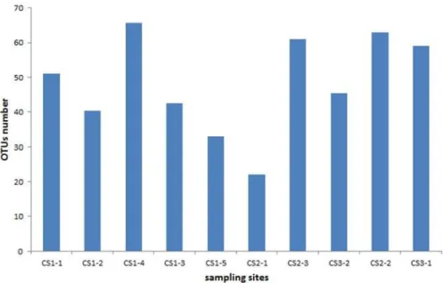
Based on Chao-1, the alpha diversity within samples varied from 22 to 66 OTUs with the most diverse sample being CS1-4 and the least diverse sample CS2-1 (Fig 7). In the PCA analysis, the first canonical axis explained 62.8% of the variability while the second axis explained 19.2% (Fig 8).
We did not obtain any sequences similar toA.macrosclerotiorumalthough thePinirhiza sclerotiaectomycorrhizal morphotype was present in the pine roots in all screened samples.
Discussion
The DSE symbiotic potential in spruce and blueberry
Fungi related toP.fortiniiare often isolated from mycorrhizal and non-mycorrhizal conifer roots and in re-synthesis experiments they frequently form pseudomycorrhizae sensu Melin [54] [4,55]. Although some authors reported thatP.fortiniis. l. formed hyphal mantles and Hartig net in re-synthesis experiments with conifers, these structures were typically accompanied by intracellular hyphal colonization [28]. To our knowledge only two papers reported distinct ectomycorrhizal morphotypes formed by fungi related toP.fortiniis. l. [32,56]. Kaldorfet al. [56] reported that a relatively frequent black unbranched ectomycorrhizal morphotype with abundant emanating hy-phae formed on hybrid aspen repeatedly yielded DNA ofP.fortiniis. l. However, the authors did not use the respective fungus in a re-synthesis experiment and the amplifiedP.fortiniis. l. DNA
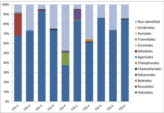
Fig 6. Relative abundances of the dominant fungal orders in Ericaceae roots detected by
pyrosequencing.Tag-encoded pyrosequencing was performed with ten Ericaceae hair roots samples from three sites in NPČeskéŠvýcarsko (CS1—five samples, CS2—three samples, CS3—two samples) where
the ectomycorrhizal morphotypePinirhiza sclerotinaformed by the DSE fungus related to PACAcephala
macrosclerotiorumwas present. The obtained data were processed as described in Materials and Methods.
OTUs with lower similarity and coverage than 88% were assigned as non-identified together withincertae
sedisspecies. Orders less abundant than 0.1% were excluded from the figure.
Fig 7. Values of Chao-1 index in equally resampled localities.The values of Chao-1 indexes for the respective localities are as follows: CS1-1 = 51, CS1-2 = 40.3, CS1-3 = 42.5, CS1-4 = 65.6, CS1-5 = 33, CS2-1 = 22, CS2-2 = 63, CS2-3 = 6CS2-1, CS3-CS2-1 = 59 and CS3-2 = 45.5.
doi:10.1371/journal.pone.0124752.g007
Fig 8. Principal component analysis of the relative abundance of OTUs.Only OTUs with component loadings for the first or second axis higher than 0.015 were visualized. For detailed information about the respective OTUs seeS2 Table.
doi:10.1371/journal.pone.0124752.g008
Root Symbiotic Potential of Dark Septate Endophytes
might as well originate from endophytic hyphae [32]. In contrast, Münzenbergeret al. [32] experi-mentally verified thatA.macrosclerotiorumformed the ectomycorrhizal morphotypeP.sclerotia. Here, under experimental conditions favorable for ectomycorrhiza formation, we showed that the tested strains representing all hitherto described PAC cryptic species failed to form ectomycorrhi-zae with spruce, in contrast toA.macrosclerotiorum. The latter species thus remains the only ex-perimentally verified DSE forming a distinct ectomycorrhizal morphotype both under natural and artificial conditions. However, its abundance in ectomycorrhizal conifers is not clear [32,57] and the ecophysiological significance ofPinirhiza sclerotianeeds to be further investigated.
Fungi related toP.fortiniis. l. are often isolated also from ericaceous hair roots and there are some reports showing that they may form intracellular hyphal coils in Ericaceae rhizoder-mis [30,31]. On the other hand, similar structures may be to some extent formed in Ericaceae hair roots also by soil saprophytic fungi [58]. Here,A.applanataandA.macrosclerotiorum formed intracellular hyphal coils inVacciniumrhizodermal cells which strongly resembled those formed in ericoid mycorrhiza. In contrast to Vohníket al. [30] this colonization pattern was neither rare nor limited to only a few rhizodermal cells, reached colonization levels of the typical ericoid mycorrhizal fungusR.ericaeand would be unambiguously considered as ericoid mycorrhiza if seen in naturally colonized roots. Additionally, the detected trend of in-creased dry shoot weight and fresh root weight connected with inoculation of blueberries with A.applanataandA.macrosclerotiorum(see below) suggests positive effects of these two DSE species on their host plants. However, this needs to be tested again under different experimen-tal conditions and with more inoculation replicates.
A.applanataandA.macrosclerotiorumsimultaneously formed intracellular hyphal coils resembling ericoid mycorrizae and typical DSE microsclerotia in the same hair roots. This parallels the observations made by Vohník and Albrechtová in roots of the European autoch-thonousRhododendron kotschyi[33] and indeed suggests the existence of morphological con-tinuum between ericoid mycorrhizal and DSE fungi.
Host response to DSE colonization
Due to their high incidence in mycorrhizal roots, the effect of DSE on host plants has already been studied for more than a century. In his classical work, Melin [54] stated that the isolated DSE related toP.fortiniis. l. (Mycelium radices atrovirens) did not form ectomycorrhiza with conifers which was necessary for their normal growth; instead, they formed the harmful pseu-domycorrhiza. Indeed, several other authors pointed out that DSE colonization in trees is harmful and its negative effects may be alleviated by ectomycorrhizal fungi [59–61].
Despite that the tested DSE strains did not form ectomycorrhizae with birch, their presence significantly increased birch shoot and root biomass in comparison with non-inoculated con-trol. In the case ofA.macrosclerotiorumAMA-1, this non-mycorrhizal functioning may be at least partly attributed to the elevated CO2concentration which stimulates net plant carbon
gain from photosynthesis. Another possible mechanism may be mineralization of the cultiva-tion substrate [23].
Host specificity in DSE
The low to none host specificity of DSE [6,7] has been challenged after the division of P.fortiniis. l. into cryptic species, and description of two new DSE species,A.macrosclerotiorumandP.glacialis. Grünig and colleagues [65] investigated three subalpine forest sites in Switzerland with simulta-neous presence ofP.abiesandVacciniumspp. and reported thatA.applanatapreferred spruce roots whileP.subalpinashowed preference for Ericaceae;P.glacialishas been so far isolated only from Ericaceae hair roots and spruce needles, but not spruce roots [66]; andA.macrosclerotiorum formed ectomycorrhizae with pine but not with hybrid aspen [32]. Here we confirmed the ectomy-corrhizal preference ofA.macrosclerotiorumfor conifers and extended the known range of its po-tential hosts for Norway spruce. On one hand,A.macrosclerotiorumhas been isolated from spruce ectomycorrhizae already by Menkiset al. [57] few years before its formal taxonomic description; on the other hand it remains unclear whether the isolatedPhialocephala6/Acephalasp. 6 (conspe-cific withA.macrosclerotiorum, seehttp://unite.ut.ee/sh/SH213469.06FU) formed the respective ectomycorrhizae or lived inside them as an endophyte. The reason for the peculiarA. macrosclero-tiorummycorrhizal incompatibility with broadleaved trees remains unknown but may parallel host preferences of other (ecto-)mycorrhizal fungi. Another reason may be in the hypothesized higher conifer tolerance to DSE colonization: Ahlich and Sieber [67] and Grünig and colleagues [3] reported that DSE colonization densities tended to be lower in broadleaf trees than in conifers and suggested that conifers might better tolerate DSE in order to prevent infection by more serious fungal pathogens.
Despite that DSE often represent the majority of mycobionts isolated from ericaceous hair roots [10,63,64] we did not isolate or detectA.macrosclerotiorumin the Ericaceae roots at sites with significant presence of its ectomycorrhizal morphotype in neighboring pines. Such absence is intriguing given the apparent ability ofA.macrosclerotiorumto form intracellular hyphal coils morphologically identical to ericoid mycorrhiza in Ericaceae rhizodermis (seeResults). However, this species has not yet been detected in Ericaceae roots which suggests that theA. macrosclero-tiorum in vitroericoid mycorrhizal potential does not need to be realized under natural condi-tions. Similar scenario might hold true also forA.applanatawhich formed ericoid mycorrhizae in vitrobut has not yet been reported in Ericaceae roots.
Possible role of DSE in common mycorrhizal networks
The above mentioned low DSE host specificity along with the ability to simultaneously colo-nize plants with different mycorrhizal preferences [6,7] opens the potential to link neighbor-ing plants with common mycelia. Such a scenario has been hypothesized for ectomycorrhizal plants and their ericaceous undergrowth because these plant guilds can form ectomycorrhizal and ericoid mycorrhizal symbioses with the same members of theR.ericaeaggregate (REA) underin vitroconditions [34,68]. However, Kohoutet al. [41] showed that the potentially shared REA mycobiont,Meliniomyces bicolor, was in fact suppressed when conifers and Eri-caceae were grown together in an open air pot experiment. Similarly, the abundance of REA members in Ericaceae roots was significantly lower at sites with ectomycorrhizal tree domi-nants when compared to sites where the dominant trees preferred arbuscular mycorrhizal
Root Symbiotic Potential of Dark Septate Endophytes
fungi [10]. Congruently, we found thatin vitro,A.macrosclerotiorumwas able to form ecto-mycorrhiza and ericoid ecto-mycorrhiza with spruce and blueberry, respectively, but this species was not detected in Ericaceae roots at natural localities with regular presence of theP. sclero-tiamorphotype which is formed byA.macrosclerotiorum. Our study thus provides another observation which suggests that mycorrhizal links between ectomycorrhizal conifers and eri-coid mycorrhizal Ericaceae are under natural conditions rare to absent and perhaps even in-hibited [10,41].
Diversity of Ericaceae root mycobionts
In comparison with arbuscular mycorrhizal and ectomycorrhizal plants, the diversity of Erica-ceae root mycobionts is tackled relatively scarcely. There are some reports from Argentina [10], Australia [69,70], Canada [64,71], China [72], Japan [73], Scandinavia [39,62], UK [74,75] and USA [63] but these are relatively scarce and studies from other regions, including central Eu-rope, are missing. Although it is premature to speculate on the global diversity of Ericaceae mycobionts it roughly seems that some REA members (M.variabilis,R.ericae) together with Oidiodendron maiusare dominant ErM fungi in temperate, boreal and subarctic Eurasia while Sebacinaceae and other mycobionts possibly form the dominant part of Ericaceae mycobionts in the North America and Southern Hemisphere [10]. In contrast, the Ericaceae hair roots in-vestigated in this study were mostly free of the prominent ErM fungi, including Sebacinaceae, and their dominant mycobionts, exceptP.fortiniis. l., belonged to hitherto undescribed fungi from Helotiales and Chaetothyriales/Verrucariales [76]. The significance of this finding remains obscure yet it has to be kept in mind that most of the screened hair roots possessed ericoid my-corrhizae, similarly to [71]. We obviously cannot prove that the dominant mycobionts detected in this study using pyrosequencing indeed formed the observed intracellular hyphal structures. This could be achieved by a method targeting single host cells, e.g., laser capture microdissection followed by DNA isolation and PCR, which however has not yet been applied on ericoid mycor-rhizae. The ITS sequence of the most common OTU 1 (Helotiales 01) clustered with many un-cultured fungi and it is thus plausible to assume that OTU 1 is a non-cultivable species; on the other hand, two similar ITS sequences (FM172779 and HM208736) belong to isolates obtained from ericaceous roots. This situation emphasizes the importance of cultivation-based methods which may, when followed by re-synthesis experiments and experiments tracking the bi-direc-tional mycorrhizal nutrient transfer, confirm the putative ericoid mycorrhizal status of domi-nant but hitherto undescribed Ericaceae mycobionts, especially from areas which have not yet been investigated.
The total diversity of Ericaceae mycobionts was relatively high but the three most abun-dant OTUs, Helotiales 01,Phialocephala fortiniis. l. and Chaetothyriales/Verrucariales 01, comprised over 60% of the total mycobiont abundance. These three OTUs showed strong preference for certain experimental sub-sites where they dominated the respective mycobiont community while being infrequent to absent at other sub-sites.M.variabilis(OTU 12), the most abundant typical ErM fungus detected in this study (yet with only 4.04% of the total mycobiont abundance) represented the most abundant mycobiont at one sub-site (23.01%) while being totally absent at other four sub-sites and nearly absent (<0.5%) at another three
sub-sites. This suggests that the community composition of ericaceous mycobionts may be significantly different even within sites with no apparent ecological gradients [38,64,74].
role is virtually unknown. Some may intracellularly colonize healthy ericaceous roots underin vitroconditions but the resulting colonization pattern does not resemble typical ericoid mycor-rhizae [35,77] and it is plausible that under natural conditions, these mycobionts colonize senes-cent or moribund hair root cells and act as opportunistic saprobes rather than true ErM fungi.
Sebacinaceae, a group of ubiquitous heterobasidiomycetous endophytes and mycorrhizal fungi [78] were proposed as common ericoid mycorrhizal fungi worldwide, based on culture-independent methods [79]. Here we detected Sebacinaceae sequences only at two sub-sites out of ten with the total abundance of 1.18%. This was a surprising finding given the absence of other common ericoid mycorrhizal fungi and the fact that Sebacinaceae often represent a major component of Ericaceae mycobiont communities [71,80]. To our knowledge, a suc-cessful isolation of a sebacinaceous strain from Ericaceae hair roots has been reported only once [35] and an experimental (re-synthetic) proof that these fungi indeed form ericoid my-corrhizae is still missing, although indirect evidence is strong [79]. The reason for the low in-cidence of Sebacinaceae (and other prominent ericoid mycorrhizal fungi) in this study is unknown and remains to be investigated.
Conclusions
Although PAC fungi are often isolated from ectomycorrhizal and ericoid mycorrhizal roots, none of the tested PAC cryptic species exceptA.applanataformed typical mycorrhizal struc-tures. Moreover, some of the tested PAC strains had negative influence on host biomass. Inter-specific variability thus likely does not explain the inconsistency of the results obtained in the DSE research during past decades when differentiation ofP.fortinii-related fungi to cryptic species was not possible. In contrast, it seems that the true reason is in variability of the many different combinations of particular DSE strains with particular host plants under particular growing conditions, as showed here and recently discussed by other authors [20,81]. On the other hand, some of the PAC close relatives apparently have ectomycorrhizal (A. macrosclero-tiorum) and ericoid mycorrhizal (A.macrosclerotiorum, perhaps alsoP.glacialis) potential, which however does not need to be realized under natural conditions as suggested by the lack ofA.macrosclerotiorumin Ericaceae hair roots in the pine fores with common occurrence of theP.sclerotiamorphotype. To our knowledge,A.macrosclerotiorumis the only documented DSE fungus forming ectomycorrhizae. Interestingly, its ectomycorrhizal potential seems to be realized only in conifers (pine, spruce) but not in broadleaved plants (birch, poplar).
Supporting Information
S1 Fig. The taxonomic position of OTU 1.For details on the phylogenetic analyses see Mate-rials and Methods.
(PDF)
S2 Fig. The taxonomic position of OTU 4.For details on the phylogenetic analyses see Mate-rials and Methods.
(PDF)
S1 Table. The identity of dark septate endophytes obtained by culture-dependent approach. The table displays the three top GenBank hits to each sequence of the DSE isolates obtained from ericaceous hair roots in this study. Sequences derived from cultured isolates with scientif-ic names were preferred.
(XLSX)
S2 Table. The identity of the mycobionts detected by tag-encoded pyrosequencing.The table provides basic information on the 380 OTUs obtained in this study by tag-encoded
Root Symbiotic Potential of Dark Septate Endophytes
pyrosequencing (seeMaterials and Methods, andResults). OTUs are in grey when the re-spective closest GenBank match had similarity<97% or there was no match in GenBank.
The column "Closest match in GenBank" lists closest matches with scientific names. (XLSX)
Acknowledgments
The authors would like to thank Babette Münzenberger and Christoph R. Grünig for provid-ing some DSE strains, Jiří Machačand TomášPicek for technical support and the authority of NPČeskéŠvýcarsko for issuing the sampling permit. Valuable comments of François Le Tacon helped to improve an earlier version of this paper and are greatly appreciated.
Author Contributions
Conceived and designed the experiments: TL PK MV. Performed the experiments: TL. Ana-lyzed the data: TL PK TV MV. Contributed reagents/materials/analysis tools: TL PK TV MV. Wrote the paper: TL PK TV MV.
References
1. Brundrett M. Understanding the roles of multifunctional mycorrhizal and endophytic fungi. In: Schulz B, Boyle C, Sieber TN, editors. Microbial root endophytes. Soil Microbiology. Berlin: Springer; 2006. pp. 179–190.
2. Rodriguez R, White J. Fungal endophytes: diversity and functional roles. New Phytol. 2009; 182: 314–330. doi:10.1111/j.1469-8137.2009.02773.xPMID:19236579
3. Grünig C, Queloz V, Sieber T, Holdenrieder O. Dark septate endophytes (DSE) of thePhialocephala
fortiniis. l.—Acephala applanataspecies complex in tree roots: classification, population biology, and
ecology. Botany. 2008; 86: 1355–1369.
4. Wang CJK, Wilcox HE. New species of ectendomycorrhizal and pseudomycorrhizal fungi:Phialophora
finlandia,Chloridium pucisporum, andPhialocephala fortinii. Mycologia. 1985; 77: 951–958.
5. Duó A, Bruggmann R, Zoller S, Bernt M, Grünig C. Mitochondrial genome evolution in species belong-ing to thePhialocephala fortiniis. l.—Acephala applanataspecies complex. BMC Genomics. 2012; 13: 166. doi:10.1186/1471-2164-13-166PMID:22559219
6. Jumpponen A, Trappe JM. Dark septate endophytes: a review of facultative biotrophic root-colonizing fungi. New Phytol. 1998; 140: 295–310.
7. Addy H, Hambleton S, Currah R. Distribution and molecular characterization of the root endophyte
Phialocephala fortiniialong an environmental gradient in the boreal forest of Alberta. Mycol Res. 2000;
104: 1213–1221.
8. Kohout P, Těšitelová T, Roy M, Vohník M, Jersáková J. A diverse fungal community associated with
Pseudorchis albida(Orchidaceae) roots. Fungal Ecol. 2013; 6: 50–64.
9. Vohník M, Mrnka L, Lukešová T, Bruzone MC, Kouhout P, Fehrer J. The cultivable endophytic
commu-nity of Norway spruce ectomycorrhizas from microhabitats lacking ericaceous hosts is dominated by er-icoid mycorrhizalMeliniomyces variabilis. Fungal Ecol. 2013; 6: 281–292.
10. Bruzone MC, Fontenla SB, Vohník M. Is the prominent ericoid mycorrhizal fungusRhizoscyphus ericae absent in the Southern Hemisphere’s Ericaceae? A case study on the diversity of root mycobionts in
Gaultheriaspp. from northwest Patagonia, Argentina. Mycorrhiza. 2015; 25: 25–40. doi:10.1007/
s00572-014-0586-3PMID:24838300
11. Kohout P, Sýkorová Z,Čtvrtlíková M, Rydlová J, Suda J, Vohník M, et al. Surprising spectra of root-as-sociated fungi in submerged aquatic plants. FEMS Microbiol Ecol. 2012; 80: 216–235. doi:10.1111/j. 1574-6941.2011.01291.xPMID:22224638
12. Selosse M-A, Vohník M, Chauvet E. Out of the rivers: are some aquatic hyphomycetes plant endo-phytes? New Phytol. 2008; 178: 3–7. doi:10.1111/j.1469-8137.2008.02390.xPMID:18315696
13. Klymiuk A, Taylor TN, Taylor EL, Krings M. Paleomycology of the Princeton Chert II. Dark-septate fungi in the aquatic angiospermEorhiza arnoldiiindicate a diverse assemblage of root-colonizing fungi during the Eocene. Mycologia. 2013; 105: 1100–1109. doi:10.3852/13-025PMID:23709575
14. Newsham KK.Phialophora graminicola, a dark septate fungus, is a beneficial associate of the grass
15. Vohník M, Albrechtová J, Vosátka M. The inoculation withOidiodendron maiusandPhialocephala forti-niialters phosphorus and nitrogen uptake, foliar C : N ratio and root biomass distribution in Rhododen-droncv. Azurro. Symbiosis. 2005; 40: 87–96.
16. Usuki F, Narisawa K. A mutualistic symbiosis between a dark septate endophytic fungus, Heteroco-nium chaetospira, and a nonmycorrhizal plant, Chinese cabbage. Mycologia. 2007; 99: 175–184. PMID:17682770
17. Wu L, Lv Y, Meng Z, Chen J, Guo S. The promoting role of an isolate of dark-septate fungus on its host plantSaussurea involucrataKar. et Kir. Mycorrhiza. 2010; 20: 127–135. doi: 10.1007/s00572-009-0268-8PMID:19707800
18. Wilcox HE, Wang CJK. Mycorrhizal and pathological associations of dematiaceous fungi in roots of 7-month-old tree seedlings. Can J For Res. 1987; 17: 884–899.
19. Stoyke G, Currah R. Resynthesis in pure culture of a common subalpine fungus-root association using
Phialocephala fortiniiandMenziesia ferruginea(Ericaceae). Arct Alp Res. 1993; 25: 189–193.
20. Tellenbach C, Grünig CR, Sieber TN. Negative effects on survival and performance of Norway spruce seedlings colonized by dark septate root endophytes are primarily isolate dependent. Environ Micro-biol. 2011; 13: 2508–2517. doi:10.1111/j.1462-2920.2011.02523.xPMID:21812887
21. Newsham K. A metaanalysis of plant responses to dark septate root endophytes. New Phytol. 2011; 190: 783–793. doi:10.1111/j.1469-8137.2010.03611.xPMID:21244432
22. Mayerhofer MS, Kernaghan G, Harper KA. The effects of fungal root endophytes on plant growth: a meta-analysis. Mycorrhiza. 2013; 23: 119–128. doi:10.1007/s00572-012-0456-9PMID:22983627
23. Reininger V, Sieber TN. Mitigation of antagonistic effects on plant growth due to root co-colonization by dark septate endophytes and ectomycorrhiza. Environ Microbiol Rep. 2013; 5: 892–898. doi:10.1111/ 1758-2229.12091PMID:24249297
24. Vohník M, Sadowsky JJ, Lukešová T, Albrechtová J, Vosátka M. Inoculation with wood decomposing
basidiomycete, but not with root symbiotic ascomycetes, positively affects growth of highbush blueberry (Ericaceae) grown in a pine litter substrate. Plant Soil. 2012; 355: 341–352.
25. Schulz B, Boyle C. The endophytic continuum. Mycol Res. 2005; 109: 661–686. PMID:16080390
26. Zimmerman E, Peterson R. Effect of a dark septate fungal endophyte on seed germination and proto-corm development in a terrestrial orchid. Symbiosis. 2007; 43: 45–52.
27. Tellenbach C, Sieber TN. Do colonization by dark septate endophytes and elevated temperature affect pathogenicity of oomycetes? FEMS Microbiol Ecol. 2012; 82: 157–168. doi:10.1111/j.1574-6941. 2012.01415.xPMID:22587673
28. Peterson R, Wagg C, Pautler M. Associations between microfungal endophytes and roots: do structural features indicate function? Botany. 2008; 86: 445–456.
29. Stoyke G, Currah RS. Endophytic fungi from the mycorrhizae of alpine ericoid plants. Can J Bot. 1991; 69: 347–352.
30. Vohník M, LukančičS, Bahor E, Regvar M, Vosátka M, Vodnik D. Inoculation ofRhododendroncv. Belle-Heller with two strains ofPhialocephala fortiniiin two different substrates. Folia Geobot. 2003; 38: 191–200.
31. Usuki F, Narisawa K. Formation of structures resembling ericoid mycorrhizas by the root endophytic fungusHeteroconium chaetospirawithin roots ofRhododendron obtusumvar.kaempferi. Mycorrhiza. 2005; 15: 61–64. PMID:15517420
32. Münzenberger B, Bubner B, Wöllecke J. The ectomycorrhizal morphotypePinirhiza sclerotiais formed
byAcephala macrosclerotiorumsp. nov., a close relative ofPhialocephala fortinii. Mycorrhiza. 2009;
19: 481–492. doi:10.1007/s00572-009-0239-0PMID:19415343
33. Vohník M, Albrechtová J. The co-occurrence and morphological continuum between ericoid mycorrhiza and dark septate endophytes in roots of six European Rhododendron species. Folia Geobot. 2011; 46: 373–386.
34. Grelet GA, Johnson D, Paterson E, Anderson IC, Alexander IJ. Reciprocal carbon and nitrogen transfer between an ericaceous dwarf shrub and fungi isolated fromPiceirhiza bicolorataectomycorrhizas. New Phytol. 2009; 182: 359–366.
35. Vohník M, Sadowsky JJ, Kohout P, Lhotáková Z, Nestby R, Kolařík M. Novel root-fungus symbiosis in Ericaceae: sheathed ericoid mycorrhiza formed by a hitherto undescribed basidiomycete with affinities to Trechisporales. PLOS ONE. 2012; 7: e39524. doi:10.1371/journal.pone.0039524PMID:22761814
36. Simard SW, Durall DM. Mycorrhizal networks: a review of their extent, function, and importance. Can J Bot. 2004; 82: 1140–65.
37. Beiler KJ, Durall DM, Simard SW, Maxwell SA, Kretzer AM. Architecture of the wood-wide web:
Rhizo-pogonspp. genets link multiple Douglas-fir cohorts. New Phytol. 2010; 185: 543–553. doi:10.1111/j.
1469-8137.2009.03069.xPMID:19878460
Root Symbiotic Potential of Dark Septate Endophytes