Conservation, Variability and the Modeling of Active
Protein Kinases
James D. R. Knight1,2,3, Bin Qian4, David Baker4, Rashmi Kothary1,2,3,5*
1Molecular Medicine Program, Ottawa Health Research Institute, Ottawa, Ontario, Canada,2The University of Ottawa Centre for Neuromuscular Disease, Ottawa, Ontario, Canada,3Department of Cellular and Molecular Medicine, University of Ottawa, Ottawa, Ontario, Canada,4Department of Biochemistry, University of Washington, Seattle, Washington, United States of America,5Department of Medicine, University of Ottawa, Ottawa, Ontario, Canada
The human proteome is rich with protein kinases, and this richness has made the kinase of crucial importance in initiating and maintaining cell behavior. Elucidating cell signaling networks and manipulating their components to understand and alter behavior require well designed inhibitors. These inhibitors are needed in culture to cause and study network perturbations, and the same compounds can be used as drugs to treat disease. Understanding the structural biology of protein kinases in detail, including their commonalities, differences and modes of substrate interaction, is necessary for designing high quality inhibitors that will be of true use for cell biology and disease therapy. To this end, we here report on a structural analysis of all available active-conformation protein kinases, discussing residue conservation, the novel features of such conservation, unique properties of atypical kinases and variability in the context of substrate binding. We also demonstrate how this information can be used for structure prediction. Our findings will be of use not only in understanding protein kinase function and evolution, but they highlight the flaws inherent in kinase drug design as commonly practiced and dictate an appropriate strategy for the sophisticated design of specific inhibitors for use in the laboratory and disease therapy.
Citation: Knight JDR, Qian B, Baker D, Kothary R (2007) Conservation, Variability and the Modeling of Active Protein Kinases. PLoS ONE 2(10): e982. doi:10.1371/journal.pone.0000982
INTRODUCTION
Protein kinases are the most ubiquitous single family of signaling molecules in the cell, accounting for approximately 2% of the proteins encoded by the human genome [1]. The simple mechanism of attaching an ATP-derived phosphate to a protein involves kinases in every aspect of cell behavior, from apoptosis to survival, proliferation to differentiation, maturation etc. Protein kinases provide a unique opportunity for understanding proteins in general by presenting us with a seeming paradox: wide scale similarity of sequence and structure combined with a diversity of behavioral consequences to their activity. The vast majority of protein kinases have readily detectable sequence similarity, which translates into structure. But even those known protein kinases that show no significant algorithm-detectable similarity at the level of sequence are believed to have very typical structures, as is evidenced by specific examples [2,3]. As they all have a shared function in transferring the terminal phosphate of ATP to another protein, similarity is understandable. Evidence to date also suggests a common catalytic mechanism (the possible exception may be the integrin-linked kinase [4]), whereby ATP and an active site divalent cation are bound in identical fashions and phospho-transfer is achieved by a shared set of amino acids. Studies in yeast [5,6] have shown that kinases can be promiscuous, phosphorylating hundreds of proteins, but they also have clear specificities. How is this specificity attained by one family of highly similar proteins? This paradox suggests the perfection of the kinase as an enzyme: a region ideally suited for the common function of catalysis, with another region(s) uniquely modifiable to attain substrate specificity without altering fold, compromising ligand binding or the subsequent reaction mechanism. A thorough understanding of this family of proteins would generate a tremendous knowledge base for discovering and predicting protein interactions, for designing highly specific and potent inhibitors, and, as a consequence of these facts, for understanding the cell and disease.
As protein kinases are the key players in cell signaling, aberrations in their activity have been directly correlated with
numerous disease states (for example, breast cancer [7] and chronic myeloid leukemia [8]) and made them potential targets for drug design in many other diseases (for example, Crohn’s [9] and cerebral vasospasm [10]). This has made the kinase the drug target of choice [11]. However, there is an inherent flaw in traditional kinase inhibitor design. Almost all inhibitors target the ATP binding pocket based on a simple principle: if ATP cannot be bound, phosphorylation cannot occur. Building a molecule that can occupy this pocket is relatively simple, but since the ATP binding pocket and the regions in its immediate vicinity are the areas of greatest conservation, building a specific inhibitor is impossible. The inherent multi-target nature of inhibitors has been demonstrated by Fabianet al. [12], where the twenty compounds tested had multi-target coverage with only 23% of the kinome screened. Other ATP binding proteins could very likely display affinities for these compounds as well, making these inhibitors not just multi-kinase but multi-enzyme. In the laboratory, how can the effect of treating cells with such inhibitors be dissected? And when used for disease, what non-intended effects may arise in the targeted cell type or others over the long term? In the hopes of
Academic Editor:Sebastian Fugmann, National Institute on Aging, United States
of America
ReceivedJuly 20, 2007;AcceptedAugust 23, 2007;PublishedOctober 3, 2007
Copyright:ß2007 Knight et al. This is an open-access article distributed under
the terms of the Creative Commons Attribution License, which permits unrestricted use, distribution, and reproduction in any medium, provided the original author and source are credited.
Funding:This work was supported by a Canadian Institutes of Health Research
Canadian Graduate Scholarship and a Multiple Sclerosis Society of Canada (MSSC) Studentship (JDRK), by an American Leukemia and Lymphoma Society Fellowship (BQ), the Howard Hughes Medical Institute (DB) and an MSSC grant (RK).
Competing Interests:The authors have declared that no competing interests
exist.
producing specific inhibitors, what is needed is a new approach to kinase drug design, one which logically targets the region of greatest dissimilarity.
True dissimilarity can be known if similarity or conservation is understood in detail. For this, structure-based comparative approaches are needed to fully extract the information hidden in the three-dimensional protein-structure space. Traditional struc-ture-driven alignment studies concentrate on maximizing fold overlap, and for the highly-similar protein kinase family which has a largely conserved fold, this can be a useful approach. But it is not necessarily the correct one, particularly where inhibitor design is concerned. Due to the information available in a three-di-mensional space, structures can be aligned in other ways, for example by using geometry independent of connectivity. Fold can be ignored and focus directed upon residues free from their covalent associations. The positioning of side-chains and those functional groups involved in enzymatic catalysis and protein interactions can be directly overlain for studying similarity and variability. This type of alignment, and not that of fold, is of greater relevance for understanding protein interactions and therefore in designing small molecules or peptides to act as inhibitors.
Understanding the similar/conserved and dissimilar/non-con-served aspects of protein kinases allows for effective drug design. In addition, conservational studies will aid especially in structure prediction. There are at least 518 known human protein kinases [1] and deriving crystal structures for them all would involve a great deal of time and effort. As all known protein kinases have similar structures, homology-driven approaches to structure pre-diction that incorporate knowledge of conservation should prove fertile. Having a reliable predicted structural kinome would be of great practical use.
In this paper we report on a structural analysis of and a modeling approach to active-conformation protein kinases. We describe the variability found between these kinases in terms of fold and amino-acid side-chain positioning. These results were
produced using a novel structural alignment algorithm that will also be described. This algorithm superimposes structures in-dependent of fold to maximize side-chain similarity. The result is not only an alignment but a consensus structure that depicts residue conservation as a distribution of amino acids and amino-acid categories. This consensus can be used to guide structure prediction, and we report here on its successful use with Rosetta [13] in predicting the structure of 3-phosphoinositide dependent protein kinase-1 (PDK1) and the atypical protein kinase Rio2.
RESULTS
Alignment
To examine conservation and variability between protein kinases we focused on a group of active-conformation structures. Obviously, it is important to examine like conformations so that any observed variability is in fact real. We defined an active kinase structure as one with ATP or a non-hydrolysable ATP analog, at least one divalent cation (always Mg2+ or Mn2+), and any necessary phosphorylations. Kinases can be constitutively active or be regulated positively or negatively by phosphorylation, which is ultimately kinase specific. Information regarding the kinase structures used in our analysis can be found inTable 1, and an example of an active-conformation protein kinase is shown in
Figure 1.
Fifteen kinase structures were aligned using the sequence-order independent algorithm outlined in the Methods section to yield a consensus set of forty-four fully and partially conserved residues. The complete set is listed inTable 2. The structural alignment produced is shown inFigure 2. From such an image it can be seen that the overall kinase shape is a highly conserved feature. The active site occurs between two lobes: the small lobe above ATP and the large lobe below. Of particular importance for later discussion is the conservation of the substrate-binding groove, located between the catalytic loop, the P+1 loop, helix D, helix F, helix G and helix H. Conserved residues are shown inFigure 3,
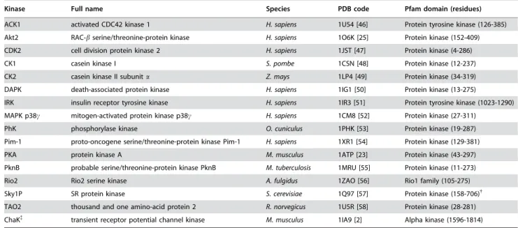
Table 1.Active-conformation kinase structures.*
. . . .
Kinase Full name Species PDB code Pfam domain (residues)
ACK1 activated CDC42 kinase 1 H. sapiens 1U54 [46] Protein tyrosine kinase (126-385) Akt2 RAC-bserine/threonine-protein kinase H. sapiens 1O6K [25] Protein kinase (152-409)
CDK2 cell division protein kinase 2 H. sapiens 1JST [47] Protein kinase (4-286)
CK1 casein kinase I S. pombe 1CSN [48] Protein kinase (12-237) CK2 casein kinase II subunita Z. mays 1LP4 [49] Protein kinase (34-319)
DAPK death-associated protein kinase H. sapiens 1IG1 [50] Protein kinase (13-275)
IRK insulin receptor tyrosine kinase H. sapiens 1IR3 [51] Protein tyrosine kinase (1023-1290) MAPK p38c mitogen-activated protein kinase p38c H. sapiens 1CM8 [52] Protein kinase (27-311)
PhK phosphorylase kinase O. cuniculus 1PHK [53] Protein kinase (19-287)
Pim-1 proto-oncogene serine/threonine-protein kinase Pim-1 H. sapiens 1XR1 [54] Protein kinase (129-381) PKA protein kinase A M. musculus 1ATP [23] Protein kinase (43-297)
PknB probable serine/threonine-protein kinase PknB M. tuberculosis 1MRU [55] Protein kinase (11-273)
Rio2 Rio2 serine kinase A. fulgidus 1ZAO [56] Rio1 family (105-275) Sky1P SR protein kinase S. cerevisiae 1Q97 [57] Protein kinase (158-706){
TAO2 thousand and one amino-acid protein 2 R. norvegicus 1U5R [58] Protein kinase (28-281)
ChaK{
transient receptor potential channel kinase M. musculus 1IA9 [2] Alpha kinase (1596-1814)
*The average pair-wise sequence identity between this set as computed by ClustalW using its default parameters is 17%.
{
Residues 304-541 constitute a large spacer within the kinase domain [59].
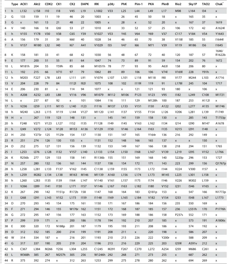
in what we term a consensus structure. This is a distribution of amino acids and amino-acid categories conserved between the protein kinases we have examined. The consensus structure has three principle parts: 1) a region of hydrophobic residues clustered around the adenosine of ATP; 2) an area around thec-phosphate of ATP – the active site – enclosed primarily by charged residues; and 3) a region in the large lobe, situated below ATP, of both hydrophobic and polar residues. The hydrophobic region around the adenosine creates a binding pocket for ATP. The charged residues in the active site bind and position thec-phosphate, as well as the divalent cation, and participate in the catalytic mechanism. The conserved residues located in the large lobe serve to stabilize that region, and may play a role in mediating substrate interactions. Only five specific amino acids are fully conserved in all the kinases. These residues play critical parts in positioning ATP, stabilizing the active-conformation and in the catalytic mechanism. These are lysine 8, which interacts with thea- andb -phosphates of ATP, thereby stabilizing it. Glutamic acid 9, which forms a salt bridge with lysine 8 further stabilizing ATP. Aspartic acid 24 is the catalytic base that initiates phosphotransfer by deprotonating the acceptor serine, threonine or tyrosine. Aspar-agine 27 interacts with a secondary divalent cation, thereby positioning thec-phosphate of ATP. And the final fully conserved residue is aspartic acid 34, which chelates the primary divalent
cation, indirectly positioning ATP at the same time. Although these are the only residues fully conserved in terms of function, location and amino-acid type, there is one other residue with functional conservation but not locational or type, and in other kinases there is variability in the origin of lysine 8 and the type of amino acid fulfilling its role.
Rio2, ChaK and conserved residue variability
Rio2 is the only atypical protein kinase amongst those included in our multiple alignment. No significant sequence similarity can be detected between Rio2 and conventional protein kinases. As such, it is classified with a distinct domain name (see Table 1). The structure of the Rio family, as initially determined by [14], shares significant similarity with serine/threonine and tyrosine kinases in the small lobe and in the regions of the large lobe directly adjacent to the active site (Figure 4A). Dissimilarity in structure occurs predominantly in the large lobe, in regions involved in substrate specificity, suggesting Rio2 has evolved a novel mechanism of substrate recognition [14]. Our structural alignment algorithm concurs with these findings, showing high similarity in and around the active site (consensus residues 1–36) but not in the large lobe (consensus residues 37–44).Slightly different results are produced for another atypical protein kinase: channel kinase (ChaK). The structure of this kinase was not included in our initial data set because it lacked a divalent cation in the active site. To examine structural similarity between ChaK and conventional protein kinases, we did align the partially active structure of ChaK (Figure 4B) with the consensus structure generated from fully active-conformation kinases (seeTable 2). Most similarity is found in the region directly around the c -Figure 2. Multiple kinase alignment.The fifteen active-conformation kinase structures listed in Table 1 were aligned using our modified Procrustes approach. Shown in green sticks is the ATP or ATP analog molecule of each structure. Each kinase is colored uniquely.
doi:10.1371/journal.pone.0000982.g002 Figure 1. The structure of protein kinase A (PKA).PKA is shown in its
active conformation with ATP in green sticks and Mn2+as black spheres. b-strands, helices and loops are labeled as in Knightonet al. [45]. The active site is situated between the small and large lobes, located above and below ATP respectively. CL: catalytic loop; MPL: magnesium-positioning loop.
doi:10.1371/journal.pone.0000982.g001
Table 2.Conserved residues found in the active-conformation kinases listed in Table 1.
. . . .
Type ACK1 Akt2 CDK2 CK1 CK2 DAPK IRK p38c PhK Pim-1 PKA PknB Rio2 Sky1P TAO2 Chak*
1 h L132 L158 I10 I18 V45 L19 L1002 V33 L25 L44 L49 L17 M98 L164 I34 x
2 G 133 159 11 19 46 20 1003 x 26 45 50 18 x 165 35 x
3 G x 161 13 21 48 22 1005 x 28 x 52 20 x 167 37 1619
4 V 140 166 18 I26l 53 27 1010 41 33 52 57 25 106 172 42 A1624l
5 h V155 Y178 V30 V38 C65 Y39 V1027 V53 Y45 V64 Y69 V37 C117 V184 V54 Y1643 6 A 156 179 31 39 I66l 40 1028 54 46 65 70 38 V118l 185 55 I1644l
7 h V157 M180 L32 I40 I67 A41 V1029 I55 V47 I66 M71 V39 V119 M186 I56 I1645
8 K 158 181 33 41 68 42 1030 56 48 67 72 40 120 187 57
1646/ R1622b
9 E 177 200 51 55 81 64 1047 74 73 89 91 59 154 202 76 1672
10 L M181h 204 55 Y59h 85 68 M1051h 78 77 93 95 A63l 158 206 80 x
11 L 192 215 66 V71l 97 79 1062 89 89 106 106 V74l V169l 228 Y91h x 12 h M203 F227 L78 L83 L111 L91 V1074 L107 L101 L118 M118 I90 V177 M244 L103 A1716
13 V 204 228 79 84 I112l I92l 1075 108 102 I119l 119 91 L178l 245 104 1717
14 E 206 230 81 x 114 94 1077 x x 121 121 93 180 x 106 x
15 h A208 A232 L83 L88 V116 V96 M1079 M112 M106 P123 V123 V95 I182 L249 C108 M1721
16 L x 237 87 92 x 101 1084 116 111 129 M128h 100 187 253 A112l x
17 h V236 I259 L111 M115 L140 I123 I1116 M137 L133 V151 I150 A122 I202 L277 A135 M1746 18 h M240 L263 L115 V119 L144 V127 M1120 L141 I137 V155 F154 L126 V206 L281 L139 F1749
19 H x 267 119 123 148 131 x 145 141 159 158 130 x 285 143 T1753p
20 h F248 V271 V123 L127 I152 I135 F1128 I149 I145 V163 L162 I134 I214 I290 M147 A1678 21 h I249 V272 L124 V128 M153 A136 V1129 I150 V146 L164 I163 I135 V215 I291 I148 x
22 H 250 Y273r 125 Y129r 154 137 1130 151 147 165 Y164r 136 216 292 149 x
23 R 251 274 126 130 155 x 1131 152 148 166 165 137 x x 150 x
24 D 252 275 127 131 156 139 1132 153 149 167 166 138 218 294 151 1765
25 l L253 I276 L128 I132 V157 L140 L1133 L154 L150 I168 L167 V139 L219 I295 V152 L1766
26 K R256b 277 129 133 158 141 R1136b 155 151 169 168 140 S220p 296 153 1727 27 N 257 280 132 136 161 144 1137 158 154 172 171 143 223 299 156 Q1767p
28 h L258 L281 L133 F137 V162 I145 C1138 L159 I155 I173 L172 I144 V224 V300 I157 x
29 h L259 M282 L134 L138 M163 M146 M1139 A160 L156 L174 L173 M145 L225 L301 L158 x
30 h L260 L283 I135 I139 I164 L147 V1140 V161 L157 I175 I174 I146 V226 M302 L159 x 31 l V266 I289 I141 I150 L171 I157 V1146 L167 I163 L182 I180 V152 I231 I546 V165 x
32 K 267 290 142 Y151p R172b 158 1147 168 164 183 Q181p 153 x 547 166 N1772p
33 l I268 I291 L143 V152 L173 I159 I1148 I169 L165 L184 V182 V154 I233 I548 L167 L1773 34 D 270 293 145 154 175 161 1150 171 167 186 184 156 235 550 169 x
35 F 271 294 146 155 W176r 162 1151 172 168 187 185 157 236 L551h 170 P1776h
36 G 272 295 147 156 177 163 1152 173 169 188 186 158 P237s 552 171 x 37 P 299 319 171 x 200 186 1178 194 192 210 207 185 x 573 191 A1806s
38 E 300 320 172 N188p 201 187 1179 195 193 211 208 186 x 574 192 x
39 D 312 332 185 200 214 199 1191 208 211 x 220 198 x 586 207 x
40 W 314 334 187 x 216 201 1193 210 213 226 222 Y200r x 588 209 x
41 G 317 337 190 205 219 204 1196 213 216 229 225 203 I259l A591v 212 x
42 h C367 L384 M266 Y256 L304 L255 C1245 M291 F267 C270 L272 A254 I259 M686 C261 x 43 L W368h 385 267 M257h 305 256 W1246h 292 268 271 273 255 x 687 262 x
44 R 375 392 274 x 312 263 1253 299 275 278 280 262 x 694 269 x
*ChaK was aligned directly onto the consensus generated from the other fifteen samples. See Results section.
The amino acid or amino-acid category of the conserved residue is listed under ‘‘type’’. For each structure the residue identifier corresponding to the conserved point is indicated (listed as x if the point is absent). For category types the amino acid present in each structure is indicated before the identifier. If a sample is missing a conserved amino acid but has a similar residue in the same location then the shared category is listed after the identifier. a, acidic; l, aliphatic; r, aromatic; b, basic; c, charged; h, hydrophobic; p, polar; s, small; v, very small.
doi:10.1371/journal.pone.0000982.t002
...
...
...
....
...
...
....
...
...
....
...
...
....
...
...
....
...
...
....
...
...
....
...
...
....
...
...
....
...
...
....
...
...
....
...
...
....
...
...
....
...
...
....
...
...
....
...
...
....
...
...
....
...
...
....
...
...
....
phosphate of ATP, although there is some elsewhere. The large lobe, like Rio2, is quite distinct. One of the few differences between this kinase and the others is that the fully conserved asparagine 27 is replaced by the very similar carboxamide containing glutamine. The fully conserved aspartic acid 34 is present but does not align, likely due to the absence of a divalent cation.
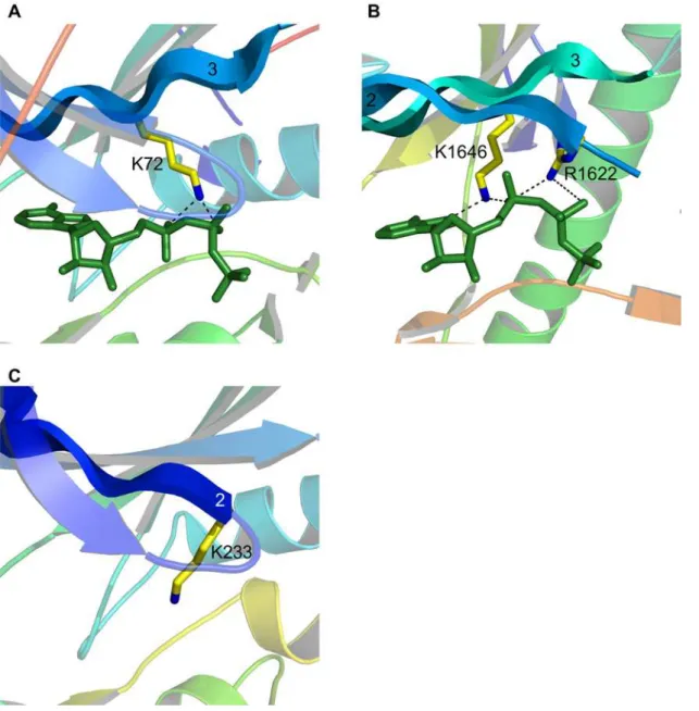
We highlight these two kinases for the additional reason that they demonstrate structural variation within the bounds of certain functional constraints. An interesting case of this is lysine/arginine 26. In almost all serine/threonine kinases there is a lysine residue located two positions downstream of the catalytic aspartic acid (consensus residue 24). This lysine aids in orientating the c -phosphate of ATP and it is thought may also act to neutralize the negative charge on this phosphate during catalysis. The position and orientation of these two residues with respect to ATP is shown in protein kinase A (PKA) in Figure 5A. Tyrosine kinases, like activated CDC42 kinase (ACK1), do not have this lysine but instead have an arginine four positions downstream from the catalytic residue (Figure 5B). This residue is orientated perpendicular to the lysine but occupies the same location, and since both are positively charged basic residues, both can fulfill the same function. ChaK, a serine/threonine kinase like PKA, utilizes a lysine for this function, which again occupies the same geometric position (Figure 5C). However, this residue is not located two or four positions downstream, but thirty-eight positions upstream on ab-strand running adjacent to the catalytic loop. This is a good demonstration of the value behind a sequence-order independent alignment algorithm that ignores fold and residue connectivity. This lysine is located on part of a novel fold not found in any of the
other kinases examined. Rio2 presents a fourth variation. Our alignment did not reveal Rio2 as having a basic residue at consensus point 26, as it did for all of the other kinases. Further examination led us to the conclusion that the function of this residue is conserved in Rio2 but the location of the residue accomplishing it is not (see also [14]). This is known as functional residue hopping [15,16]. The function of consensus residue 26 is performed by a histidine located in the small lobe (Figure 5D). This histidine is largely unique to the Archaeoglobus fulgidus Rio2 ortholog, from which the structure was derived – in most other species it is substituted by an arginine. As A. fulgidus is a hyperthermophile, the preference for histidine may be due to the extreme temperature environments in which it is found.
The only other variability we have found in conserved residues is for the so-called catalytic lysine (lysine 8). The function of this residue is somewhat debated [17,18,19], but at the very least it appears to position the phosphates of ATP and is absolutely crucial for catalysis. This residue is normally found on b-strand 3 and interacts with thea- andb-phosphates of ATP (Figure 6A). In ChaK there is a homologous lysine present but it interacts with the a-phosphate and the adenine ring of ATP (Figure 6B). Depend-ing upon the alignment parameters used, this lysine can align structurally with that found in other kinases, although for the parameters we have chosen it does not. Instead there is an arginine residue that aligns and this very similar residue interacts with both thea- andb-phosphates of ATP (Figure 6B), just as the catalytic lysine normally does. Although ChaK lacks a divalent cation, this is unlikely to affect the positions of these residues. The arginine in question, R1622, is fully conserved in the alpha kinase family [20]. Figure 3. A kinase consensus structure.Each sphere represents a conserved residue. Red indicates full conservation of a particular amino acid in all fifteen kinase structures; orange, conservation in thirteen or fourteen structures; and yellow, conservation in eleven or twelve structures. Blue spheres indicate full conservation of an amino-acid category. The ATP molecule of protein kinase A is shown in green sticks. (A) The consensus structure consisting of the forty-four points listed in Table 2. (B) The consensus structure overlaid on the multiple alignment.
doi:10.1371/journal.pone.0000982.g003
It is not known which residue in ChaK is functionally homologous to the catalytic lysine in typical protein kinases. Likely both share the function, and this represents a distinct feature of the alpha-kinase family. One other alpha-kinase is known to have a novelty in this area. This is the protein kinase with no lysine (WNK) kinase, named for the apparent absence of the catalytic lysine onb-strand 3. It was predicted by [21] that a lysine on b-strand 2 could be structurally equivalent (Figure 6C), and the absolute requirement for a lysine at this position was confirmed by this group. A subsequent crystal structure appears to confirm these predictions [22]. It is interesting that this lysine originates from the same position as the arginine in ChaK, showing that variation in conserved residues is possible and highlighting how proteins can incorporate novel features within certain functional constraints.
Substrate specific variation
The conservation we are detecting is found first and foremost in and around the active site, but is present elsewhere in the protein kinase domain. The single exception is in the cleft formed by the catalytic loop, the P+1 loop, helix D and the residues from the end of helix F through to the beginning of helix H. This is known as the substrate-binding cleft, and residues in this region have been shown crucial to substrate binding and regulatory protein-protein interactions [23,24,25,26,27,28,29]. The absence of conservation in this cleft is entirely expected. Protein kinases are substrate specific and that specificity is at least in part conferred through residue variability within this groove.
The crystal structures of PKA and Akt we have used in our analysis contain substrate peptides bound in this region. InFigure 7
we show PKA and Akt with bound substrate peptides and display variability in the context of proximity to the substrate and distance
from conserved residues. In both PKA and Akt, there are a number of atoms in the substrate-binding cleft adjacent to or near the substrate peptide that are distant from conserved residues. We highlight this for the purpose of discussing inhibitor design. Successful inhibitor design requires a region where small molecules or peptides can be bound with high specificity. The absence of specificity comes from drug targets hitting regions of residue conservation. The substrate-binding cleft obviously has a binding capacity and, as shown, this region is highly variable. Designing inhibitors that target the atoms in this region, mimicking those residues present in substrates or regulatory proteins, would be a fruitful approach.
Structure prediction
The conservation we are finding can be used for purposes other than understanding protein function, evolution and guiding drug design. It can also be used to predict the structure of protein kinases (and to other proteins if applied). The consensus structure shown inFigure 3represents the typical position of specific and conserved amino acids or amino-acid categories. If this is a distribution that active protein kinases tend to adopt, then predicted structures of active-conformation protein kinases should be made to meet these criteria. At the most basic level, models can be generated through any method and then screened against a consensus structure to discriminate between good and bad models, or, alternatively, residues known to be equivalent from a sequence alignment can be forced to meet the constraints seen in the consensus structure and the rest of the protein can be modeled within this framework. We have explored these possibilities with the structure prediction method Rosetta [30] and attempted to model two kinases, the typical 3-phosphoinositide dependent protein kinase-1 and the atypical Rio2 kinase.
We used fourteen active-conformation kinases (omitting Rio2) and generated a 52 residue consensus structure (consensus residues are listed inSupplementary Table S1). A sequence alignment of these fourteen kinases and PDK1 was then generated using ClustalW [31]. Residues from PDK1 apparently equivalent with those of the consensus were selected based on this sequence alignment (seeSupplementary Table S1). These residues were then constrained geometrically in accordance with the consensus while PDK1 was modeled with Rosetta as described in the Methods section. When compared against the partially active-conformation structure of PDK1 (PDB code: 1H1W, [32]), the top ranked prediction had 198 of 285 side chains positioned within 2 A˚ of their actual location, a CaRMSD of 1.3 A˚ and an all-atom RMSD of 1.6 A˚ (Figure 8A). The floor of the active site, where most conservation occurs, is highly accurate, with 24 of 25 side chains positioned with 2 A˚ , CaRMSD of 0.5 A˚ and an all-atom RMSD of 0.7 A˚ (Figure 8B). In non-conserved regions, such as
the substrate-binding groove, good modeling is dependent upon the ability of the prediction method applied. A well-proven method like Rosetta is, therefore, a good complement. 24 of 38 residues in the substrate-binding groove are within 2 A˚ of their actual position, a CaRMSD of 0.9 A˚ and an all-atom RMSD of 1.4 A˚ (Figure 8C). Accuracy in this region may be due in part to the constraints applied elsewhere, which would reduce the potential conformational space to search.
Rio2 kinase was modeled initially without constraints as these cannot be derived from a sequence alignment. 80,000 models were generated and the lowest 5% (in full atom energy) were screened against the consensus structure. The top ten were then scrutinized for potential constraints. All ten of the top models unambiguously agreed on the likely equivalent residues for the fully conserved lysine 7 (K120 in Rio2), aspartic acid 24 (D218), asparagine 27 (N223) and aspartic acid 34 (D235). In none of the models could a residue equivalent to the fully conserved glutamic acid 8 be Figure 5. Residue variability in positioning thec-phosphate of ATP.In each kinase the catalytic aspartic acid is shown in red and the
positively-charged residue that interacts with thec-phosphate of ATP is shown in yellow with nitrogen atoms colored blue. ATP is shown in green sticks. (A) Protein kinase A, (B) activated CDC42 kinase 1, (C) channel kinase, and (D) Rio2 kinase.
doi:10.1371/journal.pone.0000982.g005
found (it should be E154). We then proceeded with a second modeling phase using constraints from the consensus for K120, D218, N223 and D235, and allowing two candidates for the conserved glutamic acid: E134 or E154. Although models generated with E134 as the conserved glutamic acid scored equally well when compared against the consensus structure, they would likely be deemed implausible by visual inspection as helix C was distorted upwards away from the active site instead of lining the back of the ATP binding pocket as it does in other kinases. The best model for E154 as determined against the consensus structure is shown inFigure 9Aalongside the actual active-conformation structure (PDB code: 1ZAO). Fifty four of 180 side-chains are positioned within 2 A˚ of their true location. Ca and all-atom RMSD are 6.1 A˚ and 7.1 A˚ respectively. However, these values
do not accurately reflect the quality of the model. The N-terminus of protein kinases is a non-conserved region and the C-terminal lobe of Rio2 differs greatly from that of other kinases. It should not be expected that a structural-based consensus approach would be able to distinguish a correct model from an incorrect model in these regions. Looking solely at the conserved kinase regions, comprising residues 93–242 of Rio2, the Caand all-atom RMSD drop to 3.0 A˚ and 3.7 A˚ respectively. And all of the correctly positioned residues are found in this region. The floor of the active site has 16 of 24 side chains positioned with 2 A˚ , CaRMSD of 1.0 A˚ and an all-atom RMSD of 1.7 A˚ (Figure 9B). A final note: although residues located in the non-conserved large lobe helices are not correctly positioned, this region is topologically correct.
DISCUSSION
The kinase domain can be conceptualized as two functional modules. A highly conserved ATP-binding and catalytic module, located between the small and large lobes, found in all typical and atypical protein kinase structures. Little variation is found here, with only a few kinases, such as Rio2, ChaK and WNK, containing structural (but not functional) novelties. The second module is involved in substrate binding and evidence suggests is localized primarily to the large lobe, in the region named the substrate/peptide binding groove (named from kinase-peptide co-crystal structures). Very little is known about this region of kinases
as detailed knowledge would require many kinase structures co-crystallized with full-length substrates, and as yet no single such structure exists. Conservation of the groove fold suggests a common substrate-binding mechanism, while the absence of residue conservation in this region across the kinase family suggests the means by which substrate specificity is determined. The part of this groove located near the active site (between the catalytic and P+1 loops) does contain conserved residues and is entirely consistent with the broad peptide-substrate specificity of kinases. But peptides are not the same as full-length proteins, and studies on the mitogen-activated protein kinase p38 have shown that proteins can be phosphorylated at a hundred to thousand fold Figure 7. Substrate-specific variability.Cross-eyed stereo views of (A) protein kinase A and (B) Akt2. Bound substrate peptides are shown in red sticks. Atoms belonging to non-conserved residues within 10 A˚ of the substrate peptide are shown as colored spheres. Red: atoms within 2 A˚ of a conserved residue; orange: atoms between 2 and 4 A˚ of a conserved residue; yellow: atoms between 4 and 6 A˚ of a conserved residue; green: atoms between 6 and 8 A˚ of a conserved residue; cyan: atoms between 8 and 10 A˚ of a conserved residue; and blue: atoms more than 10 A˚ from a conserved residue.
doi:10.1371/journal.pone.0000982.g007
greater rate than peptides [33]. More than just the residues immediately attached to and surrounding the target serine, threonine or tyrosine are involved in the kinase-substrate in-teraction, and the general mechanism of interaction needs to be determined.
Although the substrate/peptide binding groove may be the key feature for understanding substrate specificity in most kinases, it is and need not be in all. The substrate-binding module can vary in three ways: typical kinases that preserve fold and merely vary the residues in the substrate-binding groove, typical kinases than rely on other regions/regulatory proteins in addition to the substrate-binding groove, and atypical kinases that vary the substrate-binding mechanism all together. It should be added that even in very typical kinases substrate interactions are not localized purely to the substrate-binding groove (see [34]). Much work needs to be done on this area of protein kinases, and current knowledge of substrate-binding mechanisms is rudimentary.
A thorough understanding of substrate interactions will help in discovering new proteins targeted by kinases. Insights into binding can guide computational docking approaches that test the fit of different structures onto kinases as a new means of substrate finding. More experimentally determined structures or advances in structure prediction would be a necessary requirement for this to have broad applicability. This may be a ways off yet, but a closer goal may be in applying structural binding information to the design of specific kinase inhibitors. Currently, inhibitors are primarily designed to target the ATP binding pocket, the region of greatest conservation amongst kinases. The ATP binding pocket does contain unique features between certain subsets of the kinome (the gatekeeper residue is an example [35,36]), and these novelties can be exploited by drugs. However, this type of design
approach will always be plagued by problems of specificity on the simple foundation that this region has evolved to bind a single thing: ATP. On the other hand, the substrate-binding groove has evolved to bind kinase-specific substrates and our analysis demonstrates that this is accompanied by large variability in the substrate-binding groove. Mimicking these substrates with pep-tides or small molecules that compete with substrates has proven effective in several cases (reviewed in [37]), and a peptide inhibitor that mimics a substrate interaction domain has been shown useful in vivoat reducing tumor mass [38].
tion can help in generating or selecting appropriate protein kinase models. The atypical Rio2 kinase, which possesses no significant sequence similarity to other protein kinases, would be a difficult target for standard approaches at homology modeling. As few as five geometric residue constraints derived from a consensus structure and further screening can select a highly accurate model. For a typical kinase, such as PDK1, where many constraints can be used (50 in our case), it was a simple matter to generate a highly accurate model. Residue conservation and other types of structural similarity quantified over protein families can provide the basis for model selection and act as guides that reduce the target search space for the computationally demanding task of structure prediction.
Understanding the individual role of every protein kinase in the genome is a daunting task, but necessary because of the important signaling role played by this family of enzymes and the potential for disregulation and subsequent disease. Ultimately, cellular studies that dissect the role of kinasesin vivoand the treatment of disease will require a library of specific and multi-target inhibitors. Such an aim will require a strong union between computational, molecular and structural approaches, but this goal may be close at hand. Although empirically determined structures of all kinases are a long way off, quality models can be built. Kinome wide data on substrate pools will require a great deal of work, but the techniques are now becoming available. The mechanism(s) of substrate binding will need to be elucidated in detail. This may be the greatest challenge, and towards which maximum focus should be directed, as not a single kinase-substrate structure exists. But, if such can be achieved in a representative sample of cases, kinase-substrate specific docking algorithms may be able to do the rest. The attainment of these aims would allow for the quick and reliable design of specific inhibitors, thereby allowing the precise function(s) of kinases to be determined and creating the means by which signaling networks can be precisely manipulated.
MATERIALS AND METHODS
Structural alignment overview
The results presented in this paper were generated through the use of a multiple structure alignment algorithm we have developed. The motivation was to have a method for superimposing protein structures independent of residue connectivity, thereby facilitating comparisons between proteins that have distinct folds and maximize side-chain as opposed to main-chain overlap. The field of shape analysis, specifically the Procrustes methods, provided the founda-tion for accomplishing this task. What follows is an overview of Procrustes and a description of its modification and application to protein structure comparison as we have implemented it.
Procrustes
Procrustes is a method for comparing matrices and thereby any group of objects that can be represented in such a way [40,41]. It is part of the broader field of shape analysis which has its motivation in describing and understanding variation between objects through multivariate analysis. Procrustes is often applied in biological and anthropological studies of morphology, for example in studying the relationship between turtle skull shape and life style across species [42]. It has been used in agriculture, genetics, geography, geology and psychology to name a few fields. Although multivariate analysis is not of interest, the Procrustes method is valuable for its ability to efficiently superimpose objects. Procrustes superimposition initiates from a set of points, each of which has a representative in every object under consideration. These points, known as landmarks, must have some level of correspondence and Figure 9. Modeling an atypical kinase, Rio2.(A) The best model is in
blue and the actual structure in red (PDB code: 1ZAO). Structures are shown aligned based on CaRMSD. Residues 128–142 from the model are hidden because they are disordered in the actual structure. (B) Residues 214–237 are highlighted as aligned on side-chains. ATP is shown in green sticks.
doi:10.1371/journal.pone.0000982.g009
be located within a geometric space. By manipulating scale, location and orientation, Procrustes aims to produce an optimal superimposition as defined by some criteria that acts to minimize distance between corresponding points. The term partial Procrus-tes specifically refers to the subset of methods that maintain scale, i.e. those approaches that only perform rigid body transformations [43].
Application and algorithm
An application for Procrustes in protein structure alignment is evident, where landmarks could be viewed as equivalent residues in a space of three dimensions. The term equivalent accounts for functional equivalence and/or homology, although emphasis should be placed on the former. Specifically it is the partial Procrustes method that is applicable since initial aspects of scale should be maintained in the resulting alignment.
Standard implementations of Procrustes require a set of user-specified landmarks with representatives in every input object. These two conditions are inadequate for multiple structure alignments. The primary motivation behind a structural alignment should be to find conserved residues, and a good algorithm must give leeway for less than total conservation. The algorithm we have developed allows for both. It proceeds in two phases. The first is a series of pair-wise comparisons to find residues conserved across all samples. Residue conservation means in terms of both location and amino-acid type (identity/similarity). The second phase begins with an initial multiple alignment using the landmarks found in the first phase. From the multiple alignment the set of landmarks is then extended to include any additional fully conserved residues not found in the first phase, and residues with less than total conservation. The multiple alignment and extension steps are repeated until a final non-extendable set is obtained or upon completion of a predefined number of iterations. In this approach, each ofnstructures is represented by ali63 coordinate matrix, where li equals the number of residues in structure i. The mean of the non-hydrogen side chain atoms is used as the coordinate for each residue, with the exception of glycine residues for which the alpha carbon (Ca) is used. Most structure alignment algorithms use the Cafor all residues. This is inadequate. Residue function should be viewed largely in terms of side-chain characteristics: what atoms make up the side chain and where they are positioned. It is the side chain that is principally used to interact with ligands, substrates and other residues [44], making knowledge of its position the crucial feature. Superimposed Ca’s of similar residues may have side chains positioned in different locations and are therefore unlikely to have the same function. For these reasons focus is placed on side-chain positioning.
One object (structure) can be superimposed onto a second in three dimensions if at least three equivalent residues are known. It is possible these residues may or could be known beforehand, although this is not ideal. An alternative involves a search and score approach: choose three residues from each sample that have corresponding amino-acid labels and share some geometric feature, then score the alignment produced from superimposing these residue triplets. The highest scoring alignment produced from a series of triplet superimpositions can then be used to produce a set of residues conserved across a pair of samples (we term this a pair-wise consensus set). This approach has been employed in a hash table. For all samples every triplet of residues is stored based on the amino-acid labels of its members and the inter-residue distances are indexed. Each entry of the table can be thought of as a triangle with vertices labeled according to amino acid. Between pairs of samples highly congruent triangles with matching vertices are used as candidate triplets likely to produce
high scoring superimpositions. Such criteria reduce the time spent on triplets least likely to be part of the final pair-wise consensus set. We have used congruency criteria of 1–10% as indicated below, i.e. corresponding vertices must have magnitudes within 1–10% of each other.
The method of superimposition is taken from Procrustes. Candidate triplets from each sample i are stored in an 363 coordinate matrix Xi. These matrices are first Helmertized (centered), removing translational differences:
_ X
Xi~HT HXi, ð1Þ
whereXX_iis the centered matrix andHthe (m21)6mHelmert sub-matrix whose jth row is equal to (hj,…, hj, 2jhj,0,…,0) for hj=2[j(j+1)]21/2. Other approaches for removing translation are available. The optimal rotation ofXX_iontoXX_jis found by minimizing
_ X Xj{XX_iC
2
, ð2Þ
whereCis a 363 rotation matrix. It can be shown that ifVDUTis the singular value decomposition ofXX_jTXX_i, the optimal rotationC=UVT (provisions must be made to ensure C does not also encompass a reflection).
The translation resulting from centering triplets is then applied to each structure and the appropriate sample is rotated byC. This superimposes the equivalent triplet residues and pulls the rest of each structure along. The result is that the initial pair-wise consensus set a triplet comprises may be extended from the structure-wide superimposition. Residues from the superimposed structures are considered to be equivalent (i.e. in the pair-wise consensus set) if they are identical or similar amino acids within two angstroms of one another. Similar means sharing a category, with those allowed including acidic (D, E, Z), aliphatic (A, G, I, L, V), aromatic (F, H, W, Y), basic (H, K, R), charged (D, E, H, K, R, Z), hydrophobic (A, C, F, G, I, L, M, P, V, W, Y), polar (C, D, E, H, K, N, Q, R, S, T, Y, Z), small (A, C, D, G, N, P, S, T, V) and very small (A, G, S), where Z refers to a phosphorylated serine, threonine or tyrosine.
This procedure is performed on all triplets that match the congruency criteria and all alignments are scored based on the frequency with which the amino acids of any equivalent residues occur. The frequency of occurrence for each amino acid was obtained from the protein knowledge-base release statistics of Swiss-Prot (Release 49.5, 18/04/06). Tryptophan is the lowest occurring amino acid and superimposed tryptophans are therefore given a score of one. All other scores are relative to that of tryptophan: score(amino acid x) = occurrence(W/x). The occur-rence of each category is equal to the sum of the occuroccur-rences of its members. Formequivalent residues, the alignment score is
Xm
i~1
occurrence Wð =xiÞ, ð3Þ
where xi is the amino acid or amino-acid category of the ith equivalent residue.
Computational time and memory are minimized in three ways. First, as has been mentioned, congruency between triangles is used to reduce the number of triplets tested. Second, the vertices of these triplets are only indexed by amino acid and not by category as well. If vertices could be indexed by amino-acid category, such as hydrophobic, the number of candidate triplets to search scales tremendously, as does demands upon memory if a hash table is employed. Third, we restrict triplets to a core of residues. For a structure of 300 residues there are nearly 27 million candidate triplets. Storing all in a hash table is too demanding, and is not necessary either. The best candidate triplets will be clustered in a core, which may be comprised of residues around an active site, ligand binding domain or phosphorylation site. Only searching this core will provide good candidate triplets and minimize time. By default a geometric core of fifty residues is used.
From the input structures a consensus set Sn is sought for of residues fully conserved across allnsamples (a full consensus set). This can be done quickly through a series of pair-wise comparisons. First a consensus set S2 of conserved residues is sought for between samples 1 and 2 using the superimposition procedure outlined above. A second setS3 of conserved residues found in S2 and sample 3 is then sought for using the same procedure of selecting triplets from the consensus set and the sample. This process is repeated until a final full consensus setSnis produced. From experience the procedure for derivingSnis generally sample order independent, except in some cases where an extreme outlier is present. This is a structure with few of the conserved residues found across the othern21 samples. In this case the outlier may align well with a set of residues conserved across the firstx samples but not across the finaln2x21. By aligning well with a subset ofSxnot inSn, the outlier skews the derivation process away from the true setSn. This can be accounted for by leaving such outliers until the end, by which time the consensus set has been reduced to a true group of highly conserved residues.
The multiple alignment procedure is a modification of the pair-wise superimposition method outlined above. The full consensus setSnfound through the pair-wise superimpositions is comprised of a set ofmlandmarks that can be used to simultaneously align the group of n structures. First, the conserved residues from each samplei are stored in anm63 matrixXi and Helmertized. The optimal rotation matrix for each structureCi is calculated by an iterative process that seeks to minimize the following value:
G~1 n
X
n
i~1 X
n
j~iz1
_ X
XiCi{XX_jCj
2
: ð4Þ
The rotation Ci is calculated from the singular value
decompo-sition ofXX_iwith the mean of the rest,X
T
iXX_i, for
Xi~ 1 n{1 X j=i _ X
Xj: ð5Þ
However, with each successiveXX_irotated, the previousXjforj,i will change and the previously calculated rotation ofXX_j will be incorrect. To account for this, after every sample has been rotated Gis calculated and the process of rotation is repeated until the change in G falls below some arbitrary small threshold (i.e. convergence occurs). For psteps until convergence, the optimal rotation of each structure is equal to the product of the successive
Cirotations.
This procedure superimposes the conserved residues found in the setSn. The next goal is to extend this set. Multiple alignment is
more accurate than a series of pair-wise alignments and this allows forSnto be extended to include additional fully conserved residues. At the same time residues with less than full conservation can be incorporated. We have defined conserved residues as identical or similar amino acids from different structures within two angstroms of some point, termed a consensus point. Consensus points are considered redundant if they contain more than 50% of the same residues. The point closer to the relevant landmarks is retained. Partially conserved residues must only be present in a threshold of samples and the less than full conservation of an amino acid takes precedence over the conservation of a category. Leeway in the conservation of residues allows for evolutionary freedom, but is also required for practical reasons such as x-ray resolution, model construction, side-chain movement and non-significant sample-dependent positionings. As the superimposition procedure requires all landmarks in all samples, any samples with missing landmarks are temporally given the consensus point as a place filler. If new residues can be added following an alignment then the expanded set Sn is used to re-align the structures. The extension and alignment process is repeated until a final consensus set is converged upon or until the completion of a predetermined number of iterations.
Aligning active-conformation kinases
Active-conformation kinases were aligned using the above pro-cedure with a congruency criteria of 1% and a distance constraint for conservation of two angstroms. The partially active-conforma-tion structure of the transient receptor potential channel kinase was aligned directly onto the consensus using congruency criteria of 10% and a distance constraint of 2.25 angstroms.
Availability and Performance
The program is written in C and Fortran. Full source code is freely available under the GNU General Public License by contacting the authors. PDB formatted files are required for input. For multiple alignments output consists of PDB files modified according to the alignment, a consensus structure in PDB format, a csv file indicating the conserved and aligned residues, and a script for viewing the alignment and consensus in the open-source program PyMOL (http://www.pymol.org). For a pair-wise alignment against a consensus structure, output is as above except no consensus structure is produced. The alignment and consensus structure of the fifteen kinases presented in the Results section were generated in,twenty seconds using a single processing core
of an AMD Athlon 64 X2 4200 processor with 1GB of RAM. A single pair-wise alignment takes ,2–8 seconds depending upon
the degree of similarity between input samples. Comparisons between a sample and a consensus structure are very rapid (,1 second due to the reduced set of residues in a consensus set).
Structure prediction
output by Rosetta was selected as the best model. For Rio2, the lowest 5% of models were used for screening against the consensus structure as described in the Results section. Models were ranked based on their score from Equation.
SUPPORTING INFORMATION
Table S1
Found at: doi:10.1371/journal.pone.0000982.s001 (0.23 MB DOC)
ACKNOWLEDGMENTS
We thank Miguel A. Andrade for critical reading and discussions during manuscript preparation.
Author Contributions
Conceived and designed the experiments: JK RK. Performed the experiments: JK BQ. Analyzed the data: JK BQ. Wrote the paper: JK. Other: Developed and implemented the structure prediction method described: DB BQ JK.
REFERENCES
1. Manning G, Whyte DB, Martinez R, Hunter T, Sudarsanam S (2002) The protein kinase complement of the human genome. Science 298: 1912–1934. 2. Yamaguchi H, Matsushita M, Nairn AC, Kuriyan J (2001) Crystal structure of
the atypical protein kinase domain of a TRP channel with phosphotransferase activity. Mol Cell 7: 1047–1057.
3. LaRonde-LeBlanc N, Wlodawer A (2005) A family portrait of the RIO kinases. J Biol Chem 280: 37297–37300.
4. Dedhar S, Williams B, Hannigan G (1999) Integrin-linked kinase (ILK): a regulator of integrin and growth-factor signaling. Trends Cell Biol 9: 319–323. 5. Ubersax JA, Woodbury EL, Quang PN, Paraz M, Blethrow JD, et al. (2003)
Targets of the cyclin-dependent kinase Cdk1. Nature 425: 859–864. 6. Ptacek J, Devgan G, Michaud G, Zhu H, Zhu X, et al. (2005) Global analysis of
protein phosphorylation in yeast. Nature 438: 679–684.
7. Slamon DJ, Clark GM, Wong SG, Levin WJ, Ullrich A, et al. (1987) Human breast cancer: correlation of relapse and survival with amplification of the HER-2/neu oncogene. Science 235: 177–182.
8. Sawyers CL (1999) Chronic myeloid leukemia. N Engl J Med 340: 1330–1340. 9. Hommes D, van den Blink B, Plasse T, Bartelsman J, Xu C, et al. (2002) Inhibition of stress-activated MAP kinases induces clinical improvement in moderate to severe Crohn’s disease. Gastroenterology 122: 7–14.
10. Ono-Saito N, Niki I, Hidaka H (1999) H-series protein kinase inhibitors and potential clinical applications. Pharmacol Ther 82: 123–131.
11. Sebolt-Leopold JS, English JM (2006) Mechanisms of drug inhibition of signaling molecules. Nature 441: 457–462.
12. Fabian MA, r d Biggs WH, Treiber DK, Atteridge CE, Azimioara MD, et al. (2005) A small molecule-kinase interaction map for clinical kinase inhibitors. Nat Biotechnol 23: 329–336.
13. Rohl CA, Strauss CE, Misura KM, Baker D (2004) Protein structure prediction using Rosetta. Methods Enzymol 383: 66–93.
14. LaRonde-LeBlanc N, Wlodawer A (2004) Crystal structure ofA. fulgidusRio2 defines a new family of serine protein kinases. Structure 12: 1585–1594. 15. Grishin NV (2001) Fold change in evolution of protein structures. J Struct Biol
134: 167–185.
16. Todd AE, Orengo CA, Thornton JM (2002) Plasticity of enzyme active sites. Trends Biochem Sci 27: 419–426.
17. Carrera AC, Alexandrov K, Roberts TM (1993) The conserved lysine of the catalytic domain of protein kinases is actively involved in the phosphotransfer reaction and not required for anchoring ATP. Proc Natl Acad Sci U S A 90: 442–446.
18. Robinson MJ, Harkins PC, Zhang J, Baer R, Haycock JW, et al. (1996) Mutation of position 52 in ERK2 creates a nonproductive binding mode for adenosine 59-triphosphate. Biochemistry 35: 5641–5646.
19. Iyer GH, Garrod S, r Woods VL J, Taylor SS (2005) Catalytic independent functions of a protein kinase as revealed by a kinase-dead mutant: study of the Lys72His mutant of cAMP-dependent kinase. J Mol Biol 351: 1110–1122. 20. Drennan D, Ryazanov AG (2004) Alpha-kinases: analysis of the family and
comparison with conventional protein kinases. Prog Biophys Mol Biol 85: 1–32. 21. Xu B, English JM, Wilsbacher JL, Stippec S, Goldsmith EJ, et al. (2000) WNK1, a novel mammalian serine/threonine protein kinase lacking the catalytic lysine in subdomain II. J Biol Chem 275: 16795–16801.
22. Min X, Lee BH, Cobb MH, Goldsmith EJ (2004) Crystal structure of the kinase domain of WNK1, a kinase that causes a hereditary form of hypertension. Structure 12: 1303–1311.
23. Zheng J, Trafny EA, Knighton DR, Xuong NH, Taylor SS, et al. (1993) 2.2 A˚ refined crystal structure of the catalytic subunit of cAMP-dependent protein kinase complexed with MnATP and a peptide inhibitor. Acta Crystallogr D Biol Crystallogr 49: 362–365.
24. Lei M, Lu W, Meng W, Parrini MC, Eck MJ, et al. (2000) Structure of PAK1 in an autoinhibited conformation reveals a multistage activation switch. Cell 102: 387–397.
25. Yang J, Cron P, Good VM, Thompson V, Hemmings BA, et al. (2002) Crystal structure of an activated Akt/protein kinase B ternary complex with GSK3-peptide and AMP-PNP. Nat Struct Biol 9: 940–944.
26. Huang J, Tu Z, Lee FS (2003) Mutations in protein kinase subdomain X differentially affect MEKK2 and MEKK1 activity. Biochem Biophys Res Commun 303: 532–540.
27. Tu Z, Mooney SM, Lee FS (2003) A subdomain of MEKK1 that is critical for binding to MKK4. Cell Signal 15: 65–77.
28. Felberg J, Lefebvre DC, Lam M, Wang Y, Ng DH, et al. (2004) Subdomain X of the kinase domain of Lck binds CD45 and facilitates dephosphorylation. J Biol Chem 279: 3455–3462.
29. Kim C, Xuong NH, Taylor SS (2005) Crystal structure of a complex between the catalytic and regulatory (RIalpha) subunits of PKA. Science 307: 690–696. 30. Das R, Qian B, Raman S, Vernon R, Thompson J, et al. Prediction of the structures of CASP7 targets using extensive all-atom refinement with Rosetta@home. Proteins; In press.
31. Thompson JD, Higgins DG, Gibson TJ (1994) CLUSTAL W: improving the sensitivity of progressive multiple sequence alignment through sequence weighting, position-specific gap penalties and weight matrix choice. Nucleic Acids Res 22: 4673–4680.
32. Biondi RM, Komander D, Thomas CC, Lizcano JM, Deak M, et al. (2002) High resolution crystal structure of the human PDK1 catalytic domain defines the regulatory phosphopeptide docking site. EMBO J 21: 4219–4228. 33. Hawkins J, Zheng S, Frantz B, LoGrasso P (2000) p38 map kinase substrate
specificity differs greatly for protein and peptide substrates. Arch Biochem Biophys 382: 310–313.
34. Biondi RM, Nebreda AR (2003) Signaling specificity of Ser/Thr protein kinases through docking-site-mediated interactions. Biochem J 372: 1–13.
35. Liu Y, Bishop A, Witucki L, Kraybill B, Shimizu E, et al. (1999) Structural basis for selective inhibition of Src family kinases by PP1. Chem Biol 6: 671–678. 36. Blencke S, Zech B, Engkvist O, Greff Z, Orfi L, et al. (2004) Characterization of
a conserved structural determinant controlling protein kinase sensitivity to selective inhibitors. Chem Biol 11: 691–701.
37. Bogoyevitch MA, Barr RK, Ketterman AJ (2005) Peptide inhibitors of protein kinases-discovery, characterization and use. Biochim Biophys Acta 1754: 79–99. 38. Perea SE, Reyes O, Puchades Y, Mendoza O, Vispo NS, et al. (2004) Antitumor effect of a novel proapoptotic peptide that impairs the phosphorylation by the protein kinase 2 (casein kinase 2). Cancer Res 64: 7127–7129.
39. Shah K, Shokat KM (2003) A chemical genetic approach for the identification of direct substrates of protein kinases. Methods Mol Biol 233: 253–271. 40. Goodall C (1991) Procrustes methods in the statistical analysis of shape. J R Statist
Soc B 53: 285–339.
41. Dryden IL, Mardia KV (1998) Statistical shape analysis. Chichester: Wiley. 42. Claude J, Pritchard P, Tong H, Paradis E, Auffray JC (2004) Ecological
correlates and evolutionary divergence in the skull of turtles: a geometric morphometric assessment. Syst Biol 53: 933–948.
43. Kent JT (1992) New directions in shape analysis. In: Mardia KV, ed. The art of statistical science. Chichester: Wiley. pp 115–127.
44. Gutteridge A, Thornton JM (2005) Understanding nature’s catalytic toolkit. Trends Biochem Sci 30: 622–629.
45. Knighton DR, Zheng JH, Eyck LFT, Ashford VA, Xuong NH, et al. (1991) Crystal structure of the catalytic subunit of cyclic adenosine monophosphate-dependent protein kinase. Science 253: 407–414.
46. Lougheed JC, Chen RH, Mak P, Stout TJ (2004) Crystal structures of the phosphorylated and unphosphorylated kinase domains of the Cdc42-associated tyrosine kinase ACK1. J Biol Chem 279: 44039–44045.
47. Russo AA, Jeffrey PD, Pavletich NP (1996) Structural basis of cyclin-dependent kinase activation by phosphorylation. Nat Struct Biol 3: 696–700.
48. Xu RM, Carmel G, Sweet RM, Kuret J, Cheng X (1995) Crystal structure of casein kinase-1, a phosphate-directed protein kinase. EMBO J 14: 1015–1023. 49. Yde CW, Ermakova I, Issinger OG, Niefind K (2005) Inclining the purine base
binding plane in protein kinase CK2 by exchanging the flanking side-chains generates a preference for ATP as a cosubstrate. J Mol Biol 347: 399–414. 50. Tereshko V, Teplova M, Brunzelle J, Watterson DM, Egli M (2001) Crystal
structures of the catalytic domain of human protein kinase associated with apoptosis and tumor suppression. Nat Struct Biol 8: 899–907.
51. Hubbard SR (1997) Crystal structure of the activated insulin receptor tyrosine kinase in complex with peptide substrate and ATP analog. EMBO J 16: 5572–5581.
53. Owen DJ, Noble ME, Garman EF, Papageorgiou AC, Johnson LN (1995) Two structures of the catalytic domain of phosphorylase kinase: an active protein kinase complexed with substrate analogue and product. Structure 3: 467–482. 54. Qian KC, Wang L, Hickey ER, Studts J, Barringer K, et al. (2005) Structural
basis of constitutive activity and a unique nucleotide binding mode of human Pim-1 kinase. J Biol Chem 280: 6130–6137.
55. Young TA, Delagoutte B, Endrizzi JA, Falick AM, Alber T (2003) Structure of Mycobacterium tuberculosis PknB supports a universal activation mechanism for Ser/Thr protein kinases. Nat Struct Biol 10: 168–174.
56. LaRonde-LeBlanc N, Guszczynski T, Copeland T, Wlodawer A (2005) Autophosphorylation ofArchaeoglobus fulgidusRio2 and crystal structures of its nucleotide-metal ion complexes. FEBS J 272: 2800–2810.
57. Nolen B, Ngo J, Chakrabarti S, Vu D, Adams JA, et al. (2003) Nucleotide-induced conformational changes in theSaccharomyces cerevisiaeSR protein kinase, Sky1p, revealed by X-ray crystallography. Biochemistry 42: 9575–9585. 58. Zhou T, Raman M, Gao Y, Earnest S, Chen Z, et al. (2004) Crystal structure of
the TAO2 kinase domain: activation and specificity of a Ste20p MAP3K. Structure 12: 1891–1900.
59. Nolen B, Yun CY, Wong CF, McCammon JA, Fu XD, et al. (2001) The structure of Sky1p reveals a novel mechanism for constitutive activity. Nat Struct Biol 8: 176–183.