P2X
3
Receptors Mediate Visceral
Hypersensitivity during Acute
Chemically-Induced Colitis and in the Post-Inflammatory
Phase via Different Mechanisms of
Sensitization
Annemie Deiteren1, Laura van der Linden1, Anouk de Wit1, Hannah Ceuleers1,
Roeland Buckinx2, Jean-Pierre Timmermans2, Tom G. Moreels1,3, Paul A. Pelckmans1,3, Joris G. De Man1, Benedicte Y. De Winter1*
1Laboratory of Experimental Medicine and Pediatrics, Division of Gastroenterology, University of Antwerp, Antwerp, Belgium,2Laboratory of Cell Biology and Histology, University of Antwerp, Antwerp, Belgium, 3Antwerp University Hospital, Department of Gastroenterology and Hepatology, Antwerp, Belgium
Abstract
Objectives
Experiments using P2X3knock-out mice or more general P2X receptor antagonists suggest
that P2X3receptors contribute to visceral hypersensitivity. We aimed to investigate the
ef-fect of the selective P2X3antagonist A-317491 on visceral sensitivity under physiological
conditions, during acute colitis and in the post-inflammatory phase of colitis.
Methods
Trinitrobenzene sulphonic-acid colitis was monitored by colonoscopy: on day 3 to confirm the presence of colitis and then every 4 days, starting from day 10, to monitor convales-cence and determine the exact timepoint of endoscopic healing in each rat. Visceral sensi-tivity was assessed by quantifying visceromotor responses to colorectal distension in controls, rats with acute colitis and post-colitis rats. A-317491 was administered 30 min prior to visceral sensitivity testing. Expression of P2X3receptors (RT-PCR and
immunohis-tochemistry) and the intracellular signalling molecules cdk5, csk and CASK (RT-PCR) were quantified in colonic tissue and dorsal root ganglia. ATP release in response to colorectal distension was measured by luminiscence.
Results
Rats with acute TNBS-colitis displayed significant visceral hypersensitivity that was dose-dependently, but not fully, reversed by A-317491. Hypersenstivity was accompanied by an increased colonic release of ATP. Post-colitis rats also displayed visceral hypersensitivity that was dose-dependently reduced and fully normalized by A-317491 without increased
OPEN ACCESS
Citation:Deiteren A, van der Linden L, de Wit A, Ceuleers H, Buckinx R, Timmermans J-P, et al. (2015) P2X3Receptors Mediate Visceral
Hypersensitivity during Acute Chemically-Induced Colitis and in the Post-Inflammatory Phase via Different Mechanisms of Sensitization. PLoS ONE 10 (4): e0123810. doi:10.1371/journal.pone.0123810
Academic Editor:Yvette Tache, University of California, Los Angeles, UNITED STATES
Received:July 31, 2014
Accepted:March 7, 2015
Published:April 17, 2015
Copyright:© 2015 Deiteren et al. This is an open access article distributed under the terms of the
Creative Commons Attribution License, which permits unrestricted use, distribution, and reproduction in any medium, provided the original author and source are credited.
Data Availability Statement:All relevant data are within the paper and its Supporting Information files.
release of ATP. A-317491 did not modify visceral sensitivity in controls. P2X3mRNA and
protein expression in the colon and dorsal root ganglia were similar in control, acute colitis and post-colitis groups, while colonic mRNA expression of cdk5, csk and CASK was in-creased in the post-colitis group only.
Conclusions
These findings indicate that P2X3receptors are not involved in sensory signaling under
physiological conditions whereas they modulate visceral hypersensitivity during acute TNBS-colitis and even more so in the post-inflammatory phase, albeit via different mecha-nisms of sensitization, validating P2X3receptors as potential new targets in the treatment of
abdominal pain syndromes.
Introduction
Abdominal pain is a common symptom in many gastrointestinal disorders, among which the irritable bowel syndrome (IBS) and inflammatory bowel disease (IBD). In IBD, the continuous release of inflammatory mediators during flares can sensitize peripheral nerve endings in the gut wall, resulting in disturbed sensitivity and abdominal pain, which is not only present during acute flares of inflammation but also during remission [1,2]. In contrast in IBS, abdominal pain and visceral hypersensitivity persist in the absence of overt colonic pathology [3]. Of note, a specific subgroup of patients develops IBS after an episode of acute gastroenteritis, termed post-infectious IBS [4]. Current treatments for IBD and IBS are mainly aimed at dampening the intestinal inflammation (IBD) or normalizing the associated motility dysfunctions (IBS) to indirectly reduce abdominal pain; in contrast, few therapeutics specifically target the afferent nerves.
Extracellular adenosine 5’-triphospate (ATP) has been established as a key sensory signaling molecule that activates purinergic P2X ligand-gated ion channel receptors and P2Y G-protein coupled receptors [5]. There are seven P2X receptor subunits (P2X1-7) that can be assembled as
homo- or heteromultimers [6]. Especially P2X3receptor units, expressed as homomeric P2X3
or heteromeric P2X2/3receptors, are considered to play a major role in visceral sensory
func-tion and have been put forward as an interesting target in the pursuit of new treatments for vis-ceral pain such as in IBS and IBD [7,8]. ATP, released from the epithelium lining cells upon distension of hollow organs (e.g. gut, urinary and gall bladder and lung), acts on these P2X3
channels which are expressed on subepithelial nerves relaying the sensory information to the central nervous system [5]. In addition, P2X3receptors are the predominant purinergic
recep-tor subtypes present in the dorsal root ganglia (DRGs) [9,10]. They are found mainly on small and medium-sized neurons, which are considered to be nociceptive C-fibers, but also on a sub-population of Aδ-fibers [9,11,12]. Considering the predominant expression on nociceptive af-ferent fibers, the role of purinergic signaling in visceral pain and hypersensitivity has been intensively studied. However, so far the functional evidence implicating the P2X3and P2X2/3
receptor subtypes in gut sensory signaling has come mainly from experiments using either knock-out mice or nonselective P2X antagonists such as TNP-ATP, that acts on P2X3and
P2X2/3receptors but also displays nanomolar affinity for P2X1, or PPADS that blocks all types
of P2X receptors [13–20]. In contrast, the potential of specific P2X3unit targeted therapy has
been hardly explored in animal models of gut hypersensitivity [20]. Nonetheless, such antago-nists have already progressed to phase II clinical trials for hypersensitivity-associated disorders
such as interstitial cystitis, osteoarthritis and idiopathic chronic cough (http://clinicaltrials.gov) [11]. In addition, most studies have been conducted under physiological conditions or in ani-mal models of acute chemically-induced visceral hypersensitivity [14–17,19,20]. Despite its rel-evance from a clinical point-of-view, littlein vivoevidence is available on the role of P2X3
receptor modulation of visceral sensory function in models of longer-lasting visceral pain. Therefore, in this study we investigated the effect of A-317491, a selective and potent antag-onist of P2X3channels and evaluated its antinociceptive potential under physiological
condi-tions, during acute colitis and in a model for post-inflammatory visceral hypersensitivity. We demonstrated that A-317491 dose-dependently reduced visceral hypersensitivity with a differ-ent potency during acute colitis and in the post-inflammatory phase, without affecting visceral nociception under physiological conditions.
Material and Methods
Animals
Male Sprague-Dawley rats (200–225 g) were obtained from Charles River. Animals were kept in pairs at a constant room temperature (22 ± 2°C) and humidity (60%) and on a 12h:12h day-night cycle. They were allowed to acclimatize to housing conditions for 1 week prior to experi-mentation. All animal procedures were approved by the Ethical Committee for use of Experi-mental Animals at the University of Antwerp (2010–18). Except for the assessment of visceral sensitivity which requires rats to be fully awake, all procedures were performed under pento-barbital anesthesia (45–100 mg/kg). All efforts were made to minimize animal suffering.
Induction of colitis
TNBS-colitis was induced after an overnight fast and under pentobarbital anesthesia (60 mg/kg) by intrarectal administration of a 0.5 ml enema containing 15 mg of TNBS, dissolved in 50% eth-anol [21,22]. Control rats were administered an intrarectal saline enema.
In vivo
markers of inflammation
Colonoscopy was performed to confirm the extent of colitis and to monitor convalescence lon-gitudinally in time as previously described [21]. The lubricated tip of a baby gastroscope (Olympus Europa GmbH) was inserted through the anus of the sedated rat and advanced under endoscopic control for 10 cm. During withdrawal, TNBS-induced mucosal damage was assessed using a standardized scoring system, taking into account the degree of ulceration, dis-ease extent and the presence of edema, bleeding or stenosis (total score 0–19) [23].
Post-mortem markers of inflammation
2.9 ml ofo-dianisidine solution (16.7 mg ofo-dianisidine in 1 ml saline, 98 ml of 50 mmol po-tassium phosphate buffer, pH 6.0 and 1 ml of a 0.05% H2O2solution as a substrate for MPO
enzyme). The change in absorbance was read at 460 nm over 60 s using a Spectronic Genesys 5 spectrophotometer (Milton Roy). One unit of MPO activity was defined as the quantity able to convert 1μmol H2O2to H2O per min at 25°C and was expressed as units per gram of tissue
(U/g tissue).
Visceral sensitivity
Visceral sensitivity was assessed by quantifying the VMRs to colorectal distension [25,26]. The VMR is a nociceptive reflex that integrates both peripheral and central mechanisms and con-sists of the contraction of the abdominal musculature in response to noxious colorectal disten-sion. Three to five days prior to VMR assessment, two Teflon-coated EMG electrodes were sutured into the external oblique abdominal muscle and exteriorized at the base of the neck for future access. On the day of visceral sensitivity testing, a lubricated balloon (5 cm length) was carefully inserted in the colorectum up to 0.5 cm passed the anal verge. The balloon catheter was secured to the tail and connected to a barostat system (Distender Series II Barostat, G&J Electronics) for pressure-controlled, graded colorectal distensions (10–80 mmHg, 20 s, 4 min interval). The EMG electrodes were relayed to a data acquisition system and the abdominal EMG signal was recorded (NL100AK headstage), amplified (NL104), filtered (NL 125/126, Neurolog, Digitimer Ltd, bandpass 50–5000 Hz) and digitized (CED 1401, Cambridge Elec-tronic Design,) to a PC for off-line analysis using Spike2 version 5.16 for Windows (Cambridge Electronic Design). The analog EMG signal was rectified and integrated. To quantify the mag-nitude of the VMR at each distension pressure, the area under the curve (AUC) during disten-sion (20 s) was corrected for the baseline activity (AUC pre-distendisten-sion, 20 s).
Colonic compliance
Colonic compliance was studied in the same animal to exclude pharmacologically mediated changes in the viscoelastic properties of the colonic wall as the mechanism of antinociception. Rats were anesthetized (pentobarbital 45 mg/kg) and graded volumes of saline (0–2.5 ml) were applied to the balloon inserted in the colorectum while recording the corresponding intracolo-nic pressure.
ATP assay
ATP release was assayed as previously published [16,17]. Under deep pentobarbital anesthesia (100 mg/kg) and after intrathoracic exsanguination, the distal colon was rapidly excised and transferred to an organ bad, which was continuously perfused with oxygenated Krebs solution (118 mM NaCl, 4.75 mM KCl, 1 mM NaH2PO4,22 mM NaHCO3, 1.2 mM MgSO4, 2.5 mM
CaCl2, 11 mM D-glucose; 10 ml/min). The proximal and distal ends of the colon were secured
Quantitative RT-PCR
Expression of P2X3receptors and of cdk5, csk and CAS, molecular determinants of P2X3
-mediat-ed signaling, were quantifi-mediat-ed in the colon and dorsal root ganglia (DRGs). Colonic segments were harvested from sites of representative macroscopic appearance. The DRGs contain the primary afferent neurons conveying sensory information from the colon to the spinal cord and were har-vested bilaterally at Th12-L2 and at L6-S1. Total RNA was extracted from colon using the RNeasy Minikit (Qiagen) and from DRGs using the Absolutely RNA microprep kit (Stratagene). RNA was then converted to cDNA (Transcriptor First Strand cDNA Synthese Kit, Roche). A Taqman gene expression assay (Applied Biosystems) was performed for P2X3(Rn00579301_m1), cdk5
(Rn04219635_m1), csk (Rn01418228_m1) and CASK (Rn00573365_m1) on a ABIPrism 7300 sequent detector system (Applied Biosystems) in a 25μl reaction volume containing 2μl cDNA, 12.5μl TaqMan Universal PCR master mix (Applied Biosystems), 1.25μl Taqman assay probe and 9.25μl RNase-free H2O. The parameters for PCR amplification were 50°C for 2 min, 95°C
for 10 min, followed by 40 cycles of 95°C for 15 s and 60°C for 1 min. Expression was normalized against the reference geneβ-actin (Rn00667869_m1; Applied Biosystems; stable across tissues and experimental conditions) for calculation of comparative cycle thresholds [ΔCT= CT(target
gene)—CT(β-actin)]. Relative expression of mRNA species was then determined as 2-ΔΔCTwith
ΔΔCT =ΔCT(TNBS)-ΔCT(control) [27].
Immunohistochemistry
Animals were transcardially perfused with 4% paraformaldehyde in phosphate buffer (pH 7.4). The DRGs (Th13-L2 to L6-S1) were dissected out and post-fixed for 30 min. After PBS wash-ing, DRGs were incubated in 0.01M PBS with 20% sucrose at 4°C overnight and embedded in OCT medium for cryopreservation. Cryosections (9μm) were thaw-mounted on poly-L-lysine-coated microscope slides. As a blocking step, cryosections were incubated for 1 h with 0.01M PBS (pH 7.4) containing 0.05% thimerosal (PBS) supplemented with 10% normal
horse serum (NHS) and 1% Triton X-100. Goat polyclonal anti-CGRP (calcitonin gene-related peptide; AB36001, 1:2500; Abcam) and rabbit polyclonal anti-P2X3(AB5895, 1:500; Millipore)
were diluted in PBSsupplemented with 10% NHS and applied overnight at room temperature.
After rinsing in PBS, tissues were incubated with Cy3-labeled donkey anti-goat (1:4000) and FITC-labeled donkey anti-rabbit (1:300; both Jackson Immunoresearch) diluted in 0.01M PBS (pH 7.4) and again rinsed in PBS. Cryosections were mounted in citifluor (Citifluor Ltd) and visualized with fluorescence microscopy. For quantification, 3 thoracolumbal (Th13-L2) and 3 lumbosacral (L6-S1) DRGs were evaluated per animal (n = 3 animals per experimental condi-tion: control, acute colitis, post-colitis). Three non-sequential cryosections were counted per DRG for expression of CGRP, P2X3and co-expression levels, as these were previously reported
to be enhanced in another animal model for visceral hypersensitivity [17].
Experimental protocol
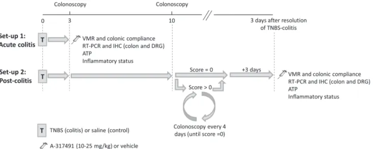
Experiments were performed either during acute colitis or in the post-colitis phase (Fig 1). In set-up 1, the role of P2X3receptors in sensory signaling was investigated during acute colitis.
Rats were randomized to receive a saline (control) or TNBS enema and all experiments were conducted 3 days later, during acute inflammation. In set-up 2, the contribution of P2X3to
colonoscopy still showed signs of mucosal inflammation, the animal was allowed to recover further and colonoscopy was repeated 4 days later. If colonoscopy showed complete mucosal healing, all experiments were conducted 3 days later.
In both the acute and post-colitis set-ups, rats received a single injection with A-317491, a selective P2X3receptor antagonist (10–25 mg/kg ip) or vehicle (saline ip) 30 min prior
VMR assessment, which was followed immediately by an evaluation of colonic compliance. In each rat, colonoscopy, as well as macroscopic and microscopic evaluation of the colonic tissue were performed in addition to an MPO activity assay to quantify inflammatory mar-kers. To assess P2X3receptor expression at the mRNA and protein level and quantify the
ex-pression of the intracellular signaling molecules of P2X3signaling, the colon and DRGs were
harvested from drug-naive rats in the control, acute colitis and post-colitis groups. In addition, colorectal distension was performedin vitroand the distension fluid was collected to assay ATP release.
Statistical analysis
Data are presented as mean ± sem for n the number of animals per group. Variables were ana-lyzed using unpaired Student’st-test and one-way or two-way ANOVA followed by Student-Newman-Keuls (SNK) post-hoc test when appropriate. Analysis of VMR and compliance data was performed by the generalized estimating equation (GEE) model followed by least signifi-cant difference (LSD) post-hoc test when appropriate. Statistical analysis was executed using SPSS 20.0 software. Statistical significance was set at p<0.05.
Fig 1. Scheme of the two experimental set-ups.In set-up 1 (acute colitis), rats were instilled with TNBS (colitis) or saline (control) and further experiments were conducted 3 days later, during the acute phase of colitis. In set-up 2 (post-colitis), rats were instilled with TNBS or saline and the extent of colitis and the healing process were monitored individually by repeated colonoscopy: first on day 3 to confirm the presence of colitis and thereafter, starting from day 10, every 4 days until complete mucosal healing (score = 0) occurred. Further experiments were conducted 3 days after colonoscopically proven mucosal healing. A-317491 (10–25 mg/kg) or vehicle (saline), denoted by the injection needle, was administered 30 min prior to the start of the VMR protocol. Evaluation of the inflammatory status entailed colonoscopy, macroscopic and microscopic assessment of the colonic tissue in addition to a myeloperoxidase activity (MPO) assay. ATP, adenosine 5’-triphosphate; DRG, dorsal root ganglion; IHC, immunohistochemistry; RT-PCR, reverse transcription polymerase chain reaction; TNBS, trinitrobenzene sulphonic acid; VMR, visceromotor response.
Results
Effect of P2X
3receptor blockade on visceral hypersensitivity during
acute TNBS-colitis
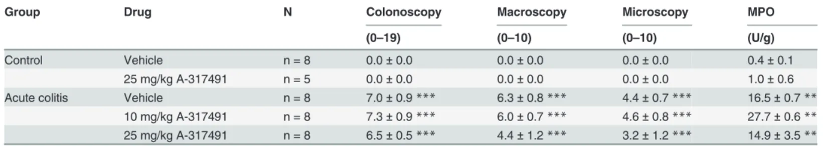
Three days after TNBS-instillation distal colitis was present, characterized by the presence of multiple serpiginous ulcers, whereas no colonic damage was seen in controls. Inflammatory markers were significantly increased in TNBS-instilled rats (Table 1).
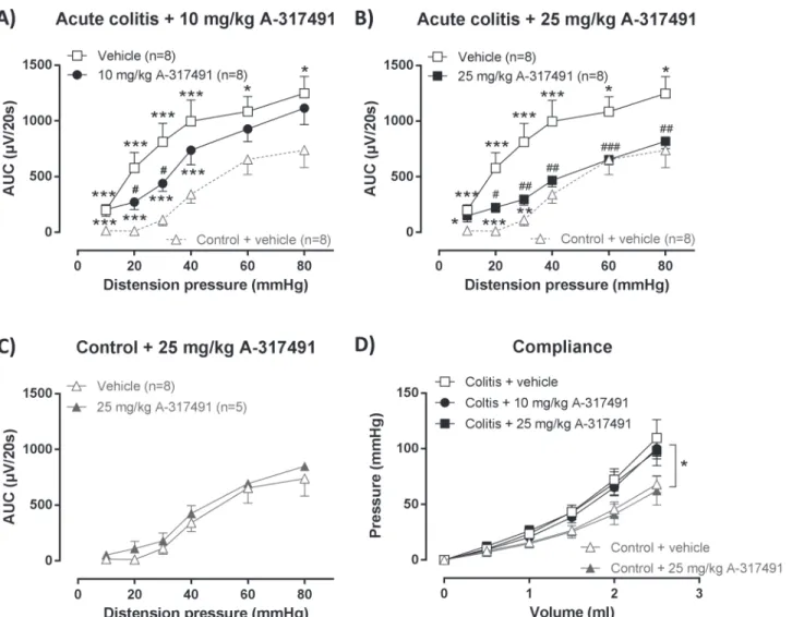
VMRs to colorectal distension were markedly increased during acute TNBS-colitis com-pared to controls, indicating the presence of acute inflammation-induced visceral hypersensi-tivity (Fig 2A). This hypersensihypersensi-tivity was dose-dependently reduced by A-317491. After 10 mg/ kg of A-317491 VMRs were not significantly reduced at the highest distension pressures of 40–
80 mmHg but were markedly attenuated at the lower distension pressures (20 and 30 mmHg). In contrast, 25 mg/kg significantly reduced hypersensitivity for almost the full range of disten-sion pressures (20 to 80 mmHg) (Fig 2B). However, VMRs remained significantly increased compared to controls at 10 to 30 mmHg distension. The 25 mg/kg dose did not affect VMRs in the control group (Fig 2C).
Colonic compliance was assessed by distending the balloon in the colorectum with increas-ing volumes of water which increased the intracolonic pressure in a volume-dependent fashion (Fig 2D). Intracolonic pressures increased more rapidly during acute colitis, indicating reduced colonic compliance compared to controls. The pressure-volume relationship was not modulat-ed by either dose of A-317491. In addition, the single administration of A-317491 30 min be-fore assessment of the VMR did not affect the colonoscopic, macroscopic or microscopic appearance of the colonic tissue (Table 1).
Effect of P2X
3receptor blockade on post-inflammatory visceral
hypersensitivity
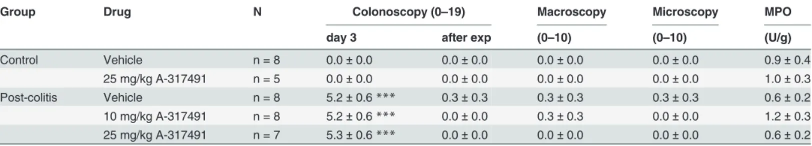
TNBS-instillation resulted in acute colitis, as evidenced by the colonoscopic scores on day 3 (Table 2). Colitis resolved spontaneously after a median of 14 days (range 10–14 days). Analy-sis of the inflammatory markers confirmed the post-inflammatory status at the time of the ex-periment (Table 2).
After resolution of TNBS-colitis, VMRs were significantly increased in post-colitis rats com-pared to controls for the full range of distension pressures, indicating the presence of marked post-inflammatory visceral hypersensitivity (Fig 3A). These increased VMRs were reduced by A-317491. After a single administration of 10 mg/kg A-317491, VMRs were markedly
Table 1. Inflammatory status during acute TNBS-colitis.
Group Drug N Colonoscopy Macroscopy Microscopy MPO
(0–19) (0–10) (0–10) (U/g)
Control Vehicle n = 8 0.0±0.0 0.0±0.0 0.0±0.0 0.4±0.1
25 mg/kg A-317491 n = 5 0.0±0.0 0.0±0.0 0.0±0.0 1.0±0.6
Acute colitis Vehicle n = 8 7.0±0.9*** 6.3±0.8*** 4.4±0.7*** 16.5±0.7**
10 mg/kg A-317491 n = 8 7.3±0.9*** 6.0±0.7*** 4.6±0.8*** 27.7±0.6**
25 mg/kg A-317491 n = 8 6.5±0.5*** 4.4±1.2*** 3.2±1.2*** 14.9±3.5**
Results are presented as mean±sem. Two-way ANOVA followed by SNK post-hoc test **p<0.01
***p<0.001, significant effect of the factor colitis; no significant effect of the factor drug; no interaction.
attenuated (Fig 3A). However VMRs remained significantly increased compared to controls at 20 mmHg distension. After treatment with 25 mg/kg A-317491, VMRs normalized over the full range of distension pressures and were no longer significantly different from those of con-trols at any of the distension pressures applied (Fig 3B). The highest dose of 25 mg/kg did not affect VMRs in control rats (Fig 3C).
After resolution of TNBS-colitis, colonic compliance was similar in the control and post-co-litis rats and was not altered by either dose of A-317491 (Fig 3D). In addition, A-317491 treat-ment had no effect on inflammatory markers (Table 2).
Fig 2. Effect of A-314791 on VMRs and colonic compliance during acute TNBS-colitis.VMRs were assessed 3 days post-TNBS enema, immediately followed by an evaluation of colonic compliance. A-314791 (filled symbols; 10–25 mg/kg) or vehicle (open symbols) was administered 30 min prior to VMR assessment. Increased VMRs were present in rats with acute TNBS-colitis compared to controls and were dose-dependently reduced by 10 mg/kg (A) and 25 mg/kg (B) A-317491. The 25 mg/kg dose did not affect VMRs in healthy controls (C). To facilitate comparison, VMRs for vehicle-treated control rats are also shown in A and B (gray dashed line). Generalized estimating equations, LSD post-hoc test, n = 5–8;*p<0.05,**p<0.01,***p<0.001, significantly
different from control + vehicle;#p<0.05,##p<0.01,###p<0.001, significantly different from acute colitis + vehicle. Colonic compliance was reduced during acute TNBS-colitis, but remained unaffected by A-317491 (10–25 mg/kg) (D). Generalized estimating equations, LSD post-hoc test, n = 5–8;*p<0.05, significant effect of the factor colitis.
Effect of P2X
3receptor blockade in acute inflammatory compared to
post-inflammatory visceral hypersensitivity
During acute colitis, A-317491 dose-dependently, but not fully reversed visceral hypersensitivi-ty, even at the highest dose of 25 mg/kg (Fig 2). However, in post-colitis rats the 10 mg/kg dose already normalized VMRs (Fig 3). To substantiate a difference in potency of A-317491 in re-ducing visceral hypersensitivity between the acute phase of colitis and the post-inflammatory phase, the effect of A-317491 on VMRs was expressed as the percentage of improvement or normalization (Fig 4): 0% indicates no improvement (compared to vehicle-treated acute colitis or post-colitis rats) whereas 100% accounts for full normalization of the VMRs (to the level of vehicle-treated controls). In post-colitis rats both the 10 and the 25 mg/kg dose of A-317491 were more effective in reducing the increased VMRs compared to the effect of A-317491 in rats in the acute phase of colitis (Fig 4).
ATP release in response to 60 mmHg of colorectal distension was significantly increased in the distal colon of rats with acute TNBS-colitis (20.0 ± 6.4 pmol/ml; n = 11; p<0.05) compared to controls (3.4 ± 2.2 pmol/ml; n = 8). In the post-colitis condition, ATP release was no longer significantly different compared to controls (2.6 ± 0.9 pmol/l; n = 6).
The relative expression of P2X3unit mRNA in the colon and DRGs was comparable in
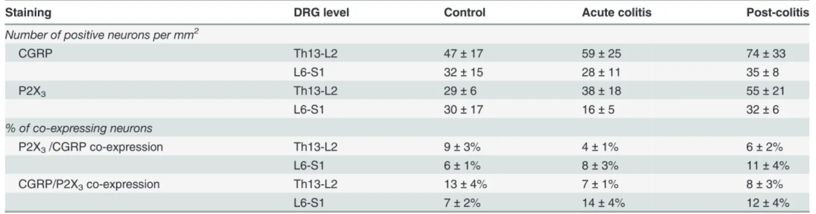
con-trol, acute colitis and post-colitis rats (Table 3). Immunohistochemical staining of DRGs also revealed no difference in expression of CGRP and P2X3between control, acute colitis and
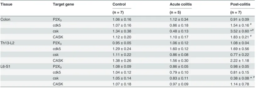
post-colitis conditions (Table 4;Fig 5). In addition, co-expression levels were similar between all groups. However, RT-PCR did reveal a difference in the mRNA expression of cdk5, csk and CASK, important molecular determinants of P2X3receptor-mediated signaling (Table 3).
Co-lonic mRNA expression of all three targets was increased in the post-colitis group, whereas mRNA levels were similar in the colon of acute colitis and control rats. The increased expres-sion in post-colitis rats was specific for the colon and was not seen at the DRG level, although we did find evidence of reduced csk mRNA expression in the DRGs L6-S1 (Table 3).
Discussion
This study was designed to evaluate P2X3receptor contribution to visceral mechanosensitivity.
Our results indicate that P2X3-mediated signaling is not involved in colonic
mechanosensitiv-ity under physiological conditions, but contributes to a different extent to visceral hypersensi-tivity during acute TNBS-colitis and in the post-inflammatory phase.
Table 2. Inflammatory status after the resolution of TNBS-colitis.
Group Drug N Colonoscopy (0–19) Macroscopy Microscopy MPO
day 3 after exp (0–10) (0–10) (U/g)
Control Vehicle n = 8 0.0±0.0 0.0±0.0 0.0±0.0 0.0±0.0 0.9±0.4
25 mg/kg A-317491 n = 5 0.0±0.0 0.0±0.0 0.0±0.0 0.0±0.0 1.0±0.3
Post-colitis Vehicle n = 8 5.2±0.6*** 0.3±0.3 0.3±0.3 0.3±0.3 0.6±0.2
10 mg/kg A-317491 n = 8 5.2±0.6*** 0.0±0.0 0.3±0.3 0.0±0.0 1.2±0.3
25 mg/kg A-317491 n = 7 5.3±0.6*** 0.0±0.0 0.0±0.0 0.0±0.0 0.6±0.2
Results are presented as mean±sem. Two-way ANOVA followed by SNK post-hoc test
***p<0.001, significant effect of the factor colitis; no significant effect of the factor drug; no interaction. Exp, experiment.
P2X3
receptors do not contribute to colonic mechanosensitivity under
physiological conditions
In our experiments, A-317491 did not affect VMRs in control rats. A-317491 is a potent and selective antagonist with nanomolar affinity for P2X3units [20]. A-317491 shows very poor
central nervous system penetration and can thus be considered a peripherally acting blocker [28,29]. Therefore, our results suggest that at least peripheral P2X3receptors do not contribute
to colonic mechanosensitivity under physiological conditions, which is in line with previous evidence [14,20]. Shinoda et al. [14] reported that pelvic afferent nerve discharge in response to colorectal distension remained unaffected in P2X3-/-mice. In the same study, VMRs in P2X3
-/-mice were significantly attenuated whereas ATP release to colorectal distension remained unal-tered, leading the authors to conclude that under physiological conditions only central, but not peripheral P2X3receptors contribute to colonic mechanosensitivity [14]. Such a central Fig 3. Effect of A-314791 on VMRs and colonic compliance after the resolution of TNBS-colitis.VMRs were assessed 3 days after colonoscopically-proven resolution of TNBS-colitis, immediately followed by an evaluation of colonic compliance. A-314791 (filled symbols; 10–25 mg/kg) or vehicle (open symbols) was administered 30 min prior to VMR assessment. Increased VMRs were present in rats that had recovered from colitis compared to controls and were normalized by 10 mg/kg (A) and 25 mg/kg (B) A-317491. The 25 mg/kg dose did not affect VMRs in healthy controls (C). To facilitate comparison, VMRs for vehicle-treated control rats are also shown in A and B (gray dashed line). Generalized estimating equations, LSD post-hoc test, n = 5–8;*p<0.05, ***p<0.001, significantly different from control + vehicle;#p<0.05,##p<0.01,###p<0.001, significantly different from acute colitis + vehicle. Colonic
compliance was similar in control and post-colitis rats and remained unaffected by A-317491 (10–25 mg/kg) (D). Generalized estimating equations, LSD post-hoc test, n = 5–8.
mechanism of action could be located at the level of the spinal cord where presynaptic P2X3
channels facilitate the release of excitatory glutamate [11]. The lack of a peripheral contribution of P2X3to sensory signaling under physiological conditions is further suggested by the finding
that double P2X2-/-P2X3-/-mice show similar increases in jejunal afferent nerve firing in
re-sponse to gut distension compared to controls [13]. Likewise, the P2X1/P2X3receptor
antago-nist TNP-ATP, which is also devoid of central nervous system penetration [30], did not affect
Fig 4. Effect of A-317491 in acute TNBS-colitis compared to the post-inflammatory phase of colitis.To substantiate a difference in potency of A-317491 to reduce visceral hypersensitivity during acute TNBS-colitis compared to the post-inflammatory phase of colitis, data are expressed as the percentage of improvement of normalization: 0% means no improvement and thus the same level of hypersensitivity as vehicle-treated rats during acute TNBS-colitis or in the post-inflammatory phase, whereas 100% means complete normalization of the increased VMRs (reaching the level of vehicle-treated controls). Both the 10 mg/kg (A) and the 25 mg/kg (B) dose of A-317491 more potently reduced visceral hypersensitivity in the post-inflammatory phase of colitis compared to the acute inflammatory phase of colitis. Generalized estimating equations, LSD post-hoc test, n = 7–8;*p<0.05, significantly different compared to acute colitis.
doi:10.1371/journal.pone.0123810.g004
Table 3. mRNA expression of P2X3, cdk5, csk and CASK in the colon and DRGs (Th13-L2 and L6-S1).
Tissue Target gene Control Acute colitis Post-colitis
(n = 7) (n = 5) (n = 7)
Colon P2X3 1.06±0.16 1.12±0.34 0.91±0.09
cdk5 1.07±0.16 0.86±0.18 1.54±0.16#
csk 1.34±0.38 0.48±0.13 3.52±0.60*#
CASK 1.12±0.20 1.10±0.17 1.83±0.21#
Th13-L2 P2X3 0.95±0.05 1.06±0.12 1.08±0.04
cdk5 1.29±0.24 1.60±0.12 1.69±0.56
csk 1.11±0.22 0.86±0.08 0.77±0.22
CASK 1.38±0.26 1.56±0.30 2.22±1.18
L6-S1 P2X3 1.08±0.09 0.86±0.05 0.98±0.05
cdk5 1.04±0.12 0.79±0.10 0.81±0.15
csk 1.05±0.14 0.83±0.11 0.38±0.08*#
CASK 1.07±0.18 0.97±0.09 1.14±0.78
Data presented as mean±sem for n = 5–10 animals per group. One-way ANOVA followed by SNK.
*p<0.05, significantly different from control
# p<0.05, significantly different from acute-colitis.
in vivonociceptive behavior to colorectal distension in rats under control conditions [18]. However in contrast, pelvic afferent nerve discharge to gut distension was significantly attenu-ated by TNP-ATP in healthy control rats [16].
P2X3
receptors contribute to visceral hypersensitivity during acute
TNBS-colitis and in the post-inflammatory phase
Ourin vivodata of increased purinergic signaling via P2X3channels during acute TNBS-colitis
are in line with previousin vitrofindings of Wynn et al. [17] who showed that pelvic afferent nerve firing in response to colorectal distension was significantly increased in an isolated colon segment from rats with acute TNBS colitis and that this increase could be attenuated by the P2X receptor antagonist PPADS and the P2X1/P2X3receptor antagonist TNP-ATP. Our study
elaborates on these results and now provides evidence from anin vivoset-up demonstrating that the selective P2X3antagonist A-317491 dose-dependently, though not fully, reversed the
increased VMRs to colorectal distension in rats with acute TNBS-colitis. This suggests that re-ceptors containing the P2X3unit contribute to visceral hypersensitivity during acute colitis.
This antinociceptive effect of the P2X3receptor antagonist was not mediated by changes in
co-lonic compliance. This is important considering other antagonists such as TNP-ATP and PPADS also block P2X1receptors expressed on smooth muscle cells in the rat distal colon [31]. Table 4. Protein expression of CGRP and P2X3in the dorsal root ganglia.
Staining DRG level Control Acute colitis Post-colitis
Number of positive neurons per mm2
CGRP Th13-L2 47±17 59±25 74±33
L6-S1 32±15 28±11 35±8
P2X3 Th13-L2 29±6 38±18 55±21
L6-S1 30±17 16±5 32±6
% of co-expressing neurons
P2X3/CGRP co-expression Th13-L2 9±3% 4±1% 6±2%
L6-S1 6±1% 8±3% 11±4%
CGRP/P2X3co-expression Th13-L2 13±4% 7±1% 8±3%
L6-S1 7±2% 14±4% 12±4%
Data presented as mean±sem for n = 3 animals per group and 3 DRGs per animal for each level. DRGs, dorsal root ganglia. Two-way ANOVA, no significant effects.
doi:10.1371/journal.pone.0123810.t004
Fig 5. Expression of CGRP and P2X3in dorsal root ganglia.Immunohistochemical detection of CGRP (left panel) and P2X3(middle panel) on
cryosections of a dorsal root ganglion. The right panel shows the merged picture; the white arrows indicate neuronal co-expression of CGRP and P2X3.
Moreover, P2X3receptors on myenteric neurons contribute to the initiation of reflex
contrac-tions when the intestinal pressure rises and could therefore modulate colonic compliance [32]. In a rat model for non-inflammatory visceral pain, induced by intraperitoneal injection of acetic acid, A-317491 has previously been shown to reduce nociception with an IC50of
27μmol/kg, which is approximately 15 mg/kg [20]. On the other hand, A-317491 had no effect on VMRs in a zymosan-induced model of acute colonic inflammatory hypersensitivity, even after administration of 100μmol/kg (approximately 56.6 mg/kg) [20]. We can only speculate on the reasons underlying the discrepancy in the results between the latter study and our own findings. Firstly, the difference in experimental models for visceral hypersensitivity should be considered (TNBSvszymosan). Secondly, in the zymosan-study, Sprague-Dawley rats were subjected to 3 colorectal distension protocols: the first before the induction, the second during acute colitis and the third 30 min after the second distension. In our hands, repeating disten-sion protocols after a 30 min interval in male Sprague-Dawley rats induced hypersensitivity in control rats (data not shown), which might relate to differences in the distension protocol (6 distensions ranging from 10 to 80 mmHgvsthree 60mmHg distensions). Therefore, only one distension protocol was performed in each rat in our set-up. In support of our results, A-317491 also exerted antinociceptive effects in models of somatic and neuropathic pain [20,29,33–35].
We additionally studied the effect of blockade of P2X3receptor units in a rat model for
post-inflammatory visceral hypersensitivity. In this model, we confirmed the resolution of TNBS-colitis by colonoscopic, macroscopic and microscopic evaluation of the colonic tissue, in addition to MPO activity. Despite the absence of inflammation, the VMRs to colorectal disten-sion were increased in post-colitis rats. These increased VMRs were dose-dependently reduced and even normalized by A-317491. This strongly suggests an important role for P2X3receptor
units in post-inflammatory visceral hypersensitivity. A contribution of purinergic P2X recep-tors to post-infectious visceral hypersensitivity was previously suggested by Rong et al. [13] who demonstrated that the P2X receptor antagonist PPADS reduced increased jejunal afferent firing afterTrichinella spiralisinfection. Yet the exact P2X receptor subtype involved remained to be identified. Our study now provides evidence of enhanced P2X3unit-mediated signaling
in the post-inflammatory phase.
Evidence for a role for purinergic modulation of nociceptive responses, most likely mediated by P2X3receptor units, has also been reported in other animal models of visceral
hypersensitiv-ity. Xu et al. [18] instilled acetic acid in the colon of neonate rats, which resulted in visceral hypersensitivity at the adult age; TNP-ATP reduced these increased VMRs. In addition, the intracolonic instillation of zymosan in control mice induced visceral hypersensitivity in the ab-sence of overt colonic inflammation, while VMRs remained unaffected by zymosan exposure in P2X3-/-mice [14]. Therefore, it seems that peripheral P2X3receptor units contribute to
vis-ceral hypersensitivity, not only during and after the resolution of TNBS-colitis but also in other types of chemically-induced colonic hypersensitivity.
Several mechanisms could underlie the difference in potency of A-317491 in reducing vis-ceral hypersensitivity between the acute phase of colitis and the post-inflammatory phase. Firstly, there was a significant increase in distension-induced release of ATP, the endogenous ligand for P2X3receptors, during acute colitis whereas ATP release had returned to control
val-ues in the post-inflammatory condition. ATP is present in the cell in millimolar concentrations and during inflammation, extracellular ATP levels increase due to active release as well as pas-sive leakage from damaged or dying cells, in combination with a downregulation of ATP break-down [36]. Being a competitive antagonist for the P2X3unit, A-317491 can be displaced from
the binding site by excess ATP [20]. In addition, at the level of the primary sensory afferent, ATP potentiates the effect of other known nociceptive mediators such as protons, capsaicin and 5-hydroxytryptamine [37]. Therefore the increased availability of extracellular ATP during acute TNBS-colitis, most likely contributes to the enhanced P2X3-mediated signaling during
the acute inflammatory phase. Secondly, differences in the P2X3receptor expression in the
acute versus the post-inflammatory phase may also contribute to the difference in A-317491 potency. However, we found no evidence of altered P2X3expression at the mNRA or protein
level in the colon or DRGs, neither during acute colitis nor in the post-inflammatory phase, ar-guing against a significant increase in P2X3receptors in our study. In addition, we could not
demonstrate enhanced colocalization between P2X3and CGRP either. In contrast, enhanced
colocalization during acute TNBS-colitis as was previously reported by Wynnet al. [17]. Be-sides increased mediator release or increased receptor expression, sensitization of P2X3
recep-tors may provide a plausible explanation for these results. The signaling of extracellular ATP binding to P2X3receptor is transduced in the intracellular environment by a series of adaptor
and scaffold molecules among which are kinases such as Cdk5, Csk, and CASK. Cdk5 and Csk modulate the electrical properties of P2X3receptors whereas CASK interacts with the P2X3
receptor to positively enforce the receptor’s stability and functional responsiveness [38]. To investigate their involvement, we performed RT-PCR for Cdk5, Csk and CASK mRNA expression in the colon and DRGs (Th13-L2 and L6-S1) of control, acute colitis and post-colitis rats. We found that in the post-colitis group, colonic mRNA expression of all three targets was significantly increased, whereas mRNA levels were similar in acute colitis and controls. The in-creased expression in the post-colitis group was specific for the colon and was not seen at the DRG level. These findings point towards P2X3receptor sensitization in the post-colitis phase,
that may underlie—or at least contribute to—the persistent post-inflammatory visceral hyper-sensitivity in our study.
The observation of an increased potency of A-317491 to reduce hypersensitivity in the post-colitis phase compared to the acute inflammatory event is an interesting observation that war-rants further study. In fact few studies compare mediator contribution to hypersensitivity in an acute versus a post-inflammatory setting. Similar evidence was presented for acid sensing ion channels type 3 (ASIC3), as these were upregulated during active Crohn’s disease and contrib-uted to peripheral sensitization by inflammatory mediators but were not involved in non-in-flammatory visceral hypersensitivity according to another group [39–41]. To further
investigate whether this increased potency in the post-inflammatory phase is specific for P2X3
receptor mediated signaling, we conversely compared the antinociceptive effect of a selective histamine H1receptor antagonist levocetirizine in the acute and post-inflammatory setting (S1
Fig). We recently demonstrated that levocetirizine fully reversed post-inflammatory visceral hypersensitivity in the post-inflammatory phase at a dose of 1 mg/kg [21]. Similar to our results for A-317491, levocetirizine was more effective in reducing hypersensitivity in the post-inflam-matory phase compared to the effect during acute TNBS-colitis (S1 Fig). As H1receptors
con-tribute to afferent nerve signaling in the gut wall [42], this suggests a more general
In summary, we evaluated the antinociceptive effects of A-317491, a selective P2X3-unit
an-tagonist, on visceral pain. A-317491 dose-dependently reduced visceral hypersensitivity during acute TNBS-colitis and fully abolished increased VMRs after resolution of colitis without af-fecting visceral sensitivity in controls. The antinociceptive effect of A-317491 was more pro-nounced in the post-inflammatory phase of colitis compared to the acute inflammatory phase, most likely due to displacement of A-317491 by excess ATP release during acute TNBS-colitis and sensitization of P2X3receptors in the post-inflammatory phase of colitis.
Hence, purinergic P2X3receptor units do not seem to be involved in sensory signaling
under physiological conditions but mediate visceral hypersensitivity with a different potency during acute TNBS-colitis and in the post-inflammatory phase. These findings validate P2X3
receptors as a potential new target in the treatment of abdominal pain syndromes such as IBS and IBD.
Supporting Information
S1 Fig. Levocetirizine in acute TNBS-colitis compared to the post-inflammatory phase of colitis.Data are expressed as the percentage of improvement of normalization: 0% means no improvement and thus the same level of hypersensitivity as vehicle-treated rats during acute TNBS-colitis or in the post-inflammatory phase, whereas 100% means complete normalization of the increased VMRs (reaching the level of vehicle-treated controls). Levocetirizine (1 mg/kg) more potently reduced visceral hypersensitivity in the post-inflammatory phase of colitis com-pared to the acute inflammatory phase of colitis. Generalized estimating equations, LSD post-hoc test, n = 5–8;p<0.05, significantly different compared to acute colitis.
(TIF)
Acknowledgments
We would like to thank our lab technicians P. Aerts, A. Jürgens, E. Theuns, S. Thys, M. Vinckx and R. Van Den Bossche for their technical assistance.
Author Contributions
Conceived and designed the experiments: AD TGM PAP JGDM BYDW. Performed the exper-iments: AD LvdL AdW HC RB JPT. Analyzed the data: AD LvdL AdW HC RB JPT JGDM BYDW. Contributed reagents/materials/analysis tools: RB JPT PAP JGDM BYDW. Wrote the paper: AD RB JGDM BYDW. Revised the manuscript critically for important intellectual con-tent: LdvL AdW HC JPT TGM PAP. Final approval of the manuscript: AD LvdL AdW HC RB JPT TGM PAP JGDM BYDW.
References
1. Vermeulen W, De Man JG, Pelckmans PA, De Winter BY (2014) Neuroanatomy of lower gastrointesti-nal pain disorders. World J Gastroenterol 20: 1005–1020. doi:10.3748/wjg.v20.i4.1005PMID:
24574773
2. De Schepper HU, De Man JG, Moreels TG, Pelckmans PA, De Winter BY (2008) Review article: gas-trointestinal sensory and motor disturbances in inflammatory bowel disease—clinical relevance and pathophysiological mechanisms. Aliment Pharmacol Ther 27: 621–637. doi:10.1111/j.1365-2036. 2008.03624.xPMID:18221407
3. Longstreth GF, Thompson WG, Chey WD, Houghton LA, Mearin F, Spiller RC (2006) Functional bowel disorders. Gastroenterology 130: 1480–1491. PMID:16678561
4. Spiller R, Garsed K (2009) Postinfectious irritable bowel syndrome. Gastroenterology 136: 1979–
5. Burnstock G (2001) Purine-mediated signalling in pain and visceral perception. Trends Pharmacol Sci 22: 182–188. PMID:11282418
6. Ford AP, Gever JR, Nunn PA, Zhong Y, Cefalu JS, Dillon MP, et al. (2006) Purinoceptors as therapeutic targets for lower urinary tract dysfunction. Br J Pharmacol 147 Suppl 2: S132–143. PMID:16465177
7. Galligan JJ (2004) Enteric P2X receptors as potential targets for drug treatment of the irritable bowel syndrome. Br J Pharmacol 141: 1294–1302. PMID:15051631
8. Yiangou Y, Facer P, Baecker PA, Ford AP, Knowles CH, Chan CL, et al. (2001) ATP-gated ion channel P2X(3) is increased in human inflammatory bowel disease. Neurogastroenterol Motil 13: 365–369. PMID:11576396
9. Burnstock G (2009) Purines and sensory nerves. In: Canning BJ, Spina D, editors. Sensory Nerves. 2009 ed: Springer Berlin Heidelberg. pp. 333–392.
10. Cockayne DA, Dunn PM, Zhong Y, Rong W, Hamilton SG, Knight GE, et al. (2005) P2X2 knockout mice and P2X2/P2X3 double knockout mice reveal a role for the P2X2 receptor subunit in mediating multiple sensory effects of ATP. J Physiol 567: 621–639. PMID:15961431
11. Ford AP (2012) In pursuit of P2X3 antagonists: novel therapeutics for chronic pain and afferent sensiti-zation. Purinergic Signal 8: 3–26. doi:10.1007/s11302-011-9271-6PMID:22095157
12. Robinson DR, McNaughton PA, Evans ML, Hicks GA (2004) Characterization of the primary spinal af-ferent innervation of the mouse colon using retrograde labelling. Neurogastroenterol Motil 16: 113–
124. PMID:14764211
13. Rong W, Keating C, Sun B, Dong L, Grundy D (2009) Purinergic contribution to small intestinal afferent hypersensitivity in a murine model of postinfectious bowel disease. Neurogastroenterol Motil 21: 665–
671, e632. doi:10.1111/j.1365-2982.2008.01259.xPMID:19220757
14. Shinoda M, Feng B, Gebhart GF (2009) Peripheral and central P2X receptor contributions to colon mechanosensitivity and hypersensitivity in the mouse. Gastroenterology 137: 2096–2104. doi:10. 1053/j.gastro.2009.06.048PMID:19549524
15. Shinoda M, La JH, Bielefeldt K, Gebhart GF (2010) Altered purinergic signaling in colorectal dorsal root ganglion neurons contributes to colorectal hypersensitivity. J Neurophysiol 104: 3113–3123. doi:10. 1152/jn.00560.2010PMID:20861433
16. Wynn G, Rong W, Xian Z, Burnstock G (2003) Purinergic mechanisms contribute to mechanosensory transduction in the rat colorectum. Gastroenterology 125: 1398–1409. PMID:14598256
17. Wynn G, Ma B, Ruan HZ, Burnstock G (2004) Purinergic component of mechanosensory transduction is increased in a rat model of colitis. Am J Physiol Gastrointest Liver Physiol 287: G647–657. PMID:
15331354
18. Xu GY, Shenoy M, Winston JH, Mittal S, Pasricha PJ (2008) P2X receptor-mediated visceral hyperal-gesia in a rat model of chronic visceral hypersensitivity. Gut 57: 1230–1237. doi:10.1136/gut.2007. 134221PMID:18270243
19. Honore P, Mikusa J, Bianchi B, McDonald H, Cartmell J, Faltynek C, et al. (2002) TNP-ATP, a potent P2X3 receptor antagonist, blocks acetic acid-induced abdominal constriction in mice: comparison with reference analgesics. Pain 96: 99–105. PMID:11932066
20. Jarvis MF, Burgard EC, McGaraughty S, Honore P, Lynch K, Brennan TJ, et al. (2002) A-317491, a novel potent and selective non-nucleotide antagonist of P2X3 and P2X2/3 receptors, reduces chronic inflammatory and neuropathic pain in the rat. Proc Natl Acad Sci U S A 99: 17179–17184. PMID:
12482951
21. Deiteren A, De Man JG, Ruyssers NE, Moreels TG, Pelckmans PA, De Winter BY (2014) Histamine H4 and H1 receptors contribute to postinflammatory visceral hypersensitivity. Gut 63: 1873–1882. doi:10. 1136/gutjnl-2013-305870PMID:24561612
22. Vermeulen W, De Man JG, De Schepper HU, Bult H, Moreels TG, Pelckmans PA, et al. (2013) Role of TRPV1 and TRPA1 in visceral hypersensitivity to colorectal distension during experimental colitis in rats. Eur J Pharmacol 698: 404–412. doi:10.1016/j.ejphar.2012.10.014PMID:23099257
23. Vermeulen W, De Man JG, Nullens S, Pelckmans PA, De Winter BY, Moreels TG (2011) The use of en-doscopy to follow the inflammatory time course of TNBS-colitis in rats. Acta Gastroenterol Belg 74: 304–311. PMID:21861315
24. Wallace JL, Keenan CM (1990) An orally active inhibitor of leukotriene synthesis accelerates healing in a rat model of colitis. Am J Physiol 258: G527–534. PMID:1970708
25. Ness TJ, Gebhart GF (1988) Colorectal distension as a noxious visceral stimulus: physiologic and phar-macologic characterization of pseudaffective reflexes in the rat. Brain Res 450: 153–169. PMID:
26. Deiteren A, Vermeulen W, Moreels TG, Pelckmans PA, De Man JG, De Winter BY (2014) The effect of chemically induced colitis, psychological stress and their combination on visceral pain in female Wistar rats. Stress 17: 431–444. doi:10.3109/10253890.2014.951034PMID:25089934
27. Abad C, Juarranz Y, Martinez C, Arranz A, Rosignoli F, Garcia-Gomez M, et al. (2005) cDNA array analysis of cytokines, chemokines, and receptors involved in the development of TNBS-induced colitis: homeostatic role of VIP. Inflamm Bowel Dis 11: 674–684. PMID:15973123
28. Sharp CJ, Reeve AJ, Collins SD, Martindale JC, Summerfield SG, Sargent BS, et al. (2006) Investiga-tion into the role of P2X(3)/P2X(2/3) receptors in neuropathic pain following chronic constricInvestiga-tion injury in the rat: an electrophysiological study. Br J Pharmacol 148: 845–852. PMID:16770326
29. Wu G, Whiteside GT, Lee G, Nolan S, Niosi M, Pearson MS, et al. (2004) A-317491, a selective P2X3/ P2X2/3 receptor antagonist, reverses inflammatory mechanical hyperalgesia through action at periph-eral receptors in rats. Eur J Pharmacol 504: 45–53. PMID:15507220
30. Cavaliere F, Amadio S, Dinkel K, Reymann KG, Volonte C (2007) P2 receptor antagonist trinitrophenyl-adenosine-triphosphate protects hippocampus from oxygen and glucose deprivation cell death. J Phar-macol Exp Ther 323: 70–77. PMID:17620457
31. Van Crombruggen K, Van Nassauw L, Timmermans JP, Lefebvre RA (2007) Inhibitory purinergic P2 re-ceptor characterisation in rat distal colon. Neuropharmacology 53: 257–271. PMID:17612577
32. Bian X, Ren J, DeVries M, Schnegelsberg B, Cockayne DA, Ford AP, et al. (2003) Peristalsis is im-paired in the small intestine of mice lacking the P2X3 subunit. J Physiol 551: 309–322. PMID:
12813150
33. Noma N, Shinoda M, Honda K, Kiyomoto M, Dezawa K, Nakaya Y, et al. (2013) Interaction of IL-1beta and P2X(3) receptor in pathologic masseter muscle pain. J Dent Res 92: 456–460. doi:10.1177/ 0022034513483770PMID:23520364
34. Ma B, Yu LH, Fan J, Cong B, He P, Ni X, et al. (2011) Estrogen modulation of peripheral pain signal transduction: involvement of P2X(3) receptors. Purinergic Signal 7: 73–83. doi: 10.1007/s11302-010-9212-9PMID:21484099
35. Hsieh YL, Chiang H, Lue JH, Hsieh ST (2012) P2X3-mediated peripheral sensitization of neuropathic pain in resiniferatoxin-induced neuropathy. Exp Neurol 235: 316–325. doi:10.1016/j.expneurol.2012. 02.013PMID:22391132
36. Cauwels A, Rogge E, Vandendriessche B, Shiva S, Brouckaert P (2014) Extracellular ATP drives sys-temic inflammation, tissue damage and mortality. Cell Death Dis 5: e1102. doi:10.1038/cddis.2014.70
PMID:24603330
37. Wynn G, Burnstock G (2006) Adenosine 5'-triphosphate and its relationship with other mediators that activate pelvic nerve afferent neurons in the rat colorectum. Purinergic Signal 2: 517–526. doi:10. 1007/s11302-005-5305-2PMID:18404489
38. Volonte C, Burnstock G (2013) P2X3 receptor: a novel 'CASKade' of signaling? J Neurochem 126: 1–
3. doi:10.1111/jnc.12282PMID:24024236
39. Matricon J, Muller E, Accarie A, Meleine M, Etienne M, Voilley N, et al. (2013) Peripheral contribution of NGF and ASIC1a to colonic hypersensitivity in a rat model of irritable bowel syndrome. Neurogastroen-terol Motil 25: e740–754. doi:10.1111/nmo.12199PMID:23902154
40. Jones RC 3rd, Xu L, Gebhart GF (2005) The mechanosensitivity of mouse colon afferent fibers and their sensitization by inflammatory mediators require transient receptor potential vanilloid 1 and acid-sensing ion channel 3. J Neurosci 25: 10981–10989. PMID:16306411
41. Yiangou Y, Facer P, Smith JA, Sangameswaran L, Eglen R, Birch R, et al. (2001) Increased acid-sens-ing ion channel ASIC-3 in inflamed human intestine. Eur J Gastroenterol Hepatol 13: 891–896. PMID:
11507351