Original Article
Investigation of antioxidative, antityrosinase and cytotoxic effects
of extract of irradiated oyster mushroom
Nutsuda Banlangsawan
1, Bungorn Sripanidkulchai
2*, and Niwat Sanoamuang
1,31 Department of Plant Science and Agricultural Resources, Faculty of Agriculture,
2 Center for Research and Development of Herbal Health Products, Faculty of Pharmaceutical Sciences,
3 Applied Taxonomic Research Center, Faculty of Sciences,
Khon Kaen University, Mueang, Khon Kaen, 40002 Thailand.
Received: 20 May 2015; Accepted: 12 August 2015
Abstract
Oyster mushroom (Pleurotus ostreatus Fries.) is rich in nutrition and has many medicinal properties such as anti-oxidant and anticancer activities. It also contains a high amount of ergosterol which can be converted to vitamin D2 when
exposing to UV light. Oyster mushroom powder was irradiated with UV-B for 180 min and extracted with 95% ethanol. Mushroom extract was determined for vitamin D2 concentration, total phenolic compound, antioxidative activity, tyrosinase
inhibitory property and cytotoxicity effect on human keratinocytes (HaCaT) and murine melanoma cells (B16F10) by MTT assay. The results demonstrated that the concentration of vitamin D2 of irradiated oyster mushroom extract was 153.96 µg/g,
which is 13 times higher than that of non-irradiated mushroom extract. Total phenolic content, antioxidative and tyrosinase inhibitory activities of the two mushroom extracts were not significantly different. Neither oyster mushroom extract had a cytotoxic effect on keratinocytes, but on the other hand both inhibited the growth of murine melanoma cells.
Keywords:oyster mushroom (Pleurotus ostreatus), vitamin D2, antioxidant effect, cytotoxic effect,
tyrosinase inhibition activity
1. Introduction
Oyster mushroom (Pleurotus ostreatus Fries.) is a white rot or wood decay fungus. This fungus can grow in a wide range of temperatures and it is cultivated in many parts of the world including Thailand (Abdurrahman et al., 2009). This mushroom is a food with great nutritional value accord-ing to its high protein and dietary fiber content. It also provides a valuable source of minerals and vitamins (Gajendra
et al., 2014). Vitamin D has been found in mushrooms and can be formed from the plant steroid called ergosterol. Vitamin D has several important functions in the body such
as the regulation of calcium and phosphorus absorption and facilitating normal immune system function (Heaney, 2003). Moreover, vitamin D may play a role in protecting against diabetes, heart disease, high blood pressure, multiple sclerosis and cancer (Rivera et al., 2010).
Cultivated oyster mushrooms are rich sources of ergosterol, the precursor of vitamin D2. Ergosterol in mushrooms can be converted to vitamin D2 after exposure to UV light (Ko et al., 2008). When mushrooms are exposed to UV light, ergosterol undergoes photolysis to yield a variety of photo-irradiation products, principally previtamin D2, tachysterol and lumisterol. The previtamin D2 is sponta-neously rearranged into vitamin D2(Teichmann et al., 2007). In general, the cultivated mushrooms contain low vitamin D2. They may not be exposed to the sunlight, which is essential in the natural production of vitamin D2 (Mattila et al., 2002).
* Corresponding author.
Email address: bungorn@kku.ac.th
Previous research showed that the concentration of vitamin D2 in cultivated mushrooms could be increased after they
were exposed to sunlight, artificial light such as UV light or Pulsed UV light (PUV) (Jasinghe and Perera, 2006). The effects of UV light on vitamin D2 production among various
cultivated mushroom species have been recently reported by a number of authors (Jasinghe and Perera, 2005; Koyyalamudi et al., 2009; Mau et al., 1998).
Oyster mushrooms contain several compounds with antioxidative activity such as vitamin A, C, E, carotenoids, polyphenolic compounds and flavonoids (Nuhu et al., 2011). Oxidative stress caused by excessive free radicals lead to aging and diseases, such as atherosclerosis, diabetes, cancer, and cirrhosis (Ilgaz et al., 2012). Although human body is designed to have its own defense and repair systems to protect against oxidative damage, these systems are insuffi-cient to entirely prevent damages (Nuhu et al., 2010). Food supplements or diets containing antioxidants such as fruit, vegetable and mushrooms may be useful to reduce the oxida-tive damage (Arbaayah and Umi, 2013). Tyrosinase, a multi-functional copper-containing enzyme, is widely distributed in fungi, plant and animal and it is responsible for melaniza-tion in animals and enzymatic browning of fruit. This enzyme catalyzes two distinct reactions involving molecular oxygen: the hydroxylation of monophenols to o-diphenols and the oxidation of o-diphenols to o-quinones (Nuhu et al., 2011). Tyrosinase inhibitors from natural sources were found to inhibit the activities of both monophenolase and diphenolase (Kim and Uyama, 2005), and oyster mushroom was reported to have tyrosinase inhibitory effect (Nuhu et al., 2010; Riani
et al., 2012).
Oyster mushrooms have beneficial effects on health and also possess a number of therapeutic properties like anti-inflammatory, immunostimulatory, immunomodulatory and anticancer activity (Iris et al., 2006). Previous reports showed that the ethanol extract of oyster mushroom had high anti-cancer activity on several cell lines such as breast, colon, liver and lung cancer cells. Moreover, the oyster mushroom extracts also showed direct anti-tumor activity against carci-nomas possibly through the activation of the body’s immune system. (Andrej and Daniel, 2008; Seema and Goyal, 2012).
Although there are many studies on the conversion of ergosterol to vitamin D2 after UV-B irradiation of oyster
mushrooms, there are no reports on the biological activities and cytotoxicity effect of the extract from irradiated oyster mushrooms. Therefore, the objectives of this research were to investigate the content of vitamin D2 and phenolic
com-pounds, the antioxidative and tyrosinase inhibitory proper-ties, and the cytotoxic effect on keratinocytes and melanoma cells of irradiated oyster mushroom extract.
2. Materials and Methods
2.1 Chemicals
Ninety-five percent ethanol (analytical grade) was
purchased from Labscan Asia Co.,Ltd. (Bangkok, Thailand). Methanol and acetonitrile (HPLC grade) were obtained from Merck (Darmstadt, Germany). 1,1-diphenyl 2-picryl hydrazyl (DPPH), 3-(4, 5-Dimethyl thiazol-2-yl)-2, 5-diphenyl tetrazo-lium bromide (MTT), ergosterol, ergocalciferol (vitamin D2),
fetal bovine serum (FBS), Folin-Ciocalteus phenol reagent, kojic acid, L-tyrosine, penicillin, streptomycin, tyrosinase, vitamin C and vitamin E were purchased from Sigma (Steinheim, Germany). Dulbecco’s Modified Eagle Medium (DMEM) powder was purchased from Invitrogen corpora-tion. The other chemicals used were of analytical grade.
2.2 Irradiation procedure and mushroom extraction
Fresh fruiting bodies of oyster mushrooms obtained from local farms in Khon Kaen province, Thailand were sliced into pieces, dried with hot air at 50C for 48 hr and finely pulverized. The obtained mushroom powder was stored at -20C until UV-B treatment. Prior to the treatment, mushroom powder was left at room temperature for 4 hr and placed on an aluminium tray. The samples were placed 15 cm away from the irradiation source and exposed to the UV-B in an irradia-tion chamber. The UV-B unit was set with 8 UV-B lamps (313 ±12 nm, Philips TL-D 18W) 604 cm in length and the total treatment area was 100x120 cm2. Mushroom powder was
treated under UV-B lamp, at an irradiation dose of 550.32 J/cm2 at 25-28C for 180 min then the irradiated mushrooms
powder was stored at -20C. The un-irradiated mushroom powder was used as a control.
Ten grams of powder samples were extracted with 100 mL of 95% ethanol for 24 hr. The ethanolic extracts were filtered through Whatman No.1 filter paper, rotary evapo-rated at 40°C then freeze-dried.
2.3 Validation of HPLC method
The method validation ensures vitamin D2 and
ergos-terol analysis credibility. The criteria of accuracy, precision, linearity, limit of detection (LOD) and limit of quantitation (LOQ) were considered. The accuracy of this HPLC method was determined by using samples spiked with five different concentrations of vitamin D2 and ergosterol standards and
defined in terms of percent recovery. Intra-day and inter-day precision were done to assure the precision of the method. Linearity was tested by eight different concentrations, which were prepared by diluting the standard stock solution. LOD was determined as the lowest concentration of vitamin D2
and ergosterol giving a response of 3 times the baseline noise defined from the analysis of control sample. LOQ was deter-mined as the lowest concentration giving a response of 10 times the baseline noise (Lloyd et al., 2012).
2.4 Analysis of vitamin D2 and ergosterol
The content of vitamin D2 was analyzed according to
mushroom extracts (0.5 g) were weighed into a 250 mL round-bottomed flask and mixed with 1 g of L-ascorbic acid, 50 mL of 99% ethanol and 25 mL of 50% potassium hydroxide. The mixture was shaken and saponified under reflux at 85C for 30 min. It was immediately cooled to room temperature and poured into a separating funnel. The mixture was firstly extracted with 10 mL of de-ionised water and subsequently with 30 mL of n-hexane. The organic layers were washed 3 times with de-ionised water until neutralized. The organic layer was transferred into a round bottom flask, rotary evapo-rated to dryness at 50C and re-dissolved in 2 mL of a mixed solution of eluent (methanol/acetonitrile = 75:25 v/v) and isopropyl alcohol (2:1 v/v). The sample was passed through a 0.45 µm non-pyrogenic filter. A volume of 20 µl of filtered sample was injected into the HPLC system (LC 20A, Shimadzu, Japan) and eluted through a reversed phase C18 column (Maxsil 5 C18, 250×4.6 mm, Phenomenex, Torrance, CA, USA). The mobile phase was methanol/acetonitrile (75: 25 v/v), at a flow rate of 1 mL/min. The UV detection of elute was performed at 264 nm. Ergosterol and vitamin D2 were
qualitatively analyzed by comparing the retention times of standards and their quantifications were done by using a calibration curve.
2.5 Scavenging effect on 1, 1-Diphenyl-2-picrylhydrazyl radical
Free radical scavenging is one of the mechanisms in inhibiting lipid oxidation commonly used to estimate anti-oxidant activity (Pornariya and Kanok-Orn, 2009). The radical scavenging activity of mushroom extracts was evaluated using 1,1-diphenyl-2-picrylhydrazyl (DPPH) radical accord-ing to the previous report (Shimada et al.,1992). Various con-centrations (0.1-5 mg/mL) of mushrooms extracts were added with 200 µl of 1 mM DPPH radical solutions in methanol, vigorously mixed and allowed to stand for 15 min at room temperature. The DPPH solution without extract solution was used as control. The scavenging effect on the DPPH radical was measured at 515 nm and read using UV/VIS spectrophotometer (UV-1700 PharamSpec). The assay was carried out in triplicate and half maximal effective concentra-tion (EC50) value was calculated. The scavenging effect on
the DPPH inhibition in term of percent (%) was calculated according to the equation: Percentage (%) of DPPH radical scavenging = (Accontrol-Assample/Accontrol)) × 100, where Ac is
the absorbance of control blank and As is the absorbance of extract sample. Vitamin C and vitamin E were used as positive controls.
2.6 Ferric reducing ability of plasma assay (FRAP)
Ferric reducing ability of plasma (FRAP) assay was also determined to evaluate antioxidant potential. The modi-fied micro-assay method was carried out (Benzie and Strain, 1996; Sripanidkulchai and Junlatat, 2014). The FRAP reagent
containing 10 mM TPTZ, 20 mM FeCl3 and 300 mM acetate
buffer, pH 3.6 in a ratio of 10:1:1 (v/v/v) was freshly prepared. The mushroom extracts were dissolved in 50% ethanol (1-10 mg/mL concentration) and (1-10 µl of extract solution was mixed with 200 µl of FRAP reagent. The mixture was left to stand at room temperature for 4 min and the absorbance was measured at 600 nm using a UV/VIS spectrophotometer (UV-1700 PharamSpec). The standard ferrous sulfate solution dissolved in 40 mM HCl was tested in a parallel process. The FRAP values were calculated from the standard curve.
2.7 Determination of total phenolic content (TPC)
Total phenolic content in extracts was determined using Folin-Ciocalteau reagent based on the method of Singleton et al. (1999). Mushroom extracts were dissolved with methanol (0.1-5 mg/mL), then the mushroom extract solution (0.5 mL) was mixed with 0.25 mL of Folin-Ciocalteau reagent and 1.25 mL of 20% sodium carbonate. The sample was incubated for 40 min at room temperature. The absor-bance (Abs) was measured at 725 nm in a UV/VIS spectro-photometer (UV-1700 PharamSpec). The standard curve of tannic acid was performed. The total phenolic contents were expressed as mg tannic acid equivalent (TAE)/g dry basis.
2.8 Tyrosinase inhibitory activity
Tyrosinase inhibitory activity was measured accord-ing to the modified dopachrome method and used L-Tyrosine as a substrate (Momtaz et al., 2008). Mushroom extracts were dissolved in DMSO and diluted with 50 mM phosphate buffer (pH 6.5) (50-5,000 µg/mL).
The mixture solution of each well contained of 70 µL of sample, 100 µL of 2 mM L-tyrosine and 30 µL of tyrosinase (167 units/mL) and was incubated at room temperature for 30 min and the absorbance measured at 492 nm using a Micro-plate reader (Anthos, Austria). Each sample was accompanied by a blank containing all components except L-tyrosine. Kojic acid was used as a positive control. The percentage of tyrosinase inhibition was calculated as follows: % inhibition = ((Abscontrol-Abssample)/Abscontrol)×100 and the
values were expressed as 50% inhibitory concentration (IC50).
2.9 Cytotoxicity tests
Human keratinocytes (HaCaT) were obtained from CLS-cell lines service (Germany) and murine melanocytes, which are melanoma cells (B16F10) were purchased from ATCC (JR Scientific U.S.A.).The cells were maintained as monolayer cultures in Dulbecco’s modified Eagle’s medium (DMEM) supplemented with 10% FBS, 100 µg/mL of strepto-mycin, and 100 U/mL of penicillin in a 5% CO2 humidified
Cell viability was measured using 3-(4, 5-Dimethyl thiazol-2-yl)-2, 5-diphenyl tetrazolium bromide (MTT) assay (Mosmann, 1983). Briefly, each cell line (1×10⁵cells/well)
was seeded onto a 96-well plate for 24 hr. The cultivated cells were separately treated with non-irradiated mushroom and UV-B irradiated mushroom extracts (12.5-2,000 µg/mL) for 24 hr. 50 µl of MTT solution was added and incubated at 37°C for 3 hr in a humidified incubator with 5% CO2. After
3 h of incubation, the formazan crystals were dissolved by adding 100 µL DMSO and the plate was further incubated for 5 min at room temperature. The optical density of the wells was measured at a wavelength of 570 nm using a Micro-plate reader (Anthos, Austria). The data were expressed as the concentration of sample required to kill 50% (IC50) of the cells
compared to the controls.
2.10 Statistical analysis
All results were expressed as mean ± SD (standard deviation). One-Way ANOVA and LSD test were used to analyze significant differences (p<0.05).
3. Results
3.1 Validation of HPLC analysis
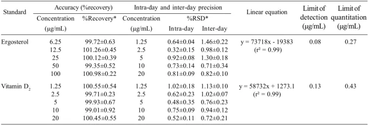
As shown in Table 1, the accuracy (percentage recovery) of ergosterol and vitamin D2 were in the range of
99.35-100.98 and 99.01-100.45, respectively. The precision of this method was assessed based on the percent relative standard deviation (%RSD) and values of inter-day and intra-day precision were lower than 2%. By plotting between the peaks area versus concentration (1.25-100 µg/mL), the linearity of standard ergosterol and vitamin D2 was obtained.
LOD and LOQ values were 0.08 and 0.27 µg/mL and 0.13 and 0.43 µg/mL for ergosterol and vitamin D2, respectively.
3.2 Levels of vitamin D2 and ergosterol in mushroom extracts
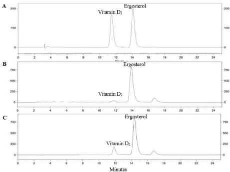
The average yields from two separated extractions of control and irradiated oyster mushroom were 4.23 and 5.66% (w/w), respectively. The chromatographic condition used in this study gave a good resolution for the analysis of vitamin D2 and ergosterol (Figure 1). The retention times of vitamin
D2 and ergosterol were 11.94 and 14.36 min, respectively.
As shown in Table 2, irradiated oyster mushroom extract contained significantly higher level of vitamin D2 than
non-irradiated oyster mushroom extract (p<0.05).
3.3 Antioxidative activity and total phenolic compounds
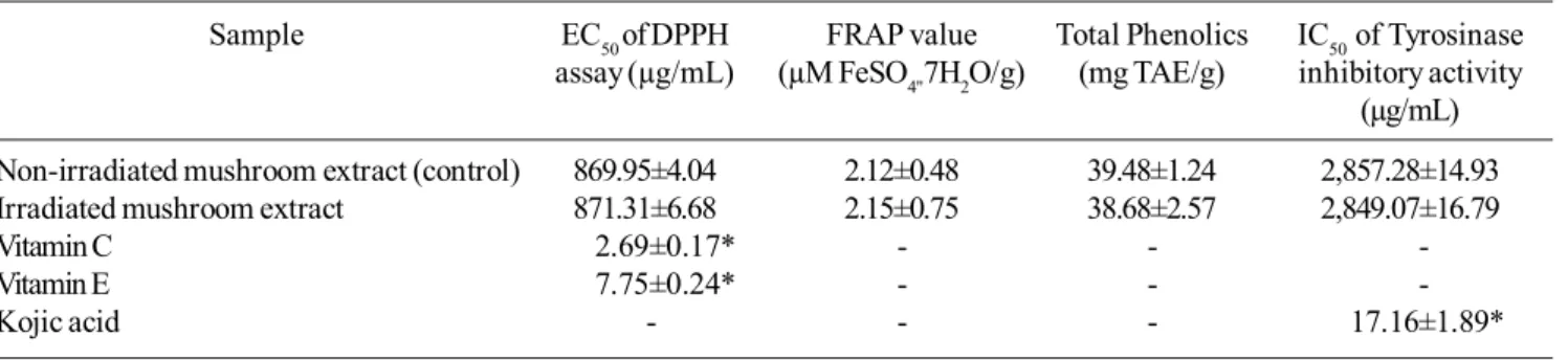
The ethanolic extracts of oyster mushroom were screened for their antioxidative activity by two assays, namely 1,1-diphenyl-2-picrylhydrazyl (DPPH) assay and the ferric reducing ability of plasma (FRAP) or reducing power assay. Oyster mushroom extract showed positive antioxida-tive activity by fading the violet color of DPPH solution to yellow and pale violet. The radical scavenging activities were in direct proportion with the concentrations of the extracts. The inhibition of DPPH radical of vitamin C, vitamin E and mushroom extracts are showed in Figure 2. As the concen-tration of extract increased, the scavenging activity towards DPPH radicals was elevated. The crude extracts of non-irradiated and non-irradiated oyster mushrooms showed similar ability to inhibit free radicals from DPPH at half maximal effective concentration (EC50) of 869.95 and 871.3 µg/mL,
respectively (Table 3). In addition, both oyster mushroom extracts had significantly weak ability of free radical inhibi-tion as compared with vitamin C and vitamin E. For FRAP assay, both mushroom extracts showed similar appreciable reducing power activities. At a concentration of 10 mg/mL, the greatest ability of reducing power inhibition was 2.12 and 2.15 µM FeSO4"7H2O/g for non-irradiated and irradiated
Table 1. Method validation of HPLC analysis of ergosterol and vitamin D2
Accuracy (%recovery) Intra-day and inter-day precision
Standard Linear equation
Concentration %Recovery* Concentration %RSD* (µg/mL) (µg/mL) Intra-day Inter-day
Ergosterol 6.25 99.72±0.63 1.25 0.64±0.04 1.46±0.22 y = 73718x - 19383 0.08 0.27 12.5 101.26±0.45 2.5 0.32±0.15 0.98±0.12 (r² = 0.99)
25 100.12±0.39 5 0.92±0.08 1.30±0.18 50 99.35±0.52 10 0.73±0.14 0.71±0.34 100 100.98±0.22 20 0.81±0.09 0.82±0.10
Vitamin D2 1.25 100.55±0.54 1.25 1.02±0.18 1.13±0.10 y = 58732x + 1273.1 0.13 0.43 2.5 99.71±0.23 2.5 0.62±0.23 1.02±0.07 (r² = 0.99)
5 99.93±0.67 5 0.48±0.35 0.76±0.23 10 99.01±0.92 10 0.75±0.09 0.94±0.12 20 100.45±0.55 20 0.52±0.11 0.72±0.21 *Values are expressed as means ± SD (n=5).
Limit of detection
(µg/mL)
Limit of quantitation
mushroom extracts, respectively. The total phenolic contents of non-irradiated and irradiated mushroom were 39.48 and 38.68 mg TAE/g, respectively.
3.4 Tyrosinase inhibition
At concentrations of 0.125-5.0 mg/mL, the extracts of non-irradiated and irradiated mushrooms moderately inhibited tyrosinase activity in a concentration dependent manner in the range of 10.23-52.28% and 12.77-53.40%, respectively. The half maximal inhibitory concentrations (IC50)
of non-irradiated and irradiated mushroom extracts were 2,857.28 and 2,849.07 µg/mL (Table 3). In contrast, at 0.3-5.0 µg/mL of the positive control, kojic acid showed strong inhibition (5.92-78.23%) with IC50 of 17.16 µg/mL.
3.5 Cytotoxicity test of mushroom extracts
Both non-irradiated and irradiated oyster mushroom extracts were subjected to in vitro cytotoxicity assay in human keratinocytes (HaCaT) and murine melanoma cells (B16F10) (Figure 3). After 24 h incubation, neither oyster
Figure 1. HPLC chromatograms of standard vitamin D2 (ergocalciferol) and ergosterol (A), control oyster mushroom extract (B) and irradiated oyster mushroom extract (C).
Table 2. The levels of ergosterol and vitamin D2 in ethanolic extracts of
oyster mushroom.
Extract Ergosterol (mg/g) Vitamin D2 (µg/g)
Non-irradiated mushroom extract 23.24±1.65 12.25±2.73
Irradiated mushroom extract 19.79±0.82* 153.96±4.86*
Values are expressed as means ± SD (n=3),
* Significant difference from non-irradiated mushroom extract (p<0.05)
mushroom extract (12.5-2,000 µg/mL) was toxic to keratino-cytes cell line and gave high level of cell viability (more than 90%), therefore the half inhibition concentration (IC50) could
not be determined (Figure 4A). Both oyster mushroom extracts had slight toxic effects on melanoma cells with the half inhibition concentrations (IC50) of 1,556.19 and 1,212
µg/mL for non-irradiated and irradiated mushroom extracts respectively (Figure 4B).
4. Discussion
The result of method validation demonstrated that the HPLC method used for analysis of ergosterol and vitamin D2 in this study is suitable with the percentage recovery close
to 100 percent, with values of inter-day and intra-day
pre-cision of lower than 2%. Irradiated mushroom extract had higher vitamin D2 and lower ergosterol values than
non-irradiated mushroom extract. Exposing oyster mushrooms to UV light before extraction with ethanol can increase the vitamin D2 concentration by a multiple of 13 in the mushroom
extract. UV light induced the production of vitamin D2 as it
was observed that the level of vitamin D2 increased, whereas
the level of ergosterol decreased. It is suggested that after irradiation some of ergosterols might be partially converted to vitamin D2 (Jasinghe and Perera, 2005). However, the
conversion of ergosterol to vitamin D2 was rather low of
quantity. Even though ergosterol in mushroom extracts was found in terms of milligrams, the yield of vitamin D2 from this
conversion was only in terms of micrograms. The reason for this low vitamin D2 yield could possibly indicate the
limita-Table 3. DPPH radical scavenging ability, ferric reducing ability (FRAP), total phenolic content and tyrosinase inhibition activity of oyster mushroom extracts.
Sample EC50 of DPPH FRAP value Total Phenolics IC50 of Tyrosinase
assay (µg/mL) (µM FeSO4"7H2O/g) (mg TAE/g) inhibitory activity
(µg/mL)
Non-irradiated mushroom extract (control) 869.95±4.04 2.12±0.48 39.48±1.24 2,857.28±14.93
Irradiated mushroom extract 871.31±6.68 2.15±0.75 38.68±2.57 2,849.07±16.79
Vitamin C 2.69±0.17* - -
-Vitamin E 7.75±0.24* - -
-Kojic acid - - - 17.16±1.89*
Values are expressed as means ± SD (n=3), *Significant differences from control at p<0.05.
Figure 3. Morphology of keratinocytes (A, C, E) and murine melanoma cells (B, D, F) after incubation for 24 h in DMEM (A, B), with 2,000 µg/mL UV-B irradiated oyster mushroom extract (C, D) and 2,000 µg/mL non-irradiated oyster mushroom extract (E, F).
tion of UV penetration into the mushroom tissues as earlier reported that UV-B was able to penetrate through the epider-mal layer not deeper than 50 µm approximately from the surface of the mushroom (Teichmann et al., 2007). Therefore, the further modification of UV irradiation method should be conducted to obtain higher vitamin D2 production.
Oyster mushroom extracts were determined for free radical scavenging efficacy by the DPPH method and reduc-ing power by the ferric reducreduc-ing-antioxidant power method. Moreover, the total phenolic content and tyrosinase inhibi-tory effects of oyster mushroom extracts were investigated. Our result showed the low antioxidative activity of the extracts by both DPPH and FRAP assay. The FRAP values (2.12 and 2.15 µM FeSO4"7H2O/g), were relatively low (<10
µM Fe2+/g) as previously reported by Wong et al. (2006)
(Wong et al, 2006; Ratchadaporn et al, 2008; Ayub et al.,
2010). In overall comparisons of DPPH radical scavenging ability, ferric reducing ability, total phenolic content and tyrosinase inhibitory activity, both non-irradiated and irradiated mushroom extracts gave similar data. This suggests that UV-B irradiation condition did not affect antioxidative activity and tyrosinase inhibitory properties of oyster mushrooms. In contrast, the previous report on Cordyceps militaris by Agnes et al. (2012) demonstrated that pulsed UV light enhanced more vitamin D2 production but lower
antioxidative properties than UV-B irradiation. The loss in antioxidative properties could be attributed to the thermal damage caused by the use of high intensity. Moreover, our results showed that both oyster mushroom extracts had higher antioxidative activity and total phenolic content than the previous studies on this mushroom (Arbaayah and Umi, 2013; Pornariya and Kanok-Orn, 2009). Our results showing moderate tyrosinase inhibitory effect of the mushroom extracts is similar to that of Riani et al. (2012).
The cytotoxicity assay of oyster mushroom extracts on human keratinocytes cells (HaCaT) and murine melanoma cells (B16F10) were measured using MTT assay. Non-irradi-ated and irradiNon-irradi-ated oyster mushroom extracts had nearly the same cytotoxic effect. This result revealed that both mushroom extracts could inhibit growth of melanoma cells without significant effect on keratinocytes. Previous studies demonstrating that several derivatives of vitamin D2, such as
20(OH)D2 and 1,2(OH)2D2 had potent antiproliferative
activity in normal and malignant cells (Slominski et al, 2011) and 1,24(OH)2D2 exerted growth inhibition against breast
cancer (Zinser et al, 2005) indicate that the metabolites of vitamin D2 may play roles in their bioactivities. Our finding
that the increasing of vitamin D2 in the irradiated mushroom
extract did not enhance its cytotoxicity to melanoma cells suggests that further study on the bioactivities of vitamin D2
and its metabolites is necessary.
All the obtained data suggest that UV-B irradiation can induce higher vitamin D2 production without affecting
the mushroom biological activities including antioxidation, tyrosinase inhibition and cytotoxicity. Moreover, the increas-ing of vitamin D2 in mushroom extract did not influence the
biological activities of cytotoxicity. However, further studies should consider identifying related mechanisms and investi-gate other biological activities such as immune modulation and anti-inflammatory activities.
Acknowledgements
This work was supported by the Higher Education Research Promotion and National Research University Project of Thailand, Office of the Higher Education Commission, through the Food and Functional Food Research Cluster, Applied Taxonomic Research Center and Center for Research and Development of Herbal Health Products, Faculty of Pharmaceutical Sciences, Khon Kaen University.
References
Abdurrahman, D., Acay, H. and Yildiz, A. 2009. Effect of using different lignocellulosic wastes for cultivation of
Pleurotus ostreatus (Jacq.) P. Kumm. on mushroom yield, chemical composition and nutritional value. African Journal of Biotechnology. 8, 662-666.
Agnes, P., Arif, R. and Shu Y. 2012. Optimization of pulsed UV light irradiation for the production of vitamin D2,
bioactive metabolites and antioxidant activity of
Cordyceps militaris mycelia. Proceeding of Inter-national Conference on Nutrition and Food Sciences, Singapore, July 23-24, 2012, 39, 76-81.
Andrej, J. and Daniel, S. 2008. Pleurotus ostreatus inhibits proliferation of human breast and colon cancer cells through p53-dependent as well as p53-independent pathway. International Journal of Oncology. 33, 1307-1313.
Ayub, M.A., Devi, I.L., Nayan, V., Chanu, V.K. and Ralte, L. 2010. Antioxidant activity of fruits available in Aizawl market of Mizoram, India. International Journal of Biological and Pharmaceutical Research. 1, 76-81. Arbaayah, H.H. and Umi, K.Y. 2013. Antioxidant properties
in the oyster mushrooms (Pleurotus spp.) and split gill mushroom (Schizophyllum commune) ethanolic extracts. Mycosphere. 4, 661-673.
Benzie, F.F.I and Strain, J.J. 1996. The Ferric Reducing Ability of Plasma (FRAP) as a Measure of “Antioxidant Power”: The FRAP Assay. Analytical Biochemistry. 239, 70-76.
Gajendra, S.J., Punetha, H., Prakash, O., Chandra, M. and Kushwaha, K.P.S. 2014. Study on in vitro antioxidant potential of some cultivated Pleurotus species (Oyster mushroom). Indian Journal of Natural Products and Resources. 5, 56-61.
Gohari, A.R., Hajimehdipoor, H., Saeidnia, S., Ajani, Y. and Hadjiakhoondi, A. 2010. Antioxidant activity of some medicinal species using FRAP assay. Journal of Medicinal Plants Research. 10, 54-60.
Clinical Nutrition. 78, 912-919.
Ilgaz, A., Ergonil, B. and Kalyoncu, F. 2012. Chemical com-position and antioxidant activities of 16 wild edible mushroom species grown in Anatolia. International Journal of Pharmaceutics. 8, 134-138.
Iris, L., Friesemb, D., Gereshc, S., Hadarb, Y. and Schwartz, B. 2006. An aqueous polysaccharide extract from the edible mushroom Pleurotus ostreatus induces anti-proliferative and pro-apoptotic effects on HT-29 colon cancer cells. Cancer Letters. 244, 61–70.
Jasinghe, V.J. and Perera, C.O. 2005. Distribution of ergosterol in different tissue of mushrooms and its effect on the conversion of ergosterol to vitamin D2 by UV
irradia-tion. Food Chemistry. 92, 541-546.
Jasinghe, V.J. and Perera, C.O. 2006. Ultraviolet irradiation: The generator of Vitamin D2 in edible mushrooms.
Food Chemistry. 95, 638-643.
Kim, Y.J. and Uyama, H. 2005. Tyrosinase inhibitors from natural and synthetic sources: structure, inhibition mechanism and perspective for the future. Cellular and Molecular Life Sciences. 62, 1707-1723.
Ko, J.A., Lee, B.H., Lee, J.S. and Park, H.J. 2008. Effect of UV-B exposure on the concentration of vitamin D2 in
sliced shitake mushroom (Lentinus edodes) and white button mushroom (Agricus bisporus). Journal of Agricultural and Food Chemistry. 56, 3671-3674. Koyyalamudi, S.R., Jeong, S.C., Song, C.H., Cho, K.Y and
Pang, G. 2009. Vitamin D2 formation and bioavailability
from Agaricus bisporus button mushrooms treated with ultraviolet irradiation. Journal of Agricultural and Food Chemistry. 58, 3351-3355.
Lloyd, R.S., Joseph, J.K. and Joseph, L.G. 2012. Practical HPLC Method Development, John Wiley and Sons, New York, U.S.A., pp. 685-713.
Mattila, P., Piironen, V., Uusi-Rauva, E., Koivistoinen, P. 1994. Vitamin D contents in edible mushrooms Journal of Agricultural and Food Chemistry. 42, 2449-2453 Mattila, P., Lampi, A.M., Ronkainen, R., Toivo, J. and Piironen,
V. 2002. Sterol and vitamin D2 contents in some wild
and cultivated mushrooms. Food Chemistry. 76, 293-298.
Mau, J.L., Chen, P.R. and Yang, J.H. 1998. Ultraviolet irradia-tion increased vitamin D2 content in edible mushrooms.
Journal of Agricultural and Food Chemistry. 46, 5269-5272.
Momtaz, S., Mapunya, B.M., Houghton, P.J., Edgerly, C., Hussein, A., Naidoo, S. and Lall, N. 2008. Tyrosinase inhibition by extracts and constituents of Sideroxylon inerme L. stem bark, used in South Africa for skin lightening. Journal of Ethnopharmacology. 119, 507-512.
Mosmann, T. 1983. Rapid colorimetric assay for cellular growth and survival: application to proliferation and cytotoxicity assays. Journal of Immunological Methods. 65, 55-63.
Nuhu, A., Yoon, K.N., Lee, K.R., Shin, P.G., Cheong, J.C., Yoo, Y.B., Shim, M.J., Lee, M.W., Lee, U.Y. and Lee, T.S. 2010. Antioxidant activities and tyrosinase inhibitory effects of different extracts from Pleurotus ostreatus
fruiting bodies. Mycobiology. 38, 295-301.
Nuhu, A., Yoon, K.N. and Lee, T.S. 2011. Evaluation of the antioxidant and antityrosinase activities of three extracts from Pleurotus nebrodensis fruiting bodies. African Journal of Biotechnology. 10, 2978-2986. Pornariya, C. and Kanok-Orn, I. 2009. Amino acids and
anti-oxidant properties of the oyster mushrooms, Pleurotus ostreatus and Pleurotus sajorcaju. Science Asia. 35, 326–331.
Ratchadaporn, O., Mario, G.F. and Suwayd, N. 2008. Anti-oxidant activity and cytotoxicity of Rang Chuet (Thunbergia laurifolia Lindl.) extracts. Journal of Agricultural and Food Industrial Organization. 1, 116-128.
Riani, H., Elya, B. and Amin, J. 2012. Formulation and evalua-tion of antioxidant and tyrosinase inhibitory effect from gel containing the 70% ethanol Pleurotus ostreatue extract. Journal of Medicinal and Aromatic Plants. 2, 135-140.
Rivera, J.R., Piedra, C.D.L., Ramos, A., Ortiz, A. and Egido, J. 2010. The expanding spectrum of biological actions of vitamin D, Nephrology Dialysis Transplantation. 25, 2850-2865.
Seema, P. and Goyal, A. 2012. Recent developments in mushrooms as anti-cancer therapeutics: a review. Journal of Biotechnology. 2, 1–15.
Shimada, K., Fujikawa, K., Yahara, K. and Nakamura T. 1992. Antioxidative properties of xanthone on the auto oxidation of soybean in cylcodextrin emulsion. Journal of Agricultural and Food Chemistry. 40, 945-948. Singleton, V.L., Orthofer, R. and Lamuela, R.R.M. 1999.
Analy-sis of total phenols and other oxidation substrates and antioxidants by means of FolinCiocalteu reagent. Methods of Enzymology. 299, 152-178.
Slominski, T.A, Kim, T., Janjetovic, Z., Tuckey, C.R., Bieniek, R., Yue, J., Li, W., Chen, J., Nguyen, N.M., Tang, K.Y.E, Miller, D., Chen, C.T. and Holick, M. 2011. 20-Hydroxyvitamin D2 is a noncalcemic analog
of vitamin D with potent antiproliferative and pro-differentiation activities in normal and malignant cells. American Journal of Physiology Cell Physiolology. 3, 526-541.
Sripanidkulchai, B. and Junlatat, J. 2014. Bioactivities of alcohol based extracts of Phyllanthus emblica
branches: antioxidantion, antimelanogenesis and anti-inflammation. Journal of Natural Medicines. 68, 615-622.
Teichmann, A., Dutta, C.P., Staffas, A. and Jagerstad, M. 2007. Sterol and vitamin D2 concentration and wild grown
Wong, C.C., Li, H.B., Cheng, K.W. and Chen, F. 2006. A systematic survey of antioxidant activity of 30 Chinese medicinal plants using the ferric reducing antioxidant power assay. Food Chemistry. 97, 705-711.
Zinser, G.M., Tribble, E., Valrance, M., Urben, C.M., Knutson, J.C., Mazess, R.B., Strugnell, S.A. and Welsh, J. 2005. 1,24(S)-dihydroxyvitamin D2, an endogenous vitamin
D2 metabolite, inhibits growth of breast cancer cells