Expression—A Potential Anti-Tumor Mechanism
Cecilia Magnusson1, Astrid M. Bengtsson1, Minghui Liu1, Jian Liu1, Yvonne Ceder3, Roy Ehrnstro¨m2, Anita Sjo¨lander1*
1Cell and Experimental Pathology, Department of Laboratory Medicine, Lund University, Ska˚nes University Hospital, Malmo¨, Sweden,2Pathology, Department of Laboratory Medicine, Lund University, Ska˚nes University Hospital, Malmo¨, Sweden,3Clinical Chemistry, Department of Laboratory Medicine, Lund University, Ska˚nes University Hospital, Malmo¨, Sweden
Abstract
Background:The cysteinyl leukotrienes receptors (CysLTRs) are implicated in many different pathological conditions, such as inflammation and cancer. We have previously shown that colon cancer patients with high CysLT1R and low CysLT2R expression demonstrate poor prognosis. Therefore, we wanted to investigate ways for the transcriptional regulation of CysLT2R, which still remains to be poorly understood.
Methodology/Principal Findings:We investigated the potential role of the anti-tumorigenic interferona(IFN-a) and the mitogenic epidermal growth factor (EGF) onCysLT2Rregulation using non-transformed intestinal epithelial cell lines and
colon cancer cells to elucidate the effects on the CysLT2R expression and regulation. This was done using Western blot, qPCR, luciferase reporter assay and a colon cancer patient array. We found a binding site for the transcription factor IRF-7 in the putative promoter region of CysLT2R. This site was involved in the IFN-a induced activity of theCysLT2R luciferase
reporter assay. In addition, IFN-a induced the activity of the differentiation marker alkaline phosphatase along with the expression of mucin-2, which protects the epithelial layer from damage. Interestingly, EGF suppressed both the expression and promoter activity of the CysLT2R. E-boxes present in the CysLT2R putative promoter region were involved in the
suppressing effect. CysLT2R signaling was able to suppress cell migration that was induced by EGF signaling.
Conclusions/Significance:The patient array showed that aggressive tumors generally expressed less IFN-a receptor and more EGFR. Interestingly, there was a negative correlation between CysLT2R and EGFR expression. Our data strengthens the idea that there is a protective role against tumor progression for CysLT2R and that it highlights new possibilities to regulate theCysLT2R.
Citation:Magnusson C, Bengtsson AM, Liu M, Liu J, Ceder Y, et al. (2011) Regulation of Cysteinyl Leukotriene Receptor 2 Expression—A Potential Anti-Tumor Mechanism. PLoS ONE 6(12): e29060. doi:10.1371/journal.pone.0029060
Editor:Patricia T. Bozza, Fundac¸a˜o Oswaldo Cruz, Brazil
ReceivedJune 14, 2011;AcceptedNovember 20, 2011;PublishedDecember 15, 2011
Copyright:ß2011 Magnusson et al. This is an open-access article distributed under the terms of the Creative Commons Attribution License, which permits unrestricted use, distribution, and reproduction in any medium, provided the original author and source are credited.
Funding:This work was supported by grants for AS from the Swedish Cancer Foundation (www.cancerfonden.se), the Swedish Medical Research Council (www. vr.se), the Foundations at Skanes University Hospital, Gunnar Nilsson Foundation (www.cancerstiftelsen.com), the O¨sterlund Foundation (www.medmicro.se/ stiftelse.htm), and for CM and AMB from the Royal Physiographic Society in Lund (www.fysiografen.se). The funders had no role in study design, data collection and analysis, decision to publish, or preparation of the manuscript.
Competing Interests:The authors have declared that no competing interests exist. * E-mail: anita.sjolander@med.lu.se
Introduction
Prolonged inflammation is known to increase the risk of developing cancer [1,2]. It has been estimated that 15% of all cancers can be accredited to infectious agents [3]. Tumor microenvironment has often been associated with infiltrating leukocytes in the tumor tissue and the surrounding stroma [4]. Moreover, patients with ulcerative colitis display a 30 to 50% increased risk of developing colon cancer [5]. Elevated levels of inflammatory mediators, such as the cysteinyl leukotrienes (cysLTs), have been found in these patients [6]. CysLTs is the collective name for LTC4, LTD4 and LTE4. They are lipid mediators derived from arachidonic acid through the 5-lipoox-ygenase (5-LO) pathway [7]. In addition to inflammatory bowel disease (IBD) [7], they have been implicated in the pathogenesis of several chronic inflammatory diseases, such as asthma, pulmonary fibrosis, and atherosclerosis [8,9]. The known biological effects of
the differentiation of Caco-2 cells [17] and is involved in vascular permeability [19]. This indicates that CysLT2R has a more protective role in cancer development while CysLT1R favors tumor progression.
Interferons (IFNs) are part of our defense system against viruses, bacteria, parasites, and malignant cells [20]. Both type I (for example IFN-a and IFN-b), and type II (IFN-c) IFNs are produced during the early innate immune response. However, while IFN-cis secreted mainly by natural killer cells and T-cells, the type I IFNs are produced by most cell types following virus infection or toll-like receptor activation [21]. IFN activation results in the increased activity of the transcription factor interferon regulatory factor 7 (IRF-7), which is involved in a positive feedback loop to augment a larger induction of IFN-aand IFN-b
genes [22]. Even though both types of IFNs have been implicated to have anti-carcinogenic effects, the type I IFNs exhibit stronger anti-proliferative and anti-angiogenic effects and have been proposed to have apoptotic effects on cancer cells [23,24]. In a mouse model, the type I IFNs have also been implicated to have anti-inflammatory functions in colitis [25]. In addition, they have demonstrated some effectiveness in the treatment of IBD patients [26]. We located a predicted IRF-7 binding site in the putative promoter region of theCysLT2R, which suggests that theCysLT2R promoter might be regulated by the IFN-a. This is interesting given that the anti-proliferative and anti-tumorigenic effects attributed to the IFN-a are consistent with the observed characteristics of the CysLT2R function [17]. IFN-chas previously been reported to increase the expression of CysLT2R mRNA and protein in eosinophils [27]. In addition it enhances the cysLT-induced inflammatory response of primary endothelial cells [28].
Epidermal growth factor (EGF) signaling via the EGFR is a known inducer of cell proliferation and tumor cell invasion [29,30]. Examining the putative promoter region for the CysLT2R, we located four conserved E-box elements (consensus sequence CANNTG). EGF signaling has been seen to induce transcriptional repression through E-boxes [31].
In this study, we found novel regulation factors for theCysLT2R. The anti-tumorigenic cytokine IFN-a is able to increase the transcription of theCysLT2Rwhile EGF, a known inducer of cell proliferation and migration, suppresses the CysLT2R expression. This is consistent with the proposed anti-tumorigenic role for CysLT2R in colon cancer.
Materials and Methods
Chemicals
The rabbit polyclonal anti-human CysLT2R antibody (diluted 1:1000) was purchased from Innovagen (Lund, Sweden). The ligand LTC4and Montelukast were from Cayman Chemicals Co. (Ann Arbor, MI). The mouse anti-actin (diluted 1:2000) and goat anti-IFNa/bR1 antibodies (diluted 1:100, immunohistochemistry) were purchased from Sigma Chemicals Co. (St. Louis). The IFNaR1 antibody (diluted 1:1000) was from Novus Biologicals (Litteleton, CO). The EGFR (diluted 1:10, immunohistochemistry) was purchased from Zymed Laboratories Inc. (San Francisco, CA). EGFR (diluted 1:800, Western blot) while the pEGFR (diluted 1:1000) antibodies were from BD Transduction Labora-tories (Erembodegem, Belgium). The total Snal 1 antibody (diluted 1:500) was from Proteintech Group Inc. (Chicago, IL) and the pSnail antibody (diluted 1:500) was from AbCam (Cambridge, UK). The secondary peroxidase linked goat rabbit and anti-mouse antibodies (diluted 1:5000) were from Dako (Glostrup, Denmark). Methyl-3H-thymidine, the enhanced chemilumines-cence (ECL) reagents and the Western blot detection reagents as
well as the hyperfilm were from Amersham International (Buckinghamshire, UK). TaqMan primers and master mix for real-time PCR were purchased from Applied Biosystems (Cam-bridge, UK). The RNeasy Plus Mini kit was from Qiagen GmbH (Hilden, Germany). Unless otherwise stated, all other reagents were of analytical grade and purchased from Sigma Chemicals Co. (St. Louis, MO) or from ICN (Temecula, CA).
Cell Culture
The non-transformed human intestinal epithelial cell line (Int 407) [32], which exhibits typical epithelial morphology and growth, was cultured as a monolayer for approximately five days in Eagle’s basal medium supplemented with 15% newborn calf serum. The two colon cancer cell lines, Caco-2 (DSMZ No: ACC 169) and SW 480 (DSMZ No: ACC 313), were respectively grown in Dulbecco’s modified Eagle medium and RPMI 1640 supple-mented with 10% and 20% fetal bovine serum respectively. All media was supplemented with 2 mM L-glutamine, 55 IU/mL penicillin and 55mg/mL streptomycin. The cell lines were cultured at 37uC in a humidified atmosphere with 5% CO2. The cells were regularly tested to ensure the absence of mycoplasma contamination.
Cell Lysates
Cells were washed, scraped with ice-cold 16PBS, and centrifuged for 5 min at 1000 g. The cell pellets were lysed with lysis buffer [33] for 30 min on ice, homogenized 10 times with a 20G needle, and centrifuged for 5 min at 500 g.
Gel Electrophoresis and Immunoblotting
To ensure equal loading, all samples were evaluated and compensated for protein concentration using the Coomassie blue protein assay. Proteins were denatured by boiling in sample buffer for 5 min [34]. The samples were subjected to electrophoresis on 8% or 10% polyacrylamide gels in the presence of 10% SDS. The immunoblotting and developing were performed as described in Nielsenet al.[34].
Real-Time PCR Analysis
Cells were washed in PBS and immediately frozen at280uC. Thereafter, they were scraped in lysis buffer provided in the kit and homogenized 10 times with a 20G needle. RNA was purified on RNeasy MinElute Spin Columns and dissolved in RNase free H2O. cDNA synthesis was performed using RevertAid H Minus M-MuLV reverse transcriptase (Fermentas Life Sciences). The mRNA expression levels of CysLT2R, CysLT1R, mucin-2 and the endogenous housekeeping gene, hypoxanthine phosphoribosyl-transferase 1 (HPRT-1), were quantified using real-time PCR analysis (TaqMan Chemistry). cDNA was mixed with 0.9mM TaqMan primers and master mix and amplified at 60uC in an Mx3005P thermocycler (Stratagene). The following primers were used: CysLT2R (Hs00252658_s1), CysLT1R (Hs00929113_m1), mucin-2 (Hs00159374_m1), and HPRT-1 (Hs99999909_m1). The samples were analyzed and normalized against HPRT-1 using the MxPro software (Stratagene).
Plasmid Constructs
forward 59-ACATCAGGCAGCATTAATGT-39and reverse 59 -GGACATAAATTTTCTCTCCAT-39, followed. This was fol-lowed by a PCR that use the following primers; forward 59 -TTATGAGCTCGTTTCAAAACATTAAATG TAAC-39 and reverse 59-TTCTAAGCTTGCTGGGTTAAAAAGAAAC-39. All PCR products were gel purified using the QIAquick gel extraction kit (Qiagen). They were digested with restriction enzymes and ligated into the pGL3-enhancer reporter vector using the Quick ligation kit (New England BioLabs). The plasmids were transformed into SoloPack Gold super competent cells (Stratagene). To produce the clone with the deleted IRF-7 binding site (construct II), the site directed mutagenesis with the following primers were used: forward 59 -ATGGCTATTCTACATT-CAAAAATTATGAAATGTAATGCAGCATGT-39 and reverse 59 -ACATGCTGCATTACATTTCATAATTTTTGAATGTA-GAATAGCCAT-39. The Quickchange XL kit (Stratagene) was used according to standard protocol. Three deletion constructs were produced by digesting construct I with restriction enzymes. Construct III was digested with SacI and SpeI to produce a construct from 21 to 2412. Construct IV was digested with
HindIIIandSpeIto remove the downstream part of the promoter, thereby creating a construct between sites21012 and2413 of the CysLT2R promoter. Blunt end digestion was performed using T4 DNA polymerase (Promega) and the vector was ligated back together. The promoter construct V was digested with SacIand
SmiI to give a construct from 21 to 2187. All plasmids were extracted using Endofree plasmid maxi kit (Qiagen) and were sequenced accordingly.
Transient Transfections and Measurement of Luciferase Activity
Transient transfections of Int 407 cells were carried out using Lipofectamine LTX and Plus Reagent (Invitrogen) in serum free medium according to the manufacturer’s instructions. The final DNA concentration used for transfections was 1mg/ml for all plasmids except for the luciferase transfection control vectorRenilla
(0.2mg/ml). The transfections were carried out for 4–6 h in 37uC, after which the medium was changed to 15% serum containing the medium. Forty-eight hours after transfection, the cells were stimulated with IFN-a (500–2000 U/ml) or EGF (100 ng/ml) in serum free medium for 24 h. The cells were lysed with passive lysis buffer (Promega) and stored in 280uC. Thawed lysates were centrifuged for 5 min at 1000 g and analyzed using the Dual Luciferase Reporter Assay System from Promega, following the manufacturer’s instructions.
Electromobility Shift Assay (EMSA)
Nuclear extracts were prepared from Int 407 cell treated with IFN-a1000 U/ml or EGF 100 ng/ml for 24 h using the Nuclear Extraction Kit (Chemicon International). The process was per-formed according to the manufacturers’ instructions. In additional, 10 mM 1-Naphthyl phosphate monosodium salt monohydrate, a broad phosphatase inhibitor, (Sigma Aldrich) was added into both cytoplasmic and nuclear lysis buffer before use. Biotin 39 end-labeled single-stranded oligonucleotide corresponding to the IRF-7 binding region ofCysLT2Rputative promoter (sense: 59-AAT CAG GAA ATT TAA ATT TAT TAT-39and antisense: 59-ATA ATA AAT TTA AAT TTC CTG ATT-39) or E-box binding site (sense: 59-TTC TTT CAG CAT TTG AGA AAT GTG-39and antisense 59-CAC ATT TCT CAA ATG CTG AAA GAA-39) were purchased from TAG Copenhagen A/S. DNA probes were then annealed in buffer containing 10 mM Tris, 1 mM EDTA, and 50 mM NaCl (pH 8.0) by incubating the oligonucleotides at 95uC for 5 min then gradually reducing the heat until the DNA reached
room temperature. Binding reactions for EMSA were prepared using the LightShift Chemiluminescent EMSA kit (Pierce Thermo Scientific). The binding buffer for IRF-7 contained 10 mM Tris-HCl (pH 7.5), 1 mM EDTA, 50 mM NaCl, 2 mM dithiothreitol, 5% glycerol, 0.5% NP-40, and 10mg/ml BSA. To reduce nonspecific binding, 62.5mg of poly(dI-dC) was added per ml. Each reaction contains 20mg nuclear extract. After 20 min of incubation with the probe at room temperature, extracts were loaded on a 5% polyacrylamide gel. After 1 h at 100 V, the gel was transferred onto a positive charged Nylon Membrane, (Thermo scientific) and detected according to kit instruction. The binding buffer for E-box contained 20 mM HEPES, pH 7.6, 150 mM KCl, 3 mM MgCl2, 10% glycerol, 0.2 mM ZnSO4, 0.3 mg/ml BSA) and 1mg/20mL of poly(dI:dC). The binding reaction was performed on ice for 30 min before being loaded onto the gel. To demonstrate the specificity of protein-DNA complex formation, a 1,000-fold molar excess of unlabeled oligonucleotides or IRF-7 (G-8) antibody (Santa Cruz) or SNAI 1 (E-18) antibody (Santa Cruz) was added to the binding reaction mixtures before the probe.
Alkaline Phosphatase Activity
The assay was performed as previously described [35]. Briefly, alkaline phosphatase activity was measured using disodium p-nitrophenyl phosphate as substrate. Caco-2 cells were seeded in Petri dishes and incubated for 24 h at 37uC. Five hundred to 2000 U/ml of IFN-aand/or 40 nM of LTC4was added to the cells and subsequently incubated for 72 h at 37uC. New LTC4 was added every 24 h. Sodium butyrate (2 mM) was used as a positive control (data not shown). The alkaline phosphatase activity was estimated by measuring the level of p-nitrophenol at 405 nm.
Thymidine Incorporation Assay
Caco-2 cells were grown in flat-bottomed 96 well plates for five days. The medium was changed to serum-free 2 h prior to stimulation. Cells were stimulated with LTC4 (40 nM), IFN-a (1000 U/ml) or EGF (100 ng/ml) for 48 h. DNA synthesis was assessed by adding 0.5mCi of [3H] thymidine during the final 18 h of stimulation. The wells were washed once with PBS and incubated with 0.05% trypsin-EDTA solution/well for 5 min at 37uC. Cells were harvested and collected on a filter paper in a Perkin Elmer harvester. The filter paper was dried and [3H] thymidine incorporation was measured in a 1450 Microbeta Trilux liquid scintillation counter (Wallac).
Cell Migration Assay
Cell migration was analyzed in a modified Boyden chamber, which consisted of two chambers separated by a polycarbonate PVDF membrane (pore size of 8mm) that was covered with a collagen I gel (3 mg/ml). Int 407 cells were grown for 18 h in the presence or absence of LTC4(40 nM) and/or EGF (100 ng/ml) in medium containing 1.5% serum. Fifteen percent serum was used as a positive control. Cells were added to the upper well and allowed to migrate into the chamber for 18 h at 37uC. The migrated cells were fixed in 4% paraformaldehyde and the membranes were stained with a 1% crystal violet/10% in a methanol solution. The membranes were washed in PBS and the remaining dye was removed with 10% SDS. The absorbance was measured at 590 nm.
Patient Samples
collected in 1990 were obtained from the biobanks of Malmo¨ University Hospital. Tissues from 78 patients with varying grades and stages of the disease were included. Grading of the tumors was performed using Dukes’ classification [36]. The matched control samples of normal colon tissue in this investigation were taken from the borders of the surgical specimens.
Ethics Statement
This study was performed after ethical permission from the Regional Ethical Review Board, Lund University # 367/2005. Archival tissue specimens from 78 colon cancer patients that were operated between 1990 and 1991 were used in the present study. Since the samples were old and taken from different parts of the region, it was not possible to obtain written consent from each patient. Detailed information describing the study and tissue microarray (TMA) construction was published in 2006 in a daily newspaper and patients were offered to contact us by mail or by phone if they had any objections. None of the 78 patients objected. This procedure was performed following strict guidelines from the Regional Ethical Review Board in Lund who approved the procedure.
Tissue Arrays and Immunohistochemistry
For histological assessment, the archival paraffin-embedded colorectal cancer and normal mucosa specimens were used and prepared as previously described [16]. The array was stained with anti-IFNa/bR1, EGFR and CysLT2R antibodies. Secondary peroxidase antibodies used were from Dako. After immunostain-ing, all slides were counterstained with Mayer’s hematoxylin.
Statistical Analyses
Statistical significance was determined as P,0.05 by two-tailed Student’s t test or one-way ANOVA. Pearson’s correlation test was used when comparing different sets of immunohistochemistry staining. SPSS software 16,0 (SPSS, Inc.) was used for the patient material analysis.
Results
An IRF-7 Binding Site and Four E-boxes Have Been Found to be Present in theCysLT2RPromoter Region
To increase the understanding of the role of cysteinyl leukotrienes in inflammation-induced colorectal carcinogenesis, we examined possible ways by which the expression of CysLT2R can be regulated, with one of the receptors generating the effects of leukotrienes. The promoter region of CysLT2R contains several interesting binding sites for known transcription factors and transcriptional repressors. Previous reports from our laboratory have shown that CysLT2R is down regulated in colon cancer [17,37] and that it is involved in colon cancer cell differentiation [17] as well as in decreased cell migration in breast cancer cells [18]. Therefore, it was of particular interest that a putative binding element for IRF-7 and four E-box elements were present in the promoter region of the CysLT2R. Five constructs of the CysLT2R promoter region were created and ligated into a pGL3-enhancer reporter vector to evaluate the importance of IRF-7 and the E-boxes for activation/transcription of CysLT2R (Fig. 1A). IFN-a can be used to activate the transcription factor IRF-7. However, the presence of the IFN-areceptor IFN-aR1 is needed to do this. The presence of the IFN-aR1 is confirmed in the intestinal cell lines intended for this study (Fig. 1B). Likewise, the expression of the EGFR (Fig. 1C) and the CysLT2R (Fig. 1D) was verified.
CysLT2RPromoter Activity is Activated by the IFN-avia an IRF-7 Binding Site and It is Repressed by the EGF Through the E-boxes Present in the Putative Promoter Region
We have seen that CysLT2R expressions were altered in patient’s tumor material, which might be due to a changed ‘‘microenvironment around the tumor’’. Therefore, we decided to search for a Th1 regulatory cytokine motif in the promoter region of theCysLT2R. The presence of a putative IRF-7 binding site in the CysLT2R promoter region indicated that the Th1 pro-inflammatory cytokine IFN-aought to induceCysLT2Rpromoter activity and transcription of the CysLT2R encoding gene. A dilution series with IFN-a showed that 1000 U/ml IFN-a
generated an optimal activation of CysLT2R (Fig. 2A). To elucidate whether IFN-a induces the CysLT2R through IRF-7 binding to its putative binding site in the promoter region, the promoter activity for deletion mutant constructs I-V were tested (Fig. 2B). IFN-ainduced a significant two-fold activation of the
CysLT2R in the non-transformed cell line Int 407 (construct I). This activation was partly reduced in the DIRF-7 mutant (construct II). IFN-awas unable to induce theCysLT2Rpromoter activity in constructs IV and V, which both lacked full-length IRF-7 response elements. However, a significant reduction in activation compared to wild-type construct I was also observed for construct III, which contains an intact IRF-7 response element but lacks the part upstream of bp2412. Taken together, this indicates that the IFN-a-induced activation of theCysLT2R requires the IRF-7 binding site for complete activation, but also needs additional unidentified binding elements located up-stream of the IRF-7 site for full activation.
Further examination of the CysLT2R promoter sequence revealed the presence of four conserved E-box elements. EGF signaling can induce repressor elements that bind to mentioned sequences. Four of the CysLT2R luciferase reporter constructs were used to investigate the effects of EGF on theCysLT2R. We found that EGF suppressedCysLT2Rluciferase reporter activity (Fig. 2C). The importance of the E-boxes was investigated by testing the ability of the EGF to suppress CysLT2R promoter activity in three deletion constructs, (construct III, with only E-box 4 present, construct IV containing three E-E-boxes, and construct V without any E-boxes). The EGF-induced reduction of
CysLT2R promoter activity significantly declined with a decreas-ing number of E-boxes present in the putative promoter region. There was a statistically significant difference between the EGF-induced reductions of construct IV and construct V. However, the reduction is probably mediated both through an E-box-dependent as well as an E-box-inE-box-dependent pathway in the intestinal cells.
Analysis of IRF-7 and E-box DNA binding activities on the
CysLT2Rputative promoter region in Int 407 cells were performed to compliment the luciferase experiments (Fig. 2D). The EMSA results showed that IRF-7 containing complexes were detected in both untreated and IFN-a treated cells. We found an increased association upon the stimulation of the complexes. The addition of cold probe and anti-IRF-7 antibody competed with IRF-7 in binding the IRF-7 binding site on the promoter region. The addition of cold probe could completely inhibit probe-protein complex formation. Likewise, Snail-containing complexes were detected in both untreated and EGF treated cells. We found increased association upon stimulation of the complex. The addition of cold probe could completely inhibit the formation of probe-protein complex. Moreover, additional anti-Snail antibody reduced the probe-protein complex formation.
activity also affected CysLT2R mRNA and protein expression. Consistent with the results from the gene activation assay, the
IFN-asignificantly up-regulated both the CysLT2R mRNA (peaking at 12 h) and the protein (maximum increase at 24 h) (Fig. 2E). The EGF induced a significant suppression of both the CysLT2R mRNA and the protein expression (Fig. 2F).
EGFR and Snail Activation is Involved in EGF Repressing
CysLT2R
Stimulation with EGF leads to the activation of the EGFR (Fig. 3A) and to the activation of Snail (Fig. 3B), a transcriptional repressor known to bind to E-box elements. It is likely that the EGF-induced suppression of the CysLT2R is generated by Snail that binds to the E-box elements present in the promoter region of theCysLT2R. Interestingly, an induction of CysLT1R mRNA is observed after EGF treatment of intestinal cells at the same time as CysLT2R mRNA is suppressed (Fig. 3C), indicating, as earlier suggested, opposing effects of the CysLTRs [37].
IFN-aCan Induce Differentiation of Caco-2 Cells
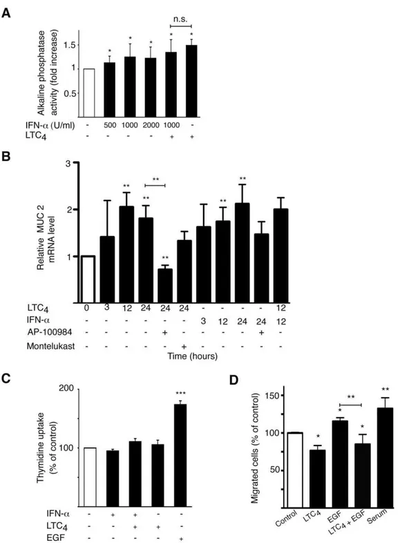
Sodium butyrate is a known inducer of differentiation in Caco-2 cells [38], which is frequently used as a model system for differentiation in cell culture [39]. The activation of different luminal brush border proteins, such as alkaline phosphatase, can be used as a measurement for epithelial cell differentiation. Previously, we have shown that the CysLT2R ligand LTC4is able to induce differentiation in Caco-2 cells [17]. IFN-a induced a small but significant increase in the activation of alkaline phosphatase, indicating increased differentiation of Caco-2 cells. IFN-a had, however, no additive effect to the alkaline phosphatase activity induced by LTC4 (Fig. 4A). To confirm the effect of alkaline phosphatase activity, an additional differentiation assay was used. Mucin-2 is a marker for intestinal cell differentiation [40]. Both LTC4 and IFN-a could significantly induce mucin-2 mRNA in Caco-2 cells (Fig. 4B). The CysLT2R antagonist AP-100984 could significantly block both LTC4induction of mucin-2, which was not seen with the specific CysLT1R antagonist Montelukast. This strengthens the hypothesis that CysLT2R might have a protective role in colon cancer and in normal intestinal mucosa. We have previously shown that LTC4does not induce proliferation in Caco-2 cells [17]. Here, we show that IFN-adoes not induce a proliferative response either. EGF, on the other hand, is a known mitogen inducer [41] and was used here as a positive control (Fig. 4C). This corresponds well to the previous reported roles of the CysLTRs.
We next investigated the effect of CysLT2R signaling on Int 407 cell migration. We found that LTC4-induced activation of CysLT2R reduced Int 407 cell migration (Fig. 4D). Furthermore, we also found that EGF-induced Int 407 cell migration could be suppressed by CysLT2R activation (Fig. 4D).
EGFR Inversely Correlates with CysLT2R in Colorectal Cancer
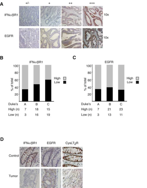
Next, we wanted to clarify if the levels of IFNa/bR1 and EGFR correlated with the CysLT2R pattern in a colon cancer patient array. The TMA contains material from 78 colon cancer patients and has previously been thoroughly described [16]. Representative stainings from the array for IFNa/bR1 and EGFR are shown in Fig. 5A. We have previously shown that CysLT2R is down-regulated in colon tumor tissue in patients from this array and that more aggressive tumors expressed less of the CysLT2R [17]. We observed that patients with more aggressive tumors generally had less expression of IFNa/bR1 while patients with less aggressive tumors generally exhibited a higher expression of IFNa/bR1. A more distinct trend was visualized when the patients were divided into subgroups with high (++, +++) or low (+/2, +) IFNa/bR1 staining. Duke’s A patients had a higher percentage of increased IFNa/bR1 staining than Duke’s B and Duke’s C patients (Fig. 5B). The expression of the EGFR was generally up-regulated in colon cancer patients in this study, which is coherent with earlier published studies [42] (Fig. 5C). Representative pictures from a Figure 1. There is an IRF-7 binding site and four E-boxes
present in theCysLT2Rputative promoter region.(A) Schematic
visualization of theCysLT2Rpromoter. Construct I contains the wildtype
promoter region (1012 bp) and constructs II–V are various deletion constructs. Representative Western blots from three different experi-ments with the intestinal epithelial cell line Int 407 and the colon cancer cell lines Caco-2 and SW 480 were analyzed as follows. (B) The blot shows the levels of IFNaR1, and A431 cells were used as a negative control while HCT116 cells were used as a positive control. (C) The blot shows the levels of EGFR protein expression, A431 cells were used as a positive control and MCF7 cells were used as a negative control, and (D) the blot shows the levels of CysLT2R protein expression.
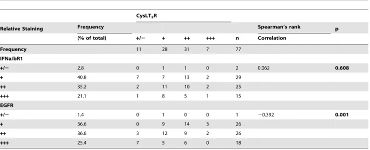
Duke’s C colon cancer patient are shown in Fig. 5D. They exhibit high expression of CysLT2R and IFNa/bR1 in control tissue and decreased expression in the tumor. Conversely, EGFR expression was enhanced in the tumor material. A statistical significant negative correlation could be observed in the expression levels between CysLT2R and EGFR in this tissue array. Although IFNa/bR1 and CysLT2R had similar expression patterns in relation to cancer staging, no significant correlation between the expression levels of these two receptors could be found (Table 1).
Discussion
Several different ways of regulating CysLT2R have previously been reported. For example, LTD4 can up-regulate the mRNA expression of CysLT2R in monocytes [43] and epithelial cells [44] and the cytokines IL-4, IL-8 and IFN-c[28,45,46] can up-regulate the mRNA expression of CysLT2R in several different cell types.
Since a binding site for IRF-7 was present in the putative promoter region of theCysLT2R, we hypothesized that the IFN-awould be able to induce CysLT2R expression via transcription factor IRF-7. The ability of the IFN-ato induce anti-proliferative and anti-tumor progression responses [24,47] made it an interesting candidate to study in relation to CysLT2R. IFNs have been reported to mediate their anti-tumor effects by altering immune responses, such as the suppression of cytokine IL-1 [48] and inducing TRAIL [49], and by inducing anti-angiogenic responses by inhibiting VEGF [50]. We show that the IFN-ainduced a two-fold activation of the wild-type
CysLT2R promoter and increased expression in the CysLT2R mRNA and protein. How the IFN-amediates its effects, however, is not completely elucidated. The IRF-7 binding site is important and required for the optimal induction of the CysLT2R promoter by the IFN-a, but it is not the only crucial activation site. Additional transcription factors are probably engaged as well. We discovered that the promoter region of theCysLT2Rcontains four conserved E-Figure 2. IFN-ainducesCysLT2Rluciferase reporter activity whereas EGF suppresses it.(A)CysLT2Rluciferase activity following transient
transfection with pGL-3-CysLT2R construct in Int 407 cells, with or without treatment with 500–2000 U/ml IFN-afor 24 h. (B) Int 407 cells transfected with wildtypeCysLT2Rpromoter orCysLT2Rpromoter deletion constructs. Cells were grown in the presence of IFN-a(1000 U/ml) or in the presence of
EGF (100 ng/ml) for 24 h (C). In brief, followed by stimulation with IFN-aor EGF for 24 h and measurement of luciferase activity (relative luciferase units). Results shown are the mean6SD of at least three independent experiments performed in triplicate. (D) Analysis of IRF-7 and E-box DNA binding activities by EMSA onCysLT2Rpromoter region in Int 407 cells. Modulation of IRF-7 DNA binding in cells stimulated with IFN-a(1000 U/ml).
For competition assays, a 1000-fold molar excess of unlabeled oligonucleotides (Lane 4) or 10mg anti-IRF-7 antibody (Lane 5) was added before
addition of probe to the binding reactions, (left panel). Effect on E-box DNA binding in cells stimulated with 100 ng/ml EGF for 24 h. A 1000-fold molar excess of unlabeled oligonucleotides (Lane 4), or 10mg anti-Snail antibody was added before addition of probe to the binding reactions (right
panel, lane 5). (E) Real-time PCR quantification of mRNA expression of CysLT2R and immunoblot analysis of CysLT2R protein following treatment with or without 1000 U/ml IFN-a. (F) Real-time PCR quantification of mRNA expression of CysLT2R and immunoblot analysis of CysLT2R protein with or without treatment with EGF (100 ng/ml). The results are shown as means6SD of at between three to teen separate experiments; *, P,0.05; **, P,0.01; ***, P,0.001 for one-way ANOVA or two tailed Student’s t-test.
box elements. Interestingly, the mitogenic EGF suppressed both CysLT2R mRNA and protein expression as well as the promoter activity, strengthening the hypothesis that the CysLT2R is a potential tumor suppressor. To validate the importance of the E-boxes, CysLT2R promoter deletion constructs were used. EGF stimulation significantly decreased theCysLT2Rpromoter activity in all promoter constructs, but to a significantly reduced capacity in the deletion constructs. These results indicate that the E-box elements are important but are not the only mechanisms that allow the EGF to suppress the transcription of theCysLT2R. We cannot rule out that other transcription factors can also be activated by the EGF to reduce theCysLT2Rluciferase reporter activity of construct V that lack the E-boxes. Mannet al.report that EGF, through the EGFR, can repress the proposed tumor suppressor prostaglandin dehydro-genase (PGDH), allowing PGE2 to accumulate and activate the pathway repeatedly in a positive feedback loop. They show that EGFR signaling induces Snail, which binds to the conserved E-box elements in thePGDHpromoter and represses transcription [31]. It is likely that the repression of theCysLT2Rin the presence of EGF is a result of EGF signaling via the EGFR, thereby activating the Snail, which then functions as a transcriptional repressor when bound to the E-boxes present in the CysLT2R promoter region. Earlier, we have shown that LTC4 is able to induce the differentiation of Caco-2 cells [17]. In this study, we show that IFN-a is also able to induce differentiation in colon cancer cells. This was demonstrated by the induction of mucin-2 after IFN-a
stimulation. Mucins are highly glycosylated proteins that build up the mucus layer protecting the underlying epithelial cells in the intestine. The colon epithelium mainly secretes mucin-2 [51], which is known to play a protective role against colorectal diseases, such as
colon cancer [52] and colitis [53]. Mucin-2-deficient mice frequently develop adenomas, which progress into invasive adenocarcinomas, demonstrating that mucin-2 is involved in colon cancer suppression [52]. Interestingly, we demonstrate that both LTC4and IFN-a significantly induced the mRNA expression of mucin-2. Taken together, this implicates that both LTC4and IFN-a are involved in the stimulation of mucus synthesis and are important in protecting against mucosal damage in the colon. We have previously shown that endogenous signaling by overexpressed CysLT2R is enough to suppress cell migration in breast cancer cells [18]. Here, we show that LTC4signaling mediates a suppression of cell migration and reduces the cell migration induced by EGF signaling. The ability of CysLT2R signaling to suppress cell migration is probably one of the reasons why high CysLT2R expression is connected to a better prognosis for colorectal cancer patients. It is possible that the balance between positive and negative gene regulators, such as repressors and transcription factors and their co-factors, are disrupted in the tumor microenvironment. This could explain the manner by which the down-regulation of CysLT2R plays a role in colon cancer progression.
We observed a decrease of IFNa/bR1 in tumors in colon cancer patients. Considering the protective role that IFNs play in the innate immune system (i.e. participating in the cell defense against infections and tumor cells), we speculate that tumor cells have an impaired innate immune response. There are epidemi-ological associations between pathogen invasion leading to chronic inflammation and malignant transformations [2]. For example, virus infections have been connected to increased incidences of cancer. Infection with hepatitis B or C viruses can result in chronic hepatitis and liver cirrhosis, which are believed to be a major cause Figure 3. EGF induced activation of EGFR and snail in Int 407 cells.(A) Western blot of Int 407 cells showing phosphorylation of EGFR after EGF (100 ng/ml) treatment for 5 or 15 min. (B) Western blot of Int 407 cells showing phosphorylation of snail after EGF treatment (100 ng/ml) for 12– 24 h. (C) Real-time PCR quantification of mRNA expression of CysLT1R and CysLT2R with and without EGF (100 ng/ml) treatment. The results are shown as means6SD of at least three separate experiments; *, P,0.05; **, P,0.01 and ***, P,0.001.
of hepatocellular carcinoma [2]. In addition, the IFN-a can achieve anti-tumorigenic effects by affecting tumor cell differen-tiation [47]. The observation that IFNa/bR1 had lower expression in more aggressive tumors agrees with an
anti-tumorigenic role for IFN-a. Furthermore, the expression of IFNa/bR1 was reduced in tumor tissue compared to normal tissue from the surgical borders of the tumor in the patients, contributing to the notion of altered IFN signaling in tumor cells. Figure 4. CysLT2R signaling mediates anti-tumorigenic effects. (A) An alkaline phosphatase activity assay was used to determine the
differentiation of Caco-2 cells. Cells were treated with IFN-a(500–2000 U/ml) and/or LTC4(40 nM) for 72 h. The alkaline phosphatase activity was determined by measuring the absorbance at 405 nm due to formation ofp-nitrophenol. (B) QPCR quantification of mRNA expression of MUC2 with or without treatment with LTC4(40 nM), IFN-a(1000 U/ml), pretreatment for 30 min with CysLT2R inhibitor AP-100984 (1mM) or CysLT1R inhibitor Montelukast (1mM), in Caco-2 cells. (C) Caco-2 cells were incubated with LTC4(40 nM) and/or IFN-a(1000 U/ml) for 48 h in medium containing 1.5% serum. To determine proliferation by thymidine uptake, [methyl-3H] thymidine (0.5
mCi/well) was added to the wells during stimulation. (D) Cell
migration was analyzed with Int 407 cells grown in the presence or absence of EGF (100 ng/ml) and/or LTC4 (40 nM). The cells were allowed to invade a collagen gel on top of a Boyden chamber for 18 hrs. The results are shown as means6SD of at least three different experiments; *, P,0.05; **, P,0.01; ***, P,0.001.
The same clinical material displayed overexpression of EGFR in tumor tissue. The expression pattern for CysLT2R [17] is similar to the expression of anti-tumorigenic IFNa/bR1while the expression of CysLT1R is increased in colon cancer [16] similar to the expression of EGFR. Both CysLT1R and EGFR exhibit pro-tumorigenic effects by driving the processes of cell migration and proliferation [29,30,33,54]. The nuclear localization of EGFR has been linked to more aggressive tumor types [29] and increased nuclear localization of CysLT1R has also been observed in colon cancer cells [34]. We show that EGF signaling induces CysLT1R
mRNA while suppressing CysLT2R. Interestingly, EGFR and CysLT2R display a significant inverse correlation in their expression pattern among the colon cancer patients in the array. In conclusion, we have located one response element for IRF-7 and four E-boxes in the promoter region of CysLT2R. We demonstrate that the anti-tumorigenic IFN-a is able to induce
CysLT2R promoter activity and expression while EGF, a known inducer of mitogenic effects, is able to suppress it. These data support the hypothesis that CysLT2R might have a protective role in colon cancer.
Figure 5. Representative IFNa/bR1 and EGFR staining in normal human colon tissue and colorectal adenocarcinomas.(A) Top row, shows the degree of IFNa/bR1 staining of carcinomas. Bottom row shows the degree of EGFR staining of carcinomas (+/2to+++, 106objective). (B) Distribution of high (++,+++) and low (+/2,+) IFNa/bR1 staining intensities of tumors in Duke’s A, B and C, and (C) of EGFR. Samples are assessed
according to total IFNa/bR1 and EGFR staining. Statistics are based on tumors from 78 patients that were included in the array. (D) Representative pairs of control and tumor immunostaining from a Duke’s C patient stained with IFNa/bR1, EGFR and CysLT2R.
Author Contributions
Conceived and designed the experiments: CM RE ML YC AS. Performed the experiments: CM AMB ML JL RE. Analyzed the data: CM AMB ML
JL RE AS. Contributed reagents/materials/analysis tools: AS CM AMB. Wrote the paper: CM ML YC AS.
References
1. Coussens LM, Werb Z (2002) Inflammation and cancer. Nature 420: 860–867.
2. Karin M, Lawrence T, Nizet V (2006) Innate immunity gone awry: linking microbial infections to chronic inflammation and cancer. Cell 124: 823–835.
3. Balkwill F, Mantovani A (2001) Inflammation and cancer: back to Virchow? Lancet 357: 539–545.
4. Negus RP, Stamp GW, Hadley J, Balkwill FR (1997) Quantitative assessment of the leukocyte infiltrate in ovarian cancer and its relationship to the expression of C-C chemokines. Am J Pathol 150: 1723–1734.
5. Ekbom A, Helmick C, Zack M, Adami HO (1990) Ulcerative colitis and colorectal cancer. A population-based study. N Engl J Med 323: 1228–1233. 6. Stenson WF (1990) Role of eicosanoids as mediators of inflammation in
inflammatory bowel disease. Scand J Gastroenterol Suppl 172: 13–18. 7. Samuelsson B (1983) Leukotrienes: mediators of immediate hypersensitivity
reactions and inflammation. Science 220: 568–575.
8. Kanaoka Y, Boyce JA (2004) Cysteinyl leukotrienes and their receptors: cellular distribution and function in immune and inflammatory responses. J Immunol 173: 1503–1510.
9. Back M, Hansson GK (2006) Leukotriene receptors in atherosclerosis. Ann Med 38: 493–502.
10. Lynch KR, O’Neill GP, Liu Q, Im DS, Sawyer N, et al. (1999) Characterization of the human cysteinyl leukotriene CysLT1 receptor. Nature 399: 789–793. 11. Heise CE, O’Dowd BF, Figueroa DJ, Sawyer N, Nguyen T, et al. (2000)
Characterization of the human cysteinyl leukotriene 2 receptor. J Biol Chem 275: 30531–30536.
12. Ciana P, Fumagalli M, Trincavelli ML, Verderio C, Rosa P, et al. (2006) The orphan receptor GPR17 identified as a new dual uracil nucleotides/cysteinyl-leukotrienes receptor. Embo J 25: 4615–4627.
13. Sjo¨stro¨m M, Johansson AS, Schroder O, Qiu H, Palmblad J, et al. (2003) Dominant expression of the CysLT2 receptor accounts for calcium signaling by cysteinyl leukotrienes in human umbilical vein endothelial cells. Arterioscler Thromb Vasc Biol 23: e37–41.
14. Lotzer K, Spanbroek R, Hildner M, Urbach A, Heller R, et al. (2003) Differential leukotriene receptor expression and calcium responses in endothelial cells and macrophages indicate 5-lipoxygenase-dependent circuits of inflamma-tion and atherogenesis. Arterioscler Thromb Vasc Biol 23: e32–36. 15. Jiang Y, Borrelli LA, Kanaoka Y, Bacskai BJ, Boyce JA (2007) CysLT2 receptors
interact with CysLT1 receptors and down-modulate cysteinyl leukotriene dependent mitogenic responses of mast cells. Blood 110: 3263–3270. 16. O¨ hd JF, Nielsen CK, Campbell J, Landberg G, Lo¨fberg H, et al. (2003)
Expression of the leukotriene D4 receptor CysLT1, COX-2, and other cell survival factors in colorectal adenocarcinomas. Gastroenterology 124: 57–70. 17. Magnusson C, Ehrnstrom R, Olsen J, Sjo¨lander A (2007) An increased
expression of cysteinyl leukotriene 2 receptor in colorectal adenocarcinomas correlates with high differentiation. Cancer Res 67: 9190–9198.
18. Magnusson C, Liu J, Ehrnstro¨m R, Manjer J, Jirstro¨m K, et al. Cysteinyl leukotriene receptor expression pattern affects migration of breast cancer cells and survival of breast cancer patients. Int J Cancer.
19. Hui Y, Cheng Y, Smalera I, Jian W, Goldhahn L, et al. (2004) Directed vascular expression of human cysteinyl leukotriene 2 receptor modulates endothelial permeability and systemic blood pressure. Circulation 110: 3360–3366. 20. Pestka S, Krause CD, Walter MR (2004) Interferons, interferon-like cytokines,
and their receptors. Immunol Rev 202: 8–32.
21. Kawai T, Akira S (2006) Innate immune recognition of viral infection. Nat Immunol 7: 131–137.
22. Takaoka A, Yanai H (2006) Interferon signalling network in innate defence. Cell Microbiol 8: 907–922.
23. Solis M, Goubau D, Romieu-Mourez R, Genin P, Civas A, et al. (2006) Distinct functions of IRF-3 and IRF-7 in IFN-alpha gene regulation and control of anti-tumor activity in primary macrophages. Biochem Pharmacol 72: 1469–1476.
24. Belardelli F, Ferrantini M, Proietti E, Kirkwood JM (2002) Interferon-alpha in tumor immunity and immunotherapy. Cytokine Growth Factor Rev 13: 119–134. 25. Katakura K, Lee J, Rachmilewitz D, Li G, Eckmann L, et al. (2005) Toll-like receptor 9-induced type I IFN protects mice from experimental colitis. J Clin Invest 115: 695–702.
26. Sandborn WJ, Targan SR (2002) Biologic therapy of inflammatory bowel disease. Gastroenterology 122: 1592–1608.
27. Fujii M, Tanaka H, Abe S (2005) Interferon-gamma up-regulates expression of cysteinyl leukotriene type 2 receptors on eosinophils in asthmatic patients. Chest 128: 3148–3155.
28. Woszczek G, Chen LY, Nagineni S, Alsaaty S, Harry A, et al. (2007) IFN-gamma induces cysteinyl leukotriene receptor 2 expression and enhances the responsiveness of human endothelial cells to cysteinyl leukotrienes. J Immunol 178: 5262–5270.
29. Lo HW, Hung MC (2006) Nuclear EGFR signalling network in cancers: linking EGFR pathway to cell cycle progression, nitric oxide pathway and patient survival. Br J Cancer 94: 184–188.
30. Lu Z, Ghosh S, Wang Z, Hunter T (2003) Downregulation of caveolin-1 function by EGF leads to the loss of E-cadherin, increased transcriptional activity of beta-catenin, and enhanced tumor cell invasion. Cancer Cell 4: 499–515.
31. Mann JR, Backlund MG, Buchanan FG, Daikoku T, Holla VR, et al. (2006) Repression of prostaglandin dehydrogenase by epidermal growth factor and snail increases prostaglandin E2 and promotes cancer progression. Cancer Res 66: 6649–6656.
32. Henle G, Deinhardt F (1957) The establishment of strains of human cells in tissue culture. Journal of Immunology 79: 54–59.
33. Paruchuri S, Sjo¨lander A (2003) Leukotriene D4 mediates survival and proliferation via separate but parallel pathways in the human intestinal Epithelial cell line Int 407. J Biol Chem 278: 45577–45585.
Table 1.CysLT2R immunoreactivity vs. IFNa/bR1 and EGFR.
CysLT2R
Relative Staining Frequency Spearman’s rank p
(% of total) +/2 + ++ +++ n Correlation
Frequency 11 28 31 7 77
IFNa/bR1
+/2 2.8 0 1 1 0 2 0.062 0.608
+ 40.8 7 7 13 2 29
++ 35.2 2 11 10 2 25
+++ 21.1 1 8 5 1 15
EGFR
+/2 1.4 0 1 0 0 1 20.392 0.001
+ 36.6 0 9 14 3 26
++ 36.6 3 12 9 2 26
+++ 25.4 7 5 6 0 18
34. Nielsen CK, Campbell JI, O¨ hd JF, Morgelin M, Riesbeck K, et al. (2005) A novel localization of the G-protein-coupled CysLT1 receptor in the nucleus of colorectal adenocarcinoma cells. Cancer Res 65: 732–742.
35. Hossain Z, Konishi M, Hosokawa M, Takahashi K (2006) Effect of polyunsaturated fatty acid-enriched phosphatidylcholine and phosphatidylserine on butyrate-induced growth inhibition, differentiation and apoptosis in Caco-2 cells. Cell Biochem Funct 24: 159–165.
36. Dukes CE (1932) The classification of cancer of the rectum. J Pathol Bacteriol 35: 323–332.
37. Magnusson C, Mezhybovska M, Lorinc E, Fernebro E, Nilbert M, et al. Low expression of CysLT1R and high expression of CysLT2R mediate good prognosis in colorectal cancer. Eur J Cancer 46: 826–835.
38. Wang Q, Wang X, Hernandez A, Kim S, Evers BM (2001) Inhibition of the phosphatidylinositol 3-kinase pathway contributes to HT29 and Caco-2 intestinal cell differentiation. Gastroenterology 120: 1381–1392.
39. Peterson MD, Mooseker MS (1992) Characterization of the enterocyte-like brush border cytoskeleton of the C2BBe clones of the human intestinal cell line, Caco-2. J Cell Sci 102(Pt 3): 581–600.
40. Augenlicht L, Shi L, Mariadason J, Laboisse C, Velcich A (2003) Repression of MUC2 gene expression by butyrate, a physiological regulator of intestinal cell maturation. Oncogene 22: 4983–4992.
41. Jorissen RN, Walker F, Pouliot N, Garrett TP, Ward CW, et al. (2003) Epidermal growth factor receptor: mechanisms of activation and signalling. Exp Cell Res 284: 31–53.
42. Rego RL, Foster NR, Smyrk TC, Le M, O’Connell MJ, et al. Prognostic effect of activated EGFR expression in human colon carcinomas: comparison with EGFR status. Br J Cancer 102: 165–172.
43. Shirasaki H, Seki N, Fujita M, Kikuchi M, Kanaizumi E, et al. (2007) Agonist-and T(H)2 cytokine-induced up-regulation of cysteinyl leukotriene receptor messenger RNA in human monocytes. Ann Allergy Asthma Immunol 99: 340–347.
44. Yudina Y, Parhamifar L, Bengtsson AM, Juhas M, Sjo¨lander A (2008) Regulation of the eicosanoid pathway by tumour necrosis factor alpha and
leukotriene D(4) in intestinal epithelial cells. Prostaglandins Leukot Essent Fatty Acids 79: 223–231.
45. Thompson C, Cloutier A, Bosse Y, Poisson C, Larivee P, et al. (2008) Signaling by the cysteinyl-leukotriene receptor 2. Involvement in chemokine gene transcription. J Biol Chem 283: 1974–1984.
46. Early SB, Barekzi E, Negri J, Hise K, Borish L, et al. (2007) Concordant modulation of cysteinyl leukotriene receptor expression by IL-4 and IFN-gamma on peripheral immune cells. Am J Respir Cell Mol Biol 36: 715–720. 47. Pfeffer LM, Dinarello CA, Herberman RB, Williams BR, Borden EC, et al.
(1998) Biological properties of recombinant alpha-interferons: 40th anniversary of the discovery of interferons. Cancer Res 58: 2489–2499.
48. Tilg H (1997) New insights into the mechanisms of interferon alfa: an immunoregulatory and anti-inflammatory cytokine. Gastroenterology 112: 1017–1021.
49. Luo JL, Maeda S, Hsu LC, Yagita H, Karin M (2004) Inhibition of NF-kappaB in cancer cells converts inflammation- induced tumor growth mediated by TNFalpha to TRAIL-mediated tumor regression. Cancer Cell 6: 297–305.
50. Borden EC, Sen GC, Uze G, Silverman RH, Ransohoff RM, et al. (2007) Interferons at age 50: past, current and future impact on biomedicine. Nat Rev Drug Discov 6: 975–990.
51. Ho SB, Niehans GA, Lyftogt C, Yan PS, Cherwitz DL, et al. (1993) Heterogeneity of mucin gene expression in normal and neoplastic tissues. Cancer Res 53: 641–651.
52. Velcich A, Yang W, Heyer J, Fragale A, Nicholas C, et al. (2002) Colorectal cancer in mice genetically deficient in the mucin Muc2. Science 295: 1726–1729.
53. Van der Sluis M, De Koning BA, De Bruijn AC, Velcich A, Meijerink JP, et al. (2006) Muc2-deficient mice spontaneously develop colitis, indicating that MUC2 is critical for colonic protection. Gastroenterology 131: 117–129.