Synthesis and Evaluation of Chloramphenicol
Homodimers: Molecular Target, Antimicrobial
Activity, and Toxicity against Human Cells
Ourania N. Kostopoulou1¤, George E. Magoulas2, Georgios E. Papadopoulos3,
Athanasia Mouzaki4, George P. Dinos1, Dionissios Papaioannou2*, Dimitrios L. Kalpaxis1*
1Department of Biochemistry, School of Medicine, University of Patras, Patras, Greece,2Laboratory of Synthetic Organic Chemistry, Department of Chemistry, University of Patras, Patras, Greece,3Department of Biochemistry and Biotechnology, University of Thessaly, Ploutonos, Larissa, Greece,4Division of Hematology, Department of Internal Medicine, School of Medicine, University of Patras, Patras, Greece ¤ Current address: Center for Molecular Medicine, L8, Karolinska Universitetssjukhuset, Solna, Stockholm, Sweden
*dimkal@med.upatras.gr
Abstract
As fight against antibiotic resistance must be strengthened, improving old drugs that have fallen in reduced clinical use because of toxic side effects and/or frequently reported resis-tance, like chloramphenicol (CAM), is of special interest. Chloramphenicol (CAM), a proto-typical wide-spectrum antibiotic has been shown to obstruct protein synthesis via binding to the bacterial ribosome. In this study we sought to identify features intensifying the bacterio-static action of CAM. Accordingly, we synthesized a series of CAM-dimers with various linker lengths and functionalities and compared their efficiency in inhibiting peptide-bond formation in anEscherichia colicell-free system. Several CAM-dimers exhibited higher activity, when compared to CAM. The most potent of them, compound5, containing two CAM bases conjugated via a dicarboxyl aromatic linker of six successive carbon-bonds, was found to simultaneously bind both the ribosomal catalytic center and the exit-tunnel, thus revealing a second, kinetically cryptic binding site for CAM. Compared to CAM, com-pound5exhibited comparable antibacterial activity against MRSA or wild-type strains of Staphylococcus aureus,Enterococcus faeciumandE.coli, but intriguingly superior activity against some CAM-resistantE.coliandPseudomonas aeruginosastrains. Furthermore, it was almost twice as active in inhibiting the growth of T-leukemic cells, without affecting the viability of normal human lymphocytes. The observed effects were rationalized by footprint-ing tests, crosslinkfootprint-ing analysis, and MD-simulations.
Introduction
The rapid and progressive prevalence of antibiotic resistance urges for intensified research in the development of compounds with potent antimicrobial activities. Along these lines, the
a11111
OPEN ACCESS
Citation:Kostopoulou ON, Magoulas GE, Papadopoulos GE, Mouzaki A, Dinos GP, Papaioannou D, et al. (2015) Synthesis and Evaluation of Chloramphenicol Homodimers: Molecular Target, Antimicrobial Activity, and Toxicity against Human Cells. PLoS ONE 10(8): e0134526. doi:10.1371/journal.pone.0134526
Editor:Hendrik W. van Veen, University of Cambridge, UNITED KINGDOM
Received:March 30, 2015
Accepted:July 9, 2015
Published:August 12, 2015
Copyright:© 2015 Kostopoulou et al. This is an open access article distributed under the terms of the
Creative Commons Attribution License, which permits unrestricted use, distribution, and reproduction in any medium, provided the original author and source are credited.
Data Availability Statement:All relevant data are within the paper and its Supporting Information files.
Funding:This work was partially supported by the University of Patras. The funder had no role in study design, data collection and analysis, decision to publish, or preparation of the manuscript.
improvement of the structural and physicochemical properties of existing antibiotics consti-tutes an extremely effective approach in the reduction of both toxic side effects and reported resistance.
Peptidyl transferase (PTase) activity, i.e. the activity of ribosomes to catalyze the peptide-bond formation, resides in the large ribosomal subunit, and in prokaryotes is one of the most thoroughly validated targets for antibiotics, including chloramphenicol (CAM) [1,2]. CAM is a broad—spectrum bacteriostatic agent, consisting of ap-nitrophenyl ring attached to a dichlor-oacetyl tail via a 2-amino-1,3-propanediol moiety (Fig 1). As detected by crystallographic anal-ysis in bacteria [3–5], it binds within the catalytic crevice of the PTase center (CAM1),
blocking essential ribosomal functions, such as peptide-bond formation [6], termination of translation [7], and translational accuracy [8]. Contrary to bacteria, the chloramphenicol bind-ing site in the archaealHaloarcula marismortui50S subunit is located at the entrance to the peptide exit-tunnel (CAM2), which is overlapping with the binding site of macrolide antibiot-ics [9].
Earlier equilibrium dialysis studies, reviewed by Pongs [10], have reported two binding sites for CAM onEscherichia coliribosomes; a high affinity site (KD= 2μM) verified by earlier and recent kinetic studies [6,11], and a low affinity site (KD= 200μM). Cross-linking of CAM to ribosomes of the bacteriumE.coliand the archaealHalobacterium halobiumidentified interac-tions of the drug with nucleotides clustered around the entrance to the peptide exit-tunnel [12]. However in this study, high concentrations of CAM (1.2 mM) were required in order to produce crosslinking with 23S rRNA. Consequently, the functional significance of the second binding site of CAM (CAM2) remains elusive, whereas it has been firmly demonstrated that binding of CAM adjacent to the A-site of the PTase center inhibits the accommodation of the 3΄-aminoacyl end of tRNA within the catalytic crevice [11]. Nevertheless, the CAM2 site, if it really exists, could be exploited for the binding of CAM dimers bearing a correctly adjusted linker. Specifically, an optimally designed CAM dimer could promote binding of the first phar-mocophore to the high affinity site and of the second one to the low affinity site. This could be easily achieved, since the unbound, but tethered pharmacophore acquires a very high local con-centration from seeking out its cognate target within a sphere having a radius that corresponds to the length of the linker [13].
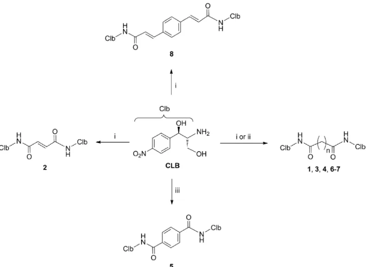
dicarboxylic acids was either aliphatic of variable length (compounds1,3,4,6and7), olefinic (compound2), or aromatic (compounds5and8). With these particular dimers, we wanted to
examine the effect of (i) the length of the aliphatic chain (linker) connecting the two CLB units (e.g.1and7) and (ii) the nature and the flexibility of the linker (e.g.4and5) on the inhibitory
activity of the homodimers on peptide bond formation in a cell-free system and the antibacte-rial activity against wild type or resistant bacteantibacte-rial strains. Compound5, ranking among the most potent members in the group of CAM dimersin vitro, was further studied for its ability to reduce the viability of human peripheral blood cells and to restrain the proliferation of human leukemic cells. The promising findings for compound5show that its structure can be fruitfully used for designing more potent, but less toxic antibacterials.
Results and Discussion
Synthesis of ribosome-targeting CAM dimers
The synthesis of CAM dimers1–8is depicted inFig 2(see alsoS1 Supplemental Procedures). Compounds1,2,4and6–8were readily assembled by the condensation of the commercially Fig 1. Structures of CAM, CLB, and the synthesized CAM dimers.Abbreviations: CAM, chloramphenicol; CLB, chloramphenicol base.
available CLB with the corresponding carboxylic acids malonic, fumaric, adipic, suberic, azelaic and 1,4-phenylenediacrylic, in the presence of the coupling agentO-(benzotriazol-1-yl)-N,N,
N’,N’-tetramethyluronium hexafluorophosphate (HBTU) and ethyldiisopropylamine, in 55–
89% yields. Compound5was obtained in 80% yield by condensing CLB and terephthaloyl chloride in the presence of triethylamine. Finally, compound3was assembled in 80% yield by first acylating CLB with glutaric anhydride and then coupling the resulting acid with additional CLB in the presence of HBTU and ethyldiisopropylamine.
Inhibition of peptide-bond formation by CAM dimers
The inhibitory effect of CAM dimers on peptide-bond formation was studied using the puro-mycin reaction, a model reaction between puropuro-mycin and a post-translocation ribosomal com-plex (comcom-plex C) derived fromE.coli[6]. Since puromycin, a pseudo-substrate of PTase which binds to the A-site of the catalytic center, was present in excess, the reaction obeyed first-order kinetics. The first-order rate constant,kobs, at each concentration of puromycin was
Fig 2. Synthesis of compounds studied in the present work.Reagents and conditions: (i) malonic acid (for compound 1), fumaric acid (for compound 2), adipic acid (for compound 4), suberic acid (for compound 6), azelaic acid (for compound 7), 1,4-phenylenediacrylic acid (for compound 8), HBTU,iPr
determined by fitting thexvalues intoEq 1,
ln 100
100 x¼kobst ð1Þ
wherexis the product AcPhe-puromycin, expressed as the percentage of complex C added in the reaction mixture, andtis the time of the reaction.
A representative time plot obtained at 400μM puromycin, in the absence of inhibitor, is illustrated inFig 3A(upper line) and, as expected, is characterized by linearity. However, when the reaction proceeded in the presence of a CAM dimer, e.g. compound5, the time plots were characterized by two unique features that distinguish them from a typical kinetic behavior. First, biphasic logarithmic time plots were obtained, with the second phase exhibiting stronger inhibition characteristics than the first one. Second, the slopes of both progress curves varied as a function of the inhibitor concentration (Fig 3A, four lower curves). When analyzed by double
Fig 3. AcPhe-puromycin synthesis in the presence or absence of compound 5.(A) First-order time plots; complex C reacted at 25°C in buffer A, with (black) 400μM puromycin or with a mixture containing 400μM puromycin and compound 5 at concentrations of 4μM (magenta), 8μM (green), 15μM (blue), and 30μM (red). (B) Variation of the apparent equilibration rate constant,keq, as a function of compound 5 concentration (I). The reaction was carried out in buffer A, in the presence of puromycin at concentrations of 200μM (red), 400μM (black), or 2 mM (blue). Thekeqvalues were determined by non linear regression fitting of the kinetic data toEq 2[11]: (C) Kinetic model for the inhibition of the puromycin reaction by CAM dimers. Symbols: C, poly(U)-programmed ribosomes fromE.coli, bearing AcPhe-tRNAPheat the P-site of the catalytic center and tRNAPheat the E-site; I, CAM dimer; S, puromycin; C’, ribosomal complex not recycling; P, AcPhe-puromycin. See alsoS1 Fig.
reciprocal plotting (1/kobsversus 1/[S]; [S] is the concentration of puromycin), both phases exhibited characteristics of simple competitive inhibition (S1 Fig, panels A and B). These kinetic results are consistent with compound5operating through an induced fit mechanism (Fig 3C), in which the inhibitor first binds rapidly to complex C to form the encounter complex CI, which then undergoes a slow conformational change to produce a final, tighter complex CI. Corroborative evidence for the consistency of this model is provided by the hyperbolic
shape of the equilibration plots (keqversus [I]), in whichkeqrepresents the apparent rate con-stant for the attainment of equilibrium among C, CI, and CI (Fig 3B). If one-step mechanism
of inhibition was applicable (C+I⇄CI),keqshould be a linear function of [I] [27]. Yet the
apparent association rate constant, (kon+koff)/Ki, was found to be 3×10⁴M1s-1that is lower than the upper limit 106M-1s-1set for the characterization of a compound as a slow-binding inhibitor [27]. Because the isomerization constantkon/koffwas calculated to be 3.6, the inhibi-tion process was finally associated with high overall inhibiinhibi-tion of peptide-bond formainhibi-tion (Ki
= 0.3μM;S1 Fig, panel C). In addition, the slowkoffrate (0.64 min-1) provided prolonged resi-dence time for compound5at the ribosome, a behavior potentially predicting good efficacyin vivo[28]. Although a direct comparison is not accurate, compound5is ~10-fold more potent than homodimers of CAM previously synthesized by Berkov-Zrihen et al. [25].
ln 100
100 x¼kobsðlateÞtþ
½kobsðearlyÞ kobsðlateÞ keq
ð1 ekeqtÞ ð2Þ
Except for compound8that was inactive, all compounds shown inFig 1, including CAM, exhibited a similar kinetic behavior to that adopted by compound5. The values of the kinetic parameters involved in the inhibition of the puromycin reaction by these compounds are sum-marized inTable 1. To examine if the effects seen are due to the presence of CAM dimers, we synthesized (seeS1 Supplemental Procedures) three additional derivatives in which only one CAM molecule is attached to one molecule of linker, i.e. glutaric-CAM (9), adipoyl-CAM (10),
and terephthaloyl-CAM (11), and tested them as inhibitors of peptide bond formation. Approximately, three- to six-fold lower inhibitory activity was recorded for these compounds (S1 Table), justifying the necessity of the presence of both CAM molecules for optimal potency.
Table 1. Equilibrium and kinetic constants involved in the inhibition of AcPhe-puromycin synthesis by CAM dimersa.
Compound Ki(μM) Ki*(μM) kon/koffb kon(min-1)c koff(min-1)c
CAMd 3.10±0.30 0.88±0.07 2.52±0.44 2.29±0.13 0.99±0.04
1 2.40±0.18 0.60±0.05 3.00±0.46 1.80±0.16 0.60±0.05
2 2.70±0.25 0.75±0.06 2.60±0.44 1.70±0.15 0.70±0.05
3 4.50±0.36 1.20±0.09 2.75±0.41 2.90±0.23 1.00±0.03
4 6.00±0.54 1.87±0.15 2.21±0.38 2.14±0.16 1.00±0.04
5 1.40±0.12 0.30±0.03 3.67±0.61 2.23±0.10 0.63±0.03
6 10.50±0.90 2.30±0.20 3.56±0.56 2.80±0.25 0.80±0.04
7 12.00±1.20 2.80±0.27 3.28±0.59 2.89±0.23 0.84±0.06
8e - - - -
-aData denote the mean±S.E. values obtained from three independently performed experiments, with two replicates per experiment. bThekon/k
offratio was calculated through Equation:kon/koff=Ki/Ki*−1, [11].
cThe individual values ofk
onandkoffwere calculated by nonlinear regressionfitting of the kinetic data to Equation:keq¼koffþkon
½I Ki 1þ½S
KSþ ½I Ki
dData taken from Xaplanteri et al., [6]
eIn the range of concentrations 1–20μM, compound8was not active in inhibiting the puromycin reaction.
Consistently, compounds9–11were almost inactive in inhibiting the growth ofStaphylococcus aureusorE.colicells, at concentrations up to 100μM. Taking into account that in a closed sys-tem, like the cell-free system used in our study, the inhibitory constant is an adequate metrics for differentiating compound potency [28], we used theKiconstant for ranking compounds
1–8;Kiby definition (Eq 3) represents the overall inhibition constant engaged in both sequen-tial reactions of the two-step mechanism shown inFig 3C.
K
i ¼Ki
koff
konþkoff
¼ ½C½I
½CI þ ½CI ð3Þ
Accordingly, we estimated that compound5is 3-fold more potent than CAM. The rest of
CAM dimers exhibited either comparable (compounds1and2), lower (compounds3,4,6, and7), or no activity (compound8). At afirst glance, it is surmised that CAM dimers
possess-ing a rigid aromatic linker of 6–7 Å, estimated byin silicoanalysis to equal the distance between the—NH- groups of CAM bound at the CAM1 and CAM2 sites, display the best activity in
inhibiting the puromycin reaction. Among the CAM dimers tested, only compound5meets by the best balance of these structural properties. Compound8also bears a rigid linker. However,
its length exceeds the ideal distance of 6–7 Å and therefore its ability to engage the CAM2 bind-ing site is compromised. Compound4, possessing an aliphatic linker of similar length,
func-tioned 6-fold less efficiently than compound5. In terms of the free energy of binding, compound4needs to pay a higher entropic cost upon binding than compound5. This is
because multiple rotatable bonds in compound4allow more conformational degrees of free-dom than those of compound5. A similar hypothesis can be adopted in explaining the low
potency of compounds3and6. Compounds1and2that possess a short linker cannot simulta-neously bind the catalytic crevice (CAM1) and the entrance to exit-tunnel (CAM2) and show a comparable activity to CAM.
Structural characterization of the RI and R
*
I complexes by time-resolved
footprinting analysis and MD simulations
The interactions between compound5, the most potent inhibitor of the puromycin reaction among the tested CAM dimers, and theE coliribosome were dissected by time-resolved foot-printing analysis. The behavior of compound5was compared to that of compound4, which is bearing a flexible linker of the same length. The footprinting analysis exploits the slow-binding character of the dimers and has been successfully applied in studying various slow-binding inhibitors of the PTase [11,29–31]. To overcome potential drawbacks resulting from protec-tions caused by natural PTase substrates, naked 70S ribosomes were used instead of complex C. To footprint the RI complex, compounds4or5used in excess, and ribosomes were incu-bated at 25°C for 2 s, and then treated with chemical probes for 3 min to modify accessible nucleosides in 23S rRNA. Because the first step of binding,R+I⇄RI, equilibrates rapidly
while the formation of RI occurs slowly, the main product formed during such a short time
interval was complex RI (>93%). To footprint the RI complex, each dimer and ribosomes
were incubated for 10 min, a time interval that is over than ten half-lives required for the attainment of the steady state. Because the isomerization constant,kon/koff, is at least 2.2 (see
Table 1), most of the ribosomes added in the reaction mixture (>70%) were in the form of RI
complex at the end of this time interval.
differ from that previously published for CAM [11,32–34]. This similarity suggests that both
compounds occupy, via one of their symmetrical CAM portions, a pocket near the A-site of the PTase catalytic center (CAM1). Compound5, compared with4, exhibits stronger protections
at nucleosides A2451 and U2506, a fact that is consistent with its lowerKivalue (Table 1). However, larger differences were recorded when footprinting analysis was performed in the RI complex; a protection seen at A2058 by compound5did not appear in the footprinting
pattern of compound4. Moreover, all the protections due to compound4were generally weaker than those caused by5. This may be associated with the flexibility of the linker tethered by each compound.
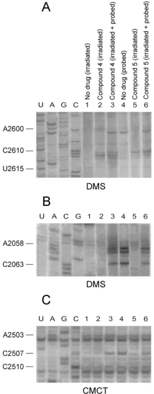
MD simulations confirmed that both compounds bind through one of their edges to the cat-alytic crevice (Fig 4). Compound5was additionally hydrogen bonded through its second CAM moiety to the 2OH- group of C2610, a fact compelling thep-nitrophenyl ring of this edge to bend and insert a hydrophobic crevice, formed by nucleosides A2058 and A2059 at the entrance to the exit tunnel (Fig 4A). Noteworthy, nucleoside C2610 has been considered as a part of a signal relay pathway linking the exit tunnel sensors to the PTase active site [35]. In contrast, compound4was revealed to bind C2611. Nevertheless, this interaction is not stable enough, nor it orientated the edge of compound4towards the A2058-A2059 crevice (Fig 4B). Due to technical limitations, certain interactions detected by MD simulations cannot be revealed by footprinting analysis; C2610 does not react with dimethyl sulfate (DMS), while C2611 is based paired with G2057 [36]. Binding models for the remaining CAM dimers, as generated by MD simulations, are presented inS3 Fig.
To experimentally demonstrate that compounds4and5are capable of binding nucleosides C2611 and C2610, respectively, a crosslinking approach was applied. Specifically, a mixture of
E.coliribosomes together with either4or5, each added to the incubation mixture at concen-tration equal to 10×Ki, were irradiated for 30 min with 365 nm light. Following purification, the irradiation products were analyzed by primer extension. As shown inFig 5(panels A and B), footprints of RI complex having bound compound5mapped to nucleoside C2610, and
less to nucleosides A2058 and A2059. Footprinting analysis of the whole irradiated mixture, before purification, indicated that compound5was firmly attached to the catalytic crevice of PTase, but did not form crosslinks with this region (Fig 5, panel C). In contrast, compound4
Table 2. Relative reactivity of nucleosides in the central loop of Domain V of 23S rRNA, when a CAM dimer (I) binds toE.coliribosomes (R) in the initial (RI) and the final (R*I) binding sitesa.
Compound 4 Compound 5
23S rRNA residue R RI R*I R RI R*I
A2058 1 0.99±0.07 0.90±0.09 1 0.93±0.06 0.32±0.04b,c
A2059 1 0.94±0.06 0.70±0.07b,c 1 0.96±0.08 0.33±0.08b,c
A2062 1 0.88±0.08b 0.67±0.05b,c 1 0.80±0.06b 0.40±0.09b,c
A2451 1 0.67±0.04b 0.77±0.05b 1 0.40±0.03b 0.41±0.05c
G2505 1 0.43±0.03b 0.44±0.03b 1 0.38±0.05b 0.35±0.06c
U2506 1 0.70±0.05b 0.70±0.04b 1 0.47±0.05b 0.33±0.05b,c
U2609 1 1.00±0.10 1.00±0.07 1 1.00±0.08 0.79±0.08b,c
aRelative reactivity of nucleosides denotes the ratio between the normalized intensity of a band of interest and the normalized intensity of the homologous
band in the control lane (R) (see alsoS2 Fig).
bSignificantly different in relation to R (P<0.05). cSigni
crosslinked to C2611, without raising any modification signal in the A2058-A2059 region. It should be mentioned that the concentrations used for compounds4and5in this series of experiments were much lower (<60μM) than those of CAM utilized previously by Long &
Porse [12]. The most plausible explanation for this enhanced affinity is that binding of a dimer to the primary high-affinity site (catalytic crevice) facilitates targeting of a cryptic, low-affinity site (entrance to the exit tunnel) via the second edge of the homodimer. Such a site cannot be easily detected by kinetic analysis of CAM binding to the ribosome, due to its highKivalue (~300μM) [12], and it is the first time that such a position is revealed by using the drug at micromolar concentrations.
Antibacterial activity of the CAM dimers and correlation with inhibitory
activity on the puromycin reaction
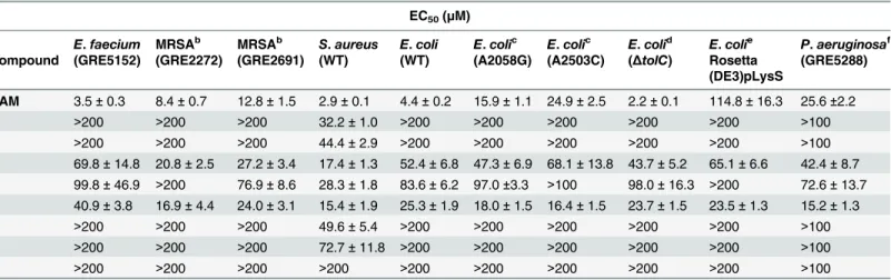
The antimicrobial potency of the synthesized CAM dimers was tested against a panel of Gram-negative and Gram-positive bacteria. Two laboratory strains ofE.colipossessing the A2058G or A2503C mutations in 23S rRNA, the Rosetta(DE3)pLysSE.colistrain expressing the CAM acetyltransferase (cat) gene, and one clinical isolate ofPseudomonas aeruginosaexhibiting resistance against CAM due both to a constitutively expressed efflux system (MexAB-OprM) and an inducible efflux system (MexXY) [37] were included as representative CAM-resistant strains. In addition, wild-typeEnterococcus faecium,E.coliandStaphylococcus aureusstrains, along with two multi-drug resistant (MDR)S.aureusisolates (MRSA), were examined in our study. Compared to the parent antibiotic, none of the CAM dimers exhibited stronger inhibi-tory activity on the growth of wild-typeE.faecium,S.aureusandE.colicells (Table 3). How-ever, better results were obtained when compound5was examined against the two multi-drug resistant MRSA isolates. The resistance of these isolates against a variety of antibiotics includ-ing methicillin, is reported inS2 Table. Interestingly, both MRSA isolates showed comparable Fig 4. Binding positions of compounds 4 and 5 on theE.coliribosome, as detected by Molecular Dynamics simulations.Compounds 4 and 5 have been docked into the 50S ribosomal subunit, by positioning one of their CAM moieties within the CAM crystallographic pocket [4]. (A) Binding position of compound 5 (yellow); hydrogen bonding with residues of the catalytic center is shown by black dots. Other residues of 23S rRNA placed adjacently to the binding pocket of 5 are ignored for clarity. (B) Binding position of compound 4 (yellow).
Fig 5. CAM dimer crosslinking at the entrance to the exit tunnel, upon UV-irradiation.Ribosomes from
E.coliwere irradiated with 365 nm light for 30 min (panels A-C), in the absence (lane 1) or the presence of compound 4 (lanes 2 and 3) or compound 5 (lanes 5 and 6). The irradiation products were analyzed by probing with DMS (panels A and B) or CMCT (panel C) and primer extension, before (lanes 3 and 6) or after discharging from excess CAM dimer (lanes 2 and 5). Probing and primer extension analysis were also applied to non-treated ribosomes (lane 4). Numbering of nucleosides for the sequencing lanes is indicated at the left. (A) Analysis of the A2600-U2615 region of 23S rRNA. (B) Analysis of the C2055-A2065 region (entrance to the exit tunnel) of 23S rRNA. (C) Analysis of the A2500-U2506 region (PTase catalytic center) of 23S rRNA.
susceptibility to compound5and CAM. Given that compound5causes less severe toxicity than CAM in human neutrophils (see below), this dimer seems to be a well promising lead can-didate for the design of efficacious drugs against MDR Gram-positive bacteria. Intriguingly, compound5was approximately 2-fold more active than CAM in inhibiting the A2503C
mutant and equivalent to CAM in inhibiting the growth of the A2058G mutant. Notably, pre-vious studies have demonstrated that the incorporation of an interactive side chain into a macrolide scaffold can significantly improve the efficacy of this drug against bacteria that exhibit resistance conferred by changes in the PTase catalytic center [38].
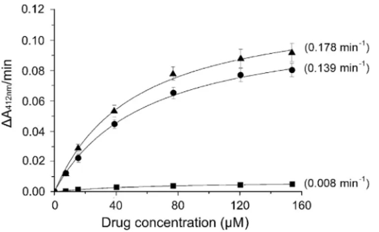
Compared to CAM, compound5displayed a five-fold higher activity in inhibiting the growth of Rosetta(DE3)pLysSE.colicells expressing thecatgene. This was a truly interesting
finding, immediately raising the question of how this would work from a biochemical point of view. Expression of CAM acetyltransferase, encoded by thecatgene, is a major mechanism by
which bacteria become resistant to CAM. This enzyme catalyzes transfer of the acetyl moiety from acetyl coenzyme A to CAM [40]. The O-acetoxy derivatives of CAM fail to act as antibi-otics, because they do not bind to bacterial ribosomes. Therefore, we tested compounds4and
5against the purified enzyme, by calculating the ratio Vmax/Km. These calculations allowed the direct evaluation of the capacity of each compound to behave as acceptor of acetyl groups. As shown inFig 6, compound4behaves like CAM as substrate of CAM acetyltransferase, while
compound5was almost inactive. However, it should be noted that the number of available hydroxyl groups in both CAM dimers is twice that of CAM. Therefore, they should confer Table 3. Determination of EC50for CAM and CAM dimers, that indicates how much concentration of each compound is needed to produce 50% of the maximal inhibitory effect of that compounda.
EC50(μM)
E.faecium MRSAb MRSAb S.aureus E.coli E.colic E.colic E.colid E.colie P.aeruginosaf Compound (GRE5152) (GRE2272) (GRE2691) (WT) (WT) (A2058G) (A2503C) (ΔtolC) Rosetta
(DE3)pLysS
(GRE5288)
CAM 3.5±0.3 8.4±0.7 12.8±1.5 2.9±0.1 4.4±0.2 15.9±1.1 24.9±2.5 2.2±0.1 114.8±16.3 25.6±2.2 1 >200 >200 >200 32.2±1.0 >200 >200 >200 >200 >200 >100 2 >200 >200 >200 44.4±2.9 >200 >200 >200 >200 >200 >100 3 69.8±14.8 20.8±2.5 27.2±3.4 17.4±1.3 52.4±6.8 47.3±6.9 68.1±13.8 43.7±5.2 65.1±6.6 42.4±8.7 4 99.8±46.9 >200 76.9±8.6 28.3±1.8 83.6±6.2 97.0±3.3 >100 98.0±16.3 >200 72.6±13.7 5 40.9±3.8 16.9±4.4 24.0±3.1 15.4±1.9 25.3±1.9 18.0±1.5 16.4±1.5 23.7±1.5 23.5±1.3 15.2±1.3 6 >200 >200 >200 49.6±5.4 >200 >200 >200 >200 >200 >100 7 >200 >200 >200 72.7±11.8 >200 >200 >200 >200 >200 >100 8 >200 >200 >200 >200 >200 >200 >200 >200 >200 >100
aData represent the mean±SE values obtained from three independently performed experiments, with two replicates per experiment. EC50values were
determined by nonlinear regressionfitting of the observed optical density values (Y) into Hill Equation,y¼minþ max min
1þ x EC50
n(seeMaterials and Methods).
bMethicillin-resistantS.aureus(MRSA) isolates belong to the ST80 clone and exhibit multi drug resistance behavior (see alsoS2 Table)
cE.coliTA531 cells lacking chromosomalrrnalleles, but containing pKK35 plasmids that possess wild-type 23S rRNA display the same EC50value for
each drug, like those of wild-type (WT)E.coliK12 cells. However, when pKK35 plasmids possess mutated 23S rRNA (A2058G or A2503C), the cells are resistant to CAM because of target mutation [14,15].
dDeletions intolCgene result in an increased sensitivity ofE.colito a wide range of antibiotics, including CAM [39].
eRosetta(DE3)pLysSE.colicells express thecatgene that encodes CAM acetyltransferase, an enzyme that inactivates CAM [40] (see alsoFig 6). fP.aeruginosaexhibits reduced susceptibility to CAM, in part due to the intrinsically expressed MexAB-OprM ef
flux system, and additionally to an inducible by CAM efflux system (MexXY) [37].
double initial velocity to the reaction at low substrate concentration, if they were just as effi-cient as CAM.
The preferential activity of CAM dimers against the growth ofS.aureuscells thanE.coli
cells is the first evidence that penetration of the outer cellular membrane may be a significant limitation in the efficacy of CAM dimers as antibiotics. It is known that CAM gains access to the periplasm through pore-forming porins [19], and that utilization of the porin pathway by antibiotics depends on the molecular dimensions of the drugs [41]. Therefore, we suggested that CAM dimers are too large for effective diffusion through porins. The second evidence was provided when the antimicrobial activity of CAM dimers was correlated with their ability to inhibitin vitropeptide-bond formation. By using IC50(puro)as a criterion of the efficiency of compounds in targeting the ribosome, where IC50(puro)is defined as the compound concentra-tion causing 50% inhibiconcentra-tion in peptide-bond formaconcentra-tion at the presence of 2 mM puromycin, and calculating the ratio EC50(cell growth)/IC50(puro), we realized that the value of this ratio is much lower for CAM than for any CAM dimer (S3 Table). This suggests that CAM dimers are prone to transport limitations. It should be kept in mind that a second bacterial barrier, the plasma membrane, may also contribute in obstructing CAM dimers from accumulating into the cells. There are more than seven efflux systems inE.colithat can pump out toxic com-pounds, such as antibiotics, detergents, organic solvents etc [39]. An important efflux system inE.coliis the AcrAB-TolC multidrug resistance tripartite pump [42]. Deletions inacrABand/ ortolCgenes result in an increased sensitivity ofE.colito a wide range of antibiotics, including CAM [39]. To investigate the effect of this efflux system on the intracellular accumulation of our compounds, we determined the EC50values against anE.colistrain BL21 DE3 lacking the
tolCgene that codes TolC, the outer membrane component of the AcrAB-TolC efflux pump. We observed that this efflux system does not appear to affect the antibacterial activity of CAM dimers, as the EC50values regarding this strain were similar to those of wild-typeE.coli (Table 3). Notably,E.coliBL21 DE3 (ΔtolC) strain was approximately 2-fold more sensitive to
Fig 6. Kinetic analysis of the CAM acetyltransferase reaction using CAM or compounds 4 and 5 as substrates.The reaction was carried out in 3 ml of 94 mM Tris/HCl pH 7.8, containing 0.083 mM 5,5’ -dithio-bis(2-nitrobenzoic acid), 0.16 mM acetyl coenzyme A, 25 units CAM acetyltransferase, and either CAM (●), compound 4 (▲), or compound 5 (&) at the concentrations indicated. The product of the enzymatic reaction, coenzyme A, reacted with 5,5’-dithio-bis(2-nitrobenzoic acid) to yield 5-thio-2-nitrobenzoate which absorbs at 412 nm, with a micromolar extinction coefficient equal to 0.0136. The Vmaxand Kmvalues were determined by fitting the substrate concentrations [S] and the obtainedΔA412nm/min (Vo) values into equationV0=
Vmax[S]/(Km+ [S]). The obtained Vmaxvalues were divided by 0.0136 to convert their units inμMmin-1(http://
www.sigmaaldrich.com/technical-documents/protocols/biology/enzymatic-assay-of-chloramphenicol-acetyltransferase.html). The ratio Vmax/Kmfor each curve is given in parenthesis.
CAM than wild-typeE.coli(Table 3). In contrast, another Gram-negative bacterium,P. aerugi-nosa, possessing both inducible and constitutively expressed efflux pumps showed 6-fold higher EC50value for CAM than wild-typeE.coli, maintaining the EC50values for compounds
3,4, and5almost unchanged. This means that CAM dimers are neither recognized by the con-stitutively expressed MexAB-OprM efflux system, nor induce the MexXY efflux system of this bacterium [37].
Toxicity of CAM dimers against Human peripheral blood cells and
leukemic cell lines
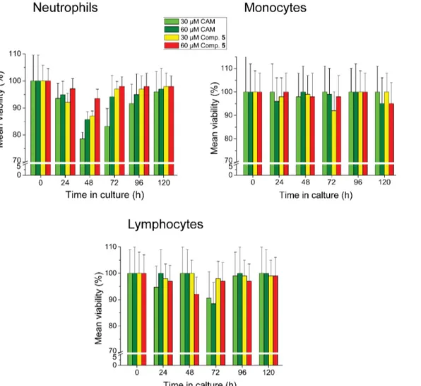
Accumulating evidence has shown that CAM causes adverse effects to the hematopoietic sys-tem [21,43–45]. This prompted us to test the CAM dimers for potential toxicity against human
peripheral blood cells and leukemic cell lines. Compound5, the most potent member of the synthesized CAM dimers, displayed a mild and transient toxicity on neutrophils during a 120 h exposure of blood cells to this compound, peaked at 48 h (Fig 7). Nevertheless, this toxicity
Fig 7. Toxicity assays in human peripheral blood cells.Peripheral blood was collected in EDTA-coated tubes from 5 healthy volunteers (age range: 25– 30 years). Concentration was adjusted to 1.8×10⁹cells/L using RPMI-1640 medium containing 1% penicillin/streptomycin. Cells were cultured in triplicate in the presence or the absence of 30 or 60μM CAM or compound 5, under a humidified 5% CO2atmosphere for 5 days, at 37°C. Cultures were counted daily by a CELL-DYN 3700 Hematology Analyzer and values were expressed as a percentage of cells measured in controls.
was less severe when compared with that caused by CAM. Toxicity of compound5against other types of leukocytes was negligible.
The toxicity effects of compound5on leukemic cell lines were tested, using HS-Sultan, Jur-kat and U937 cells. Preliminary records, produced by counting daily the cells in a CELL-DYN 3700 Hematology Analyzer, showed that Jurkat cells grew exponentially at all the tested con-centrations of compound5; however, the rate of growth was reduced proportionally to the
con-centration of compound5(S4 Fig). In contrast, HS-Sultan or U937 cells were insensitive to compound5. Therefore, the effect of compound5on Jurkat cells was further studied by flow
cytometric analysis. The results showed that compound5at 60μM failed to induce necrosis, but did induce 43% apoptosis to Jurkat cells, expressed as a percentage of total cells (Fig 8). In comparison, CAM at 60μM did not induce any necrosis/apoptosis effect to these cells under the same conditions of treatment. This different response confers compound5a comparatively
significant advantage over CAM, since the anti-apoptotic behavior of CAM plays a critical role in CAM-induced leukemogenesis by allowing proliferating cells to continuously survive [21]. Fig 8. Toxicity assays in Jurkat cells.Jurkat cells were adjusted to 1×10⁹cells/L in RPMI-1640 medium containing 1% Penicillin/Streptomycin and 10% fetal bovine serum. The cells were grown in triplicate in the presence or absence of compound 5 at the indicated concentrations for 4 days at 37°C, under a humidified 5% CO2atmosphere. CAM was used as a reference compound. For cell necrosis and apoptosis assays, samples (106cells) were collected daily and determined by flow cytometry. Apoptotic and necrotic cells were expressed as a percentage of total cells.
Conclusions
To explore the existence and utilization of multiple binding sites of CAM within the central loop of domain V of 23S rRNA, we constructed eight homodimers of CAM, tethered via a linker of varying length and flexibility. Compared to CAM, three of the CAM dimers inhibited the AcPhe-puromycin synthesis, a model reaction for peptide-bond formation, more effi-ciently. Footprinting and crosslinking analysis, combined with computational modeling, revealed that the enhanced binding affinity exhibited by these constructs resulted from their unique architecture and ability to recognize multiple binding sites within the ribosome’s three-dimensional structure. It was realized that multiple interactions synergize in order to enhance the apparent affinity and lead to a prolonged residence time of the constructs at their targets. Specifically, dissection of the mechanism of action of compound5binding to the ribosome allowed us to verify a kinetically cryptic binding site of CAM at the entrance to the exit tunnel and gave us the opportunity to clarify previous uncertainties related to the number and exact localization of CAM binding sites in the ribosome. The remarkablein vitroinhibitory activity of compound5on bacterial protein synthesis, combined with its ability to bypass some resis-tance mechanisms, its low toxicity against human peripheral blood cells and promising activity against human T-leukemic cells provide the impetus to further improve its design. Realizing the serious problems met in internalizing CAM dimers into bacterial cells, future efforts will focus on strengthening the capacity of these compounds in penetrating the outer and plasma membrane barriers.
Materials and Methods
Materials, bacterial strains, peripheral blood samples, leukemic cell
lines, biochemical preparations, and instrumentation
CAM free base [D-(-)threo-1-(p-nitrophenyl)-2-amino-1,3-propanediol]), tRNAPhefromE.
coli, dimethyl sulfate (DMS), DMS stop solution, puromycin dihydrochloride, and tRNAPhe fromE.coliwere from Sigma-Aldrich. 1-Cyclohexyl-3-(2-morpholinoethyl)-carbodiimide metho-p-toluene sulfate (CMCT) and kethoxal were purchased from Fluka Biochemicals and MP Biomedicals, respectively. AMV reverse transcriptase was supplied by Roche, dNTPs by HT Biotechnology, and ddNTPs by Jena Bioscience. L-[2,3,4,5,6 -3H] Phenylalanine was from Amersham Biosciences and [α-32P] ATP from Izotop. The HS-Sultan (Burkitt’s lymphoma)
poly(U)-programmed ribosomes (complex C) fromE.coliK12, bearing tRNAPheat the E-site and Ac [3H]Phe-tRNA at the P-site were prepared, as previously described [46]. The percentage of active ribosomes in AcPhe-tRNA binding was 75%.
Melting points were determined with a Buchi SMP-20 apparatus and are uncorrected. IR spectra were recorded as KBr pellets on a Perkin Elmer 16PC FT-IR spectrophotometer.1H NMR spectra were obtained at 400.13 MHz and13C NMR spectra at 100.62 MHz on a Bruker DPX spectrometer. Chemical shifts are reported inδunits, parts per million (ppm) downfield from TMS. Electron-spray ionization (ESI) mass spectra were recorded at 30V, on a Micro-mass-Platform LC spectrometer using MeOH as solvent. Analytical HPLC was used to deter-mine the purity of final products, confirming95% purity. Analytical RP-HPLC was performed on a Waters system (2695 Alliance). Elution of the compounds was determined from the absorbance at 254 nm (Waters 2996 Photodiode array detector). Compound purity was assessed using a LiChrospher C8 column (5μm, 125 x 4.0 mm) and a linear gradient of 5%-60% acetonitrile (containing 0.05% TFA) in water (containing 0.05% TFA) over 20 min at a flow rate of 1 ml/min. Flash column chromatography (FCC) was performed on Merck silica gel 60 (230–400 mesh) and TLC on 60 Merck 60F254films (0.2 mm) precoated on aluminium foil. Spots were visualized with UV light at 254 nm and charring agents. All solvents were dried and/or purified according to standard procedures prior to use. All reagents employed in syn-thesis were purchased from either Aldrich or Alfa-Aesar. CLB and the required dicarboxylic acids, glutaric anhydride and terephthaloyl dichloride were obtained from Aldrich. The synthe-sis of 1,4-phenylenediacrylic acid and compounds1–11as well as physical and spectra data for the synthesized compounds are presented inS1 Supplemental Procedures.
Inhibition of peptide-bond formation by CAM dimers
The puromycin reaction, i.e. the reaction between complex C and excess puromycin (S), was carried out at 25°C in buffer A [100 mM Tris-HCl pH 7.2, 6 mM (CH3COO)2Mg, 100 mM NH4Cl and 6 mM 2-mercaptoethanol]. Under these conditions, the puromycin reaction obeys
CþS⇄ KS CS!kcat
C0þS
pseudo-first-order kinetics and was analyzed as previously described [11].
In the presence of CAM dimers, biphasic semi-logarithmic time plots were obtained. The slope of the straight line through the origin was seen as the value of the apparent rate constant,
kobs(early), at the early phase of the reaction. Similarly, the slope of the second straight line was
taken as the apparent rate constant,kobs(late), at the late phase of the puromycin reaction.
Time-resolved binding of CAM dimers to
E
.
coli
ribosomes and
characterization of RI and R
*
I complexes by footprinting analysis
70S ribosomes fromE.coli(100 nM) were incubated either alone or with each CAM dimer at concentration equal to 50×Kiin 100μl of buffer B [Hepes/KOH, pH 7.2, 6 mM Mg
[11]. Values indicated inTable 2denote the ratio between the normalized intensity of a band of interest and the normalized intensity of the corresponding band in the control lane (ribo-somes non-treated with CAM or CAM dimers).
Crosslinking of CAM dimers to
E
.
coli
ribosomes
70S ribosomes fromE.coli(100 nM) were incubated either alone or with compound4or5at concentration equal to 50×Kiin 100μl of buffer B [Hepes/KOH, pH 7.2, 6 mM Mg
(CH3COO)2, 100 mM NH4Cl, and 5 mM dithiothreitol] at 25°C, for 5 min. Following forma-tion of RI complexes, the samples were irradiated at 365 nm, for 30 min, in a Vilber Lourmat
UV Cabinet (VL-215.LC-30W) light source. The light source was placed ~5 cm over a microti-ter tray containing the sample on an ice-wamicroti-ter bath. Half of the sample was probed with DMS or CMCT, and then analyzed by primer extension as shown above, while the other half was extracted with phenol, phenol-chloroform (1:1), and chloroform, followed by ethanol precipi-tation to remove non-crosslinked agents. The isolated rRNA was then subjected to primer extension analysis.
Molecular Dynamics simulations
3D models for compounds1to8and their parameterization for the CHARMM Force field were achieved, as previously described [11], starting with the 3D structure of CAM derived from crystallographic data (PDB: 3OFC). The CAM dimers were docked into the 50S ribo-somal subunit structure, by positioning one of their CAM moieties within the drug crystallo-graphic pocket. All groups of 50S subunits in a distance of 10 Å around CAM dimers were selected, solvated with TIP3 water molecules, and then neutralized with sodium ions using the VMD program [48].
All systems derived as above were energy minimized and then subjected to canonical ensemble Molecular Dynamics (MD) simulations for 10 ns at 300K, with Particle Mesh Ewald (PME) algorithm and rigid bonds assigned using the NAMD software [49]. During MD simu-lations, all nucleic acid backbone atoms were positionally restrained. Finally, an average struc-ture over the last 100 frames of each simulation trajectory was energy minimized and used for further analysis. An H-bond was considered as existing, if hydrogen donor and acceptor atoms were closer than 0.35 nm and the angle between the line connecting these atoms and the hydro-gen bond was lower than 30°. All molecular visualizations were produced with the PyMOL Molecular Graphics System, Version 1.5.0.4 Schrödinger, LLC.
Biological evaluation of CAM dimers in bacterial cells containing
wild-type or mutant ribosomes
The antibacterial activity of CAM dimers was assessed in CAM-sensitiveE.faecium,S.aureus
andE.colistrains, as well in two CAM-resistant strains ofE.colilacking chromosomalrrn
study. Briefly,S.aureusorE.colicells (200μl of a 0.700 OD560preculture) containing wild-type or mutant ribosomes were added in 3.8 ml of LB (Luria-Bertani) medium and grown at 37°C in the presence or absence of CAM or CAM dimers until the optical density of the control culture (grown in the absence of drug) reached the value 0.700 at 560 nm. ForE.faeciumand
P.aeruginosacultures, TSB (Tryptic Soy Broth) medium was used, instead of LB medium, in order to achieve exponential doubling times between 25 and 35 minutes. From dose-response curves, the half-maximal effective concentration (EC50) for each compound and strain was esti-mated. EC50represents the molar concentration of a compound that produces 50% of the max-imal possible effect [50]. The EC50values were mathematically determined by non linear regression fitting of the observed culture optical-density values, expressed as the percentage of 0.700 (y), into Hill Equation,
y¼minþ max min
1þ x EC50
n
whereminandmaxare the lowest and highest observed values of the culture optical density, respectively,xthe concentration of the tested compound, andnthe Hill coefficient that repre-sents the largest absolute value of the curve slope. EC50is equal to thex- value of the sigmoid’s midpoint. Fitting was performed using the Four Parameter Logistic Curve of the SigmaPlot Program Version 11.0 (Systat Software, Inc) for Exact Graphs and Data Analysis.
CAM acetyltransferase assay
The activity of CAM acetyltransferase (CAT) employing CAM or CAM dimers as acetyl-accep-tor substrates was assayed by using purified enzyme fromE.coli(Sigma-Aldrich), following the manufacturer’s protocols (http://www.sigmaaldrich.com/technical-documents/protocols/ biology/enzymatic-assay-of-chloramphenicol-acetyltransferase.html).
Toxicity assays in Human peripheral blood cells and leukemic cell lines
Peripheral blood was collected in EDTA-coated tubes from 5 healthy volunteers (age range: 25–30 years). Cell concentration was adjusted to 5×10⁸cells/l using RPMI-1640 medium (GIBCO BRL) containing 1% penicillin/streptomycin. Cells were cultured in triplicate under a humidified 5% CO2atmosphere for 5 days, at 37°C, in the absence (control cultures) or the presence of CAM or CAM dimers. Counting of cells was performed daily in a CELL-DYN 3700 Hematology Analyzer (Abbott, USA) and values were expressed as a percentage of cells measured in control cultures.
Statistical analysis
All data presented in the Figures and Tables denote the mean values obtained from three inde-pendently performed experiments, with two replicates per experiment, and are expressed as means±standard error. Significant differences between mean values were measured at p<0.05
by the F-Scheffe test (SPSS program 20.0 for Windows).
Supporting Information
S1 Fig. Kinetic plots for the AcPhe-puromycin synthesis in the presence or absence of com-pound 5.
(DOCX)
S2 Fig. Protections against chemical probes in nucleotides of the central loop of domain V of 23S rRNA, caused by binding of CAM or CAM dimers (compounds 4 and 5) toE.coli
ribosomes.
(DOCX)
S3 Fig. Binding positions of CAM dimers in theE.coliribosome, as detected by Molecular
Dynamics Simulations.
(DOCX)
S4 Fig. Toxicity assays in Human leukemic cell lines.
(DOCX)
S1 Supplemental Procedures. General experimental procedure for the synthesis of com-pounds 1–8.Synthesis of 1,4-phenylenediacrylic acid,1H- and13C-NMR spectra and RP-HPLC chromatograms of CAM dimers 1–8. General procedure for the synthesis of com-pounds 9 and 10. Synthesis of compound 11.1H- and13C-NMR spectra of compounds 9
–11. Supplemental references.
(DOCX)
S1 Table. Kinetic parameters of the puromycin reaction carried out in the presence of CAM attached to linkers indicated by red.
(DOCX)
S2 Table. Bacterial strains and mechanism of resistance or hypersensitivity to antibiotics.
(DOCX)
S3 Table. Determination of the ratio EC50(cell growth)/IC50(puro)in wild-typeE.colifor CAM
and CAM dimers.
(DOCX)
Acknowledgments
This work was partially supported by the University of Patras. We thank Prof. A.S. Mankin, Prof. I. Spiliopoulou, and Dr. D.N. Wilson for providing us with resistant bacterial strains, Ioannis Panagoulias and Maria Rodi for their generous assistance in conducting the flow cytometry assays, Charalampos Anastasopoulos for his help with analytical HPLC, and Kon-stantina Nika for critical reading of the manuscript. The authors declare no conflict of interest.
Author Contributions
reagents/materials/analysis tools: GEM ONK GPD AM DP. Wrote the paper: DLK DP. Obtained permission for use of cell lines and blood cells: AM.
References
1. Wimberly BT. The use of ribosomal crystal structures in antibiotic drug design. Curr Opin Investig Drugs 2009; 10: 750–765. PMID:19649920
2. Wilson DN. Ribosome-targeting antibiotics and mechanisms of bacterial resistance. Nat Rev Microbiol. 2014; 12: 35–48. doi:10.1038/nrmicro3155PMID:24336183
3. Schlünzen F, Zarivach R, Harms J, Bashan A, Tocilj A, Albrecht R, et al. Structural basis for the interac-tion of antibiotics with the peptidyl transferase center in eubacteria. Nature 2001; 413: 814–821. PMID: 11677599
4. Dunkle JA, Xiong L, Mankin AS, Cate JHD. Structures of theEscherichia coliribosome with antibiotics bound near the peptidyl transferase center explain spectra of drug action. Proc Natl Acad Sci USA 2010; 107: 17152–17157. doi:10.1073/pnas.1007988107PMID:20876128
5. Bulkley D, Innis CA, Blaha G, Steitz TA. Revisiting the structures of several antibiotics bound to the bac-terial ribosome. Proc Natl Acad Sci USA 2010; 107: 17158–17163. doi:10.1073/pnas.1008685107 PMID:20876130
6. Xaplanteri MA, Andreou A, Dinos GP, Kalpaxis DL. Effect of polyamines on the inhibition of peptidyl-transferase by antibiotics: revisiting the mechanism of chloramphenicol action. Nucleic Acids Res. 2003; 31: 5074–5083. PMID:12930958
7. Polacek N, Gomez MJ, Ito K, Xiong L, Nakamura Y, Mankin AS. The critical role of the universally con-served A2602 of 23S ribosomal RNA in the release of the nascent peptide during translation termina-tion. Mol Cell 2003; 11: 103–112. PMID:12535525
8. Thompson J, O'Connor M, Mills JA, Dahlberg AE. The protein synthesis inhibitors, oxazolidinones and chloramphenicol, cause extensive translational inaccuracyin vivo. J Mol Biol. 2002; 322: 273–279. PMID:12217690
9. Hansen JL, Moore PB, Steitz TA. Structures of five antibiotics bound at the peptidyl transferase center of the large ribosomal subunit. J Mol Biol. 2003; 330: 1061–1075. PMID:12860128
10. Pongs O. Chloramphenicol. In: Hahn FE, editor. Antibiotics V. New York: Springer Verlag; 1979. pp. 26–42.
11. Kostopoulou ON, Kouvela EC, Magoulas GE, Garnelis T, Panagoulias I, Rodi M, et al. Conjugation with polyamines enhances the antibacterial and anticancer activity of chloramphenicol. Nucleic Acids Res. 2014; 42: 8621–8634. doi:10.1093/nar/gku539PMID:24939899
12. Long KS, Porse BT. A conserved chloramphenicol binding site at the entrance to the ribosomal peptide exit tunnel. Nucleic Acids Res. 2003; 31: 7208–7215. PMID:14654696
13. Vauquelin G. Simplified models for heterobivalent ligand binding: when are they applicable and which are the factors that affect their target residence time. Naunyn Schmiedebergs Arch Pharmacol. 2013; 386: 949–962. doi:10.1007/s00210-013-0881-0PMID:23812645
14. Vester B, Garrett RA. The importance of highly conserved nucleotides in the binding region of chloram-phenicol at the peptidyl transferase centre ofEscherichia coli23S ribosomal RNA. EMBO J. 1988; 7: 3577–3587. PMID:3061800
15. Douthwaite S. Functional interactions within 23S rRNA involving the peptidyltransferase center. J Bac-teriol. 1992; 174: 1333–1338. PMID:1531223
16. Giessing AM, Jensen SS, Rasmussen A, Hansen LH, Gondela A, Long K, et al. Identification of 8-methyladenosine as the modification catalyzed by the radical SAM methyltransferase Cfr that confers antibiotic resistance in bacteria. RNA 2009; 15: 327–336. doi:10.1261/rna.1371409PMID:19144912 17. Persaud C, Lu Y, Vila-Sanjurjo A, Campbell JL, Finley J, O'Connor M. Mutagenesis of the modified
bases, m5U1939 andψ2504, inEscherichia coli23S rRNA. Biochem Biophys Res Commun. 2010; 392: 223–227. doi:10.1016/j.bbrc.2010.01.021PMID:20067766
18. Schwarz S, Kehrenberg C, Doublet B, Cloeckaert A. Molecular basis of bacterial resistance to chloram-phenicol and florfenicol. FEMS Microbiol Rev. 2004; 28: 519–542. PMID:15539072
19. Delcour AH. Outer membrane permeability and antibiotic resistance. Biochim Biophys Acta 2009; 1794: 808–816. doi:10.1016/j.bbapap.2008.11.005PMID:19100346
21. Yuan ZR, Shi Y. Chloramphenicol induces abnormal differentiation and inhibits apoptosis in activated T cells. Cancer Res. 2008; 68: 4875–4881. doi:10.1158/0008-5472.CAN-07-6061PMID:18559535 22. Yang K, Fang H, Gong J, Su L, Xu W. An overview of highly optically pure chloramphenicol bases:
Applications and modifications. Mini Rev Med Chem. 2009; 9: 1329–1341. PMID:19929809 23. Michael K, Wang H, Tor Y. Enhanced RNA binding of dimerized aminoglycosides. Bioorg Med Chem.
1999; 7: 1361–1371. PMID:10465410
24. Kumar S, Kellish P, Robinson WE Jr, Wang D, Appella DH, Arya DP. Click dimers to target HIV TAR RNA conformation. Biochemistry 2012; 51: 2331–2347. doi:10.1021/bi201657kPMID:22339203 25. Berkov-Zrihen Y, Green KD, Labby KJ, Feldman M, Garneau-Tsodikova S, Fridman M. Synthesis and
evaluation of hetero- and homodimers of ribosome-targeting antibiotics: antimicrobial activity,in vitro
inhibition of translation, and drug resistance. J Med Chem. 2013; 56: 5613–5625. doi:10.1021/ jm400707fPMID:23786357
26. Wong IL, Chan KF, Chen YF, Lun ZR, Chan TH, Chow LM.In vitroandin vivoefficacy of novel flavonoid dimers against cutaneous leishmaniasis. Antimicrob Agents Chemother. 2014; 58: 3379–3388. doi: 10.1128/AAC.02425-13PMID:24687505
27. Morrison JF, Walsh CT. The behavior and significance of slow-binding enzyme inhibitors. Adv Enzymol Relat Areas Mol Biol. 1988; 61: 201–301. PMID:3281418
28. Lu H, Tonge PJ. Drug-target residence time: critical information for lead optimization. Curr Opin Chem Biol. 2010; 14: 467–474. doi:10.1016/j.cbpa.2010.06.176PMID:20663707
29. Kostopoulou ON, Petropoulos AD, Dinos GP, Choli-Papadopoulou T, Kalpaxis DL. Investigating the entire course of telithromycin binding toEscherichia coliribosomes. Nucleic Acids Res. 2012; 40: 5070–5087.
30. Petropoulos AD, Kouvela EC, Starosta AL, Wilson DN, Dinos GP, Kalpaxis DL. Time-resolved binding of azithromycin toEscherichia coliribosomes. J Mol Biol. 2009; 385: 1179–1192. doi:10.1016/j.jmb. 2008.11.042PMID:19071138
31. Petropoulos AD, Kouvela EC, Dinos GP, Kalpaxis DL. Stepwise binding of tylosin and erythromycin to
Escherichia coliribosomes, characterized by kinetic and footprinting analysis. J Biol Chem. 2008; 283: 4756–4765. PMID:18079110
32. Kostopoulou ON, Kourelis TG, Mamos P, Magoulas GE, Kalpaxis DL. Insights into the chloramphenicol inhibition effect on peptidyl transferase activity, using two new analogs of the drug. Open Enzyme Inh J. 2011; 4: 1–10.
33. Moazed D, Noller HF. Chloramphenicol, erythromycin, carbomycin and vernamycin B protect overlap-ping sites in the peptidyl transferase region of 23S ribosomal RNA. Biochimie 1987; 69: 879–884. PMID:3122849
34. Rodriguez-Fonseca C, Amils R, Garrett RA. Fine structure of the peptidyl-transferase centre on 23S-like rRNAs deduced from chemical probing of antibiotic-ribosome complexes. J Mol Biol. 1996; 247: 224–235.
35. Vázquez-Laslop N, Klepacki D, Mulhearn DC, Ramu H, Krasnykh O, Franzblau S, et al. Role of antibi-otic ligand in nascent peptide-dependent ribosome stalling. Proc Natl Acad Sci USA 2011; 108: 10496–10501. doi:10.1073/pnas.1103474108PMID:21670252
36. Egebjerg J, Larsen N, Garrett RA. Structural map of 23S rRNA. In: Hill WE, Moore PB, Dahlberg A, Schlessinger D, Garrett RA, Warner JB, editors. The Ribosome Structure, Function and Evolution. Washington: Am Soc Microbiol; 1991. pp. 168–179.
37. Morita Y, Tomida J, Kawamura Y. Responses ofPseudomonas aeruginosato antimicrobials. Front Microbiol. 2014; 4: 422. doi:10.3389/fmicb.2013.00422PMID:24409175
38. Hansen LH, Mauvais P, Douthwaite S. The macrolide-ketolide antibiotic binding site is formed by struc-tures in domains II and V of 23S ribosomal RNA. Mol Microbiol. 1999; 31: 623–631. PMID:10027978 39. Sulavik MC, Houseweart C, Cramer C, Jiwani N, Murgolo N, Greene J, et al. Antibiotic susceptibility
profiles ofEscherichia colistrains lacking multidrug efflux pump genes. Antimicrob Agents Chemother. 2001; 45: 1126–1136. PMID:11257026
40. Shaw WV. Chloramphenicol acetyltransferase: enzymology and molecular biology. CRC Crit Rev Bio-chem. 1983; 14: 1–46. PMID:6340955
41. Danilchanka O, Pavlenok M, Niederweis M. Role of porins for uptake of antibiotics byMycobacterium smegmatis. Antimicrob Agents Chemother. 2008; 52: 3127–3134. doi:10.1128/AAC.00239-08PMID: 18559650
43. Lokhande J, Juvekar AS, Kulkarni KP. Chloramphenicol: screening and review to evaluate its potential beneficial effects in Leukaemia. J Indian Med Assoc. 2007; 105: 224–228. PMID:17822196
44. Barnhill AE, Brewer MT, Carlson SA. Adverse effects of antimicrobials via predictable or idiosyncratic inhibition of host mitochondrial components. Antimicrob Agents Chemother. 2012; 56: 4046–4051. doi: 10.1128/AAC.00678-12PMID:22615289
45. Paez PL, Becerra MC, Albessa I. Chloramphenicol-induced oxidative stress in human neutrophils. Basic Clin Pharmacol Toxicol. 2008; 103: 349–353. doi:10.1111/j.1742-7843.2008.00290.xPMID: 18684218
46. Dinos G, Wilson DN, Teraoka Y, Szaflarski W, Fucini P, Kalpaxis D, et al. Dissecting the ribosomal inhi-bition mechanisms of edeine and pactamycin: the universally conserved residues G693 and C795 reg-ulate P-site RNA binding. Mol Cell 2004; 13: 113–124. PMID:14731399
47. Moazed D, Stern S, Noller HF. Rapid chemical probing of conformation in 16S ribosomal RNA and 30S ribosomal subunits using primer extension. J Mol Biol. 1986; 187: 399–416. PMID:2422386
48. Humphrey W, Dalke A, Schulten K. VMD-Visual Molecular Dynamics. J Mol Graph. 1996; 14: 33–38. PMID:8744570
49. Phillips JC, Braun R, Wang W, Gumbart J, Tajkhorshid E, Villa E, et al. Scalable molecular dynamics with NAMD. J Comput Chem. 2005; 26: 1781–1802. PMID:16222654
50. Neubig RR, Spedding M, Kenakin T, Christopoulos A. International Union and Pharmacology Commit-tee on receptor nomenclature and drug classification. XXXVIII. Update on terms and symbols in quanti-tative Pharmacology. Pharmacol Rev. 2003; 55: 597–606. PMID:14657418