Prophase Events to Promote Crossover Assurance
Alison J. Deshong, Alice L. Ye, Piero Lamelza, Needhi Bhalla*
Department of Molecular, Cell and Developmental Biology, University of California, Santa Cruz, Santa Cruz, California, United States of America
Abstract
Meiotic chromosome segregation relies on homologous chromosomes being linked by at least one crossover, the obligate crossover. Homolog pairing, synapsis and meiosis specific DNA repair mechanisms are required for crossovers but how they are coordinated to promote the obligate crossover is not well understood. PCH-2 is a highly conserved meiotic AAA+
-ATPase that has been assigned a variety of functions; whether these functions reflect its conserved role has been difficult to determine. We show that PCH-2 restrains pairing, synapsis and recombination inC. elegans. Loss ofpch-2results in the acceleration of synapsis and homolog-dependent meiotic DNA repair, producing a subtle increase in meiotic defects, and suppresses pairing, synapsis and recombination defects in some mutant backgrounds. Some defects inpch-2mutants can be suppressed by incubation at lower temperature and these defects increase in frequency in wildtype worms grown at higher temperature, suggesting that PCH-2 introduces a kinetic barrier to the formation of intermediates that support pairing, synapsis or crossover recombination. We hypothesize that this kinetic barrier contributes to quality control during meiotic prophase. Consistent with this possibility, defects inpch-2mutants become more severe when another quality control mechanism, germline apoptosis, is abrogated or meiotic DNA repair is mildly disrupted. PCH-2 is expressed in germline nuclei immediately preceding the onset of stable homolog pairing and synapsis. Once chromosomes are synapsed, PCH-2 localizes to the SC and is removed in late pachytene, prior to SC disassembly, correlating with when homolog-dependent DNA repair mechanisms predominate in the germline. Indeed, loss ofpch-2results in premature loss of homolog access. Altogether, our data indicate that PCH-2 coordinates pairing, synapsis and recombination to promote crossover assurance. Specifically, we propose that the conserved function of PCH-2 is to destabilize pairing and/or recombination intermediates to slow their progression and ensure their fidelity during meiotic prophase.
Citation: Deshong AJ, Ye AL, Lamelza P, Bhalla N (2014) A Quality Control Mechanism Coordinates Meiotic Prophase Events to Promote Crossover Assurance. PLoS Genet 10(4): e1004291. doi:10.1371/journal.pgen.1004291
Editor:Michael Lichten, National Cancer Institute, United States of America
ReceivedAugust 28, 2013;AcceptedFebruary 21, 2014;PublishedApril 24, 2014
Copyright:ß2014 Deshong et al. This is an open-access article distributed under the terms of the Creative Commons Attribution License, which permits unrestricted use, distribution, and reproduction in any medium, provided the original author and source are credited.
Funding:Some nematode strains used in this work were provided by the Caenorhabditis Genetics Center, which is funded by NIH Office of Research Infrastructure Programs (P40 OD010440). This research was supported by the National Center for Research Resources (NCRR) and the Office of Research Infrastructure Programs (ORIP) (Grant Number R00RR024110) and the National Institutes of General Medical Sciences (NIGMS) (Grant Number 1R01GM097144), both of the National Institutes of Health (NIH). The funders had no role in study design, data collection and analysis, decision to publish, or preparation of the manuscript.
Competing Interests:The authors have declared that no competing interests exist.
* E-mail: [email protected]
Introduction
During sexual reproduction, meiotic chromosome segregation generates haploid gametes so that fertilization restores diploidy to the resulting embryo. Meiosis halves the DNA complement by segregating homologous chromosomes in one division and follows that reductional division with an equational division that resembles mitosis, in which sister chromatids are partitioned. Proper segregation of homologous chromosomes during meiosis I requires the formation of a linkage, or chiasma, between homologs. This linkage is introduced by a series of progressively intimate associations between homologs. Homologs identify and pair with their unique partner. The assembly of a proteinaceous structure, the synaptonemal complex (SC), stabilizes this pairing in a process called synapsis. In the context of synapsis, some programmed double strand breaks (DSBs) are selectively repaired to form crossovers, which give rise to chiasmata. However, how these events are coordinated to guarantee that each homolog pair gets at least one chiasma (the obligate crossover) is poorly understood.
In all meiotic organisms studied, DSBs outnumber crossovers (CO) and can be repaired through multiple mechanisms [1,2].
Therefore, crossover formation is regulated at multiple points during meiotic DNA repair to assure the obligate crossover. Once a DSB forms, it can either be repaired using the homolog or the sister chromatid as a template. Meiotic DNA repair is biased towards interhomolog recombination to promote the formation of chiasmata [3]. Repair intermediates are then routed through crossover (CO) or non-crossover (NCO) pathways. This decision is thought to be made early, soon after DSB formation [4], and is inhibited by the presence of nearby crossovers (or CO-eligible intermediates), a phenomenon known as crossover interference [5]. Next, a subset of CO-eligible intermediates become crossovers, a process called designation [6]. In addition to these levels of control, feedback or surveillance mechanisms appear to respond to the absence of CO-eligible intermediates [7–9]. InC. elegans, continued phosphorylation of the nuclear envelope protein SUN-1 is associated with defects in synapsis and recombination, suggesting that this post-translational modification is an indicator of the activation of such a surveillance or feedback mechanism [8,10].
this recombination pathway. Budding yeast pch2mutants exhibit elevated rates of DNA repair from sister chromatids [11,12], misregulation of CO interference in some genetic intervals and an inability to buffer a reduction in DSBs [13,14]. This is in addition to other reported defects in meiotic chromosome structure [13,15] and DSB formation [16,17]. In mice, the requirement forTrip13 in meiotic chromosome metabolism is conserved [18] and cytological analysis reveals defects in DNA repair and CO formation, distribution and interference in mice deficient in Trip13function [19,20]. Work inDrosophilahas identified PCH2 as a component of a checkpoint that responds to defects in recombination and meiotic chromosome structure [21,22]. How-ever, in C. elegans, PCH-2 was identified as a component of a checkpoint that monitors synapsis independent of defects in recombination [23]. PCH-2 is conserved in organisms that undergo synapsis during meiosis [24], raising the possibility that the gene product is involved in this process.
AAA-ATPases typically couple ATP hydrolysis to the disassem-bly of macromolecular complexes [25]. However, the identity of a PCH-2 substrate has remained elusive. Recently, biochemical analysis has revealed that Pch2 purified from budding yeast is capable of removing the meiotic chromosomal protein Hop1 from DNA in an ATP-dependent reaction [26]. Hop1 is a member of the HORMA domain containing family of proteins [27] and is required for pairing, synapsis and inter-homolog recombination [28–32]. In vivo, Pch2 may remodel Hop1 on meiotic chromo-somes to promote these events [26].
We show that PCH-2 is required to restrain pairing, synapsis and recombination during meiotic prophase. Synapsis and
recombination occur more rapidly in pch-2 mutants than in
wildtype worms, producing subtle meiotic defects, and loss ofpch-2 suppresses defects in pairing, synapsis and recombination in some mutant backgrounds. PCH-2 is expressed in germline nuclei prior to meiotic entry. Once meiosis initiates and chromosomes are synapsed, PCH-2 localizes to the SC when homolog-dependent repair mechanisms are active in the worm germline and loss of PCH-2 results in premature loss of homolog-dependent repair mechanisms in mid-pachytene. We conclude that PCH-2 inhibits meiotic prophase events by introducing a kinetic barrier to pairing, synapsis and recombination, potentially by destabilizing their intermediates. In support of this model, defects in synapsis and recombination are less frequent whenpch-2mutants are incubated at lower temperature and more frequent in both wildtype and pch-2mutant worms grown at higher temperature. Since mutation of pch-2results in more severe meiotic defects when combined with
mutations that abrogate germline apoptosis or mildly affect meiotic DNA repair, we hypothesize that PCH-2 restrains meiotic prophase events to coordinate them, promoting quality control and preventing meiotic defects. We discuss how these findings contribute to our understanding of PCH-2 as a checkpoint protein.
Results
PCH-2’s role in regulating synapsis and recombination is temperature dependent
To determine what role PCH-2 plays in regulating meiotic prophase events inC. elegans, we analyzed pairing, synapsis and recombination in the hermaphrodite germline. Meiotic nuclei are arrayed in a spatiotemporal gradient in the germline, allowing for the analysis of the progression of meiotic events as a function of position in the germline (see cartoon in Figure 1A). We divided germlines into six zones of equal size and evaluated the steady-state levels of pairing of the left end of the X chromosome (as visualized by the binding of the protein HIM-8) [33] and the 5S rDNA (as visualized by FISH) in each of these zones and observed no difference in the pairing of these two loci in wildtype andpch-2 single mutants (Figure S1).
We analyzed SC assembly in wildtype andpch-2mutants. The
SC is assembled in two steps: axial element proteins, such as HTP-3 [HTP-34,HTP-35], load prior to synapsis, and central element components, such as SYP-1 [36], load concomitant with synapsis. Nuclei that have assembled full SC between all chromosome pairs exhibit complete colocalization of HTP-3 and SYP-1 while nuclei with incomplete synapsis have stretches of HTP-3 without SYP-1 (nucleus indicated by arrows in Figure 1B). We calculated the percentage of nuclei with complete synapsis as a function of meiotic progression. Meiotic nuclei in wildtype hermaphrodites grown at 20uC initiated synapsis in the transition zone, corresponding to zone 2, and 99% of meiotic nuclei had
completed SC assembly by zone 4 (Figure 1Cii). pch-2 mutants
grown at 20uC also initiated SC assembly in zone 2 but twice as many nuclei completed SC assembly in this early stage of meiotic prophase (Figure 1Cii). Furthermore, nuclei with complete
synapsis peaked at 95% in pch-2 mutants (zones 4 and 5),
indicating some slight defects in SC assembly in this mutant background (Figure 1Cii). In zone 6, SC disassembly was also
disrupted in pch-2 mutants grown at 20uC when compared to
wildtype worms (Figure 1Cii). Therefore, pch-2 is required to restrain synapsis early in meiotic prophase and promote SC disassembly in late meiotic prophase when these events occur at 20uC.
In budding yeast, temperature can modify pch2 mutant
phenotypes [13]. We investigated what effect temperature might
have on SC assembly and disassembly in pch-2 mutants by
monitoring synapsis at 15uC and 25uC. At 15uC,pch-2 mutants accelerated synapsis, albeit less dramatically than at 20uC, and exhibited no defects in assembling or disassembling the SC (Figure 1Ci). By contrast, nuclei inpch-2mutants grown at 25uC completed SC assembly at frequencies similar to wildtype meiotic nuclei (Figure 1Ciii). The similar progression of SC assembly in
wildtype andpch-2mutants was accompanied by an increase in
nuclei with unsynapsed chromosomes in zones 3 and 4 of the germline in both genotypes (Figure 1Ciii). By zone 4, when the percentage of nuclei with complete synapsis peaked in both wildtype andpch-2mutants, 16% and 11% of meiotic nuclei had
unsynapsed chromosomes in wildtype and pch-2mutant worms,
respectively. pch-2 mutants also disassembled their SCs less
efficiently at higher temperature. These data indicate that we can uncouple the acceleration of SC assembly from the defect in Author Summary
**
**
*
**
SC disassembly by incubatingpch-2 mutants at lower or higher temperature.
We dissected how recombination was affected by loss ofpch-2at different temperatures. First, we assessed the progress of meiotic DNA repair by localizing the recombination protein, RAD-51, in the germlines of wildtype andpch-2mutants grown at 15uC, 20uC and 25uC (Figures 2A and B). RAD-51 is required for DNA repair and its presence on chromosomes indicates the introduction of DSBs while its disappearance indicates progression of DSBs through a repair pathway [37]. We determined the average number of RAD-51 foci per nucleus as meiotic nuclei progressed through the germline. At both 15uC and 25uC, average number of
RAD-51 foci were similar between wildtype and pch-2 mutants
(Figure 2Bi and iii). However,pch-2mutants grown at 20uC had significantly fewer average number of RAD-51 foci in meiotic nuclei zones 4 and 5 than wildtype worms grown at the same temperature (Figure 2Bii).
The reduction in average number of RAD-51 foci on
chromosomes in pch-2 mutants at 20uC could either be the
product of a reduction in DSBs or more rapid repair of DSBs. To distinguish between these possibilities, we assayed RAD-51 foci in rad-54and rad-54;pch-2double mutants (Figure S2). Mutation of rad-54prevents the removal of RAD-51 from repair intermediates and stalls the progression of meiotic recombination [38]. rad-54 single mutants andrad-54;pch-2double mutants exhibited a similar profile of RAD-51 loading, both in terms of kinetics and number of foci (Figure S2), indicating that the reduction in average number of RAD-51 foci we quantified inpch-2single mutants at 20uC was the product of more rapid repair of DSBs and not a decrease in the introduction of DSBs.
We evaluated how these differences in meiotic DNA repair affected crossover formation. We used GFP::COSA-1 as a cytological reporter to monitor putative crossover formation [6] in wildtype and pch-2 mutants grown at different temperatures (Figure 2C). Most nuclei in wildtype worms exhibit six GFP::COSA-1 foci (Figure 2D), corresponding to the six pairs of homologous chromosomes that each undergo one crossover [6]. This was particularly striking at 15uC: no nuclei had fewer than six GFP::COSA-1 foci. At 20uC and 25uC, a low percentage of meiotic nuclei in wildtype worms had fewer than six GFP::COSA-1 foci (2.3% and GFP::COSA-1.5% respectively) (Figures 2C and E). pch-2 mutants exhibited a greater range of nuclei with fewer than six GFP::COSA-1 foci. At 15uC,pch-2mutants had none, similar to wildtype worms (Figure 2C). However, at 20uC,pch-2mutants had a significantly higher fraction of meiotic nuclei with fewer than six GFP::COSA-1 foci than wildtype worms, indicating that crossover assurance mechanisms were compromised in this mutant
back-ground. This difference between wildtype and pch-2 mutants
became less severe at 25uC.
To investigate the effects on chiasmata formation, we identified
the number of achiasmate chromosomes in wildtype and pch-2
mutants at each temperature (arrows in Figure 2F). Consistent with our analysis of GFP::COSA-1 foci, there were no achiasmate
chromosomes in wildtype and pch-2 mutants grown at 15uC
(Figure 2G). We also did not observe achiasmate chromosomes in wildtype worms grown at 20uC (Figure 2G).pch-2mutants grown
at 20uC and 25uC had a small percentage of achiasmate
chromosomes (2.4% and 1.9% respectively), as did wildtype worms grown at 25uC (1%) (Figure 2G). Thus, our analysis of meiotic DNA repair and crossover formation indicated that performing these tasks at lower temperature suppressed defects observed in pch-2 mutants grown at 20uC and 25uC. Wildtype worms exhibited defects in these events when incubated at 25uC (Figure 2G).
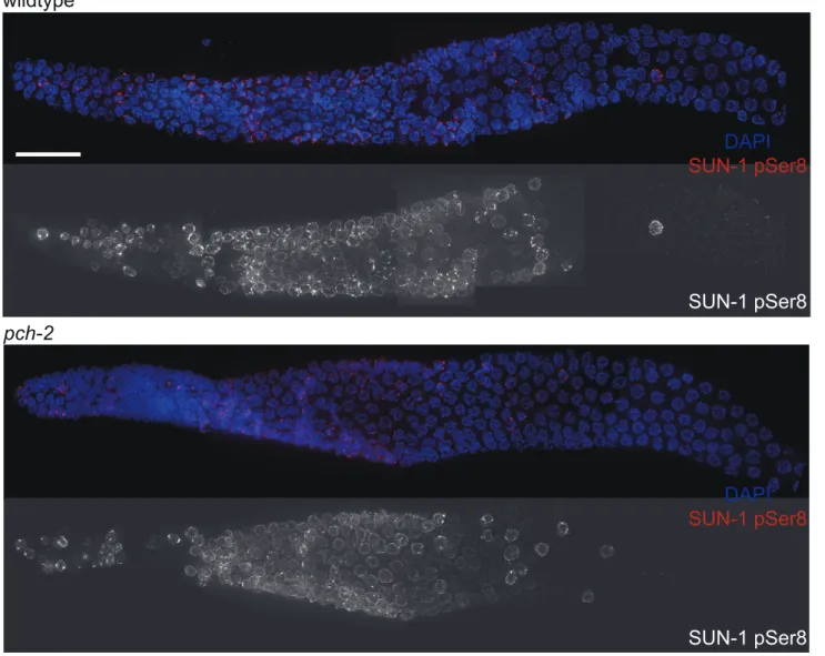
Since both synapsis and meiotic DNA repair were accelerated in pch-2mutants grown at 20uC, we tested whether meiotic nuclei in pch-2mutants entered meiosis earlier or progressed through the germline more rapidly than in wildtype worms at 20uC. Phosphorylation of the nuclear envelope protein SUN-1 occurs at meiotic entry [10,39] and persists until synapsis is complete and meiotic recombination is proceeding normally [10]. We localized SUN-1 phosphorylated on serine 8 (SUN-1 pSer8) in wildtype and pch-2mutants grown at 20uC and did not detect any difference in its appearance or disappearance between the two genotypes (Figure 3).
To determine if nuclei were progressing through the germline more rapidly, we EdU labeled [40] meiotic nuclei in both wildtype andpch-2 mutants (Figure S3A) and visualized their progression through the various stages of meiotic prophase. Both wildtype and pch-2mutants grown at 20uC exhibited a similar progression of EdU labeled nuclei throughout the timecourse (Figures S3B and C). Both of these assays lead us to conclude that the effects on
synapsis and recombination that we observed in pch-2 mutants
were not merely the result of more rapid meiotic entry or progression of nuclei through the germline but that PCH-2 is specifically required to restrain these events.
In summary, we report accelerated rates of synapsis and meiotic DNA repair, accompanied by subtle defects in synapsis and recombination, inpch-2mutants grown at 20uC (Figures 1Cii and 2Bii). Synapsis and meiotic DNA repair were less affected inpch-2 mutants cultured at 15uC (Figures 1Ci and 2Bi) and these mutant worms did not have any recombination or synapsis defects (Figures 1Ci, 2C and 2G). Any difference in the rate of SC
assembly and meiotic DNA repair between wildtype and pch-2
mutants was lost at 25uC (Figures 1Ciii and 2Biii) and this correlated with an increase in the frequency of synapsis and recombination defects in wildtype worms (Figures 1Ciii, 2C and
2G). Moreover, defects in SC disassembly in pch-2 mutants
correlated with defects in meiotic recombination (Figures 1Cii, 1Ciii, 2C and 2G).
Mutation ofpch-2suppresses pairing defects in the absence of synapsis
Our previous experiments indicated that mutating pch-2 had
subtle effects on synapsis and recombination. To more completely dissect how PCH-2 might be affecting homolog interactions, we investigated the effect of mutating pch-2 in sensitized mutant backgrounds.
Figure 1. PCH-2’s role in regulating synapsis is temperature sensitive.A. Cartoon of theC. elegansgermline divided into six zones of equal size. In all graphs, meiotic progression is from left to right. B. Meiotic nuclei in wildtype worms stained with antibodies against the axial element component HTP-3 (red) and the central element component SYP-1 (green). Most nuclei exhibit complete colocalization on HTP-3 and SYP-1. One nucleus (indicated by arrow) includes stretches of HTP-3 without SYP-1, indicating the presence of unsynapsed chromosomes. Grayscale images of HTP-3 and SYP-1 are also provided. Unless stated otherwise, scale bars in all images represent 5 microns. C. Histograms representing the percentage of nuclei that exhibit complete synapsis as a function of meiotic progression in wildtype andpch-2mutant worms at 15uC (i), 20uC (ii) and 25uC (iii). Error bars indicate 95% confidence interval. For all graphs, a * indicates a p value,0.05 and a ** indicates a p value of,0.0001. Significance was assessed by performing Fisher’s exact test.
First we analyzed pairing in syp-1 and syp-1;pch-2 double
mutants. syp-1 mutants fail to assemble SC between paired
homologs, allowing the visualization of pairing intermediates that typically precede and promote synapsis [35,36]. InC. elegans,cis -acting sites called Pairing Centers (PCs) are required for efficient pairing and synapsis [35]. In the absence of synapsis, homologous chromosomes exhibit stable pairing of PC ends but not non-PC ends of chromosomes [36]. This interaction depends on both
homologs having functional PCs [35]. We monitored
synapsis-independent, PC-dependent pairing of X chromosomes as a
function of meiotic progression by performing immunofluores-cence against HIM-8 [41] (Figure 4A). In bothsyp-1single mutants andpch-2;syp-1double mutants, PCs ofXchromosomes initiated pairing in zone 2 and maintained this pairing until late meiotic prophase (zone 6) (Figure 4B). Interestingly, the PC end of X
chromosomes was more frequently paired in pch-2;syp-1 double
Figure 2. PCH-2’s role in regulating recombination is temperature sensitive.A. Meiotic nuclei in early pachytene in wildtype andpch-2 mutant worms stained with DNA (blue) and antibodies against RAD-51 (red). B. Histograms representing the average number of RAD-51 foci per nucleus as a function of meiotic progression in wildtype andpch-2mutant worms at 15uC (i), 20uC (ii) and 25uC (iii). Error bars indicate standard error of the mean. C. Histogram representing the percentage of nuclei with less than six GFP::COSA-1 foci per nucleus in wildtype andpch-2mutant worms grown at 15uC, 20uC and 25uC. The number of nuclei assayed for each genotype are as follows: wildtype at 15uC, 303, at 20uC, 304, and 25uC, 339; pch-2at 15uC, 318, at 20uC, 293, and 25uC, 360. D. A meiotic nucleus stained with DAPI (blue) with six GFP::COSA-1 foci (green). E. A meiotic nucleus stained with DAPI (blue) with five GFP::COSA-1 foci (green). F. Representative images of late meiotic nuclei without (top) and with (bottom) achiasmate chromosomes (arrows). G. Histogram representing the percentage of meiotic nuclei with achiasmate chromosomes in wildtype andpch-2 mutant worms grown at 15uC, 20uC and 25uC. The number of nuclei assayed for each genotype are as follows: wildtype at 15uC, 91, at 20uC, 156, and 25uC, 109;pch-2at 15uC, 96, at 20uC, 170, and 25uC, 106. For all graphs, a * indicates a p value,0.05 and a ** indicates a p value of,0.0001. Significance was assessed by performing either a paired t-test (2B) or Fisher’s exact test (2C).
doi:10.1371/journal.pgen.1004291.g002
Figure 3. The appearance and disappearance of phosphorylated SUN-1 is not affected by mutation ofpch-2.Dissected germlines stained with DAPI (blue) and antibodies against SUN-1 pSer8 (red) in wildtype andpch-2mutant worms. Grayscale images of SUN-1 pSer8 for both genotypes are also provided. Scale bar represents 20 microns.
mutants in zone 2 than syp-1single mutants, at levels similar to wildtype in the same region of the germline (Figure 4B). Therefore, loss ofpch-2suppressed the pairing defect ofsyp-1mutants early in prophase. To determine if this effect was an indirect consequence of a reduction in apoptosis affecting the overall population of meiotic nuclei inhabiting hermaphrodite germlines, we also
monitored X chromosome PC pairing in spo-11;syp1 double
mutants, which display a similar level of germline apoptosis as pch-2;syp-1 mutants due to abrogation of the DNA damage
checkpoint [23]. This double mutant background did not display
more stable association of X chromosome PCs (Figure 4B),
indicating that PCH-2 destabilizes synapsis-independent, PC-dependent pairing.
Loci that are located some distance away from PCs also exhibit synapsis-independent pairing, although to a lesser extent than that observed at PCs [36]. To evaluate the effect of mutatingpch-2on pairing of an autosomal, internally located locus, we performed
FISH against the 5 s rDNA locus (Figure 4C). In syp-1 single
B
**
*
**
Figure 4. Mutation ofpch-2suppresses pairing defects insyp-1mutants.A. Meiotic nuclei insyp-1andsyp-1;pch-2mutants stained with DAPI (blue) and antibodies against theXchromosome PC protein HIM-8 (red). B. Histogram representing the percentage of nuclei with paired HIM-8 signals as a function of germline position in wildtype (from Figure S1A),syp-1,syp-1;pch-2, andsyp-1;spo-11mutants. C. Meiotic nuclei insyp-1and syp-1;pch-2mutants stained with DAPI (blue) and a FISH probe against the 5 s rDNA locus (red). D. Histogram representing the percentage of nuclei with paired FISH signals as a function of germline position in wildtype (from Figure S1B),syp-1,syp-1;pch-2, andsyp-1;spo-11mutants. Error bars in both graphs indicate 95% confidence intervals. A * indicates a p value,0.05 and a ** indicates a p value of,0.0001. Significance was assessed by performing Fisher’s exact test.
*
Figure 5. Mutation ofpch-2suppresses synapsis defects inmeDf2heterozygotes.A. Meiotic nuclei inmeDf2/+andmeDf2/+;pch-2double
mutants, there was a gradual increase in pairing of this locus until zone 3 (Figure 4D). Once again, inpch-2;syp-1double mutants, we observed higher levels of pairing at this locus in both zones 2 and 3 (Figure 4D), indicating that PCH-2 also negatively regulates pairing at a non-PC locus.
Mutation ofpch-2suppresses synapsis defects inmeDf2
heterozygotes
Given that mutation ofpch-2affected homolog interactions at both PC and non-PC loci, we determined what effect loss ofpch-2 would have on synapsis that could not initiate at two stably paired PCs. meDf2is a deficiency that removes theXchromosome PC
[35,42]. Animals heterozygous for meDf2 (meDf2/+) exhibit
unsynapsed X chromosomes in 50% of meiotic nuclei [35]
(Figure 5B), illustrating that homolog synapsis can occur, albeit inefficiently, when only one chromosome has a functional PC. We tested whether mutation ofpch-2inmeDf2heterozygotes had any effect on the synapsis defect in these hermaphrodites. When we monitored synapsis as a function of meiotic progression inmeDf2/ + animals, there was a gradual increase in meiotic nuclei with complete colocalization of HTP-3 and SYP-1, which mirrored the
gradual increase in pairing that has been reported for the X
chromosome in meDf2 heterozygotes [35]. By zone 5, 48% of
meiotic nuclei had completely assembled SC between all homolog pairs while the remainder exhibit stretches of HTP-3 without SYP-1 (arrows in Figure 5A), which are the unsynapsedXchromosomes
(Figure 5C). pch-2;meDf2/+ double mutants also exhibited a
gradual increase in the percentage of meiotic nuclei with complete synapsis (Figures 5A and B). However, in this double mutant background, most meiotic nuclei (87%) achieved complete synapsis in zone 5 (Figure 5B), indicating that mutation ofpch-2 suppressed the synapsis defect ofmeDf2heterozygotes. These data explain the reduction in apoptosis we previously reported in meDf2/+;pch-2double mutants when compared tomeDf2/+single mutants [23]. We verified that synapsis was occurring between homologous X chromosomes by performing FISH against a non-PC locus inpch-2;meDf2/+double mutants (Figures 5C and D).
We tested whether the synapsis in pch-2;meDf2/+ double
mutants was functional for crossover formation by analyzing the number of nuclei with achiasmate chromosomes inmeDf2/+single
mutants and pch-2;meDf2/+ double mutants (Figure 5E). Intro-duction of thepch-2mutant allele suppressed the appearance of
achiasmate chromosomes in meDf2 heterozygotes (Figure 5E),
indicating that synapsis in pch-2;meDf2/+ double mutants can support crossover formation.
We next evaluated the percentage of male-self progeny produced by these double mutants. Since males (with a singleX chromosome) typically arise from spontaneous non-disjunction of
Xchromosomes at a low frequency in wildtype hermaphrodites
(with twoXchromosomes), mutations that increase the frequency ofXchromosome non-disjunction will exhibit an increase in male
self-progeny. For example, meDf2/+ hermaphrodites produced
11.6% male progeny (Table 1). Consistent with our analysis of
synapsis and recombination in pch-2;meDf2/+ double mutants,
these hermaphrodites produced 0.9% male self-progeny (Table 1),
indicating that X chromosomes segregated correctly during
meiosis. Altogether, these data provide additional evidence that PCH-2 normally acts to restrain synapsis, even in situations in which synapsis cannot initiate from two stably paired PCs [35].
Mutation ofpch-2exacerbates meiotic defects in mutants defective in germline apoptosis and HTP-3 function
Thus far, the acceleration of pairing, synapsis and DNA repair in pch-2 mutants suggested a role for the gene product in coordinating these events, potentially to promote quality control. If this model is correct, we reasoned that loss of bothpch-2 gene function and other known mechanisms that contribute to quality control during meiotic prophase might increase the frequency of meiotic defects. For example, germline apoptosis removes defective nuclei in late meiotic prophase to prevent the production of aneuploid gametes [23]. To test our hypothesis, we introduced a mutation inced-4, a gene that encodes a core component of the apoptotic machinery that is required for germline apoptosis [43], into pch-2 mutants and monitored the number of nuclei with
achiasmate chromosomes. ced-4 single mutants had achiasmate
chromosomes in 1% of their late prophase nuclei (Figure 6). This frequency of nuclei with achiasmate chromosomes corresponds with the frequency of wildtype meiotic nuclei with less than six GFP::COSA-1 foci (2%), consistent with germline apoptosis
Xchromosome indicates thatXchromosomes are paired inmeDf2/+;pch-2mutants. D. Histogram representing the percentage of nuclei with pairedX FISH signals as a function of meiotic progression inmeDf2/+andmeDf2/+;pch-2mutant worms. Error bars in both graphs indicate 95% confidence
intervals. E. Histogram representing the number of late meiotic nuclei with achiasmate chromosomes inmeDf2/+(54 nuclei) andmeDf2/+;pch-2(78 nuclei). A * indicates a p value,0.05. Significance was assessed by performing Fisher’s exact test.
doi:10.1371/journal.pgen.1004291.g005
Table 1.Embryonic viability and frequency of male self-progeny of various mutants.
Genotype Embryonic Viability Frequency of male self-progeny
wildtype 100% 0.1%
pch-2(tm1458) 100% 0.7%
mnDp66/+;meDf2/+ 97.5% 11.6%*
mnDp66/+;meDf2/+;pch-2(tm1458) 98.2% 0.9%*
ced-4(n1162) 90.8% 0.2%
ced-4(n1162);pch-2(tm1458) 75.7% 0.2%
htp-3(vc75) 100% 0.1%
htp-3(vc75);pch-2(tm1458) 81.9% 1.1%
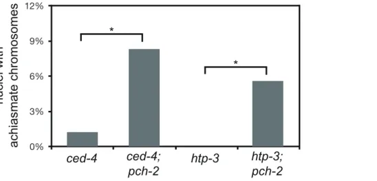
culling defective nuclei even during wildtype meiosis. 8% of nuclei in pch-2;ced-4 double mutants had achiasmate chromosomes (Figure 6), indicating that significantly more meiotic nuclei in pch-2mutants have meiotic defects than was represented by the frequency of nuclei with achiasmate chromosomes in the single mutant. Moreover, the increase in meiotic nuclei with achiasmate
chromosomespch-2;ced-4double mutants was accompanied by an
increase in the production of inviable progeny, indicating the missegregation of chromosomes during meiosis (Table 1).
We wanted to further test our hypothesis that PCH-2 was involved in quality control during meiotic prophase by introducing thepch-2mutation into another mutant background to determine if the double mutants would exhibit a more severe meiotic phenotype. However, sincepch-2mutants suppress some meiotic phenotypes (Figures 4 and 5), we reasoned that we should use a mutation in which the meiotic phenotype was relatively mild, indicating no major defects in meiosis. In this scenario, loss ofpch-2 could reveal a more severe meiotic phenotype.
HORMA domain containing proteins promote many events during meiotic prophase, including pairing, synapsis and recom-bination between homologous chromosomes [34,44–48]. There
are four genes in the C. elegans genome that encode meiotic
HORMA domain containing proteins (him-3,htp-1,htp-2, and htp-3) and recently a hypomorphic allele of htp-3,vc75, has been identified. Worms with this mutation appear to undergo meiosis normally, producing no achiasmate chromosomes (Figure 6), but exhibit minor defects in DNA repair and SC disassembly [49]. We
generated pch-2;htp-3(vc75) double mutants and monitored the
viability of its progeny and chiasmata formation in these double mutants.pch-2;htp-3(vc75)double mutants had a synthetic meiotic phenotype: Although each individual mutant produced no inviable progeny, the double mutant generated 20% inviable progeny (Table 1). This reduction in viability can be explained by the significant increase in achiasmate chromosomes (Figure 6), indi-cating thatpch-2;htp-3(vc75)mutants had a more severe defect in recombination than either single mutant. We ruled out that this synthetic meiotic phenotype could be explained by the possibility that pch-2 mutants activate an HTP-3 dependent meiotic DNA damage response by analyzing SC disassembly in double mutants. When mutations that activate an HTP-3 dependent meiotic DNA damage response are combined with thehtp-3(vc75)mutant allele,
these double mutants disassemble their SCs more rapidly than the htp-3(vc75) single mutant [49]. This did not occur in pch-2; htp-3(vc75)double mutants (data not shown). Thus, introduction of thepch-2mutation exacerbates the meiotic phenotype of a weak mutant allele ofhtp-3, consistent with a role for PCH-2 in quality control during meiotic prophase.
PCH-2 localizes to synapsed chromosomes when inter-homolog DNA repair mechanisms are active
To gain a deeper understanding of how PCH-2 might be regulating pairing, synapsis and recombination, we generated a polyclonal antibody against the PCH-2 protein and localized it in wildtype worms. In wildtype meiotic nuclei, PCH-2 was expressed in germline nuclei before they adopted the polarized morphology that is indicative of meiotic entry, termed the transition zone (Figures 7A and B). In these nuclei, PCH-2 formed foci that did not colocalize with axial elements, such as HTP-3 [34,35] (data not shown), or with HIM-8 [33] (data not shown). After meiotic entry, PCH-2 localized to meiotic chromosomes in a manner reminiscent of proteins that compose the SC (Figure 7A and B). PCH-2 was loaded into chromosomes in the transition zone (Figures 7A and B) and present in the portion of the worm germline that corresponds to pachytene (Figure 7A). However, unlike SC proteins, PCH-2 was absent from meiotic chromosomes in the most distal region of the germline, corresponding to late pachytene (Figure 6A). To confirm this localization pattern, we co-stained wildtype meiotic nuclei with antibodies against PCH-2 and the SC component SYP-1 [36]. In mid-pachytene, SYP-1 and PCH-2 colocalized (Figure 7C), consistent with PCH-2 localizing to the SC. In late pachytene, SYP-1 was present on meiotic chromosomes but PCH-2 was not (Figure 7D), indicating it was removed from the SC before the SC disassembled.
We tested the genetic requirements for PCH-2 loading on meiotic chromosomes. PCH-2 was not present in germline nuclei when pch-2 was inactivated by RNA interference in wildtype worms (data not shown). Synapsis is required for PCH-2 localization to the SC, since PCH-2 localization was lost insyp-1 mutants [36] (Figure 7E). However, homologous synapsis was not, as illustrated by PCH-2’s localization to the SC inhtp-1(Figure 7F) and sun-1 mutants (data not shown), in which chromosomes synapse non-homologously [46,50], and inieDf2mutants (data not
*
*
Figure 6. Mutation ofpch-2exacerbates meiotic defects in mutants defective in germline apoptosis and HTP-3 function.Histogram representing the percentage of late meiotic nuclei inced-4(79 nuclei),ced-4;pch-2(68 nuclei),htp-3(97 nuclei) andhtp-3;pch-2(107 nuclei) mutants. A * indicates a p value,0.05. Significance was assessed by performing Fisher’s exact test.
shown), in which chromosomes self-synapse [51]. PCH-2 was present on the SC when meiotic recombination is disrupted, such as in msh-5 [52], zhp-3 [53], spo-11 [54] (Figure S5), or rtel-1 mutants [55,56] (data not shown). We also determined that htp-3(vc75) mutants and wildtype animals grown at 25uC exhibited normal localization of PCH-2 (data not shown).
The region of the germline where PCH-2 was absent from meiotic chromosomes is associated with a shift in double strand break (DSB) repair from use of the homolog as a repair template, presumably to use of the sister chromatid [57,58]. This shift in DNA repair mechanisms is contemporaneous with the loading of the crossover-promoting factor, COSA-1 [6]. To verify that PCH-2’s absence from meiotic chromosomes coincided with these events, we performed immunofluorescence on wildtype hermaph-rodites harboring a GFP tagged COSA-1 transgene [6] with antibodies against PCH-2. PCH-2 was indeed absent from nuclei that had robust GFP::COSA-1 foci (Figure S4).
The shift in DNA repair mechanisms is dependent on the activity of the MAP kinase pathway [57], the crossover-promoting factor, msh-5, and the anti-recombinase, rtel-1 [58]. PCH-2’s removal from meiotic chromosomes in late pachytene did not
occur in mpk-1 (Figure 7G) and msh-5 mutant backgrounds
(Figures 7I and S5) but did in rtel-1 mutants (Figure 7J). This relocalization also failed to occur when we alleviate the meiotic
progression defect in mpk-1mutants by RNAi of a downstream
effector, dpl-1 [59] (Figure 7H), indicating that an inability to remove PCH-2 from meiotic chromosomes is not merely an indirect consequence of meiotic prophase arrest inmpk-1mutants. PCH-2 was also present on the SC in late pachytene nuclei in spo-11 and zhp-3 mutants, suggesting that PCH-2’s perdurance is a general response to defects in recombination, similar to the persistence of phosphorylated SUN-1 [10] (Figure S5).
PCH-2 is required to promote homolog-dependent meiotic DNA repair
The timing of PCH-2 localization and removal from the SC (Figures 7C and D), along with the effects that mutation ofpch-2 had on meiotic recombination (Figure 2), led us to speculate that PCH-2 might specifically be required to promote homolog dependent DNA repair. To test this, we first monitored the dynamics of RAD-51 loading and removal insyp-1andpch-2;syp-1 mutants (Figure 8A). The complete absence of SC between all
homologous chromosomes in syp-1 mutants [36] results in an
inability to repair programmed DSBs because of the absence of closely aligned homologous chromosomes that can be used as repair templates [37]. As a result, RAD-51 foci persist until late pachytene (zone 6 in Figure 8A), when a synapsis-independent
repair program is activated. When we compared syp-1 and
pch-2;syp-1mutants, we observed that mutation ofpch-2reduced the average number of RAD-51 foci per nucleus insyp-1mutants in zone 5 (Figure 8A). Since our previous experiments ruled out the possibility that mutation of pch-2 reduced the number of DSBs (Figure S1), these data indicated that some DSBs were being repaired in pch-2;syp-1 double mutants by synapsis-independent mechanisms, presumably by using the sister chromatid as a repair template. Despite this premature activation of
synapsis-indepen-dent DSB repair mechanisms, pch-2;syp-1 double mutants
exhibited an extended transition zone [36] and prolonged zone of meiotic nuclei with phosphorylated SUN-1 [10], indicating that meiotic progression is unaffected by mutation ofpch-2in syp-1mutants (data not shown). Moreover, our data also raise the possibility that the partial suppression of recombination defects in pch-2;syp-1 double mutants is temporally constrained to mid-pachytene (zone 5) since the average number of RAD-51 foci was not significantly different betweensyp-1 andpch-2;syp-1mutants in earlier zones of the germline (Figure 8A).
We directly tested the role of PCH-2 in promoting homolog-dependent meiotic DNA repair. We used a meiotic recombination assay recently developed to monitor when meiotic chromosomes can access their homolog for DNA repair [58]. This assay takes advantage of an insertion of theMos1transposon into theunc-5 locus on one homologous chromosome, which disrupts the gene. DSBs are generated in young adult hermaphrodites carrying this locus when heat shock induced expression of the Mos transposase
excises the Mos1 transposon. Since the other homologous
chromosome contains a mutation that inactivates theunc-5gene elsewhere in the gene, the outcome of DNA repair produces a wildtype unc-5(+) allele, allowing these recombinants to be phenotypically identified among the progeny laid by heat-shocked worms. The mode of DNA repair can be determined by
monitoring the progeny of unc-5(+) recombinants, since the
presence of a linked marker (dpy-13) can distinguish non-crossover from crossover recombinants (Figure 8B). Given the spatiotempo-ral organization of meiotic nuclei in the hermaphrodite germline, recombinants laid at different time points reflect the competence of meiotic chromosomes to use their homolog for DNA repair at different stages of meiotic prophase.
When we performed this assay with wildtype andpch-2mutant hermaphrodites, we did not recover any recombinants in the first time point (10–22 hours post-heat shock) in both genetic backgrounds (Figure 8C), which corresponds to late pachytene in the hermaphrodite germline when homolog-dependent repair is not active [57,58]. Given the subtle defects in pairing, synapsis and recombination we observed inpch-2mutants (Figures 1, 2 and 3), we divided the next twelve-hour time point (22–34 hours post heat shock) into two six hour timepoints (22–28 hours post heat shock and 28–34 hours post heat shock). Wildtype animals produced recombinant progeny in the 22–28 hour window after heat shock at a frequency of 4%, reflecting the increased competence of nuclei in mid-pachytene for DNA repair from a homologously synapsed partner. In pch-2 mutants, statistically significant fewer recombinants were generated during this period and of the nine recombinants identified, none were crossovers. In wildtype worms, 21% of recombinants recovered at this timepoint were crossovers.
The difference in the frequency of recombinants between wildtype andpch-2mutants was lost in later timepoints (Figure 8C). By the next six-hour interval (28–34 hours post heat-shock), wildtype hermaphrodites generated recombinants at a frequency of 5% andpch-2mutants at a frequency of 4%. Wildtype andpch-2 mutant hermaphrodites generated equivalent frequencies of recombinants until the end of the timecourse. Thus, PCH-2 is required to maintain access to homologous chromosomes for meiotic DNA repair during pachytene.
meiotic chromosomes. E. Meiotic nuclei insyp-1mutants stained with antibodies against HTP-3 (red) and PCH-2 (green). F. Meiotic nuclei inhtp-1 mutants stained with antibodies against HTP-3 (red) and PCH-2 (green). G. Meiotic nuclei inmpk-1mutants stained with antibodies against SYP-1 (red) and PCH-2 (green). H. Meiotic nuclei inmpk-1mutants in whichdpl-1has been knocked down by RNAi stained with antibodies against SYP-1 (red) and PCH-2 (green). I. Meiotic nuclei in late pachytene inmsh-5mutants stained with antibodies against SYP-1 (red) and PCH-2 (green). J. Meiotic nuclei in late pachytene inrtel-1mutants stained with antibodies against HTP-3 (red) and PCH-2 (green).
To determine what effect premature loss of homolog access had on crossover formation inpch-2mutants, we monitored recombi-nation genetically in wildtype andpch-2mutant animals using five single nucleotide polymorphisms (SNPs) that spanned 95% of
Chromosome IIIor the Xchromosome [60] (Figure 8D). pch-2
mutants had slightly lower rates of recombination in all intervals across both chromosomes, resulting in a reduction of the genetic length of both ChromosomeIII(from 55.4 to 43.1 cM) and theX chromosome (from 58.5 to 45.8 cM). However, the decrease in genetic distance between SNPs was less severe at the PC end of
both chromosomes. In addition, mutation of pch-2 appeared to
more strongly affect the genetic distance between SNPs in the center of ChromosomeIII. We observed several double crossovers
in wildtype animals on both Chromosome III and the X
chromosome, 8 and 10 respectively. In contrast to wildtype, pch-2mutants had significantly fewer double crossovers, only 2 on the
X chromosome and none on Chromosome III. Therefore, the
acceleration of homolog-dependent DNA repair in pch-2 single
mutants at 20uC, as assayed both by the appearance and
disappearance of RAD-51 foci (Figure 2Bii) and the ability of chromosomes to access their homolog for DNA repair (Figure 8C), reduced the genetic length of chromosomes, albeit not uniformly among genetic intervals, and the frequency of double crossovers.
Mutation ofpch-2reduces the percentage of nuclei with six GFP::COSA-1 foci inmeDf2homozygotes
The localization pattern of PCH-2 inmsh-5mutants suggested that the persistence of PCH-2 on meiotic chromosomes into late pachytene might be a cytological read-out for prolonged homolog access and that PCH-2 might be required for this extension. However, we could not directly test this possibility inpch-2;msh-5 double mutants since the assay to monitor homolog access relies on large numbers of viable progeny to assess significance [58]. Instead, we chose to address this possibility inmeDf2homozygotes. In this mutant, asynapsis ofXchromosomes produces changes in recombination frequencies in some genetic intervals and a loss of crossover control so that autosomes sometimes experience double crossovers [61]. This redistribution of recombination events in meDf2homozygotes could, in part, be the consequence of PCH-2 remaining on meiotic chromosomes in late pachytene and promoting inter-homolog recombination in response to defects in synapsis.
First we determined how PCH-2 localization was affected by
asynapsis of X chromosomes and whether any change in its
localization correlated with characterized responses to asynapsis.
We stained meDf2 hermaphrodite germlines with antibodies
against PCH-2 and phosphorylated SUN-1 [10]. The staining pattern for both was extended (Figure 9A), in contrast to their staining patterns in wildtype germlines (Figures 3 and 7A). This extension was accompanied by a delay in the appearance of GFP::COSA-1 foci (data not shown).
Next, we analyzed the number of GFP::COSA-1 foci per nucleus [6] (Figure 9B). InmeDf2mutants, the majority of germline nuclei (58%) exhibited five GFP::COSA-1 foci. However, a
substantial fraction of meiotic nuclei (34%) had six GFP::COSA-1 foci. Even taking into account that GFP::COSA-10% of these nuclei had
undergone successful synapsis of X chromosomes [35], this
suggested that about one-quarter of meiotic nuclei had an autosome with an additional putative crossover event. When we
analyze GFP::COSA-1 foci in pch-2;meDf2 double mutants, we
observed a statistically significant reduction in the number of germline nuclei that have six foci (22%) and a corresponding increase in the number of nuclei with five foci (74%). Mutation of pch-2had no effect on the percent of nuclei with complete synapsis
in homozygous meDf2 mutants (data not shown). These results
suggest that cytologically, PCH-2 is required for the increase in putative double crossovers inmeDf2mutants. However, nuclei with six GFP::COSA-1 foci were still present in pch-2;meDf2 double mutants, indicating that additional mechanisms promote
homo-log-dependent meiotic DNA repair in meDf2 mutants. This
possibility was supported by the similar average numbers of RAD-51 foci in bothmeDf2andmeDf2;pch-2mutants (Figure 9C). We also assessed double crossover formation genetically in both meDf2 and pch-2;meDf2 double mutants on Chromosome III.
Mutation of pch-2 did not ameliorate the minor shifts in
recombination pattern that we observed inmeDf2. Indeed, when represented as fractions of wildtype recombination rates, the
recombination landscape was more severely affected in
pch-2;meDf2 double mutants than meDf2 single mutants. There was no difference in the frequency of double crossovers betweenmeDf2 and meDf2;pch-2 (Figure 9D), likely because of the removal of meiotic nuclei by germline apoptosis (see Discussion).
Discussion
Homolog pairing, synapsis and meiosis specific DNA repair mechanisms are essential for the formation of chiasmata and proper meiotic chromosome segregation. Moreover, pairing synapsis and recombination can be temporally and/or mechanis-tically linked, depending on the organism in question. Therefore, a major challenge during meiotic prophase is to coordinate these events and monitor their progression to ensure that they are occurring appropriately, in the correct order and ultimately give rise to homologous chromosomes linked by at least one crossover. The acceleration of pairing (Figure 4), synapsis (Figures 1C and 5) and inter-homolog recombination (Figures 2B, 8A and 8C) in pch-2mutants results in the loss of crossover assurance (Figures 2C, 2G and Figure 8D), a defect that becomes more severe when germline apoptosis is abrogated or meiotic DNA repair is mildly disrupted (Figure 6). Therefore, we propose that PCH-2 restrains pairing, synapsis and recombination to coordinate them and promote their quality control to assure the obligate crossover. The variability in meiotic defects betweenpch-2mutants and wildtype worms grown at different temperatures suggests that PCH-2 restrains these events by introducing a kinetic barrier to the stabilization of pairing, synapsis and/or recombination interme-diates. This barrier is largely restored topch-2mutants undergoing meiosis at reduced temperature (Figures 1Ci, 2C and 2G), suppressing meiotic defects seen inpch-2mutants grown at higher Figure 8.pch-2is required to maintain inter-homolog DNA repair in mid-pachytene.A. Histogram representing the average number of RAD-51 foci per nucleus as a function of meiotic progression insyp-1andpch-2;syp-1mutant worms. Error bars indicate standard error of the mean. A ** indicates a p value,0.0001. Significance was assessed by performing a paired t-test. B. Mos1 excision based DSB repair assay. C. Time-course analysis of inter-homolog recombination induced at the Mos1 site. Chart shows the frequency ofunc-5(+)recombinants among progeny derived
from eggs laid in the indicated time intervals (hours phs) and the frequency of CO and NCO repair types. D. Genetic analysis of meiotic recombination in wildtype andpch-2mutants. Physical and genetic maps of ChromosomeIIIand theXchromosome are depicted to scale. Genetic distance is shown in centimorgans and was calculated as number of recombinants divided by total number of chromosomes assayed. For both C and D, a * indicates a p value,0.05 and significance was assessed by performing Fischer’s exact test.
temperatures, and overcome in wildtype worms incubated at higher temperature (Figures 1Ciii, 2C and 2G), producing meiotic defects even in the presence of functional PCH-2. A similar
observation has been made in budding yeast. pch2 mutants
exhibited fewer defects in recombination when cultured at 30uC
than at 33uC [13]. Given PCH-2’s membership in a family of
proteins characterized by their ability to disassemble macromo-lecular complexes [25], this kinetic barrier is likely the disassembly of pairing, synapsis and/or recombination intermediates.
The localization of PCH-2 is consistent with this model. PCH-2 is expressed when initial pairing interactions likely occur (Figures 7A and B). Once synapsis has initiated, PCH-2’s presence on the SC when meiosis specific homolog-dependent DNA repair mechanisms are active [57,58] (Figures 7A, C and D) suggests that this localization is related to its regulation of meiotic recombina-tion. Indeed, changes in PCH-2 localization when recombination is defective (Figures 7I, S5 and 9A) also support this interpretation. However, its localization to the SC is not strictly necessary for its effect on recombination, since we still observe a reliance on PCH-2 function insyp-1mutants (Figure 8A), which do not assemble SC [36] and fail to localize PCH-2 (Figure 7E). This might suggest that the localization of PCH-2 in nuclei prior to and in the transition zone (Figures 7A and B) could also have implications for the regulation of recombination.
The proposed role of PCH-2 as a factor that disassembles meiotic intermediates is best illustrated by how loss ofpch-2affects pairing and synapsis. We observe PCH-2’s requirement for pairing insyp-1mutants (Figure 4), in which the inability to synapse reveals pairing intermediates that contribute to efficient synapsis in C. elegans[36]. Homologous PC interactions appear more stable in pch-2;syp-1 double mutants, indicating that pch-2 normally destabilizes these interactions and providing a rationale for the
accelerated synapsis we observe in pch-2 single mutants
(Figure 1Cii): If efficient synapsis initiates at stably paired homologous PCs [35], enhanced stabilization of PC interactions will promote premature synapsis. However, synapsis can also initiate, albeit less efficiently, in the absence of stably paired PCs, as illustrated by the frequency of synapsis in a mutant in which one homolog has a PC and the other does not (meDf2heterozygotes) [35]. Since mutation of pch-2 suppresses the synapsis defect of meDf2heterozygotes (Figure 5) and partially suppresses the pairing defect at a non-PC locus insyp-1mutants (Figures 4C and 4D), we support a model in which PCH-2 is required to destabilize pairing intermediates that promote synapsis initiation at both PC and non-PC loci. Indeed, the accelerated synapsis inpch-2single mutants could be a consequence of PCH-2 regulating synapsis at both PC and non-PC sites.
We do not favor a model in which meiotic chromosomes in
pch-2 mutants undergo non-homologous synapsis. If accelerated
synapsis in pch-2 mutants produced non-homologous synapsis,
we predict that pairing at either PC loci, non-PC loci or both would be significantly reduced. Our pairing analysis in wildtype andpch-2mutant worms (Figure S1) does not indicate this. Either loss ofpch-2does not affect the ability of homologs to identify each
other or redundant mechanisms compensate in destabilizing non-homologous interactions.
We hypothesize that loss ofpch-2also inappropriately stabilizes recombination intermediates, producing an acceleration of mei-otic-specific DNA repair, as measured both by the disappearance of RAD-51 from meiotic chromosomes (Figure 2Bii) and the reduced ability to access a homolog as a repair template in mid-pachytene (Figure 8A and 8C). However, we have not directly demonstrated this. It is formally possible that PCH-2’seffect on synapsis indirectly affects the progress of meiotic recombination. The ability to uncouple recombination defects (Figures 2C and
2G) from the acceleration of synapsis (Figure 1C) when pch-2
mutants are incubated at different temperatures and the activation
of synapsis-independent DNA repair pathways in pch-2;syp-1
double mutants (Figures 8A) do not support this interpretation. Moreover, a model in which PCH-2 destabilizes meiotic recombination intermediates could explain the intimate associa-tion of PCH2 with early recombinaassocia-tion proteins in budding yeast
[11] and the phenotypes observed in pch2 and Trip13 mutants.
Genetically, the inability to disassemble intermediates that are precursors to crossovers may result in additional crossovers and appear as loss of interference in some genetic intervals, as reported in budding yeastpch2mutants [13,14]. Cytologically, this defect would result in an increase in the number of foci containing CO-promoting proteins per chromosome and a loss of cytological interference between these foci, as reported inTrip13mutant mice [20]. Consistent with our model, stable recombination intermedi-ates that precede crossover formation, such as the single end invasion intermediate, occur with greater frequency and persist for longer in budding yeastpch2mutants [15]. The reason thatpch-2 mutant worms manifest a different recombination defect (fewer
crossovers than wildtype) than budding yeast pch2 mutants or
Trip13mutant mice (more crossovers than wildtype) is likely due to the dramatically higher levels of DSB formation reported in budding yeast [62] and mice [1], in contrast toC. elegans[58].
If PCH-2 destabilizes both pairing intermediates that produce synapsis and recombination intermediates that promote crossovers inC. elegans, the question remains whether PCH-2 accomplishes these roles by regulating a single event or factor common to pairing, synapsis and recombination or regulates these events independently of each other. Only the identification of a PCH-2 substrate(s) will directly address this issue. Meiotic HORMA domain containing proteins are very strong candidates, given their role in promoting pairing, synapsis and recombination [34,44–48], their characterized relationship withPCH2/Trip13in yeast [13,15] and mice [10] and Pch2’s biochemical activity against Hop1/ DNA complexes in vitro [26]. If PCH-2 modulates these events independent of each other, cofactors that contribute to substrate specificity of this particular AAA-ATPase [25] may explain PCH-2’s ability to modulate multiple meiotic factors and affect multiple meiotic events.
How does the proposed function of PCH-2 explain its role in meiotic checkpoints? Checkpoint signaling is often initiated from a defective intermediate in the process being monitored. We propose Figure 9. Mutation ofpch-2reduces the percentage of nuclei with six GFP::COSA-1 foci inmeDf2homozygotes.A. A dissected germline stained with DAPI (blue) and antibodies against SUN-1 pSer8 (red) and PCH-2 (green). The extension of PCH-2 staining into late pachytene correlates with the extension of phosphorylated SUN-1 staining. Scale bar represents 20 microns. B. Histogram representing the percentage of meiotic nuclei that contain a given number of GFP::COSA-1 foci inmeDf2andmeDf2;pch-2mutants. 174meDf2nuclei and 178meDf2;pch-2nuclei were counted. Error bars represent 95% confidence interval. C. Histogram representing the average number of RAD-51 foci per nucleus inmeDf2andmeDf2;pch-2 mutants. Error bars indicate standard error of the mean. For both graphs, a * indicates a p value,0.05. Significance was assessed by performing either Fisher’s exact test (7B) or a paired t-test (7C). D. Genetic analysis of meiotic recombination in wildtype,pch-2,meDf2andmeDf2;pch-2mutants represented as fractions of wildtype recombination and color-coded according to the legend on the right.
that the synapsis checkpoint is activated by the presence of PCs that are not stably paired and is silenced by stably paired, synapsed PCs. We are not suggesting that the checkpoint is merely
monitoring unpaired PCs since homologous PCs appear to pair at significant steady-state levels insyp-1mutants but nonetheless activate the synapsis checkpoint [23]. Instead, we propose that the Figure 10. Model for role of PCH-2 during crossover formation.See discussion for details.
checkpoint specifically responds to the stability of PC pairing. Thus, PCH-2 promotes checkpoint activation by destabilizing paired homologous PCs and inhibiting synapsis. We hypothesize thatpch-2;syp-1double mutants satisfy the synapsis checkpoint by inappropriately stabilizing paired PCs despite the absence of synapsis, effectively mimicking the stabilization of PC pairing normally accomplished by synapsis. In budding yeast, Pch2 may play a similar role with recombination intermediates that contribute to synapsis initiation, potentially explaining Pch2’s initial characterization as a pachytene checkpoint component [63]. Lending support to this hypothesis, budding yeast pch2 mutants display an increase in synapsis initiation complexes [13], cytologically observed foci that include factors required for both meiotic recombination and synapsis [64,65].
PCH-2 promotes homolog dependent DNA repair to ensure the obligate crossover
PCH-2 is required to maintain interhomolog meiotic recombi-nation (Figure 8). A similar role for Pch2 has been demonstrated in budding yeast [11,12]. However, our experiments reveal that the requirement for PCH-2 in promoting interhomolog meiotic recombination is temporally constrained to mid-pachytene (Figures 8A and C). We cannot interpret any other differences between wildtype andpch-2 mutants at later timepoints (namely
the 58–70 hours phs and 70+ hours phs timepoints) since Mos
transposase may persist after induction and continue to introduce DSBs throughout the germline.
Mos-induced DSBs can be repaired by inter-homolog repair in pch-2mutants prior to this window in mid-pachytene, as indicated by the frequency of recombinants laid soon after the 22–28 hour timepoint (Figure 8C). Thus, PCH-2 appears to be redundant with additional mechanisms that promote inter-homolog meiotic recombination earlier in prophase. In addition,pch-2mutants do not load GFP::COSA-1 prematurely (data not shown), indicating that switching from meiotic inter-homolog DNA repair to a homolog-independent DNA repair mode can be uncoupled from crossover designation. Alternatively, designation may occur earlier, concomitant with loss of homolog access, and visible COSA-1 recruitment stabilizes the designated crossover.
Our model of how PCH-2 regulates meiotic recombination is illustrated in Figure 10. Nuclei begin introducing DSBs coincident with the localization of DSB-1 and DSB-2, proteins required for DSB formation and thought to identify a DSB competent period during prophase [8,9]. A subset of these DSBs become licensed as CO-eligible recombination intermediates [6]. Nuclei in which all chromosomes have a CO-eligible intermediate progress through mid-pachytene and a subset of these CO-eligible intermediates are designated as crossovers in late pachytene [6]. In a small population of nuclei that contain a chromosome without a CO-eligible intermediate, 5–10% based on our studies of GFP::COSA-1 inpch-2mutants (Figure 2C), DSB competence is thought to be prolonged to increase the likelihood that a CO-eligible interme-diate will be formed. This extension is accompanied by PCH-2-dependent promotion of interhomolog recombination, providing a potential explanation for the presence of double crossovers amongst the progeny of wildtype animals that are lost in the progeny ofpch-2mutants (Figure 8D).
Why is there a mechanism unique to mid-pachytene to maintain homolog access? Our model assumes the requirement for PCH-2 in mid-pachytene is linked to the maintenance of DSB competence when a chromosome does not have a CO-eligible intermediate. Some possibilities that could explain our results are: 1) The redundant homolog-dependent repair mechanism(s) active earlier in prophase is down-regulated in mid-pachytene, even if
DSB competence is maintained. We do not favor this possibility (see below). 2) There is something unique about DSBs or the environment in which DSBs are introduced during mid-pachytene that requires additional reinforcement to promote the formation of crossovers. These models are not mutually exclusive.
We favor another interpretation. Since nuclei in mid-pachytene continue to localize PCH-2 to meiotic chromosomes but do not exhibit other molecular markers associated with DSB competence, such as SUN-1Ser8P [7–9] (Figure S6), we suggest that the requirement for PCH-2 in mid-pachytene is independent of DSB competence. This possibility takes into account events that are thought to occur during mid-pachytene. CO-eligible intermediates have been licensed but they have not yet been designated. Villeneuve and colleagues have speculated that a reinforcement step precedes designation, whereby a single CO-eligible interme-diate per chromosome is reinforced by CO-promoting mecha-nisms and ultimately stabilized by designation [6]. Since our analysis of pairing, synapsis and meiotic DNA repair indicates that PCH-2 typically restrains meiotic events, another way to interpret the loss of homolog access inpch-2mutants is that it reveals the prematurity of another event, namely reinforcement of crossovers. Since licensed CO-eligible intermediates that are not reinforced are presumably repaired by NCO mechanisms [6], homolog access would be maintained during this process. In the absence of PCH-2 and the more rapid reinforcement of CO-eligible intermediates, homolog access during mid-pachytene would also be lost. This could explain the redistribution of crossovers inpch-2 mutants (Figure 8D). We think it unlikely that PCH-2 signals the absence of CO-eligible intermediates as its loss does not affect the staining pattern of SUN-1Ser8P [10] and DSB-2 [7–9] (data not shown) in genetic backgrounds that have defects in recombination and/or synapsis (i.e.meDf2andsyp-1). Indeed, crossover distribu-tion in pch-2;meDf2 double mutants appears to be the additive
effect of loss of pch-2 and the delay in meiotic progression
introduced by asynapsis (Figure 9D), suggesting that these events are independent of each other.
Multiple independent feedback mechanisms promote crossover assurance
In contrast to wildtype hermaphrodites, PCH-2 localizes to the SC of meiotic chromosomes in late pachytene when synapsis and/ or recombination are defective (Figures 7I, S5 and 9A). This delay in PCH-2 removal correlates with other indicators associated with a delay in meiotic prophase progression, such as phosphorylation of SUN-1 [10] (Figures S5 and 9A) and persistence of DSB-1 and DSB-2 [8,9] (see nucleus labeled defective meiosis in Figure 10). We suggest that the continued localization of PCH-2 to meiotic chromosomes in mutant backgrounds that perturb synapsis and/ or recombination (Figures S5 and 9A) indicates the existence of a feedback mechanism that maintains inter-homolog recombination to promote crossover assurance. Such a model would explain the
delay in the appearance of GFP::COSA-1 foci inmeDf2mutants
Since the number and kinetics of RAD-51 focus formation are indistinguishable betweenrad-54andrad-54;pch-2(Figure S2), we also conclude that this feedback mechanism is distinct from one that has been proposed to prolong DSB competence in response to defects in synapsis and/or recombination [7–9]. PCH-2’s proposed role in maintaining interhomolog repair mechanisms in response to defects in meiotic recombination potentially clarifies
why PCH2 is required for crossover homeostasis [13,14]. A
reduction in DSB formation increases the likelihood that chromosomes do not have a CO-eligible recombination interme-diate and would activate described feedback mechanisms, including an attempt to increase DSBs [7–9] and prolong interhomolog access for repair. Since DSB formation is compro-mised, there is a greater reliance on maintaining homolog dependent meiotic DNA repair, a pathway (partially) dependent on PCH2. This potential relationship between defective meiotic recombination and Pch2’s ability to maintain inter-homolog repair into late meiotic prophase could also explain why prophase arrest rescues a reduction in DSB activity but only affects wildtype meiosis minimally [66], especially if Pch2 activity is regulated similarly in budding yeast as inC. elegans.
However, mutation of pch-2 does not eradicate nuclei with
greater than five GFP::COSA-1 nuclei in the meDf2background
(Figure 9B). Moreover, our analysis of RAD-51 in meD2;pch-2
(Figure 9C) does not indicate the activation of homolog-independent repair mechanisms, in contrast to our studies with syp-1;pch-2mutants (Figure 8A). Therefore, we also conclude that at least one other pathway in C. elegans, in addition to PCH-2, maintains homolog access in response to defects in synapsis and recombination and that this pathway relies on synapsis for its activity. An attractive candidate for this pathway is the one that we hypothesize acts earlier in meiotic prophase and may be coupled to DSB competence [8,9].
Materials and Methods
Genetics
The wildtypeC. elegansstrain background was Bristol N2 [67]. All experiments were performed at 20uunder standard conditions unless otherwise stated. Mutations and rearrangements used were as follows:
LG I: mnDp66, rtel-1(tm1866), rad-54&snx-3(ok615), zhp-3(jf61),
hT2[bli-4(e937) let-?(q782) qIs48]
LG II: pch-2(tm1458), meIs8 [pie-1p::GFP::cosa-1+unc-119(+)] LG III: htp-3(vc75), dpy-5(e61), mpk-1(ga111), ced-4(n1162) LG IV: spo-11(ok79), htp-1(gk174), msh-5(me23), dpy-13(e184) unc-5(ox171::Mos1), unc-5(e791),ieDf2,nT1[unc-?(n754) let-?(m435)] (IV, V), nTI [qIs51]
LG V: sun-1(jf18), syp-1(me17), krIs14 [hsp-16.48::MosTransposase; unc-122::gfp; lin-15(+)]
LG X: meDf2
Some strains were provided by the CGC, which is funded by
NIH Office of Research Infrastructure Programs (P40
OD010440).
Antibodies, Immunostaining, Fluorescence in situ hybridization, EdU labeling, and microscopy
Polyclonal rabbit antibodies directed against the first 100 amino acids of PCH-2 were generated by Strategic Diagnostic Inc. (SDI, Newark, DE).
Immunostaining was performed as in [23]. Primary antibodies were as follows (dilutions are indicated in parentheses): rabbit anti-SYP-1 (1:500) [36], guinea pig anti-SYP-1 (1:500) [36], guinea pig anti-HTP-3 (1:500) [35], chicken anti-HTP-3 (1:1000)
[35], rabbit anti-RAD-51 (1:1000) [37], guinea pig anti-SUN-1S8Pi (1:700) [68] and rabbit anti-PCH-2 (1:1000). Secondary antibodies were Cy3 anti-chicken, anti-guinea pig and anti-rabbit (Jackson Immunochemicals), Alexa-Fluor 555 rabbit, anti-guinea pig, and anti-chicken (Invitrogen) and Cy5 anti-anti-guinea pig and anti-chicken (Jackson Immunochemicals).
Fluorescence in situ hybridization was performed as described in [33].
Quantification of synapsis, pairing and RAD-51 foci was performed with a minimum of three whole germlines per genotype. For each figure, the number of nuclei assayed for each genotype in each zone is provided in Table S1.
EdU labeling was performed as described in [58]. L4 hermaphrodites were aged for 8–10 hours and moved to plates withE. colilabeled with EdU to promote EdU labeling ofC. elegans meiotic nuclei. A minimum of 15 germlines was scored for each genotype at each timepoint.
All images were acquired using a DeltaVision Personal DV system (Applied Precision) equipped with a 1006N.A. 1.40 oil-immersion objective (Olympus), resulting in an effective XY pixel spacing of 0.064 or 0.040mm, and a 606oil-immersion objective (Olympus), resulting in an effective XY pixel spacing of 0.11 or 0.067mm.
Three-dimensional image stacks were collected at 0.2-mm Z-spacing
and processed by constrained, iterative deconvolution. Image scaling and analysis were performed using functions in the softWoRx software package. Projections were calculated by a maximum intensity algorithm. Composite images were assembled and some false coloring was performed with Adobe Photoshop.
Genetic analysis of meiotic recombination
The wild-type Hawaiian CB4856 strain and the Bristol N2 strain were used to assay recombination between single nucleotide
polymorphisms (SNPs) on Chromosomes III and X. The SNPs
used on Chromosome III were: pkP3081, pkP3095, pkP3101,
pkP3035, and pkP3080. The SNPs used on theXchromosome
were: pkP6139, pkP6120, pkP6157, pkP6161, and pkP6170 [60]. To measure wild-type recombination, N2 males containing bcIs39were crossed to Hawaiian CB4856 worms. Cross-progeny
hermaphrodites were identified by the presence of bcIs39 and
contained one N2 and one CB4856 chromosome. These were assayed for recombination by crossing with males containingmIs11 and N2 SNPs. Cross-progeny hermaphrodites from the resulting mate were isolated as L4s, and then cultured individually in 96-well plates in liquid S-media complete supplemented with HB101, carbenicillin, and Nystatin. Four days after initial culturing, starved populations were lysed and used for PCR and restriction digest to detect CB4856 SNP alleles. For recombination inpch-2 mutants, strains homozygous for the CB4856 background of the
relevant SNPs were created, then mated with pch-2; bcIs39.
Subsequent steps were performed as in the wild-type worms.
Mos1excision-induced DSB repair assay