Male Germline But Little Evidence for Chromosomal
Dosage Compensation or Meiotic Inactivation
Colin D. Meiklejohn*, Emily L. Landeen, Jodi M. Cook, Sarah B. Kingan, Daven C. Presgraves Department of Biology, University of Rochester, Rochester, New York, United States of America
Abstract
The evolution of heteromorphic sex chromosomes (e.g.,XYin males orZWin females) has repeatedly elicited the evolution of two kinds of chromosome-specific regulation: dosage compensation—the equalization of X chromosome gene expression in males and females— and meiotic sex chromosome inactivation (MSCI)—the transcriptional silencing and heterochromatinization of the X during meiosis in the male (orZ in the female) germline. How theX chromosome is regulated in theDrosophila melanogastermale germline is unclear. Here we report three new findings concerning gene expression from theXinDrosophila testes. First, Xchromosome-wide dosage compensation appears to be absent from most of the Drosophila male germline. Second, microarray analysis provides no evidence for X chromosome-specific inactivation during meiosis. Third, we confirm the previous discovery that the expression of transgene reporters driven by autosomal spermatogenesis-specific promoters is strongly reduced when inserted on the X chromosome versus the autosomes; but we show that this chromosomal difference in expression is established in premeiotic cells and persists in meiotic cells. The magnitude of theX-autosome difference in transgene expression cannot be explained by the absence of dosage compensation, suggesting that a previously unrecognized mechanism limits expression from the X during spermatogenesis inDrosophila. These findings help to resolve several previously conflicting reports and have implications for patterns of genome evolution and speciation inDrosophila.
Citation:Meiklejohn CD, Landeen EL, Cook JM, Kingan SB, Presgraves DC (2011) Sex Chromosome-Specific Regulation in theDrosophilaMale Germline But Little Evidence for Chromosomal Dosage Compensation or Meiotic Inactivation. PLoS Biol 9(8): e1001126. doi:10.1371/journal.pbio.1001126
Academic Editor:Michael B. Eisen, University of California Berkeley, United States of America
ReceivedDecember 23, 2010;AcceptedJuly 8, 2011;PublishedAugust 16, 2011
Copyright:ß2011 Meiklejohn et al. This is an open-access article distributed under the terms of the Creative Commons Attribution License, which permits unrestricted use, distribution, and reproduction in any medium, provided the original author and source are credited.
Funding:This work was supported by funds from an Ernst Caspari Fellowship (ELL), the NSF (www.nsf.gov; CDM, DEB-0839348), the Alfred P. Sloan Foundation (www.sloan.org; DCP), the David and Lucile Packard Foundation (www.packard.org; DCP), and the University of Rochester (DCP). The funders had no role in study design, data collection and analysis, decision to publish, or preparation of the manuscript.
Competing Interests:The authors have declared that no competing interests exist.
Abbreviations:DCC, dosage compensation complex; FDR, false discovery rate; MSCI, meiotic sex chromosome inactivation; MSUC, meiotic silencing of unpaired chromatin; RNAi, RNA interference
* E-mail: [email protected]
Heteromorphic sex chromosome systems (withXYmales orZW
females) have evolved independently many times in animals and plants [1]. The difference between the sexes in chromosome copy number—e.g., twoX’s in females but only oneXin males—and the general absence of recombination between X and Y
chromosomes have resulted in the evolution of sex chromosome-specific content and organization [2–4], rates of mutation and substitution [5], and most conspicuously, chromosome-level regu-lation. Two kinds of chromosomal regulation, in particular, have evolved repeatedly: dosage compensation, the process that equalizes
Xchromosome gene expression levels between theXYandXXsexes, and meiotic sex chromosome inactivation (MSCI), the facultative heterochromatinization and early transcriptional silencing of theX
and theY chromosome in germline cells entering meiosis in XY
individuals [6,7].
Dosage compensation, by far the better characterized of the two processes, has evolved in XY (mammals, Drosophila), XO (nema-todes), but not, it seems, inZWtaxa (birds and Lepidoptera [8,9]). While mechanisms of dosage compensation differ [10]—from silencing of a singleXinXX female cells in eutherian mammals [11] to hypertranscription of the singleXinXYmales inDrosophila
[12]—its function is to equalize the balance of X to autosomal
in ovaries and testes, consistent with X chromosome dosage compensation [2,3,22]. Together these findings have suggested that a DCC-independent mechanism of X chromosome dosage compensation occurs in theDrosophilamale germline [22,23].
MSCI, which is less well characterized, occurs in mammals, nematodes, grasshoppers (XO), and possibly in birds [24]. In mice, MSCI is observable cytologically in pachytene spermatocytes as theXandYchromosomes are sequestered into a distinct region of the nucleus [25]. During MSCI, multiple epigenetic modifications are localized to the X and Y (reviewed in [7]) and there are profound consequences forXchromosome gene expression—over 80% ofX-linked genes decrease in expression by 10-fold or more [26]. Although 33 multicopy X-linked gene families are actively transcribed post-meiotically [27], most single-copyX chromosom-al genes remain repressed in post-meiotic spermatids [26]. The function of MSCI is also less obvious than dosage compensation. The most general model posits that MSCI functions to silence selfish segregation distorter elements, which tend to accumulate preferentially on theX chromosome [28–32] (for other possible functions, see [7,33]). Surprisingly, the existence of MSCI in
Drosophila has been disputed for decades. Lifschytz and Lindsley argued that MSCI is universal and essential in all male XYtaxa [6,34]. They inferred MSCI in Drosophila from cytological and genetic findings including, but not limited to, their claim of allocyclic condensation of the X chromosome in primary sper-matocytes and the dominant male-specific sterility of ,75% of
X-autosome translocations [6]. Consistent with Lifschytz and Lindsley’s observations, Rastelli & Kuroda [18] found that H4Ac12, a histone mark enriched in heterochromatin in somatic cells, may label the X-Y cluster in late primary spermatocytes, whereas H3K4me3, a histone mark associated with active transcription, may be depleted from theX-Ycluster [35]. Kremer et al. [36], however, claim that the euchromatin of theXis entirely decondensed during a considerable period of first meiotic prophase, ‘‘contradictory to the results and the model of Lifschytz and Lindsley’’ (p. 158). McKee and Handel [33] further suggest that the cytological evidence for MSCI inDrosophila is inconclusive and the genetic data indirect.
Instead, they argue that MSCI functions to prevent harmful crossing over betweenXandYchromosomes in theXYsex, and as
Drosophilamale meiosis is achiasmate, MSCI need not occur. Two recent experiments appear to provide renewed support for MSCI inDrosophila. First, Parsch and colleagues [37,38] found that the promoter sequence ofocnus, an autosomal gene that encodes a putative sperm-specific histone (possibly a transition protein or protamine) [39], can drive strong testis-specific expression of alacZ
reporter when transgenes are inserted onto autosomes but not when inserted onto theXchromosome. Similar results have been observed for autosomal versusX-linked transgene inserts with the promoter of another testis-specific gene,b2-tubulin [40]. Second, using stage-specific microarray analyses of premeiotic, meiotic, and postmeiotic cell populations dissected from testes, Vibranovski et al. [41] found a small but significant excess of genes on theX
chromosome that show reduced expression in meiotic relative to premeiotic stages of spermatogenesis. These studies are consistent with MSCI but provide somewhat conflicting pictures of the process. The transgene reporter assays, for instance, suggest that MSCI reduces expression from theXchromosome more than 5-fold [37,40], whereas the microarray analyses suggest that MSCI is relatively weak, causing only,10% reduction in the expression of
X-linked genes in meiotic cells on average [41].
In this article, we study the regulation of the Drosophila X
chromosome in the male germline, revisiting earlier studies and reporting results from new analyses and experiments. First, we show that, contrary to previous reports, theXdoes not appear to undergo X chromosome dosage compensation in the Drosophila
male germline. Second, we find no evidence for an excess ofX -linked genes showing reduced expression in meiotic cells in the previously published microarray data [41], suggesting that MSCI inDrosophilaeither does not exist or is sufficiently weak to escape detection by microarray analysis. Finally, we find that the sperm-specific ocnus transgenes show much lower expression when X -linked versus autosomal, as previously reported [37,38], but that this marked chromosomal difference is established early, in pre-meiotic cells. In theDrosophila male germline, then, both a lack of dosage compensation and an as yet unrecognized premeiotic mechanism appear to limit expression from theXchromosome. Our results help to resolve several seemingly conflicting findings regarding the regulation of theX chromosome in theDrosophila
male germline and have implications for patterns of genome evolution and speciation inDrosophila.
Results
NoXChromosome Dosage Compensation inDrosophila
Spermatocytes
In theDrosophilamale germline, decreased expression from the
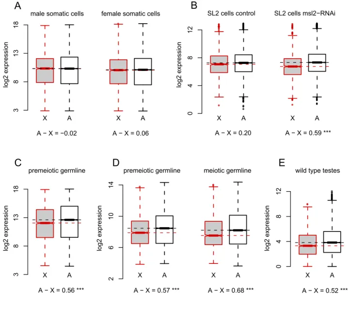
Xchromosome could plausibly reflect MSCI or the stage-specific loss ofXchromosome dosage compensation. To distinguish these possibilities, we asked if X chromosome dosage compensation occurs in premeiotic spermatocytes. As controls, we first estimated levels of X chromosome dosage compensation in male somatic tissues, using microarrays to assay gene expression in thorax dissected from adult males and females. Cells in the thorax are likely to be similar between the sexes (i.e., largely comprising flight muscle), thus minimizing the confounding effects of sex-specific gene expression. Global gene expression is indeed highly cor-related between male and female thorax (r =0.972, p,10215; Figure S1). Furthermore, the difference in median expression level between X-linked and autosomal probes is negligible, with autosomal probes showing 0.98- and 1.04-fold higher expression levels in males and females, respectively, than X-linked ones Author Summary
(Figure 1A). As expected, in these cells, theXchromosome is fully dosage compensated and there is equal expression from theXand the autosomes in both sexes.
To determine the magnitude of theX-autosome difference in expression expected in the absence of dosage compensation, we referred to data from published microarray experiments using
Drosophila male-like SL2 cells in which mRNA encoding the limiting dosage compensation protein, MSL2 [42], was knocked down by RNA interference (RNAi) [43]. In control cells treated with RNAi against GFP, autosomal genes have a slight (1.15-fold) but significantly higher median expression than X-linked genes (Mann-WhitneyPMW= 0.01; Figure 1B), whereas inmsl2
-knock-down cells, autosomal genes have a 1.51-fold higher median expression thanX-linked ones (PMW,10215; Figure 1B).
Impair-ment of the DCC in these experiImpair-ments therefore results in a 1.31-fold reduction inX-linked gene expression relative to the auto-somes. Similar RNAi knockdown ofmsl2andmofinSL2cells, with gene expression measured by RNA-seq, results in a 1.35-fold decrease inX-linked gene expression relative to autosomes [44]. Similarly, male larvae carrying mutations at theroX loci show a 1.20-fold difference betweenXand autosomal expression [45,46]. Loss of DCC-dependent dosage compensation therefore results in a 1.2- to 1.4-fold decrease in expression of X-linked genes compared to autosomal ones.
X A ● X A 38 3 18 1
A − X = −0.02 A − X = 0.06
A
male somatic cells female somatic cells
● ● ● ● ● ● ● ● ● ● ● ● ● ● ● ● ● ● ● ● ● ● ● ● ● ● ● ● ● ● ● ● ● ● ● ● ● ● ● ● ● ● ● ● ● ● ● ● ● ● ● ● ● ● ● ● ● ● ● ● ● ● ● ● ● ● ● ● ● ● ● ● ● ● ● ● ● ● ● ● ● ● ● ● ● ● ● ● ● ● ● ● ● ● ● ● ● ● ● ● ● ● ● ● ● ● ● ● ● ● ● ● ● ● ● X A ● ● ● ● ● ● ● ● ● ● ● ● ● ● ● ● ● ● ● ● ● ● ● ● ● ● ● ● ● ● ● ● ● ● ● ● ● ● ● ● ● ● ● ● ● ● ● ● ● ● ● ● ● ● ● ● ● ● ● ● ● ● ● ● ● ● ● ● ● ● ● ● ● ● ● ● ● ● ● ● ● ● ● ● ● ● ● ● ● ● ● ● ● ● ● ● ● ● ● ● ● ● ● ● ● ● ● ● ● ● ● X A 04 8 2 1
A − X = 0.20 A − X = 0.59 ***
B
SL2 cells control SL2 cells msl2−RNAi
D
X A 38 3 18 1A − X = 0.56 ***
C
premeiotic germline ● X A 26 0 14 1A − X = 0.57 *** premeiotic germline
● ●
X A
A − X = 0.68 *** meiotic germline ● ● ● ● ● ● ● ● ● ● ● ● ● ● ● ● ● ● ● ● ● ● ● ● ● ● ● ● ● ● ● ● ● X A
A − X = 0.52 ***
0
4
82
1
E
wild type testes
log2 expression
log2 expression
log2 expression
log2 expression log2 expression
To directly test forXchromosome dosage compensation in the
Drosophila male germline, we used microarrays to assay gene expression in cells dissected from the apical tip of the testes with the somatic and DCC-expressing [18] cells of the surrounding testes sheath removed. These apical dissections comprise hub cells, germline and somatic stem cells, somatic cyst cells, mitotic spermatogonia, and early primary spermatocytes, which grow for approximately 3 d following their last mitotic division prior to the first meiotic division [47]. We chose these dissected cells (for convenience, hereafter called ‘‘premeiotic’’) rather than whole testes to avoid conflating our results with meiosis-specific X
chromosome regulation, such as MSCI. In these premeiotic cells, median absolute expression of autosomal probes is 1.47-fold higher thanX-linked probes (PMW,1026; Figure 1C). The precise
magnitude of this X-autosome difference depends somewhat on the extent to which lowly expressed probes are filtered from the analysis but ranges from 1.39-fold to 1.54-fold (Figure S2).
To evaluate the generality of our estimated,1.5-fold difference in
X-autosome expression, we analyzed data from two previous studies. In the first study, Vibranovski et al. [41] dissected three populations of cells fromDrosophilatestes: apical tips enriched for premeiotic cells; proximal cells enriched for late-stage primary and meiotically dividing spermatocytes (hereafter ‘‘meiotic’’); and distal cells enriched for postmeiotic cysts and elongating spermatids (hereafter ‘‘postmeiotic’’). We observe a similar X-autosome expression difference in their premeiotic dissections that included the somatic testis sheath [41]: autosomal probes show 1.48-fold higher median expression than theX
(PMW,10210; Figure 1D). In proximal dissections (which did not
include testis sheath) [41], the autosomes show a 1.60-fold higher median expression than the X (PMW,10210; Figure 1D). In the
second study, Gan et al. generated RNA-seq data from whole testes [48]. Based on 19,849,063 uniquely mapped reads, we estimate that autosomal genes show 1.44-fold greater expression versus X-linked genes (PMW,10210; Figure 1E).
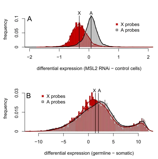
In addition to comparing expression from the X and the autosomes within a tissue, we compared differences in expression between cell types forX-linked and autosomal genes. Themsl2 -RNAi experiments [43] again provide a useful control, where the median difference in expression between msl2-knockdown cells and control cells is 1.05-fold for autosomal probes and 0.80-fold forX-linked probes (Figure 2A). The difference in expression levels between cells with and without dosage compensation in these experiments is therefore 1.32-fold lower for X-linked genes than for autosomal genes. The analogous difference in expression between germline and somatic cells is complicated by large tissue-specific differences in gene expression (Figure 2B). However, despite the confounding effects of tissue-specific expression, the difference in median expression levels between male thorax and premeiotic dissections is 1.48-fold lower forX-linked probes than for autosomal probes, a value similar to that from themsl2-RNAi experiments. Thus, across three independent experiments using differently dissected stages of spermatogenesis, whole testes, and across three different gene expression assays (Affymetrix micro-arrays, Agilent micromicro-arrays, and RNA-seq), we find that the X
chromosome has reduced expression relative to the autosomes. The magnitude of this difference is strikingly similar to that seen for experimentally manipulated cells lacking dosage compensation. We therefore conclude thatXchromosome dosage compensation is absent from most of theDrosophilamale germline.
XChromosome Expression in theDrosophilaFemale Germline and in Germline Stem Cells
To test if reduced expression from theXis a general feature of germline expression, rather than a male-specific absence of germline
X chromosome dosage compensation, we estimated X and autosomal expression levels in wildtype ovaries from the RNA-seq data of Gan et al. [48]. In contrast to the testes, autosomal genes show 0.89-fold lower median expression than X-linked genes (PMW= 0.027; Figure 3). Reduced expression from theXrelative
to the autosomes is therefore specific to the testes and not a general property of germline gene expression inDrosophila.
We also estimated X and autosomal expression levels using RNA-seq data from mutant male and female germline tissue in which development is arrested at an early stage [48]. The bag-of-marbles (bam)gene is required for male germline cells to exit the mitotic divisions and begin primary spermatocyte development, and bammutant gonads are consequently enriched for undiffer-entiated germ-line stem cells and mitotic spermatogonia [49,50]. In bam ovaries, X-linked and autosomal expression levels are similar to wild-type ovaries: autosomal genes show 0.91-fold lower median expression thanX-linked genes (PMW= 0.035; Figure 3). In
bammutant testes, however, we find that autosomal genes show a 1.13-fold higher median expression thanX-linked genes (Figure 3), a value that is significantly different from zero (PMW,0.001), but
smaller than the ,1.45-fold difference seen in wild-type testes. Notably, primary spermatocytes are absent from bam testes but likely constitute most of the premeiotic cells dissected from the apical tip of the testes. The discrepancy in the X-autosome difference in expression between bam testes (1.13-fold) and premeiotic dissections (1.45-fold) therefore suggests that the X -autosome difference in expression increases in differentiating primary spermatocytes.
XChromosome Expression in Late Meiotic
Spermatocytes—A Modest Dearth of Upregulated Genes But No Excess of Downregulated Genes
The magnitude of the X-autosome difference in expression in
Drosophila testes described above is consistent with a lack of X
chromosome dosage compensation but not with global inactiva-tion of theX. In mice, MSCI initiates at pachytene of prophase I [7], resulting in transcriptional silencing of more than 80% ofX -linked genes [26,27]. Assuming Drosophila males experience a similar stage-specific inactivation of the sex chromosomes, cells in late prophase I undoubtedly represent a small proportion even of meiotic dissections enriched for late primary spermatocytes. Any signal of MSCI might therefore only be detected by comparing the changes inXand autosomal expression across different stages of spermatogenesis [41]. As described above, Vibranovski et al. [41] dissected populations of cells from wild-type testes enriched for premeiotic, meiotic, and postmeiotic cells and assayed gene expression with microarrays. Using a novel Bayesian analysis of allX-linked and autosomal probes, these authors reported a small but significant excess ofX-linked genes downregulated in meiotic dissections relative to premeiotic dissections (56% of X-linked versus 52% of autosomal genes identified as testis-expressed in FlyAtlas [51], see Figure 3 in [41]).
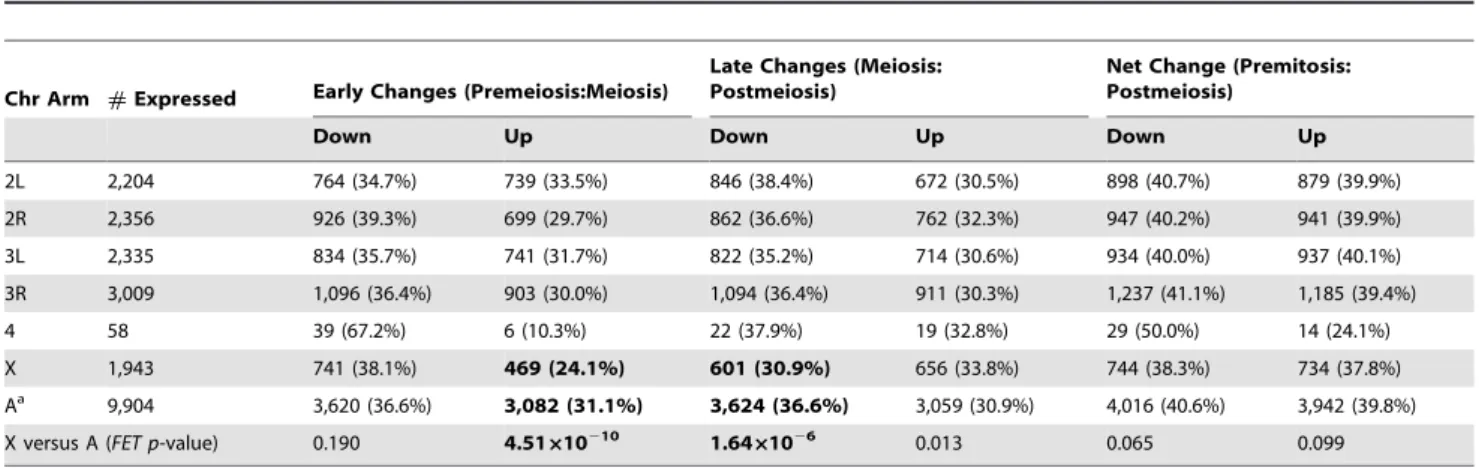
auto-somal probes, respectively, show significant decreases in expres-sion, whereas 24% and 31% of X-linked and autosomal probes show significant increases in expression, respectively (Table 1). While the proportion of genes downregulated in meiotic cells is similar for theXand autosomes (Fisher’s exact testPFET= 0.190),
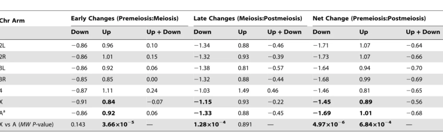
the Xhas a significant paucity of genes upregulated in meiotic cells (PFET= 4.5610210). Of those probes that show significant
changes in the early transition, the median magnitude of de-creased expression is similar for theXand autosomes (Table 2), but X-linked probes show significantly smaller increases in expression (PMW= 3.6661025). The deficit of upregulated
X-linked genes in the early transition was found by Vibranovski et al. [41], but they also reported a small but significant excess of
X-linked genes downregulated in the early transition, which we do not observe.
A different pattern emerges for the late transition (meioticR
postmeiotic cells): 31% and 37% of X-linked and autosomal probes, respectively, show significant decreases in expression, whereas 34% and 31% ofX-linked and autosomal probes show significant increases in expression, respectively (Table 1). TheX
has a significant deficit of probes downregulated in postmeiotic cells (PFET= 1.661026), and a marginally significant excess of
probes upregulated in postmeiotic cells (PFET= 0.013). During
the late transition, the magnitude of decreased expression is significantly less for the X than for the autosomes (PMW=
1.2861024; Table 2), whereas the magnitude of increased expression is similar (PMW= 0.891).
The behavior of the X chromosome in the Drosophila male germline is therefore distinct from MSCI as it occurs in mammals [26], at least at the resolution afforded by these dissections. Instead
differential expression (MSL2 RNAi − control cells)
y
c
n
e
u
q
erf
−2
−1
0
1
2
X
A
X probes
A probes
05
0.
01
.
0
A
differential expression (germline − somatic)
y
c
n
e
u
q
erf
−10
−5
0
5
10
X probes
A probes
X A
05
1
0.
0
3
0.
0
B
Figure 2. Differences in X-linked and autosomal gene expression between male-like SL2 cells with and without dosage compensation are similar to the differences between somatic and germline cells in males.(A) The distributions of expression differences betweenmsl2-RNAi and control cells forXchromosome and autosomal probes [43]. (B) The distributions of expression differences between male germline cells and male thorax tissue forXand autosomal probes. Black lines indicate the median values of each distribution; the difference between the median log2 expression of autosomal andX-linked probes is 0.398 in (A) and 0.568 in (B).
of an inactivation of the Xchromosome during prophase I that results in strong decreases in the number and magnitude of expressedX-linked genes that then largely persists throughout the remainder of spermatogenesis [26,27], we see an overall dampening of the change in gene expression on theXrelative to the autosomes: a smaller proportion ofX-linked genes change in expression at either stage of spermatogenesis and, of those that do change, the median fold-change is,10%–20% smaller than that seen on the autosomes (Tables 1 and 2, Figure S3).
In contrast to the rest of the genome, the largely heterochro-matic fourth chromosome shows an excess of downregulated genes in the meiotic dissections (Table 1): 67% of fourth chromosome probes decrease expression in the early transition (Fisher’s exact test of fourth chromosome probes versus all others:PFET= 3.56
1026), whereas only 10% increase expression (PFET= 7.66102 4
). The magnitude of expression changes at both transitions is, however, similar for the fourth and theXand autosomes (Table 2). The fourth chromosome results show that combining these testes dissections with microarray analysis [41] provides sufficient resolution to detect large-scale chromosome-wide changes in expression during spermatogenesis. The absence of a comparable pattern for theXchromosome is thus not simply due to a lack of statistical power or experimental resolution.
It is worth noting that the genes showing significant changes in expression in meiotic cells relative to premeiotic ones fit what might be expected ofDrosophila spermatogenesis. Those showing significantly elevated expression in meiotic cells, for instance, are enriched for functions in microtubule activity (e.g., dynein complex, axoneme function) and sperm development (e.g., vesicle and membrane docking), whereas those showing significantly reduced expression are enriched for transcriptional functions (e.g., RNA pol II activity, RNA splicing, mRNA processing). These findings are consistent with overall reduced postmeiotic de novo
transcriptional activity and a shift to posttranscriptional mecha-nisms of development during spermatogenesis in Drosophila
[47,49,53,54].
Differential Somatic Contamination between Premeiotic and Meiotic Cell Dissections
Our inference that there is little signal of MSCI in these dissections [41] is conservative, as the proportion ofX-linked genes downregulated in meiotic cells is likely overestimated in these microarray data. The premeiotic dissections from the apical tip of the testes included the surrounding testes sheaths—which are somatic, express the DCC [18], and are thus presumably dosage compensated—whereas the meiotic dissections from the proximal
log2 expression ● ● ● ● ● ● ● ● ● ● ● ● ● ● ● ● ● ● ● ● ● ● ● ● ● ● ● ● ● ● ● ● ● ● ● ● ● ● ● ● ● ● ● ● ● ● ● ● ● ● ● ● ● ● ● ● ● ● ● ● ● ● ● ● ● ● ● ● ● ● ● ● ● ● ● ● ● ● ● ● ● ● ● ● ● ● ● ● ● ● ● ● ● ● ● ● ● ● ● ● ● ● ● ● ● ● ● ● ● ● ● ● ● ● ● ● ● ● ● ● ● ● ● ● ● ● ● ● ● ● ● ● ● ● ● ● ● ● ● ● ● ● ● ● ● ● ● ● ● ● ● ● ● ● ● ● ● ● ● ● ● ● ● ● ● ● ● ● ● ● X A
A − X = −0.162 *
● ● ● ● ● ● ● ● ● ● ● ● ● ● ● ● ● ● ● ● ● ● ● ● ● ● ● ● ● ● ● ● ● ● ● ● ● ● ● ● ● ● ● ● ● ● ● ● ● ● ● ● ● ● ● ● ● ● ● ● ● ● ● ● ● ● ● ● ● ● ● ● ● ● ● ● ● ● ● ● ● ● ● ● ● ● ● ● ● ● ● ● ● ● ● ● ● ● ● ● ● ● ● ● ● ● ● ● ● ● ● ● ● ● ● ● ● ● ● ● ● ● ● ● ● ● ● ● ● ● ● ● ● ● ● ● ● ● ● ● ● ● ● ● ● ● ● X A
A − X = −0.139 *
● ● ● ● ● ● ● ● ● ● ● ● ● ● ● ● ● ● ● ● ● ● ● ● ● ● ● ● ● ● ● ● ● ● ● ● ● ● ● ● ● ● ● ● ● ● ● ● ● ● ● ● ● ● ● ● ● ● ● ● ● ● ● ● ● ● ● ● ● ● ● ● ● ● ● ● ● ● ● ● ● ● ● ● ● ● ● ● ● ● ● ● ● ● ● ● ● ● ● ● ● ● ● ● ● ● ● ● ● ● ● ● ● ● ● ● ● ● ● ● ● ● ● ● ● ● ● X A
A − X = 0.172 ***
0
48
2
1
wild type ovaries bam mutant ovaries bam mutant testes
Figure 3.Xchromosome and autosome expression is similar in ovaries and germline stem cells.RNA-seq data [48] from wild-type ovaries,bammutant ovaries, andbammutant testes. *p,0.05, ***p,0.001 (Mann-Whitney test).
doi:10.1371/journal.pbio.1001126.g003
Table 1.Number of genes with significant differences in expression between stages of spermatogenesis.
Chr Arm #Expressed Early Changes (Premeiosis:Meiosis)
Late Changes (Meiosis: Postmeiosis)
Net Change (Premitosis: Postmeiosis)
Down Up Down Up Down Up
2L 2,204 764 (34.7%) 739 (33.5%) 846 (38.4%) 672 (30.5%) 898 (40.7%) 879 (39.9%)
2R 2,356 926 (39.3%) 699 (29.7%) 862 (36.6%) 762 (32.3%) 947 (40.2%) 941 (39.9%)
3L 2,335 834 (35.7%) 741 (31.7%) 822 (35.2%) 714 (30.6%) 934 (40.0%) 937 (40.1%)
3R 3,009 1,096 (36.4%) 903 (30.0%) 1,094 (36.4%) 911 (30.3%) 1,237 (41.1%) 1,185 (39.4%)
4 58 39 (67.2%) 6 (10.3%) 22 (37.9%) 19 (32.8%) 29 (50.0%) 14 (24.1%)
X 1,943 741 (38.1%) 469 (24.1%) 601 (30.9%) 656 (33.8%) 744 (38.3%) 734 (37.8%) Aa 9,904 3,620 (36.6%) 3,082 (31.1%) 3,624 (36.6%) 3,059 (30.9%) 4,016 (40.6%) 3,942 (39.8%) X versus A (FET p-value) 0.190 4.51610210 1.6461026 0.013 0.065 0.099
regions of the testes included only germline cells [41] that lackX
chromosome dosage compensation (see above). The presence of contaminating sheath tissue could therefore inflateXchromosome expression levels in premeiotic samples, causing a spurious inference of downregulation in meiotic cells. To test for an effect of the presence of somatic sheath cells on the observed expression of X-linked genes in premeiotic versus meiotic cells in the microarray data, we dissected three cell populations from the testis: apical tips with testis sheath (premeiotic+sheath), apical tips without testis sheath (premeiotic), and proximal dissections without sheath (meiotic). Using quantitative reverse transcriptase-PCR (qPCR), we assayed expression of 15 genes: 12 at different cyto-logical positions on theXwith high overall expression levels in the microarray data and significant changes from meiotic cells to premeiotic cells, and three chosen as normalizing controls because they showed no significant change in expression between pre-meiotic and pre-meiotic cells (CG1440on theX,Tub84Don3R, and
CG10252on3R; see Materials and Methods).
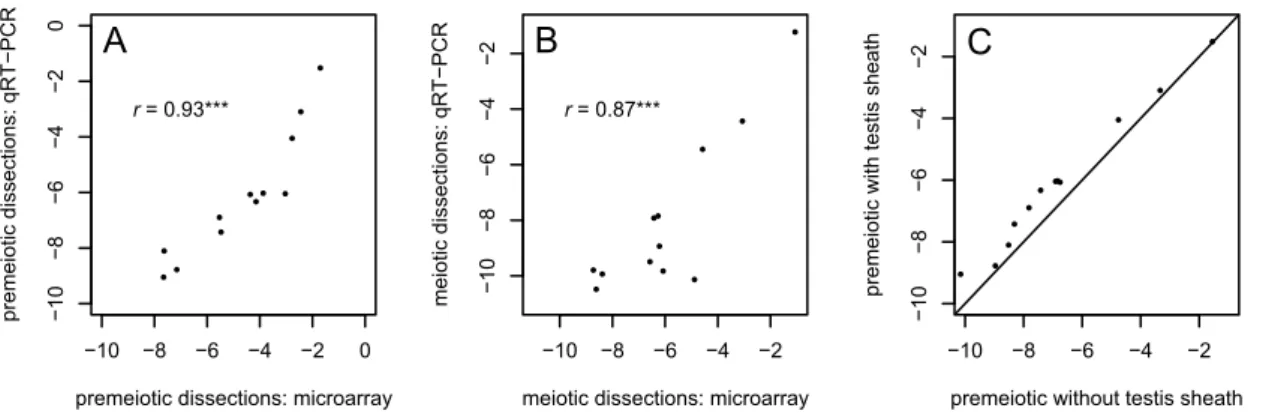
Relative expression levels of all 12 genes in both our premeiotic dissections including sheath and meiotic dissections recapitulate the previous microarray analysis well (Figure 4A,B) [41]. For 11 of the 12 genes, our qPCR results show the same direction and similar magnitude of expression change between stages (Table 3). All 12X-linked genes show greater expression in premeiotic cells with the testis sheaths than when the sheaths are removed (binomial test p= 4.961024; Figure 4C). This difference is sig-nificant for six of the 12 genes (p,0.05), and highly significant when all 12 genes are pooled (p,1028; Table 3). These results suggest that, on average, the differences in expression levels ofX -linked genes between premeiotic and meiotic cells in Vibranovski et al. [41] might be overestimated by as much as 36%. Con-sequently, the proportions and magnitudes of X-linked genes upregulated and downregulated in meiosis shown in Tables 1 and 2 are likely underestimates and overestimates, respectively. Sheath contamination also likely contributes to the greater difference between X and autosomal expression seen in the meiotic dis-sections relative to the premeiotic disdis-sections (Figure 1C).
XChromosome-Specific Reduction inWOLandYLZ Transgene Expression Is Independent of Spermatogenic Stage
We next extended the analysis of two transgenes used by Hense et al. as putative reporters of MSCI in Drosophila [37]. In both
transgene constructs, lacZ expression is driven by a 110-bp promoter-containing sequence from the 59-region ofocnus(ocn), an autosomal (3R) gene that encodes a putative sperm-specific histone [37,39]: P[wFl:ocn:lacZ:w+] and P[y+:YEStes:ocn:lacZ] (hereafter
WOL and YLZ, respectively). WOL and YLZ constructs differ from one another in two ways: YLZ possesses the ocn 39-UTR downstream oflacZas well as flanking Suppressor of Hairy-wing binding sites, which function as chromosomal insulators [37]. Previously, Hense et al. [37] showed thatX-linked inserts of the
WOLandYLZtransgenes show significantly lower expression than autosomal inserts in both mRNA and protein levels in males.
We confirmed that the transgenes show strong sex- and chromosome-specific expression differences by assaying mRNA transcript levels in whole adult females homozygous for singleX -linked or autosomal transgene inserts and in whole adult males hemizygous for single X-linked inserts and heterozygous and homozygous for autosomal inserts. Our qPCR results show, as reported by Hense et al. [37], that lacZ expression from both transgenes is much higher in males than in females (Table 4; Figure 5A), consistent with the testis-specific function ofocn. We also find a highly significant interaction between sex and chro-mosomal location (Table 4):X-linked inserts show,5-fold lower
lacZexpression than autosomal inserts in males but not in females. The reduced lacZ expression from X-linked transgenes is thus specific to males.
To investigate stage-specific expression of WOL and YLZ
transgenes in testes, we assayed reporter expression in premeiotic and meiotic cells dissected from testes with the somatic sheath removed. If the difference between X-linked and autosomal transgene insertions is due to transcriptional silencing of theXin spermatocytes during meiosis, as expected under MSCI, thenlacZ
expression from X-linked but not autosomal inserts should be strongly reduced in meiotic versus premeiotic dissections. How-ever, X-linked WOL and YLZ transgenes show no stage-specific
repression in theDrosophilamale germline. First,X-linked inserts show much lower (,30-fold) lacZ expression than autosomal inserts in both premeiotic and meiotic cells (Figure 5B; Table 5, line 1). Second, relative to the control geneRpL32,lacZexpression from both transgenes is significantly higher in meiotic cells versus premeiotic cells (Figure 5B; Table 5, line 2); this increase is likely due to reduced transcript abundance of RpL32 in meiotic dis-sections (unpublished data). However, there is no significant interaction between stage of spermatogenesis (premeiotic versus meiotic) and chromosomal location (Xversus autosome; Table 5,
Table 2.Median log2 magnitude of changes in expression between stages of spermatogenesis.
Chr Arm Early Changes (Premeiosis:Meiosis) Late Changes (Meiosis:Postmeiosis) Net Change (Premeiosis:Postmeiosis)
Down Up Up+Down Down Up Up+Down Down Up Up+Down
2L 20.86 0.96 0.10 21.34 0.88 20.46 21.71 1.07 20.64
2R 20.86 1.01 0.15 21.32 0.93 20.39 21.73 1.07 20.66
3L 20.86 0.92 0.06 21.38 0.81 20.57 21.64 0.94 20.70
3R 20.85 0.85 0.00 21.32 0.88 20.44 21.68 0.99 20.69
4 20.87 1.11 0.24 21.03 1.49 0.46 21.46 0.81 20.65
X 20.91 0.84 20.07 21.15 0.93 20.22 21.45 0.89 20.56
Aa
20.86 0.92 0.06 21.33 0.88 20.45 21.69 1.01 20.68 X vs A (MW P-value) 0.143 3.6661025 — 1.2861024 0.891 — 4.9761026 6.8461024 —
line 4): both autosomal and X-linked transgenes show similarly increased relative expression in meiotic cells (Figure 5B).
These findings show that theWOLandYLZtransgene inserts on theXchromosome have much lower expression than autosomal inserts in premeiotic cells and that this chromosomal effect persists without significant change in meiotic cells. The overall lower expression ofX-linked versus autosomal inserts reported by Hense et al. [37] cannot therefore be attributed to a meiosis I-specific inactivation of theXchromosome. Furthermore, the magnitude of lower expression ofX-linked versus autosomal inserts—,30-fold in premeiotic cells and,5-fold in whole males for hemizygousX -linked inserts versus heterozygous autosomal ones—is too large to
be explained by a lack of dosage compensation (see Figure 5A and also [37]). TheWOLandYLZtransgenes thus appear to reveal a previously uncharacterized mechanism of reduced expression from theXchromosome, distinct from the lack of dosage compensation and distinct from mammal-like MSCI, that is established early in cells of the Drosophila male germline and persists at least into meiosis.
Discussion
The findings reported here lead to several new conclusions regarding expression from theXchromosome inDrosophilatestes. ●
● ●
● ●
●
● ●
● ●
●
● ●
−10 −8 −6 −4 −2 0
0
1
−8
−6
−4
−2
−0
premeiotic dissections: microarray
R
C
P
−
T
R
q :
s
n
oit
c
e
s
si
d
cit
oi
e
m
er
p
r = 0.93***
A
● ●
● ●
●
●● ●
●
● ●
●
−10 −8 −6 −4 −2
0
1
−8
−6
−4
−2
−
meiotic dissections: microarray
R
C
P
−
T
R
q :
s
n
oit
c
e
s
si
d
cit
oi
e
m
r = 0.87***
B
● ●
●
● ●
● ●
● ●
● ●
●
−10 −8 −6 −4 −2
0
1
−8
−6
−4
−2
−
premeiotic without testis sheath
ht
a
e
h
s
sit
s
et
hti
w
cit
oi
e
m
er
p
C
Figure 4. qRT-PCR analysis indicates the contaminating effect of testis sheath has a detectable effect on gene expression.(A & B) qRT-PCR results for 12 genes from premeiotic and meiotic dissections show good correspondence with previously published microarray results [41]. (C) Apical dissections (premeiotic cells) including the testis sheath show slight but detectable increases in the expression ofX-linked genes relative to apical dissections from which the sheath has been removed. ***p,0.001.
doi:10.1371/journal.pbio.1001126.g004
Table 3.Contamination by somatic testis sheath has detectable effects on changes in gene expression between stages of spermatogenesis.
Gene
Cytological Location
XChromosome Coordinate
Ps - Ma
Microarray
Ps - Ma
qRT-PCR
Ps - Pnb
qRT-PCR
Sheath Effectc
Sheath Effect
pValued
CG14629 1E 945569 0.72 1.02 0.91 89% 0.088
CG3655 1E 967938 0.92 1.69 0.40 24% 0.149
CG14805 2B 1771351 2.03 3.80 0.81 21% 0.044
CG14806 2B 1774329 0.45 1.34 0.23 17% 0.032
Notch 3C 3028904 1.29 1.71 0.18 11% 0.324
dunce 3C 3070474 0.93 2.07 0.88 43% 0.036
Cdc42 18E 19591116 1.63 1.39 0.70 50% 0.011
CG12703 18E 19644832 1.73 1.77 0.68 38% 0.047
Cyp6v1 19E 20528810 0.56 0.89 1.10 124% 0.113
CG1835 19E 20539348 20.81 20.29 0.03 29% 0.544
penguin 19E 21217529 1.68 4.09 0.86 21% 0.055
Helicase 20A 21256541 1.90 2.60 1.08 42% 0.014
All genes 1.09 1.84 0.66 36% 6.6861029
Gene expression differences are log2 fold-change between the various dissections. qRT-PCR values were normalized by three control genes (see Materials and Methods). aPremeiotic dissections with testis sheath included
2meiotic dissections.
bPremeiotic dissections with testis sheath included2premeiotic dissections with testis sheath removed. cSheath effect is calculated as the ratio of (Ps2Pn)/(Ps2M) from qRT-PCR.
d
First, expression levels of genes on the X chromosome and the autosomes inDrosophilatestes are not equal, contrary to previous reports [3,22]. Instead, X chromosome dosage compensation appears to be absent in the Drosophila male germline, consistent with the absence of the DCC in the testes [18]. Second, we find no indication of a chromosome-wide, meiosis-specific silencing of gene expression from theXchromosome in data from microarrays or theocnustransgenes. Although we cannot formally exclude that MSCI occurs in flies, the recent expression-based assays provide little evidence for it. Instead, we show that the markedly reduced expression driven by the autosomalocnuspromoter fromX-linked versus autosomal transgenes is established in the testes well before meiosis I. Thus, expression from these X-linked transgenes is
constrained throughout much of theDrosophilamale germline by an uncharacterized mechanism, in a manner distinct from MSCI as it occurs in mammals [7].
XChromosome and Autosomal Expression of
Endogenous Genes in theDrosophilaMale Germline
Expression of endogenous X-linked genes in Drosophila testes was thought to be affected by two modes of chromosomal regulation: DCC-independentXchromosome dosage compensa-tion was thought to equalizeXand autosomal expression [3,22], and MSCI was thought to cause reduced expression from theXin early meiosis [6,37,41]. A third possible cause of X-autosome differences in expression involves evolved differences in chromo-somal gene content. We discuss all three of these possibilities below.
We have found that the X chromosome shows ,1.5-fold significantly lower overall expression than the autosomes in premeiotic cells dissected from the apical tip of the testes in our microarray data, in those of Vibranovski et al. [41], and in RNA-seq data from whole testes [48]. The magnitude of these X -autosome differences is strikingly similar to that seen in cells in which DCC-mediated dosage compensation was experimentally impaired (Figure 1;[43,44]), suggesting thatXchromosome dosage compensation is absent inDrosophilatestes. It is, however, impor-tant to distinguishXchromosome dosage compensation (like that mediated by the DCC) from other processes not specific to theX
chromosome that ameliorate gene dose differences, sometimes termed buffering or (confusingly) dosage compensation [55]. Gene expression analyses, for instance, indicate that hemizygous auto-somal genes in deficiency-bearingDrosophilaadults have,1.5-fold
Table 4.Sex, transgene, and chromosomal effects on the expression ofocntransgenes.
Source of Variation SumSq df F pValue
1. Sex (male versus female) 1,626.18 1 1,309.67 ,1610215
2. Location (Xversus A/A versus A/+) 54 2 21.74 1.7361028
3. Transgene (WOLversusYLZ) 7.31 1 5.89 0.0172
4. Sex6location 25.9 2 10.43 0.0001
5. Sex6transgene 1.28 1 1.03 0.3129
6. Location6transgene 6.59 2 2.65 0.0758
7. Sex6location6transgene 2.42 2 0.97 0.3819
Residuals 116.72 94
doi:10.1371/journal.pbio.1001126.t004
n
oi
s
s
er
p
x
e
)l
ort
n
o
c/
e
n
e
g
s
n
art
(
2
g
ol 8
1
−
5
1
−
2
1
−9
−6
−3
−0
female WOL female YLZ male WOL male YLZ
insert location autosomal autosomal/+ X−linked
A
noi
s
s
er
p
x
e
)l
ort
n
o
c/
e
n
e
g
s
n
art
(
2
g
ol
8
−6
−
4
−
2
−
024
premeiosis meiosis
B
Figure 5. Sex, chromosome, and spermatogenic stage effects on the expression ofWOLandYLZtransgenes.(A) Expression ofocn:lacZ
transgenes is low or absent in females, and is significantly lower forX-linked inserts than autosomal inserts in males. Bars indicate the mean expression measured from 8X-linked and 8 autosomalWOLinserts and 6X-linked and 5 autosomalYLZinserts. RNA was extracted from whole adult flies and expression from autosomal inserts was measured in both heterozygous and homozygous male and homozygous female genotypes. (B) The difference betweenX-linked and autosomal inserts persists from premeiotic to meiotic cells in the male germline. A subset of genotypes (twoX-linked and one autosomalWOLand twoX-linked and one autosomalYLZ) shown in panel A were used for dissections (see Materials and Methods for details). In both panels, gene expression is measured relative to a Rpl32control probe and error bars indicate 95% confidence intervals.
lower expression than wildtype [56]. These experiments and dose-response analyses in aneuploid cells [44] show that 2-fold differences in gene dose are dampened by a buffering mechanism acting at the transcriptional level, resulting in only a ,1.5-fold expression difference, on average. We speculate that this kind of buffering mitigates the 2-fold gene dose difference betweenXand autosomes in the male germline, resulting in a ,1.5-fold X -autosome difference in expression. Thus, the simplest explanation for our observations—given theX-autosome difference in expres-sion, the absence of the DCC or any known analogs, and the lack of H4Ac16 (or H4Ac5 and H4Ac8) enrichment on theX[18]—is that a dedicated X chromosome-wide mechanism of dosage compensation analogous to the somatic DCC is absent in the male germline. It is worth noting that while X chromosome dosage compensation is essential for male viability, male-like cells with compromised DCC-mediated dosage compensation are viable and show no reduction in doubling times [43].Xchromosome dosage compensation thus appears essential for somatic development but not cell viability or, we infer, germline function.
Lifschytz and Lindsley [6] inferred MSCI inDrosophilafrom two lines of evidence: the dominant chromosomal male-sterility of mostX-autosome translocations and cytological observations. The translocation data are, however, indirect [33] and the original cytological data do not appear definitive [36]. Theocnusandb 2-tubulin transgene experiments [37,40] along with microarray analyses of staged testes dissections provided what seemed to be new and complementary functional evidence for MSCI in flies. As we have shown here, however, the reduced expression ofX-linked transgenes in Drosophila testes does not reflect a meiosis-specific process, and microarray data fail to show evidence for overall reduced expression from theXchromosome in cells enriched for meiosis I-stage spermatocytes (Tables 1 and 2; see also Supporting Information). Thus, any cytological differences between the X
and the autosomes in male meiosis do not seem to result in chromosome-wide silencing of gene expression. In support of this conclusion, a recently published study of gene expression in de-veloping larval testes also failed to find evidence of MSCI in
Drosophila[57].
There are at least three caveats to our conclusion that expression of endogenous genes provides little evidence for MSCI. First, two patterns in the microarray data might be construed as evidence of MSCI. While we detect no excess ofX-linked genes downregulated in meiotic cells, there is a modest dearth of upregulated X-linked genes (Table 1); and when considering all probes on the microarrays, ignoring whether they show individ-ually significant changes in expression, there is a significant
difference between the median magnitude of change from pre-meiotic to pre-meiotic dissections forX-linked (0.97-fold) and auto-somal (1.02-fold) probes (PMW,1026). These patterns may
correspond to the effect detected by an earlier Bayesian analysis [41] and may reflect MSCI taking place in a small subset of spermatocytes in the meiotic cell dissections. However, we hesitate to take these subtle expression differences as evidence of MSCI. For one, a dearth of upregulatedX-linked genes in meiotic cells, but no corresponding excess of downregulatedX-linked genes, is not necessarily expected under MSCI. Furthermore, the weakly reduced magnitude of expression ofX-linked genes in meiotic cells could be due to the confounding effects of the presence of DCC-compensated testis sheath tissue in the premeiotic dissections but not the meiotic ones (Figure 4, Table 3).
Second, expression-based assays may have limited power to detect MSCI in flies, for two technical reasons. First, while the stage-specific premeiotic and meiotic testes dissections are likely enriched for different cell populations—mitotic spermatogonia/ premeiotic spermatocytes versus meiotic spermatocytes, respec-tively—other cell types and stages undoubtedly contaminate them [41]. Indeed, the strong signal of MSCI in mammal expression analyses, in which more than 80% of genes on theXshow greater than 10-fold reduced expression in pachytene spermatocytes [26], could result from purer samples. Second, as microarrays measure transcript abundance and not transcription per se, they may not be ideal for measuring an abrupt, stage-specific reduction in gene expression. Even if transcription on theXwere completely silenced in meiotic spermatocytes, transcripts produced earlier may persist—particularly during spermatogenesis—thus dampening any signal of MSCI. Transcript persistence does not, however, seem to suppress the signal of MSCI in mammals. Furthermore, the heterochromatic-dot fourth chromosome shows a robust excess of downregulated genes in the meiotic dissections (Table 1), sug-gesting that such an effect is detectable using the current micro-array analyses and dissections. If there is an effect of MSCI on gene expression in the Drosophila germline, its signal must be weaker than that seen for the dot fourth chromosome and heterochromatic genes.
Third, there is, in addition to MSCI and the absence of X
chromosome dosage compensation, another possible cause for the
X-autosome difference in gene expression in Drosophila testes. Genes with male-biased expression (i.e., those expressed at higher levels in males than in females) are significantly underrepresented on the Drosophila X chromosome [2,3]. This evolutionary ‘‘demasculinization’’ of theX has previously been attributed to the long-term accumulation of gene duplications from theXto the autosomes. The causes of the excess XRautosome gene
move-ment are unclear [58], but hypotheses include both mutation bias [58] and selective pressures. Two selection models suggest that XRautosome duplications are compensatory adaptations to either the suboptimal expression levels achievable by X-linked testes-expressed genes subjected to MSCI, or to the presence of sexually antagonistic genetic variation [41,59,60]. Given the present findings, the general lack ofXchromosome dosage compensation in the testes provides a more plausible impetus for the evolution of compensatory gene duplications with testes-specific expression than MSCI.
Regardless of its causes, if evolutionary demasculinization has been sufficiently powerful to shapeXchromosome gene content, then it might cause theXchromosome to show lower expression than the autosomes in the male germline as ‘‘male-biased genes’’ inDrosophila are largely comprised by those expressed in testes. The challenge, then, is to distinguish the relative contributions of evolutionary demasculinization versus the absence ofX
chromo-Table 5.Spermatogenic stage and chromosomal effects on the expression ofocntransgenes.
Source of Variation SumSq df F pValue
1. Location (Xversus autosomes) 380.54 1 54.001 1.3861029
2. Stage (premeiotic versus meiotic) 140.98 1 20.006 4.2261025
3. Transgene (WOLversusYLZ) 6.28 1 0.891 0.350
4. Stage6location 2.52 1 0.358 0.552
5. Stage6transgene 6.5 1 0.922 0.341
6. Location6transgene 1.58 1 0.224 0.638
7. Stage6location6transgene 0.17 1 0.024 0.877
Residuals 366.44 52
some dosage compensation to the X-autosome difference in expression in Drosophila testes. To highlight the difficulty of this problem, it is worth noting that in male-likeSL2cells in whichX
chromosome dosage compensation has been knocked down, theX
appears ‘‘demasculinized’’ relative to controls: msl2-RNAi cells show a significant deficit of highly expressed genes (and a corresponding excess of lowly expressed genes) on the Xdue to its overall shift towards lower expression (Figure S2).
The two alternatives—evolutionary demasculinization and the lack ofXchromosome dosage compensation—are not, however, mutually exclusive. Indeed, there is evidence for demasculinization of theXin male somatic tissue: fewer than 2% of genes encoding accessory gland proteins reside on theX[60]; and a significant, albeit much weaker, signal of a demasculinized X is found in microarray analyses of gonadectomized males [3,61]. In the
Drosophilatestes, however, there is reason to believe that the lack of
Xchromosome dosage compensation is a major determinant of the
X-autosome difference in expression. In particular, the magnitude of the X-autosome difference, whether measured in dissected premeiotic cells or in whole testes, is strikingly similar to that seen for cell lines in which X chromosome dosage compensation is experimentally removed. It is unclear why demasculinization should result in so coincidental an X-autosome difference in ex-pression. Future analyses of desmasculinization using gene expres-sion data must take into account the lack ofXchromosome dosage compensation in theDrosophilamale germline.
Our final caveat is that despite the inability to detect strong, mammal-like MSCI in flies, there is suggestive evidence from cytological analyses. In early primary spermatocytes, in which transcription is active, the heterochromatin-associated H4Ac12 is absent from the three major chromatin clusters, whereas in late spermatocytes H4Ac12 seems to be enriched on the X-Y cluster, suggestive of an increase in heterochromatin on the sex chro-mosomes [18]. Conversely, H3K4me3, a modification associated with active transcription, appears depleted on the Xand Y late spermatocytes [35]. Given these observations, we suggest that it is formally possible that some form of MSCI exists in flies, that it may even be essential for fertility, but that it simply fails to register in gene expression assays (see also [57]) or at the resolution that the current dissection approaches provide. As we argue below, how-ever, any putative effects of MSCI in meiosis I spermatocytes in
Drosophilaare distinct from those revealed by the expression of the
ocnustransgene constructs.
XChromosome and Autosomal Expression of Testes-Specific Transgenes in theDrosophilaMale Germline
Our stage-specific analysis ofocnustransgenes reveals that their striking ,30-fold X-autosome difference in expression is estab-lished prior to meiosis I and cannot therefore be attributed to a mammal-like pachytene-specific MSCI. This reduction inX-linked transgene expression is neither a consequence of transgene dose nor of meiotic silencing of unpaired chromatin (MSUC) [63], as males heterozygous for autosomal inserts express the transgenes at least as highly as homozygous males (Figure 5A). Furthermore, the X-autosome difference cannot be attributed to the absence of germline dosage compensation, for two reasons. First, the X -autosome difference is simply too large. Second, single-copy hemizygousX-linked inserts are expressed at much lower levels than single-copy heterozygous autosomal ones (Figure 5) [37]. As neither MSCI, MSUC, nor the absence of germline dosage compensation can account for theX-autosome difference, we infer that some other, previously undescribed mechanism reduces expression driven by normally autosomal testes-specific promoters
fromX-linked transgenes, a process that begins in premeiotic cells and persists into later stages of spermatogenesis.
The suppression of X-linked transgene reporters driven by autosomal testes-specific promoters is not specific toocnusas Hoyle et al. [40] reported similar findings using another autosomal testes-specific promoter, b2-tubulin. More generally still, the opposite experiment—moving normallyX-linked testes-specific promoters to autosomal sites—has revealed the opposite effect: when inserted onto autosomes, X-linked testes-specific promoters drive over -expression in both premeiotic and meiotic cells ofDrosophilatestes (J. Parsch, personal communication). These findings support the notion of a strong, general,X-autosome difference in the expres-sion of transgene reporters in the male germline. Anecdotal observations suggest that a similarly dramatic X-autosome dif-ference in transgene expression does not occur in the soma [40,64,65].
There is a conspicuous discrepancy between the expression of transgene constructs and the expression of endogenous genes in the testes as measured by microarrays or RNA-seq: ocnus trans-genes show ,30-fold lower expression from X-linked than autosomal inserts, whereas endogenousX-linked genes show only ,1.5-fold lower expression compared to autosomal ones. There are at least two possible explanations for the discrepancy. First, the promoters of spermatogenesis genes encoded on theXmay have evolved to mitigate the suppressive environment of theX. One interpretation of the transgene data, then, is that naı¨ve promoters of autosomal male germline-expressed genes, likeocnus and b 2-tubulin, are not adapted to theXand consequently suffer strongly reduced expression when moved to its suppressive environment. Second, theX-autosome difference may be specific to expression from transgenes. Such suppression might result from P-element transposon sequences that are inserted during transgene integra-tion. If so, it would be, to our knowledge, the first example of germline- and chromosome-specific regulation of expression due to transposon sequences.
Implications for Speciation inDrosophila
Sex chromosomes play a special role in speciation. InDrosophila, the sterility of hybrid males is an early and nearly obligate phase in the evolution of complete reproductive isolation between species [66,67]. The X chromosome contributes disproportionately to hybrid male sterility [68,69], and fine-scale genetic analyses show that the density of genetic factors causing hybrid male sterility is 2.5–4 times higher on theXthan on the autosomes [70,71]. One hypothesis for why theXis a hotspot for hybrid male sterility is that its regulation in the male germline may be disrupted in hybrids [6,72,73]. In the house mouse, for instance, MSCI appears to be disrupted in sterile hybrid males [74,75]. InDrosophila,the absence of dosage compensation in the male germline excludes its disruption as a contributor to hybrid male sterility [69], while disruption of MSCI (if it exists) remains a formal possibility. Gene expression studies of hybrid male sterility in Drosophila do not indicate global misregulation of the X but, for hybrid males between some species pairs, suggest a slight excess of overex-pressedX-linked genes [76].
spread within species, and later came under the control of sup-pressors [77]. Consistent with the drive model, male mice with genetically compromised MSCI preferentially transmit X chro-mosomes, producing an excess of daughters, as expected if silent distorters on theXwere released from suppression [32]. The rapid divergence between species might therefore result from antago-nistic coevolution between meiotic drivers and the loci controlling these chromosome-wide suppressive mechanisms.
The possibility that recurrent bouts of drive and suppression can cause divergence between species that contributes to hybrid sterility has now been confirmed. Two of the four known hybrid male sterility genes in Drosophila are directly involved, either causing sex chromosome drive [78] or suppressing it [79]. A third hybrid male sterility factor, theX-linkedOdysseus(Ods) gene [80], behaves like a relict driver: the ODS protein from Drosophila mauritiana binds the D. simulans—but not the D. mauritiana—Y
chromosome [81]. IfOdshad a history of drive inD. mauritianaby targeting and disrupting theYchromosome, then theD. mauritiana Ywould be expected to lose sequences targeted byOdswhile the naı¨veD. simulans Ywould not. Finally, the first hybrid sterility gene discovered in mammals, mouse Prdm9, disrupts MSCI in hybrid males between two house mouse subspecies [75]. These findings are consistent with a model in which recurrent conflict involvingX
chromosome drive elements, the MSCI machinery in mammals [74], and driver-specific genic suppressors can cause molecular genetic divergence between species that contributes to the rapid evolution of hybrid male sterility.
Materials and Methods
Fly Strains
WOLand YLZtransgene insert lines (described in [37]) were generously provided by John Parsch. All flies were raised on standard cornmeal media at room temperature.
Sample Preparation for Microarrays
Wild-type individuals of theOreRlab strain were used for tissue dissection and RNA extraction. All dissections were done on 1–6-d-old mated males or females. Testis apical tips were dissected in Ringer’s solution following [41], except that the surrounding testes sheath was removed. Thoraxes were dissected away from the head and abdomen in Ringer’s solution and the legs and wings were removed. All dissected tissue was frozen at 280 until RNA extraction. RNA was extracted using the Clonetech Nucleospin RNA kit following the manufacturer’s protocol (including a DNase treatment). Tissue from approximately 40 testis dissections and 100 thoraxes was used per extraction column, and approximately 760 testis apical tips, 100 male thoraxes, and 100 female thoraxes were used for each microarray hybridization. RNA extractions were pooled into four independent samples, and 1mg of total RNA was used as a template for cRNA synthesis with Ambion’s Amino Allyl MessageAmp aRNA amplification protocol. Cy3 labeled cRNA was hybridized to Agilent Drosophila gene expression microarrays and scanned with an Agilent G2505B scanner. cRNA synthesis and array hybridization were done at the Cornell Microarray Core Facility. Array data are available at the NCBI GEO under accession#GSE30850.
Microarray Analysis
Background subtracted probe intensities calculated by Agilent software were used as raw signal intensity values. Signal intensity was averaged across replicate spots for probes represented more than once on the array. Probe-level log2 signal intensities were used to estimate expression levels for each probe in each of the
three tissues (male thorax, female thorax, male germline). All analyses were done with the limma package [82] in R [83].
Previously published gene expression data from testes dissec-tions were obtained from Supplementary Table 1 of Vibranovski et al. [41]. Statistically significant gene expression differences between spermatogenic stages were determined by probe set t
tests, corrected for multiple tests by controlling the FDR [52]. The distributions of signal intensities on both these arrays and the Agilent arrays are distinctly bimodal (Figure S1). Vibranovski et al. [41] did not exclude genes that are lowly expressed (and thus unreliably measured on the microarray) or not significantly ex-pressed above background, and thus the lower mode likely includes noise that may obscure real differences between expres-sion ofX-linked and autosomal genes [43,56]. Therefore, all probe sets with log2 expression levels,6 in all three dissections from [41] were removed when calculatingX-autosome ratios of sion. Similarly, all probes on the Agilent arrays with log2 expres-sion levels,7 in all three tissues (male thorax, female thorax, male germline) were removed when calculatingX-autosome ratios of expression (see Figure S1).
Somatic Contamination qRT-PCR
Premeiotic germline cells in the apical tip of the testis were dissected in Ringer’s solution either including the somatic cells of the testis sheath (following [41] exactly), or they were removed from the sheath in a manner similar to the meiotic (proximal) dissections of Vibranovski et al. [41]. Meiotic cells were dissected following [41]. Approximately 50 dissections from each cell type (apical cells with and without testis sheath and proximal cells without testis sheath) were used for RNA extraction with the Clonetech Nucleospin RNA kit. 5mL of eluted RNA was used as a
template for cDNA synthesis with Superscript III (Invitrogen) and primed with oligo-dT. 1mL of cDNA was used in a 20 uL qRT-PCR reaction with ABI Taqman probes. Two replicate qRT-qRT-PCR reactions were run on a 96-well plate, and each plate was run in duplicate. Ct values were averaged across replicate wells within a plate for each probe, and the mean Ct value for the three control genes within each dissection on each plate was calculated to control for the amount of RNA in each dissection. Normalized Ct values for target genes were obtained by subtracting mean control gene Ct values.
Whole FlyocnTransgene qRT-PCR
Approximately 10 young adult male and female flies of each genotype were flash-frozen in liquid nitrogen and RNA was extracted using a standard TRIzol/chloroform protocol, followed by an EtOH precipitation. At least 3 ug of RNA was used as a template for cDNA synthesis. 1mL of cDNA was used in a 20mL
qRT-PCR reaction with ABI Taqman probes complementary to theocn::lacZtransgene orRpL32as a control (these are the same probes used by Hense et al. [37]). Three replicate reactions were run on a single plate and Ct values were averaged across replicate wells for the transgene and control probes. TheAnovafunction in thecarpackage in R was used for a factorial ANOVA with Type II sums of squares and the following model: Normalized Ct,Sex * Location (Xversus A) * Transgene (WOLversusYLZ).
Spermatogenic Stage-SpecificocnTransgene qRT-PCR
RNA was extracted from approximately 50 dissected testes of apical (premeiotic) cells with the sheath removed or proximal (meiotic) cells using the Clonetech Nucleospin RNA kit. Five replicate dissections for each spermatogenic stage were done for each genotype. Five mL of eluted RNA were used for cDNA