Vojnosanit Pregl 2014; 71(11): 1045–1048. VOJNOSANITETSKI PREGLED Strana 1045
Correspondence to: Rasuliý Lukas, Faculty of Medicine, University of Belgrade, Clinic for Neurosurgery, Clinical Center of Serbia, Višegradska 26, 11 000 Belgrade, Serbia. Phone: +381 63 205591. E-mail: lukas.rasulic@kcs.ac.rs
O R I G I N A L A R T I C L E UDC: 616-006.6::616.714.15]:[617.51:616.831-089 DOI: 10.2298/VSP1411045R
Surgical treatment and dilemmas in the treatment of basal cell
carcinomas with intracranial propagation
Hirurško le
þ
enje i dileme pri le
þ
enju bazocelularnih karcinoma sa
intrakranijalnom propagacijom
Lukas G. Rasuliü*, Milan D. Jovanoviü†
*Clinic for Neurosurgery, †Clinic for Burns, Plastic and Reconstructive Surgery, Clinical Center of Serbia, Faculty of Medicine, University of Belgrade, Belgrade, Serbia
Abstract
Background/Aim. Basal cell carcinoma (BCC) is one of the most common malignant skin tumors on the head in 90% of cases and is characterized by a high local infiltrating potential and destructive growth. The aim of this study was to show the characteristics of a correlation between patho-histological types of basal cell carcinoma and the size of this lesion, aggressiveness and infiltration of basal cell carci-noma, and its effect on the course of the therapy. Methods. We analyzed 27 patients operated on for BCC that affected the scalp and the bone. We described and considered the clinical characteristics (size, depth of invasion), duration and speed of intracranial propagation and then made compari-son with the type of BCC. We described the extent of surgi-cal treatment and the width of excision to determine the best course of the treatment. The patients went through ex-aminations during the next three years. Results. According to the histopathological type the most common tumors were: infiltrative (60.2%), noduloinfiltrative (37.2%), and morpheaform (2.6%). Tumors were clinically manifested as ulcerative lesions, ulcus rodens and ulcus terebrans. Tumor di-ameters ranged from 2 to 25 cm. The depth of intracranial propagation depended on the histological type and tumor size. Most relapses (35%) occurred with morpheaform type of BCC. In 17 of the cases, BCC affected the bone without intracranial propagation. In 10 of the cases, basalioma infil-trated intracranial space – in 8 of the cases it infilinfil-trated the dura and in 6 of the cases the brain parenchyma, of which in two of them, the superior sagittal sinus was affected and had to be surgically tied off. Conclusion. The aggressive-ness and infiltration of basal cell carcinoma into the brain parenchyma is directly linked to the histological type and the size of the tumor. The larger the basalioma or if histo-pathological findings confirm morpheaform type of basali-oma the larger surrounding healthy tissue, sometimes more than 3 cm in diameter, needs to be removed. In cases of these tumors postoperative radiotherapy is recommended.
Key words:
head and neck neoplasms; neoplasms, basal cell; neoplasm invasiveness; neurosurgical procedures.
Apstrakt
Uvod/Cilj. U predelu poglavine javlja se veliki broj tumo-roznih promena. Tumorozne promene na glavi javljaju se kod 90% sluÿajeva i odlikuju se lokalnim infiltrativnim a ponekad i destruktivnim rastom. Cilj ovoga rada bio je ana-liza bazocelularnog karcinoma sa intrakranijalnom propaga-cijom, karakteristika i korelacija izmeĀu patohistološkog ti-pa, veliÿine tumora, infiltracione agresivnosti i naÿina leÿ e-nja. Metode. Analizirali smo 27 bolesnika operisanih zbog bazocelularnog karcinoma sa zahvaýenim koštanim tkivom poglavine. Opisali smo i prouÿavali kliniÿke karakteristike (veliÿinu, dubinu invazije), vreme trajanja i brzinu intrakra-nijalne propagacije, a zatim ih uporeĀivali sa tipom bazoce-lularnog karcinoma. Razmatrali smo radikalnost operacije i širinu ekscizije i procenjivali najbolju hiruršku intervenciju. Bolesnike smo pratili tri godine nakon operacije. Rezultati. Prema patohistološkom tipu karcinoma, najzastupljeniji bili su: infiltrativni (60,2%), noduloinfiltrativni (37,2%) i morfo-eiformni (2,6%) tip. Kliniÿki su se manifestovali kao ulcero-zne lezije: ulcus rodens i ulcus terebrans. Veliÿina tumora kretala se od 2 do 25 cm u preÿniku. Dubina intrakranijalne propa-gacije zavisila je od histološkog tipa i veliÿine tumora. Naj-veýi broj recidiva (35%) bio je prisutan kod morfoeiform-nog tipa bazocelularmorfoeiform-nog karcinoma. Kod 17 bolesnika bazo-celularni karcinom je bio zahvatio kost bez propagacije in-trakranijalno, a vreme trajanja promene bilo je od jedne do dve godine. Kod 10 bolesnika bazeliom je bio prodro intra-kranijalno i to u osam infiltrisao duru, a kod šest moždani parenhim, od toga kod dva bolesnika bio je zahvaÿen sinus sagittalis koji je morao biti podvezan. Zakljuÿak. Agresiv-nost i infiltracija bazocelularnog karcinoma u moždani pa-rehhim direktno su uslovljeni histološkim tipom i veliÿinom tumora. Što je bazeliom veýi ili ako je prema patohistološ-kom nalazu morfoeiformni tip mora se izvršiti veýe otkla-njanje okolnog zdravog tkiva, nekada više od 3 cm u preÿ -niku, uz postoperativnu radiološku terapiju.
Kljuÿne reÿi:
Strana 1046 VOJNOSANITETSKI PREGLED Volumen 71, Broj 11
Rasuliý GL, Jovanoviý MD. Vojnosanit Pregl 2014; 71(11): 1045–1048. Introduction
Carcinoma basocellulare originates from pluripotent cells of the basal layer of the epidermis, the outer layer of hair follicles, sebaceous glands or other skin adnex 1–2. It is characterized by local infiltrative and sometimes destructive growth. It very rarely gives metastases. In the scalp region a large number of tumorous lesions appear. Some types of cancer can be very aggressive and by location they can be intracranial with extracranial propagation or extracranial with intracranial propagation. One type of tumor that is by localization extracranial and has intracranial propagation are basal cell carcinomas.
There are several histological types of basocelular car-cinoma 2 which may be manifested in over a dozen clinical forms. The most aggressive is infiltrating form which is usu-ally clinicusu-ally manifested as: carcinoma basocellularae ul-cerosum (exulcerans) s. ulcus rodens – it appears as a sharply defined ulcerative lesion of various sizes and depths, with irregular edges 3–4. The bottom appears as a crater cov-ered with bloody discharge, hypergranulation and crust and easily bleeds. It grows very fast and infiltratively, devastat-ing the surrounddevastat-ing tissue down to the cartilage and bone; carcinoma basocellulare terebrans, s. ulcus terebrans – it develops from ulcus rodens with a deeper and more exten-sive tissue destruction and decay. The tumor penetrates and infiltrates and destroys the subcutis, fascia, muscles and car-tilage, bones 4. Extensive mutilations and deformities appear with bleeding and secondary infections. They usually occur on the scalp and the middle of the face and are very difficult to treat 4; carcinoma basocellulare morpheaform (sclero-dermiform) – a less frequent clinical form, more common on the head as a small, raised yellow or white plate with a net-work of telangiectasia on the surface. This type of basocelu-lar cacinoma is radioresistant.
Surgical treatment largely depends on the histological type, size, location and the depth of the intracranial propaga-tion and its clinical manifestapropaga-tions.
Methods
We analyzed 27 patients operated on for basal cell car-cinoma affecting the scalp and bone, of both sexes and dif-ferent ages. We studied the clinical characteristics of basal cell carcinoma affecting the bone scalp. We described: the size, the depth of invasion, duration and speed of intracranial propagation and then made comparison with the histopatho-logic types i.e. the type of basal cell carcinoma.
We used multidisciplinary methodology, including the specialist in plastic surgery. Surgical treatment consisted of excision of basal cell changes, and closure of secondary de-fects with skin grafts and flap reconstruction depending on the location and size. We entered the following data in the protocol: age, sex, lesion diameter, duration and anatomic site. Preparations were placed in 1% formaldehyde and sent to histopathological (HP) verification. After receiving the HP findings we entered in the protocol the data on the presence of tumor tissue at the edges of the resection and the histo-pathologic type of basal cell carcinoma. On the basis of their clinical characteristics we compared the histopathologic type and the type of basal cell carcinoma and then described the radicality of surgery and the width of excision to determine the best surgical treatment. The patients were followed three years after the surgery. The diagnostic methods we used were computed tomography (CT), magnetic resonance (MR) and 64-multislice CT (MSCT) as a very accurate diagnostic procedure.
Results
Out of 27 analyzed patients with basal cell carcinoma 15 (56%) were male and 12 (44%) female. The age structure of the patients ranged from 82 years to 43 years (with the av-erage age of ʉ = 62 years). Tumor size ranged from 2 cm to 25 cm in diameter (average value of ʉ = 3.8 cm). In relation to the anatomic site basal cell carcinomas were usually pres-ent in the frontal region (22%), parietal (31%), in the tempo-ral (33%), and in the occipital region (14%).
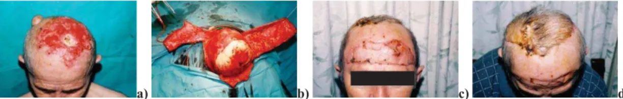
By histopathologic type the most common were: infil-trative (60.2%), noduloinfilinfil-trative (37.2%) and morpheaform (2.6%). They were clinically manifested as: ulcerative, ulcus rodens and ulcus terebrans (Figure 1). Non-infiltrative basal cell carcinoma (nodular, noduloadenoid and superficial) did
not affect the bony part of the scalp and did not make intra-cranial penetration. The duration of the changes, i.e. the time between noticing and the first consultation with the doctor ranged from 6 months up to 3 years. Tumor sizes ranged from 2 to 25 cm in diameter. The depth of the intracranial propagation depended on the histological type and tumor size. Most relapses (42%) were present in the morpheaform type of basal cell carcinoma. In 17 cases the basaliom af-fected only the bone without intracranial propagation and the duration of changes ranged from one to two years. In 10 cases the basalioma penetrated intracranially (duration of changes over three years), of which in 8 cases the duru and in 6 cases it infiltrated the brain parenchyma, of which in two
a) b) c) d)
Volumen 71, Broj 11 VOJNOSANITETSKI PREGLED Strana 1047
Rasuliý GL, Jovanoviý MD. Vojnosanit Pregl 2014; 71(11): 1045–1048. cases the sagittal sinus was affected and had to be tied off (Figure 2). In all the cases with the brain parenchyma in-vaded we removed basalioma down to the macroscopically and microscopically healthy brain parenchyma and in all the cases we implemented radiotherapy.
Discussion
Despite the fact that basal cell carcinomas are affected by radiation therapy (except for the morpheaform type), cryotherapy, curettage, local cytostatic therapy, retinoids or electrodisection, surgical treatment is the method of choice in the treatment of this type of tumor 5–10. Surgically, the tu-mor can be entirely removed with safe histopathological checkup and confirmation 11, 12.
The rate of healing in surgical excisions ranges from 85% to 95% 5. Epstein 9 finds that the visual assessment of basal cell carcinoma edges is within 1 mm from the right edge in about 94% of cases, which led him to conclude that the edges of 2 mm give 94% healing. Burg et al. 11 compared the clinical size of the tumor estimated by the naked eye with the right size determined by Mohsov’s micrographic exami-nation and received the distinction of 5.5 r 3 mm in primary basocelular carcinomas and 8.9 r 4.8 mm in recurrent basal cell carcinomas.
Based on the received results and clinical experience we believe that the width of excision and the presence of tu-morous cells on the resection lines are in direct correlation with the histological type and size of basal cell carcinoma. So, the adequacy of surgical excision is directly dependent on the type and size of basal cell carcinoma.
We found that in all our cases with the affected scalp bone tissue and intracranial propagation, it was an infiltrative basal cell carcinoma and all its histological combinations with
infiltrative component (no macroscopically visible edge com-pared to the healthy tissue), and that it required wider excision and depended on the clinical manifestation of the tumor and its size4, 13–17. If it is the infiltrative type (clinically most often with ulceration and covered with crust) and if it is larger than 1
cm (clinically most often manifested as ulcus rodens or ulcus terebrans excision should be made into the healthy tissue for about 1.5 cm to 3 cm, sometimes even more.
Noduloinfiltrative histological type is most often clini-cally manifested as nodus accompanied by tissue decay, ul-ceration. Because of the clinically present nodus it sometimes misleads in terms of the evaluation of the excision width (therefore a high percentage of the presence of tumor at the re-section edge) 17–24. But with this kind, there is no clear visual limits of tumor from healthy skin which requires, depending on the size, at least 5 mm excision into the healthy tissue.
In the morpheaform type of basal cell carcinoma (rate of the presence at resection lines 35%) excisions need to be made into the healthy tissue up to 3 cm from the tumor edge which is not macroscopicly clearly defined. In the diagnosis of the width of excision in morpheaform basal cell carcino-mas we used 64-slice MSCT.
Conclusion
The aggressiveness and infiltration of basal cell carci-noma into the brain parenchyma is directly linked to the his-tological type and size of the tumor. The larger the basal cell carcinoma the wider excisions need to be made into the healthy tissue with obligatory removal of the affected bone down to the macroscopicly and microscopicly healthy brain parenchyma sometimes up to 3 cm in diameter with obliga-tory postoperative radiological therapy.
R E F E R E N C E S
1. Yenidunya MO. Surgical treatment of auricular malignancies when the anterior or posterior skin is intact. J Craniofac Surg 2013; 24(2): 350î3.
2. Braun-Falco O, Plewig G, Wolff HH, Winkeimann RK. Malignant epithe-lial tumors. In: Braun FO, Plewig G, Wolff HH, Winkeimann RK, edi-tors. Dermatology. Berlin: Springer-Verlag; 1991. p. 1018î35.
a) b) c) d)
e) f)
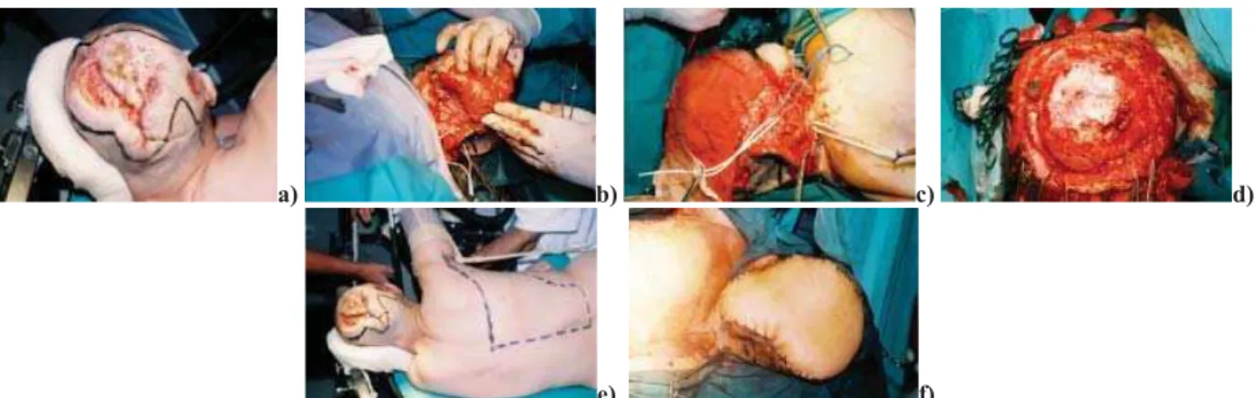
Fig. 2 – a) Clinical manifestation of infiltrated basal cell carcinoma (ulcus terebrans); b) Marked lines represent the site of delamination of free microvascular latissimus dorsi flap; c) Clear invasion of carcinoma into the tissue (which is much larger
Strana 1048 VOJNOSANITETSKI PREGLED Volumen 71, Broj 11
Rasuliý GL, Jovanoviý MD. Vojnosanit Pregl 2014; 71(11): 1045–1048. 3. Wade TR, Ackerman AB. The many faces of basal-cell
carci-noma. J Dermatol Surg Oncol 1978; 4(1): 23î8.
4. Marshall V. Premalignant and malignant skin tumours in im-munosuppressed patients. Transplantation 1974; 17(3): 272î5. 5. Araújo JL, Aguiar GB, Prado AU, Mayrink D, Saade N, Veiga JC.
Malignant chondroid syringoma with central nervous system involvement. J Craniofac Surg 2012; 23(2): 514î5.
6. Ronÿeviý R, Aleksiý V, Stojiÿiý M, Jovanoviý M, Ronÿeviý D. Invasive, aggresive basal cell carcinoma: Carcinoma basocellulare tere-brans. Eur J Plast Surg 2006; 23: 379î84.
7. Vuloviý D, Stepiý N, Pavloviý A, Miliýeviý S, Pisceviý B. Recon-struction of the columella and the tip of the nose with an is-land-shaped forehead flap. Vojnosanit Pregl 2011; 68(3): 277î80. (Serbian)
8. Beatty ME, Habal MB. De novo cutaneous neoplasm: Biologic behavior in an immunosuppressed patient. Plast Reconstr Surg 1980; 66(4): 623î7.
9. Epstein E. How accurate is the visual assessment of basal car-cinoma margins. Br J Dermatol 1973; 89(1): 37î43.
10.Longobardi G, Diana G, Poddi V, Pagano I. Follicular cyst of the jaw developing into a keratocyst in a patient with unrecognized Gorlin-Goltz syndrome. J Craniofac Surg 2010; 21(3): 833î6. 11.Burg G, Hirsch RD, Konz B, Braun-Falco O. Histographic surgery:
Accuracy of visual assessment of the margins of basal-cell epithelioma. J Dermatol Surg 1975; 1(3): 21î4.
12.Deo SV, Hazarika S, Shukla NK, Kumar S, Kar M, Samaiya A. Surgical management of skin cancers: Experience from a re-gional cancer centre in North India. Indian J. Cancer 2005; 42(3): 145î50.
13.Pennington BE, Leffell DJ. Mohs micrographic surgery: Estab-lished uses and emerging trends. Oncology (Willston Park) 2005; 19(9): 1165î71; discussion 1171î2, 1175.
14.Bojanoviý M, Zivkoviý-Marinkov E, Veselinoviý D, Bojanoviý A, Vuckoviý I. Malignant tumors of auricula and periauricular area. Vojnosanit Pregl 2009; 66(8): 611î6. (Serbian)
15.Hutcheson AC, Fisher AH, Lang PG. Basal cell carcinomas with unusual histologic patterns. J Am Acad Dermatol 2005; 53(5): 833î7.
16.McCutcheon B, White K, Kotwall C, Germolic D, Rebolloso Y, Ha-mann MS, et al. A preliminary study of imiquimod treatment in variants of basal cell carcinoma. Am Surg 2005; 71(8): 662î5. 17.Asilian A, Tamizifar B. Aggressive and neglected basal cell
car-cinoma. Dermatol Surg 2005; 31(11 Pt 1): 1468î71.
18.Steve M, Paranque AR, Barthélémy I, Bui P. Management of a ba-socellular carcinoma of the cheek. Rev Stomatol Chir Maxillo-fac 2008; 109(1): 56î60.
19.Anwar U, Ghazal AS, Ahmad M, Sharpe DT. Horrifying basal cell carcinoma forearm lesion leading to shoulder disarticula-tion. Plast Reconstr Surg 2006; 117(1): 6eî9e.
20.Eisner JM, Russell M. Cartilage hair hypoplasia and multiple ba-sal cell carcinomas 2006; 54(2 Suppl): S8î10.
21. Ríos-Buceta L. Management of basal cell carcinomas with positive margins. Actas Dermosifiliogr 2007; 98(10): 679î87. (Spanish) 22.Su SY, Giorlando F, Ek EW, Dieu T. Incomplete Excision of
Basal Cell Carcinoma: A Prospective Trial. Plast Reconstr Surg 2007; 120(5): 1240î8.
23.Wettstein R, Erba P, Farhadi J, Kalbermatten DF, Arnold A, Haug M, et al. Incomplete excision of basal cell carcinoma in the subunits of the nose. Scand J Plast Reconstr Surg Hand Surg 2008; 42(2): 92î5.
24.Gargiulo M, Papa A, Capasso P, Moio M, Cubicciotti E, Parascandolo S. Electrochemotherapy for non-melanoma head and neck cancers: clinical outcomes in 25 patients. Ann Surg 2012; 255(6): 1158î64.