48 ACTA ORTOP BRAS 10(1) - JAN/MAR, 2002
Tratamento das lesões traumáticas instáveis da
coluna torácica e lombar com retângulo de Hartshill*
Treatment of unstable thotacic spine traumatic injuries using Hartshill rectangle
J
ÚLIOC
ÉSARP
EREIRA DAC
UNHA1, J
ORGEM
AUADF
ILHO2, N
ELSONH
ELYM
IKAELB
ARSAM3, J
OSÉW
AGNER DEB
ARROS4RESUMO
Foram avaliados 14 pacientes com lesão instável da coluna torá-cica e lombar, tratados com instrumentação de Hartshill. De uma forma geral, obtivemos bons resultados com a técnica proposta.
Descritores: Lesões traumáticas instáveis da coluna, lesões trau-máticas da coluna torácica e lombar, retângulo de Hartshill.
INTRODUÇÃO
A redução cruenta com fixação interna é um procedimento de rotina no tratamento das lesões instáveis da coluna torácica e
lombar(3,19). Com o aparecimento de novas técnicas e materiais
de síntese, houve grande avanço nas cirurgias de estabilização da coluna(4,5,13,15,17).O retângulo(6), utilizado nas últimas décadas,
tem demonstrado resultados clínicos relevantes(6,19).
O objetivo deste estudo é avaliar 14 pacientes com lesões traumáticas instáveis da coluna torácica e lombar, tratados com instrumentação de Hartshill e com artrodese.
CASUÍSTICA E MÉTODOS
No período entre julho de 1990 e abril de 2000, 14 pacientes com lesão instável da coluna torácica e lombar foram submetidos a tratamento cirúrgico com instrumentação de Hartshill com artrodese. Dos pacientes avaliados, 11(78,6%) eram do sexo masculino e três (21,4%), do feminino. A idade variou de 16 a 42 anos, com média de 29 anos, aproximadamen-te. Os pacientes foram submetidos a radiografias em frente e perfil (Fig. 1A-B). A coluna torácica foi acometida em seis (42,9%) pacientes, a lom-bar em seis (42,9%) e a toracolomlom-bar em 2 (14,2%).
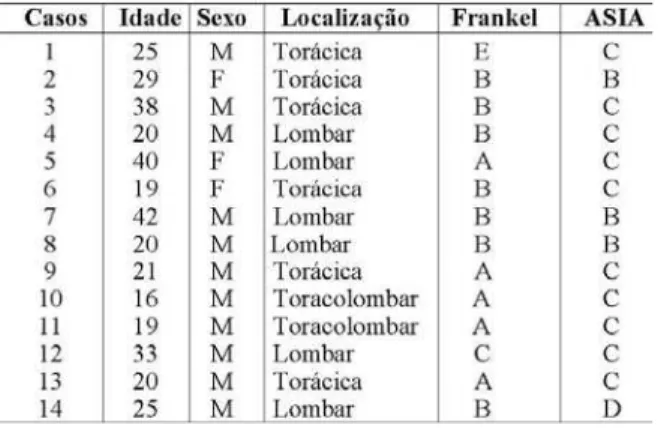
As lesões foram classificadas conforme preconizado pela American Spinal lnjury Assocíation (ASlA)(16)(Tabela 1). Dez (71,4%) eram do tipo C
SUMMARY
Fourteen patients with unstable thoracolumbar spine injuries treated with Hartshill’s instrumentation have been evaluated. Good results were generally achieved using this technique.
Key Words: unstable spine trauma, trauma thoracolumbar spine, Hartshill’s instrumentation
INTRODUCTION
Open reduction and internal fixation is routine procedure when treating unstable injuries of thoracic and lumbar spine(3, 19). New
advances in fixation devices brought significant advances to spine stabilization(4, 5,13,15,17). The rectangle(6), used in the last decades has
demonstrated relevant clinical results(6,19).
The objective of this study is to evaluate 14 patients with unsta-ble injury of thoracic and lumbar spine, who were treated with Hartshill’s instrumentation plus arthrodesis.
CASES AND METHODS
Between July, 1990 and April, 2000, 14 patients with uns-table injury of thoracic and lumbar spine underwent surgical treatment with Hartshill’s instrumentation plus arthrodesis. From these patients, 11 (78.6%) were male and three (21.4%) female. Age ranged between 16 and 42 years, average ap-proximately 29 years.
Patients underwent AP and lateral radiograph (Figures 1 A-B). Thoracic spine was involved in six patients (42.9%), lumbar in six (42.9%) and thoracolumbar in 2 (14.2%).
The injuries were evaluated according to American Spinal Injury Association (ASIA)(16) (Table 1). Ten (71.4%) were rated
*Work performed at Department of Orthopaedics and Traumatology from Hospital Escola da Faculdade de Medicina do Triângulo Mineiro – Uberaba – MG
1- Ex-Resident
2- Master in Orthopaedics and Traumatology from FMRP-USP, Head of Spine Surgery Sector
3- Ortopaedist Surgeon 4- Chairman
Adress: José Wagner de Barros – Disciplina de Ortopedia e Traumatologia Departamento de Cirurgia do HE/FMTM – R. Getúlio Guaritá S/Nº Bairro Abadia – CEP 38025-440 – Uberaba – MG
Trabalho recebido em 03/01/2001. Aprovado em 27/11/2001
A
RTIGOO
RIGINAL*Trabalho realizado no Departamento de Ortopedia e Traumatologia do Hospital Escola da Faculdade de Medicina do Triângulo Mineiro – Uberaba – MG
1- Ex-Residente
2- Mestre em Ortopedia e Traumatologia pela FMRP-USP, Responsável pelo Setor de Cirurgia da Coluna 3- Médico Ortopedista
4- Professor Titular e Responsável pela Ortopedia e Traumatologia
ACTA ORTOP BRAS 10(1) - JAN/MAR, 2002 49
as C and four (28.6%) as B. In preoperative evalu-ation, patients were cli-nically evaluated, with a c a r e f u l s c r e e n i n g f o r neurologic damage, ac-cording to the scale(8)
Table 2. General charac-teristics of these patients is in Table 3. Internal fi-x a t i o n d e v i c e w a s a Hartshill’s rectangle with 26.3 mm wide and ran-ging from 40 to 400 mm long, using Luque wires with diameters of 1 and 1.2 mm.
Patients underwent general anesthesia, and placed in ventral decubi-tus. Approach as medi-an longitudinal. After ex-posure of spinal proces-ses of involved verte-bras, and two vertebras above and two below the injury, a reduction w a s p e r f o r m e d a n d when necessary, decom-pressive laminectomy was performed. Lesions were stabili-zed with the previously molded Hartshill’s rectangle and Lu-que wires fixed to the laminas above and below the lesion (Figures 2 A-B). Arthrodesis was performed by cancellous bone graft from iliac or spinal processes.
Postoperatively patients underwent a new clinical evalua-tion according to the scale of neurologic damage(8) (Table 2)
and radiographic evaluation performed at 30, 60 and 90 days. Patients presenting walking conditions Fran-kel D and E were allo-wed early walking using a Jewet vest for 3 mon-ths, and not walking pa-tients, Fankel A, B and C were referred to Physio-therapy for rehabilitation.
RESULTS
From the 14 evalua-ted patients, postopera-tively 5 (35.8%) were Frankel A, 7 (50%) Fran-kel B, 1 (7.1%) FranFran-kel C and 1 (7.1%) Frankel E.
From the 5 Frankel A patients, 2 kept the result,
e quatro (28,6%), do tipo B. Na avaliação pré-operatória, os pacientes foram examina-dos clinicamente e realizada cuidadosa avaliação para averiguar danos neurológi-cos, conforme a escala(8)
(Ta-bela 2). As características ge-rais desses pacientes avalia-dos encontram-se na (Tabela 3). O sistema de fixação inter-na utilizado constitui-se de: Retângulo de Hartshill, com 26,3mm de largura e varian-do de 40 a 400mm de com-primento, associado a fios de Luque com diâmetros de 1mm e 1,2mm.
Os pacientes foram sub-metidos à anestesia geral e posicionados em decúbito ventral. A via de abordagem foi a mediana longitudinal. Após a exposição dos pro-cessos espinhosos das vér-tebras acometidas inclusive duas vértebras acima e duas abaixo da lesão. Foi realizada a redução e, quando
neces-sário, feita a laminectomia descompressiva. As lesões foram estabiliza-das com retângulo de Hartshill, previamente moldado, e com os fios de Luque fixados nas lâminas acima e abaixo da lesão (Fig. 2A-B). Foi realiza-da artrodese com colocação de enxerto esponjoso de crista ilíaca ou do processo espinhoso.
No pós-operatório, os pacientes foram submetidos a uma nova
avaliação clínica de acordo com a escala de danos neurológicos(8)
(Tabela 2) e feitos exames radiológicos aos 30, 60 e 90 dias. Os pacientes que
apre-sentaram condições de deambulação Frankel D e E foram liberados para marcha precoce, com uso de colete de Jewet, por 3 meses, e pacientes que não deambulavam Frankel A, B e C foram encaminha-dos ao serviço de Fisiote-rapia para reabilitação.
RESULTADOS
Dos 14 pacientes ava-liados no pós-operatório, 5 (35,8%) eram Frankel A, 7 (50%) Frankel B, 1 (7,1%) Frankel C e 1 (7,1%) Frankel E.
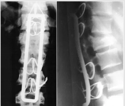
Figura 1- Fotografias de radiografias pré-operatórias da coluna lombar e torácica. A- (frente) B- (perfil). Nota-se fratura em LI, com afundamento
e angulação de 280º.
Figure 1- Photographs of preoperative radiographs of thoracic and lumbar spine. A- (front) B- (lateral). Notice L1 fracture, depressed and
with 280° deformity.
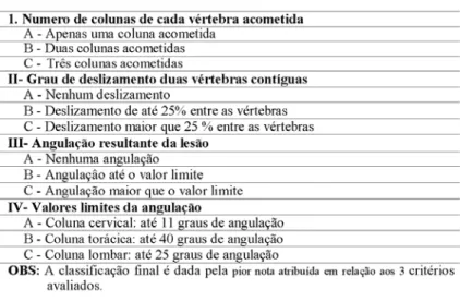
Tabela I - Classificação da American Spinal Injury Association (ASIA) para lesões da coluna vertebral(9).
50 ACTA ORTOP BRAS 10(1) - JAN/MAR, 2002
one became B, one O and the other one became E. From the 7 Frankel B, two became C, three O and two E. The Frankel C pati-en t , b e ca m e E. T he Frankel E remained as E. Postoperative clinical evo-lution of the patients, is in Table 4.
No secondary com-plication was observed, such as infection, loss of reduction or failure of the implant. Only one patient presented with persis-tent pain, remitting four weeks after treatment with NSAID.
DISCUSSION
Unstable injuries of thoracic and lumbar spi-ne have been widely dis-cussed regarding surgi-cal treatment(4,5,13,15,17).
Some authors recom-mend reconstruction and stabilization of the poste-rior part of the spine with the implant working as a tensor(20).
Others prefer anterior approach and fixation(1), not considering
the axial load to anterior spine. And others recommend a fixation both anterior and posterior, as the best stabilizing method(10,9).
Generally, all methods recommend instrumentation with arthro-desis that allow stabilizing the lesions(11,12,18), being indicated to
reduce morbidity and shorten in hospital stay(2,14).
Reduction and fixation of fractures, with anatomical res-toration of unstable injuries of thoracic and lumbar spine allow an stability that allows a better evolution in patients with a neurologic deficit(7).
The results of this series were similar to those in the litera-ture, when compared to other instrumentation techniques(13,15),
monosegmental fixation with transpedicular screws(5)and
Harms method(4).
Dos 5 pacientes Fran-kel A, 2 mantiveram-se, um evoluiu para B, um para O e outro para E. Dos 7 Frankel B, dois evoluíram para C, três para O e dois para E. O Frankel C evo-luiu para E .0 Frankel E manteve-se. A evolução clínica dos pacientes, no pré e no pós-operatório, encontra-se na (Tabela 4). Não ocorreram compli-cações secundárias como infecção, perda da redução ou falência de material de sín-tese. Apenas um paciente evoluiu com dor persistente sendo evidenciada melhora do quadro, quatro semanas após o tratamento com an-tiinflamatório não hormonal.
DISCUSSÃO
As lesões instáveis da coluna torácica e lombar têm sido amplamente discutidas, quanto ao tipo de tratamento operatório utilizado(4,5,13,15,17).
Alguns autores preconizam a reconstrução e a estabilização da parte posterior da coluna, com implante funcionando como tirante de tensão(20).
Outros autores dão preferência à abordagem e fixação anterior(1),
relevan-do a carga axial que é submetida à coluna anterior. Outros ainda recomen-dam a fixação anterior e posterior como método mais eficaz de estabiliza-ção(10,9). De uma forma geral, todos os procedimentos recomendam
instrumentação, com fusão óssea que permita estabilizar as lesões(11,12,18),
sendo indicado para reduzir a morbidade e encurtar o período de hospi-talização do paciente(2,14).
A redução e fixação das fraturas, com a restauração anatômica das lesões instáveis da coluna torácica e lombar, permitem uma es-tabilidade que proporciona uma melhora na evolução clínica dos pacientes com déficit neurológico(7).
Os resultados obtidos foram semelhantes aos da literatura, quando comparados com outras técnicas de fixação, como
ins-Figura 2- Fotografias de radiografias pós-operatórias da coluna lombar e torácica. A- (frente) B- (perfil). Nota-se a estabilização da fratura com o
sistema de Hartshill.
Figure 2- Photograph of postoperative radiographs of lumbar and thoracic spine. A- (front) B- (lateral). Notice fracture stabilization with
Hartshill’s rectangle.
Tabela 2 - Escala de danos neurológicos de Frankel(8).
ACTA ORTOP BRAS 10(1) - JAN/MAR, 2002 51
REFERÊNCIAS BIBLIOGRÁFICAS
1. Aebi, M.: Operative Behandlung von Wirbels frakturen — dorsale oder ventrale instrumentation. Op Journal 12: 182-187, 1996.
2. Bedbrook,G.M.: Use and disure of surgery in limbo-dorsal fractures. J Western Pacific Orthop Assn 6: 5-26, 1969.
3. Brum, P. R., Labronici, P. J., D’Angelo,D. S. &Amaral, C. A B.: Tratamento cirúrgico das fraturas instáveis da coluna toracolombar. RevBras Ortop 30: 125-130,1995.
4. Defino, H. L. A., Fuentes, A. E. R. & Rejaili, W. A: Tratamento das fraturas da colina toracolombar pelo método de Harms. Rev Bras Ortop 32: 546-554, 1997.
5. Defino, H. L. A., Fuentes, A. E. R., Remond, P. H. &Valim, E.: Fixação monossegmentar das fraturas da coluna toracolombar. Rev Bras Ortop 33: 119-124, 1998.
6. Dove, J: lnternal fixation of the lumbar spine. The Hartshill rectangle. Clin Orthop 203: 135-140, 1986.
7. Flesch, J.R., Leíder, L.L.,Erikson, D.L., Chou, S.N. & Bradford, D.S.: Harrington instrumentation and spine fusion for unstable fractures and fractures-dislocations of thoracic and Iumbar spine. J Bane Joint Surg [Am]59: 143-153, 1977.
8. Frankel, H. L. Hancock, O. 0. & Hyslop, G.: The spine wíth paraplegia and tetraplegia. Paraplegia7: 225, 1969.
9. Harms, J.: Screw-threaded rod system ín spinal fusion surgery. State of the art review. Spine6: 541 -577, 1992.
10. Harms, J. & Stoltze,D.: lhe indications and principIes of correction of
pot-traumatic deformitíes. Eur Spine J 1:142-151, 1992.
11. Harrington, P. R.: Treatment of scoliosis. Correction and internal fixation by spine instrumentation. J Bane Joint Surg [Am] 44: 591,1962. 12. Harrington, P.R: Instrumentation in spine instability other than scoliosis.
SAfr Surg 5: 7-12,1967.
13. Harrington, P.R. & Dickson, J.H.: The development and further prostects of internal fixatíon of the spine. Isr J Med Sci 9: 773-778, 1973. 14. Katznelson, A.M.: Stabilization of the spine in traumatic paraplegia. Paraplegia7: 33-37, 1969.
15. Luque, E., Cassis, H. & Ramirez-Wiella, G.: Segmental spinal instrumentation in the treatment of fractures of the thoracolumbar spíne. Spine7: 312, 1982.
16. Meyer, P.: New Spine Fracture Clasification System,Annual meeting of the AAPM&R, Chicago, 1996.
17. Munford, J.,Weinsten, N., Spratt, K. F. Et aI.: Thoracolumbar burst fractures: The clinical efficacy and outcome of monoperative managemente.Spine8: 955-970, 1993.
18. Oliveira, R.P., Barros Filho, T.E.P., Greve, J.M.D’A., Rodrigues, N.R. & Basíle Jr., R.: Fraturas vertebraís do sebmento toracolombar: descompressão cirúrgica. Rev Bras Ortop 27: 699-704, 1992. 19. Puertas, E. B., Chagas, J. O. M. & Kasinski, S. K.: Trauma toracolombar.
Rev Bras Ortop 27:138-140, 1992.
20. Staufer, E.S.: “The use of the AO ‘fixateur interne’ for thoracic and lumbarfractures”, in Brídwell, K. H. & Oewald, R. L. (eds).: The text-book of spinal sugery, Phíladelphia, J.B Lippincott, 1997. p. 1949-1955.
trumentação(13,15), fixação
monos-segmentar com parafusos
trans-pedicuIados(5)e método de
Har-ms(4).
O instrumental de Hartshill, por nós utilizado, proporcio-nou uma melhor estabilização no pós-operatório e demons-trou ser uma boa opção para a fixação posterior das lesões instáveis da coluna torácica e lombar nos pacientes avalia-dos.
CONCLUSÕES
A fixação interna com restau-ração anatômica das lesões ins-táveis da coluna torácica e lom-bar, com o retângulo de Hartshill, permitiu uma estabilidade das fra-turas e demonstrou ser um méto-do apropriaméto-do no tratamento méto-dos pacientes com lesão traumática grave da coluna.
The Hartshill’s rectangle used by us, allowed a best pos-toperative stabilization and de-monstrated to be a good opti-on for posterior fixatiopti-on of uns-table injuries of thoracic and lumbar spine in this group of patients.
CONCLUSIONS
Internal fixation and anato-mical restoration of unstable le-sions of thoracic and lumbar spine with Hartshill’s rectangle allowed stability to the fractu-res and proved to be adequate in treatment of patients with severe traumatic spinal injury.
Tabela 4 - Evolução clínica dos pacientes no pré e pós-operatório segundo a escala de danos neurológicos de Frankel.
Table 4 - Pre and post operative evolution of the patients according to Frankel’s rating.
Tabela 3 - Características gerais dos pacientes submetidos ao tratamento operatório quanto à idade, sexo,
localização, danos neurológicos e tipo de lesão.
Table 3 - General aspects of patients who underwent surgery regarding age, sex, location, neurological data