Iranian Journal of Basic Medical Sciences
ijbms.mums.ac.ir
)nfluence of vitamin D on cell cycle, apoptosis, and some
apoptosis related molecules in systemic lupus erythematosus
Nafise Tabasi , Maryam Rastin *, Mahmoud Mahmoudi , Mohsen Ghoryani , Zahra Mirfeizi ,
Shahrzad Zamani Taghizadeh Rabe , (adi Reihani
.)mmunology Research Center, BuAli Research )nstitute, Faculty of Medicine, Mashhad University of Medical Sciences, Mashhad, )ran .Rheumatic Disease Research Center, )mam Reza (ospital, Mashhad University of Medical Sciences, Mashhad, )ran
A R T ) C L E ) N F O A B S T R A C T Article type:
Original article Objective(s):erythematosus SLE . Autoreactive lymphocytes are cleared through apoptosis and any disturbance in Genetic and environmental factors are involved in the pathogenesis of systemic lupus the apoptosis or clearance of apoptotic cells may disturb tolerance and lead to autoimmunity. Vitamin D has anti‐proliferative effects and controls cell cycle progression. )n this study we investigated the effects of vitamin D on cell cycle and apoptosis induction in lupus patients.
Materials and Methods: )solated peripheral blood mononuclear cells PBMCs from SLE patients were cultured in the presence of nM of , O( D ; then one part of the cells were stained with F)TC labeled Annexin V and P) and were analyzed for apoptosis determination. For gene expression assessment of FasL, Bcl‐ and Bax, RNA was extracted from one another part of the cells, cDNA was synthesized and gene expression analysis was performed using Real time PCR. An additional part of the cells were treated with P) and the cell cycle was analyzed using flowcytometer.
Results: The mean number of early apoptotic cells in vitamin D treated cells decreased significantly
. ± . % compared to untreated cells . ± . % P= . . Cell cycle analysis showed a
significant increase in G phase in vitamin D treated cells . ± . % compared to non treated ones . ± . % P = . . Vitamin D up‐regulated the expression levels of Bcl‐ by . fold increase ,
and down‐regulated expression of Bax % and FasL % .
Conclusion: Vitamin D has regulatory effects on cell cycle progression, apoptosis and apoptosis related molecules in lupus patients.
Article history: Received: Oct , Accepted: Oct , Keywords:
Systemic lupus erythema‐ tosus
Apoptosis Cell cycle Vitamin D
►Please cite this article as:
Tabasi N, Rastin M, Mahmoudi M, Ghoryani M, Mirfeizi Z, Zamani Taghizadeh Rabe Sh, Reihani (. )nfluence of vitamin D on cell cycle, apoptosis, and some apoptosis related molecules in systemic lupus erythematosus. )ran J Basic Med Sci ; : ‐ .
Introduction
Systemic lupus erythematosus SLE is an autoimmune disease in the pathogenesis of which genetic and environmental factors play an important role. Breakdown of tolerance and generation of autoantibodies against self antigens and augmentation of inflammatory responses are implicated in the pathogenesis of SLE .
Autoreactive B and T lymphocytes are eliminated through apoptosis in the course of self tolerance . Any disturbance in the process of apoptosis or impairment in the clearance of apoptotic cells could lead to the accumulation of apoptotic bodies, generation of autoantibodies , and exacerbation of inflammatory responses .
)n different studies an association of vitamin D deficiency with predisposition to SLE was reported , , and in some studies in mice models of lupus it was shown that vitamin D consumption improved
the disease progression and increased the
number of Regulatory T cells . (uman studies
have also showed that vitamin D deficiency is correlated with disease activity in lupus patients , . Vitamin D could promote the production of anti
inflammatory mediators , and is considered to be
implicated in immune regulation . )t could regulate
the growth of normal and cancer cells and appears
to be involved in controlling agitated cells proliferation . The mechanism of anti‐proliferative effects of vitamin D is not yet well known , but controlling the expression of some genes that are involved in proliferation, differentiation, angiogenesis, and cell death are supposed. Vitamin D can also affect cell
cycle progression , .
As impaired apoptosis is one of the possible mechanisms involved in the development and exacerbation of symptoms in systemic lupus erythe‐ matosus, and because vitamin D ameliorates the disease symptoms in SLE we aimed to study the regulatory effect of vitamin D on the apoptosis and cell cycle progression as well as the expression levels of FasL, Bcl‐ and Bax in lupus patients.
*Corresponding author: Maryam Rastin. )mmunology Research Center, BuAli Research )nstitute, Faculty of Medicine, Mashhad University of Medical
Materials
and
Methods
Participants
Twenty five SLE patients women and men,
with a mean age of . ± . years participated in this study. All SLE patients fulfilled at least four of the revised SLE criteria of the American College of
Rheumatology revised criteria for the
classification of SLE (ochberg, . Each lupus
patient was evaluated by a rheumatologist according to the following inclusion and exclusion criteria:
)nclusion criteria:
a- Patients who were newly diagnosed before
starting treatment .
b- Patients in remission who took only a
maximum dose of mg/day prednisolone.
c- Patients with active major organ involvement
were sampled before changing treatment strategy and starting cytotoxic therapy.
Exclusion criteria:
a- Patients in remission who took hydroxyl‐
chloroquine.
Patients in remission who took higher than mg/day of prednisolone.
b- Patients who took any cytotoxic drugs. c- Patients with overlap syndromes. d- Patients with concomitant malignancy. e- Patients with infection.
f- Female patients who were pregnant.
)nformed written consent was taken from all subjects prior to blood sampling. The Ethic Council of Mashhad University of Medical Sciences Mashhad, )ran approved all protocols used with these subjects in this study.
PBMCsisolation
Peripheral blood mononuclear cells PBMCs were isolated using Ficoll G)BCO, USA density gradient centrifugation. Cells were washed in phosphate buffered saline PBS , and then resuspended and adjusted to × cells/ul in PBS.
Assessment of cell cycle by Flowcytometry using
propidiumiodidestaining
The distribution of DNA content in cell cycle was determined using propidium iodide P) as a DNA‐
binding dye. Briefly, lymphocytes × cells were
incubated with vitamin D nM for hr, the
optimal dose of vitamin D and time of incubation was optimized in our lab in previous studies . Then harvested cells were washed twice with cold phosphate‐buffered saline PBS , incubated with µg/ml RNAse A GenetBio Co., Nonsan, Korea for min at °C and subsequently incubated with P)
Sigma Chemical Co., MO, USA solution µg/ml
P), . % Triton X‐ and . % sodium citrate in dark for min at °C. The percentage of DNA
content × events in cell cycle area was
Becton Dickinson, Carlsbad, CA and the data were subsequently analyzed using FSC Express . software. Cultured cells without vitamin D were used as controls.
Detectionofapoptosisusingannexin‐V/propidium
iodide(PI)dualstaining
Death pattern was quantified using fluoroscein isothiocyanate‐labeled Annexin V Annexin V‐F)TC and propidium iodide P) staining kit Abcam, Cambridge, MA . The combination of Annexin V‐F)TC
with P) has been used widely to distinguish cells in early and late apoptosis stages. During early
apoptosis, phosphatidyl serine, which is usually
located in the inner membrane of the cells, is transported into the outer portion of the
membrane that can be detected by its strong affinity for annexin V‐F)TC, a phospholipid binding protein. The dead cells can be detected by binding P) to the cellular DNA, where the cell membrane has been totally crashed. )n this study, PMBCs × /well were treated with vitamin D nM for hr. After that, cells were washed with PBS, resuspended in binding buffer and incubated at room temperature for min in dark with Annexin‐V F)TC and P) solutions. The cells were then analyzed within hr using a
FACSCalibur Flowcytometer Becton Dickinson,
Carlsbad, CA . )n each case, a minimum of × events
were analyzed. Results were reported as percentage of Annexin V+ early apoptosis , P)+ necrosis , Annexin
V+P)+ double positive; late apoptosis , or Annexin V‐P)‐
double negative; non‐stained cell population. Cells with no treatment were used as control.
GeneexpressionanalysisofFasL,Bcl‐2andBaxby
Realtime‐PCR
Total RNA was extracted using Tripure Roche, Mannheim, Germany , employing manu‐ facturer’s instructions. cDNA was synthesized and the expression level of the genes of FasL, Bcl‐ and Bax were assessed using specific primers and probes by Real time PCR method. A comparative CT method
‐CT was used to quantify gene expression levels. All
data was expressed as mean transcript expression fold‐ change over untreated samples controls , normalized to GAPD( internal control .
Statisticalanalysis
All data are reported as mean±SD. Data of apoptosis, cell cycles and gene expression were analyzed using Student’s t‐test. P‐values less than
. were considered as significant.
Results
Twenty five SLE patients women and men, with a mean age of . ± . years participated to current study. Anti‐dsDNA was positive in . %
0 10 20 30 40 50 60 70 80
G2M S
G1
pe
rc
e
nt
control vitamin D
*
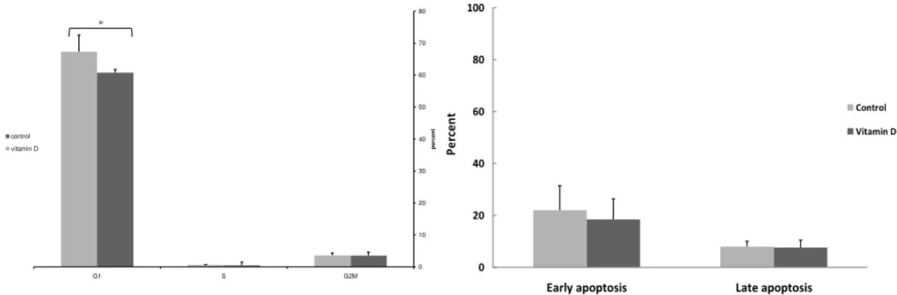
Figure 1. Comparison of cell cycle progression in nM of
, O( D treated D+ and in non treated D‐ PBMCs control of systemic lupus erythematosus patients. Vitamin D treatment significantly increased the number of cells in G phase G Gap ; growth phases of cell cycle , S Synthesis phase of cell
cycle , G M Gap and Mitosis phases of cell cycle . *P‐value< .
. % , kidney damage . % , anemia . % , and
thrombocytopenia . % .
Flowcytometryassessmentofcellcycle
To further explore the influence of vitamin D on PBMCs of SLE patients, cells were cultured as indicated above, and analyzed for DNA content by flowcytometr. The mean number of cells in the G phase in vitamin D treated PBMCs was significantly higher . . %
compared to untreated cells control group
. . % P‐value . Figure . There was not
any significant difference in the number of cells in G M and S phase between vitamin D treated cells and the control group.
InfluenceofvitaminDonapoptosis
To determine if any alteration in cell viability induced by vitamin D might be attributable to apoptosis, Annexin‐ V and P) staining was conducted. Our results showed that vitamin D dramatically decreased early apoptotic population of lymphocytes . ± . % Annexin V+ cells compared to untreated cells . ± . %
P‐value . Figure .
GeneexpressionofFasL,Bcl‐2andBax
Quantitative Real‐time PCR analyses of the expression of FasL, Bcl‐ and Bax in vitamin D
treatment of the cells of SLE patients showed that transcript expression of Bcl‐ . ± . ,
Bax . ± . and FasL . ± . underwent
fold‐change relative to the levels in untreated cells. This translated to up‐regulated expression
of Bcl‐ . fold increase , and down‐regulted
expression of Bax % and FasL % due to
culturing in the presence of nM vitamin D Figure .
0 20 40 60 80 100
Early apoptosis Late apoptosis
Pe
rc
en
t Control
Vitamin D+
Finger 2. Comparison of the effects of nM of , O( D
treatment on the early and late apoptosis in vitamin D treated D+ and in non treated D‐ PBMCs control of systemic lupus erythematosus patients. Vitamin D treatment for hr of PBMCs of SLE patients D+ significantly decreased the percent of cells in early apoptosis Annexin V‐F)TC+ compared to control untreated cells.
There was no difference in the late apoptotic cells between vitamin D treated and non treated cells. *P‐value< .
All data was expressed as mean transcript expression fold‐change over untreated samples controls , normalized to GAPD( internal control .
Statisticalanalysis
All data are reported as mean±SD. Data of cell cycles and gene expression were analyzed using
Student’s t‐test. P‐values less than . were
considered as significant.
Figure3.Real‐time PCR results using comparative CT method
Discussion
)mmunomodulatory role of vitamin D has been the subject of much interest in recent literature , , but the precise mechanisms of this modulation needs further studies. An inhibitory effect of vitamin
D on cell proliferation was previously reported ,
but it was not well characterized whether this property is exerted by arrest in cell cycle, apoptosis or both. We studied the effects of vitamin D treatment on apoptosis, cell cycle progression and expression of FasL, Bax and Bcl‐ on PBMCs of lupus patients.
Findings of our study showed that vitamin D treatment decreased the apoptosis rate in PBMCs of SLE patients, increased the DNA content in G /G , down regulated the expression rate of FasL and Bax, while up regulated the expression of Bcl‐ .
)mportance of impaired apoptosis in SLE development was frequently reported in previously
published literature , , and we found that
vitamin D by decreasing the expression of FasL and Bax and by increasing the expression rate of Bcl‐ which is an antiapoptotic molecule, could control the apoptosis rate in lupus patients. Results of current study showed that anti apoptotic and anti proliferative effects of vitamin D is also regulated partly by cell cycle arrest in G and in parallel to our findings, Ohnishi et al showed that cell cycle
arrest of vitamin D is mediated by inhibiting several
key proteins which regulate the G /S phase and
up‐regulates gene expression of P . Chen etal
reported an anti proliferative effect for vitamin D on B cells differentiation and autoantibody
production .
Apoptosis plays a controversial role in systemic lupus erythematosus. )mpaired apoptosis and decreased elimination of autoreactive lymphocytes in the developmental phase of immune system could lead to the loss of self tolerance, and some studies showed increased rate of apoptosis in lymphocytes
of SLE patients . Regulatory T cells are a subset
of lymphocytes which are highly susceptible to
apoptosis , and role of increased apoptosis in the
reduction of regulatory T cells was demonstrated in
a number of studies . )n a previous investigation
in our group we showed that consumption of vitamin D increased the number of regulatory T cells and down‐regulated the expression of )L‐ in animal models of lupus . Several studies suggested a profound role for )L‐ in the reduction of the number and activity of regulatory T cells . Decreased expression or function of regulatory T cells was correlated with active lupus . Results of this
study indicates that , O( D regulates the
processes related to the cell cycle, and apoptosis, and considering the data of our previous study, it is highly suggested that some beneficial effects of vitamin D on increasing the number of regulatory T
apoptosis and cell cycle arrest which needs further studies.
Lupus is a disease with few treatment regimens. Vitamin D as an immunomodulator has shown potential benefits in lupus patients. By decreasing the apoptosis rate in hyper activated cells and by inducing arrest in cell cycle progression, vitamin D can fine‐tune the balance and probable reestablishment of tolerance. More studies are needed to better understand the real mechanisms and the ideal dose of vitamin D needed for beneficial effects on lupus patients.
This was a cross sectional study, and further longitudinal works in future will provide more data about the immunomodulatory and anti apoptotic effects of vitamin D in systemic lupus erythematosus.
Conclusion
Our results showed that vitamin D as an immunomodulator decreased the apoptosis rate in PBMCs of lupus patients and induced arrest in cell cycle progression. Presence of , O( D decreased the expression rate of FasL and Bax and increased the expression of Bcl‐ in lupus patients.
Acknowledgment
This research was supported by the Research Council of Mashhad University of Medical Sciences
Mashhad, )ran, vice president by grant number .
References
. Nagy G, Koncz A, Perl A. T and B cell abnormalities in systemic lupus erythematosus. Rev )mmunolo
; : ‐ .
. Denny MF, Chandaroy P, Killen PD, Caricchio R, Lewis
EE, Richardson BC, et al. Accelerated macrophage
apoptosis induces autoantibody formation and organ damage in systemic lupus erythematosus. J )mmunol
; : – .
. Cline AM, Radic MZ. Apoptosis, subcellular particles,
and autoimmunity. Clin )mmunol ; : – .
. Lemire JM, )nce A, Takashima M. , ‐Dihydroxy‐ vitamin D attenuates the expression of experimental
murine lupus of MRL/l mice. Autoimmunity ;
: ‐ .
. Wahono CS, Rumini (, Soelistyoningsih D, (akim R, (andono K, Endharti AT, Kalim (, Widjajanto E. Effects
of , O( D in immune response regulation of
systemic lupus erithematosus SLE patient with
hypovitamin D. )nt J Clin Exp Med ; : ‐ .
. Marques CD, Dantas AT, Fragoso TS, Duarte AL. The importance of vitamin D levels in autoimmune
diseases. Bras J Rheumatol ; : ‐ .
. Vaisberg MW, Kaneno R, Franco MF, Mendes NF: )nfluence of cholecalciferol vitamin D on the course of experimental systemic lupus erythematosus in F
NZBxW mice. J Clin Lab Anal ; : ‐ .
. Amital (, Szekanecz Z, Sz“cs G, Danko K, Nagy
E, Csépany T, et al. Serum concentrations of
‐O( vitamin D in patients with systemic lupus erythematosus SLE are inversely related to disease activity: is it time to routinely supplement patients
with SLE with vitamin D? Ann Rheum Dis ;
: ‐ .
. Contorna MT, Zhu y, Froicu M, Wittke a. Vitamin D status, , ‐dihydroxyvitamin D , and the immune
system. Am J Clin Nutr ; : S‐ S.
. Adorini L, Penna G. Control of autoimmune diseases by the vitamin D endocrine system. Nat. Clin.
Pract. Rheumatol ; : ‐ .
. Elen G, Gysemans C, Verlinden L, Vanoirbeek E, De Clercq P, Va (aver D, Mathieu C, Bouillon R, Verstuyf A. Mechanism and potential of the growth inhibitory actions of vitamin D and analogs. Curr Med Chem
; : ‐ .
. Dusso AS, Thadhani R, Slatopolosky e. Vitamin D
receptor and analogs. Semin Neph ; : ‐ .
. Flanagan L, Packman K, Juba B, O Neill S, Tenniswood M, Welsh J. Efficacy of vitamin D compounds to modulate estrogen receptor negative breast cancer growth and invasion. J Estroid Biochem
Mol Biol ; : ‐ .
. Mantel DJ, Owens PE, Bundred NJ, Mawer EB,
Canfield AE. , ‐dihydroxy vitamin D inhibits
angiogenesis in vitro and in vivo. Circ Res ;
: ‐ .
. Verlinden L, Verstuy A, Van Camp M, Marcelis S, Sabbe K, Zhao XY, De Clercq D, Vanderwalle M,
Bouillon R. Two novel ‐epi‐analogues of , ‐
dihydroxy vitamin D inhibit the growth of human
breast cancer cells in vitro and in vivo. Cancer Res
; : ‐ .
. Muszkat P, Camargo MBR, Griz L(M, Lazaretti‐ Castro M. Evidence‐based non‐skeletal actions of vitamin D, Arquivos Brasileiros de Endocrinologia
eMetabologia ; : ‐ .
. Zhang (, Shih DQ, Zhang X. Mechanisms under‐lying effects of , ‐Dihydroxy vitamin D on the Th cells.
Eur J Microbiol. )mmunol ; : ‐ .
. Schwalfenberg GK. A review of the critical role of vitamin D in the functioning of the immune system and the clinical implications of vitamin D deficiency.
Mol Nutr Food Res ; : ‐ .
. Navratil JS, Ahearn JM: Apoptosis, clearance mechanisms, and the development of systemic lupus
erythematosus. Curr Rheumatol Rep ; : ‐ .
. Cohen PL. Apoptotic cell death and lupus.
Springer Semin )mmunopathol ; : ‐ .
. Ohnishi T, Takahashi A, Ohnishi K. Studies about space radiation promote new fields in radiation
biology. J Radiat Res Tokyo ; S ‐ .
. Chen S, Sims GP, Chen XX, etal. Modulatory effects
of , ‐dihydroxyvitamin D on human B cell
differentiation. J )mmunol ; : ‐ .
. Dhir V, Singh AP, Aggarwal A, Naik S, Misra R. )ncreased T‐lymphocyte apoptosis in lupus correlates with disease activity and may be responsible for reduced T‐cell frequency: a cross‐sectional and
longitudinal study. Lupus ; : ‐ .
. Okamoto A, Fujio K, Okamura T, Yamamoto K. Regulatory T‐cell‐associated cytokines in systemic lupus
erythematosus. J Biomed Biotechnol : .