enterocolitica
Lipid A
Mar Reine´s1,2, Enrique Llobet1, Ka¨the M. Dahlstro¨m3, Camino Pe´rez-Gutie´rrez1, Catalina M. Llompart1, Nuria Torrecabota1, Tiina A. Salminen3, Jose´ A. Bengoechea1,2*
1Laboratory Microbial Pathogenesis, Fundacio´ d’Investigacio´ Sanita`ria de les Illes Balears (FISIB), Recinto Hospital Joan March, Bunyola, Spain,2Consejo Superior de Investigaciones Cientı´ficas (CSIC), Madrid, Spain,3Structural Bioinformatics Laboratory, Department of Biosciences, A˚bo Akademi University, Turku, Finland
Abstract
Pathogenic bacteria may modify their surface to evade the host innate immune response.Yersinia enterocoliticamodulates its lipopolysaccharide (LPS) lipid A structure, and the key regulatory signal is temperature. At 21uC, lipid A is hexa-acylated and may be modified with aminoarabinose or palmitate. At 37uC, Y. enterocolitica expresses a tetra-acylated lipid A consistent with the 39-O-deacylation of the molecule. In this work, by combining genetic and mass spectrometric analysis, we establish thatY. enterocoliticaencodes a lipid A deacylase, LpxR, responsible for the lipid A structure observed at 37uC. Western blot analyses indicate that LpxR exhibits latency at 21uC, deacylation of lipid A is not observed despite the expression of LpxR in the membrane. Aminoarabinose-modified lipid A is involved in the latency. 3-D modelling, docking and site-directed mutagenesis experiments showed that LpxR D31 reduces the active site cavity volume so that aminoarabinose containing Kdo2-lipid A cannot be accommodated and, therefore, not deacylated. Our data revealed that
the expression oflpxRis negatively controlled by RovA and PhoPQ which are necessary for the lipid A modification with aminoarabinose. Next, we investigated the role of lipid A structural plasticity conferred by LpxR on the expression/function ofY. enterocoliticavirulence factors. We present evidence that motility and invasion of eukaryotic cells were reduced in the
lpxRmutant grown at 21uC. Mechanistically, our data revealed that the expressions offlhDCandrovA, regulators controlling the flagellar regulon and invasin respectively, were down-regulated in the mutant. In contrast, the levels of the virulence plasmid (pYV)-encoded virulence factors Yops and YadA were not affected in thelpxRmutant. Finally, we establish that the low inflammatory response associated toY. enterocoliticainfections is the sum of the anti-inflammatory action exerted by pYV-encoded YopP and the reduced activation of the LPS receptor by a LpxR-dependent deacylated LPS.
Citation:Reine´s M, Llobet E, Dahlstro¨m KM, Pe´rez-Gutie´rrez C, Llompart CM, et al. (2012) Deciphering the Acylation Pattern ofYersinia enterocoliticaLipid A. PLoS Pathog 8(10): e1002978. doi:10.1371/journal.ppat.1002978
Editor:Ralph R. Isberg, Tufts University School of Medicine, United States of America
ReceivedMay 15, 2012;AcceptedSeptember 5, 2012;PublishedOctober 25, 2012
Copyright:ß2012 Reine´s et al. This is an open-access article distributed under the terms of the Creative Commons Attribution License, which permits unrestricted use, distribution, and reproduction in any medium, provided the original author and source are credited.
Funding:The funders had no role in study design, data collection and analysis, decision to publish, or preparation of the manuscript. Fellowship support to C.M.L. from Govern Illes Balears is gratefully acknowledged. M.R. is the recipient of a JAE PreDOC fellowship (JAEPre_07_00250). This work has been funded by the Sigrid Juselius Foundation, and the Tor, Joe and Pentti Borgs Foundation to T.A.S. and by a grant from Consejo Superior de Investigaciones Cientı´ficas (Spain) (Intramural Program 200820I174) to J.A.B.
Competing Interests:The authors have declared that no competing interests exist.
* E-mail: [email protected]
Introduction
Lipopolysaccharide (LPS) is one of the major surface compo-nents of Gram-negative bacteria. The molecular structure of LPS is rather unique: an amphiphile with a hydrophobic region, the so-called lipid A, adjacent to a densely negatively charged polysac-charide. In Escherichia coli K-12, the lipid A is a b(19-6)-linked disaccharide of glucosamine phosphorylated at the 1 and 49 positions with positions 2, 3, 29, and 39acylated with R -3-hydroxymyristoyl groups, the so-called lipid IVA. The 29and
39R-3-hydroxymyristoyl groups are further acylated with laureate (C12) and myristate (C14), respectively, by the action of the
so-called late acyltransferases LpxL (HtrB) and LpxM (MsbB), respectively [1]. WhenE. coli is grown at 12uC, LpxP, the cold-temperature-specific late acyltransferase, acts instead of LpxL adding palmitoleate (C16:1) [1]. Although the enzymes required to
synthesize the lipid A are conserved throughout all Gram-negative bacteria there is heterogeneity on lipid A structure among Gram-negative bacteria compared to the E. coli K-12. This is due to differences in the type and length of fatty acids, in the presence of
decorations such as aminoarabinose or phosphoethanolamine and even in the removal of groups such as phosphates or fatty acids from lipid A [2].
The genusYersiniaincludes three human pathogens:Y. pestis,Y. pseudotuberculosis and Y. enterocolitica. The latter can cause food-borne infections in animals and humans (yersiniosis), with symptoms such as enteritis and mesenteric lymphadenitis [6].Y. enterocoliticais endowed with a repertoire of virulence factors that help bacteria to colonize the intestinal tract and to resist host defence mechanisms [7,8]. Temperature regulates most, if not all, virulence factors of yersiniae [7,8]. Recent studies have shown that temperature also regulates the structure of yersiniae lipid A [9–14]. Thus the number and type of the lipid A fatty acids and the substitutions of the 1- and 49-positions in the glucosamine disaccharide can vary. Rebeil and co-workers [12] elegantly demonstrated that a shift in temperature induces a change in the number and type of acyl groups on the lipid A of the threeYersinia species. At 21uC, lipid As are mainly hexa-acylated whereas at 37uC they are tetra-acylated [12]. The temperature-dependent regulation of the lipid A acyltransferases underlines the shift in lipid A acylation both in Y. pestis and in Y. enterocolitica [12,14]. Pathogenic yersiniae also express hepta-acylated lipid A due to the addition of C10, in Y. pestis and Y. pseudotuberculosis, or C16
(palmitate), inY. enterocolitica[12,14,15]. PagP is the acyltransferase responsible for the addition of palmitate to the lipid A in Y. enterocolitica [15]. Other lipid A species are consistent with the substitution of the phosphate at the 49 end of the glucosamine disaccharide with aminoarabinose [15]. The aminoarabinose content is temperature-regulated inY. pestisand inY. enterocolitica [12,15,16]; being higher in bacteria gown at 21uC than at 37uC. Similar to other Gram-negative bacteria, the products ofugdand pmrHFIJKLM (arnBCADTEF)(hereafterpmrFoperon) are required for the synthesis and addition of aminoarabinose to lipid A inY. enterocolitica[15]. Finally, we and others [9–14,17] have reported a unique tetra-acyl lipid A species (m/z 1388) found only in Y. enterocoliticagrown at 37uC. Evidence support the notion that this species lacks the ester-linkedR-3-hydroxymyristoyl group further acylated with laureate (C12) [12,14,17]. Indeed, mass spectrometry
analysis did confirm that the nonreducing glucosamine of the lipid A is substituted with only one (amide-linked)R-3-hydroxymyristoyl group further acylated with myristate (C14) [17]. Altogether, these
findings strongly suggest that the tetra-acyl lipid A species (m/z
1388) may be caused by a deacylase removing the 39-acyloxyacyl residue of the lipid A. The work described in this article gives experimental support to this hypothesis and explores the impact of the lipid A structure onY. enterocoliticavirulence traits.
Results
Identification of theY. enterocolitica39-O-deacylase of lipid A
Further confirming previous findings [14,15], lipid A isolated fromY. enterocolitica8081 serotype O:8 (hereafter YeO8; Table 1) grown at 37uC appeared to be identical to those reported by Rebeilet al.and Oerteltet al.[12,17]. The main species were a 39 -O-deacylated form (m/z1388) containing two glucosamines, two phosphates, three 3-OH-C14, and one C14; and a hexa-acylated
form (m/z1797) (Figure 1A). In bacteria grown at 21uC, a minor species (m/z 1414) was detected and may represent a 39 -O-deacylated form containing three 3-OH-C14and one C16:1[12].S.
entericaserovartyphimuriumandHelicobacter pylorialso express 39 -O-deacylated lipid A species [18,19]. A membrane located hydrolase, named LpxR, removes the 39-acyloxyacyl residue of lipid A in both organisms [18,19]. In silico analysis of the YeO8 genome (accession number AM286415; [20]) revealed that this pathogen may encode an LpxR orthologue (locus tag YE3039). The predicted YeO8 LpxR (YeLpxR) has 73% and 20% amino acid identities toS. enterica and H. pylori LpxR proteins, respectively. Furthermore, YeLpxR has 100% amino acid identity to Y. enterocolitca Y11 serotype O:3 (locus tag Y11_05741; accession number FR729477) and Y105 serotype O:9 (locus tag YE105_C2442; accession number CP002246) LpxR homologs. Analysis of the availableY. pestisandY. pseudotuberculosisgenomes revealed that they do not encode any gene similar tolpxR.
YeO8 lpxR was mutated to determine whether this gene is indeed responsible for removing the 39-acyloxyacyl residue of lipid A. MALDI-TOF mass spectrometry studies showed that, at 37uC, the lpxR mutant (YeO8-DlpxRKm) produced a lipid A which lacked the unique tetra-acyl lipid A species (m/z 1388) found in YeO8 and only contained the hexa-acylated species (m/z 1797; four 3-OH-C14, one C12and one C14) (Figure 1B). At 21uC, lipid
A isolated from YeO8-DlpxRKm was similar to that of YeO8 although without the minor species m/z 1414 (Figure 1B). Complementation of the mutant with pTMLpxR restored the presence of the tetra-acyl species (Figure 1C).
In summary, our results confirmed the predicted function ofY. enterocoliticaO:8lpxRhomolog as the lipid A 39-O-deacylase.
Expression oflpxR
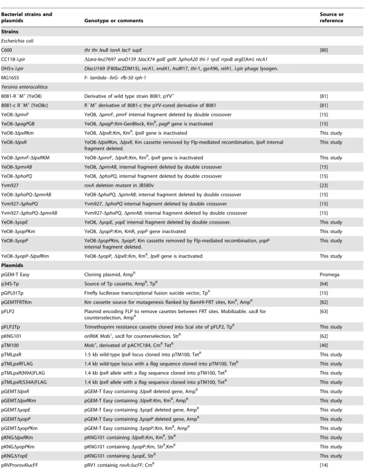
The LpxR-dependent lipid A deacylation was more evident on bacteria grown at 37uC than at 21uC, hence suggesting that the expression and/or function of the deacylase might be temperature-regulated, being higher at 37uC than at 21uC. To monitor transcription of lpxR quantitatively, a transcriptional fusion was constructed in which a promoterlesslucFF gene was under the control of thelpxRpromoter region (see Material and Methods); thereafterlpxR::lucFFwas introduced into YeO8 and the luciferase activity was determined. The expression of the fusion was higher at 21uC than at 37uC (Figure 2A). Real time (RT) quantitative PCR (RT-qPCR) experiments showed thatlpxRmRNA levels were also higher at 21uC than at 37uC (Figure 2B).
To assess LpxR levels, the C-terminus of the protein was tagged with a FLAG epitope and the construct was cloned into the medium-copy plasmid pTM100 to obtain pTMLpxRFLAG (see Materials and Methods). This plasmid restored the presence of the tetra-acyl species (m/z1414 andm/z1388) in the lipid A of YeO8-Author Summary
Lipopolysaccharide (LPS) is one of the major surface components of Gram-negative bacteria. The LPS contains a molecular pattern recognized by the innate immune system. Not surprisingly, the modification of the LPS pattern is a virulence strategy of several pathogens to evade the innate immune system. Yersinia enterocolitica
causes food-borne infections in animals and humans (yersiniosis). Temperature regulates most, if not all, virulence factors of yersiniae including the structure of the LPS lipid A. At 21uC, lipid A is mainly hexa-acylated and may be modified with aminoarabinose or palmitate. In contrast, at 37uC,Y. enterocoliticaexpresses a unique tetra-acylated lipid A. In this work, we establish that Y. enterocoliticaencodes a lipid A deacylase, LpxR, responsi-ble for the lipid A structure expressed by the pathogen at 37uC, the host temperature. Our findings also revealed that the low inflammatory response associated toY. enteroco-liticainfections is the sum of the anti-inflammatory action exerted by aYersiniaprotein translocated into the cytosol of macrophages and the reduced activation of the LPS receptor complex due to the expression of a LpxR-dependent deacylated LPS.
Table 1.Strains and plasmids used in this study.
Bacterial strains and
plasmids Genotype or comments
Source or reference
Strains
Escherichia coli
C600 thi thr leuB tonA lacY supE [80]
CC118-lpir D(ara-leu)7697araD139DlacX74 galE galKDphoA20 thi-1 rpsE rrpoB argE(Am)recA1
DH5a-lpir DlacU169 (F80lacZDM15),recA1,endA1,hsdR17,thi-1,gyrA96,relA1,lpir phage lysogen.
MG1655 F- lambda- ilvG- rfb-50 rph-1
Yersinia enterocolitica
8081-R2M+(YeO8) Derivative of wild type strain 8081; pYV+ [81]
8081-c R2M+(YeO8c) R2M+derivative of 8081-c the pYV-cured derivative of 8081 [81]
YeO8-DpmrF YeO8,DpmrF,pmrFinternal fragment deleted by double crossover [15]
YeO8-DpagPGB YeO8,DpagP::Km-GenBlock, KmR,pagPgene is inactivated [15]
YeO8-DlpxRKm YeO8,DlpxR::Km, KmR,lpxRgene is inactivated This study
YeO8-DlpxR YeO8-DlpxRKm,DlpxR, Km cassette removed by Flp-mediated recombination,lpxRinternal fragment deleted.
This study
YeO8-DpmrF-DlpxRKM YeO8-DpmrF,DlpxR::Km, KmR,
lpxRgene is inactivated This study
YeO8-DpmrAB YeO8,DpmrAB, internal fragment deleted by double crossover [15]
YeO8-DphoPQ YeO8,DphoPQ, internal fragment deleted by double crossover [15]
Yvm927 rovAdeletion mutant in JB580v [23]
YeO8-DphoPQ-DpmrAB YeO8-DphoPQ,DpmrAB; internal fragment deleted by double crossover [15]
Yvm927-DphoPQ Yvm927,DphoPQinternal fragment deleted by double crossover [15]
Yvm927-DphoPQ-DpmrAB Yvm927-DphoPQ,DpmrAB; internal fragment deleted by double crossover [15]
YeO8-DyopE YeO8,DyopE,yopEinternal fragment deleted by double crossover. This study
YeO8-DyopPKm YeO8,DyopP::Km, KmR,yopPgene inactivated This study
YeO8-DyopP YeO8-DyopPKm,DyopP, Km cassette removed by Flp-mediated recombination,yopP
internal fragment deleted.
This study
YeO8-DyopP-DlpxRKm YeO8-DyopP,DlpxR::Km, KmR,lpxRgene is inactivated This study
Plasmids
pGEM-T Easy Cloning plasmid, AmpR Promega
p34S-Tp Source of Tp cassette, AmpR, TpR [64]
pGPL01Tp Firefly luciferase transcriptional fusion suicide vector, TpR [15]
pGEMTFRTKm Km cassette source for mutagenesis flanked by BamHI-FRT sites, KmR, AmpR [82]
pFLP2 Plasmid encoding FLP to remove casettes between FRT sites. Mobilizable.sacBfor
counterselection, AmpR [63]
pFLP2Tp Trimethoprim resistance cassette cloned into ScaI site of pFLP2, TpR This study
pKNG101 oriR6K Mob+
,sacBfor counterselection, StrR [62]
pTM100 Mob+
, derivated of pACYC184, CmRTetR [40]
pTMLpxR 1.5 kb wild-typelpxRlocus cloned into pTM100, TetR This study
pTMLpxRFLAG 1.4 kb wild-type locus with aflagsequence cloned into pTM100, TetR This study
pTMLpxR(N9A)FLAG 1.4 kblpxRallele with aflagsequence cloned into pTM100, TetR This study
pTMLpxR(S34A)FLAG 1.4 kblpxRallele with aflagsequence cloned into pTM100, TetR This study
pGEMTDlpxR pGEM-T Easy containingDlpxRdeleted gene, AmpR This study
pGEMTDlpxRKm pGEM-T Easy containingDlpxR::Km, KmR, AmpR This study
pGEMTDyopE pGEM-T Easy containingDyopEdeleted gene, AmpR This study
pGEMTDyopP pGEM-T Easy containingDyopPdeleted gene, AmpR This study
pGEMTDyopPKm pGEM-T Easy containingDyopP::Km, KmR, AmpR This study
pKNGDlpxRKm pKNG101 containingDlpxR::Km, KmR, StrR This study
pKNGDyopPKm pKNG101 containingDyopP::Km, StrR,KmR This study
pKNGDYopE pKNG101 containingDyopE, StrR This study
DlpxRKm (data not shown). Western blot analysis of purified membranes from YeO8-DlpxRKm containing pTMLpxRFLAG showed that LpxR levels were higher in membranes from bacteria grown at 21uC than at 37uC (Figure 2C). Altogether, it can be concluded that the expression of lpxR is indeed temperature-regulated but, in contrast to our initial hypothesis, its expression is higher at 21uC than at 37uC.
The apparent contradiction between the mass spectrometry analysis, more deacylation at 37uC, and the Western blot data, higher levels of LpxR at 21uC than at 37uC, led us to explore whether low temperature may affect the function of the enzyme. Since E. coli has been used as surrogate host to characterize SalmonellaLpxR (StLpxR) function [18], we mobilized pTMLpxR into E. coli MG1655 to analyze lipid A species by mass spectrometry in bacteria grown at 21uC and 37uC. Results shown in figure 3 demonstrate that LpxR did deacylate theE. colilipid A from bacteria grown either at 21 or 37uC as detected by the presence of species m/z 1360 (Figure 3C–D). This species was found previously inE. coli expressing StLpxR [18]. Of note, the speciesm/z1414, which is consistent with the deacylation of the species m/z 1850 containing palmitoleate (C16:1) instead of
laureate (C12), was observed only inE. coligrown at 21uC. LpxP
is the cold-temperature-specific late acyltransferase responsible for the addition of palmitoleate [1]. Altogether, our results indicate that the reduced LpxR-dependent deacylation found in YeO8 grown at 21uC cannot be attributed to a general lack of function of the enzyme at this temperature.
Lipid A modification with aminoarabinose affects LpxR-dependent deacylation
We sought to determine why LpxR activity was not observed in YeO8 grown at 21uC despite the detection of the enzyme in the membrane. Among other possibilities, we speculated that specific features of YeO8 lipid A found only at 21uC might be responsible for the reduced LpxR activity. Furthermore, these features should be absent inE. coligrown at 21uC since LpxR-dependent activity was observed here. A conspicuous difference between YeO8 and E. colilipid As is the presence of aminoarabinose and palmitate (m/ z 1954 and 2063, respectively) only in the former [14,15]. Therefore, we explored whether any of these modifications could account for the reduced LpxR activity. In YeO8, similarly to other Gram-negative pathogens, the products of the pmrF operon are required for the synthesis and addition of aminoarabinose to lipid A whereas the acyltransferase PagP is required for the addition of palmitate to lipid A [15]. The lipid A from the pagP mutant, YeO8-DpagPGB, grown at 21uC resembled that of the wild-type strain, except that the species containing palmitate (m/z2063) was not detected (Figure 4A). In contrast, the tetra-acylated species (m/ z 1414) was clearly observed in the lipid A from YeO8-DpmrF
grown at 21uC (Figure 4C). This was dependent on LpxR activity since the peak was absent in the double mutant YeO8- DpmrF-DlpxRKm (Figure 4E). LpxR-dependent deacylation of lipid A (m/ z1388) observed in bacteria grown at 37uC was not affected in either pmrF or pagP single mutants (Figure 4B, D). Control experiments revealed that lpxR expression was not affected in YeO8-DpmrFsince the expression of thelpxR::lucFFfusion was not significantly different between YeO8 and thepmrFmutant either grown at 21uC or at 37uC (Figure 4G).
On the whole, these results are consistent with the notion that the reduced LpxR activity observed in YeO8 at 21uC is associated with the lipid A modification with aminoarabinose.
LpxR 3-D modelling
Our findings might suggest that aminoarabinose-containing LPS may directly inactivate the lipid A deacylase activity of YeLpxR. Alternatively, modification of lipid A with aminoar-abinose could inhibit the physical interaction of LPS with YeLpxR. To explore this, the 3-D structure of YeLpxR was modeled (Figure 5A). The amino acids 1–296 (following the putative signal sequence) could be modeled based on the crystal structure of StLpxR (PDB code 3FID; [21]) and the sequence alignment between StLpxR and YeLpxR (Figure S1). The fold of the resulting model is likely to be of good quality, since YeLpxR has such a high sequence identity to StLpxR (75%). Additionally, the important StLpxR amino acids identified by Rutten and co-workers [21] are conserved in YeLpxR. Six amino acids differ between the YeLpxR and the StLpxR active sites (Figure S1). Major differences are D31 and Q35 in YeLpxR, of which D31 is closer to the active site (Figure 5B). The corresponding amino acids are much smaller in StLpxR, glycine and an alanine, respectively, which cause StLpxR to have a bigger cavity. StLpxR has a protruding cavity close to K67, which cannot be found in YeLpxR (Figure 6A). The difference is induced by D31 in YeLpxR, which occupies more space than G31 in StLpxR. As a consequence, the conserved K67 adopts a different conformation in the YeLpxR model. Due to D31, the cavity in YeLpxR is divided into two parts with a narrow connection, and this amino acid also prevents YeLpxR from forming an inward protruding cavity similar to the one found near G31 in StLpxR (Figure 6A).
Docking of a modified Kdo2-lipid A molecule (see Materials and
methods) to the model of YeLpxR showed that the phosphate group, which attaches aminoarabinose to Kdo2-lipid A, binds into
the cavity in the vicinity of K67 and D31 (Figure 6B). Docking of the same molecule to the crystal structure of StLpxR yielded a result where the phosphate group was located in the protruding cavity close to K67 (Figure 6C). As expected, docking of the modified Kdo2-lipid A molecule with aminoarabinose to the Table 1.Cont.
Bacterial strains and
plasmids Genotype or comments
Source or reference
pGPL01TpYelpxR pGPL01Tp containing a 443-bp DNA fragment corresponding to the
lpxRpromoter region, TpR This study
pDHS45 pFUSE containingyplXA’::lacZYA, CmR [29]
pRSFlhDC08 pRV1 containingflhDC::lucFF, CmR [26]
pRVProrovAlucFF pRV1 containigrovA::lucFF, CmR [14]
pINP41 pEP184 containinginvD412::phoA, ClmR [32]
doi:10.1371/journal.ppat.1002978.t001
YeLpxR model did not give any valuable result. On the other hand, when the same molecule was docked to the StLpxR crystal structure, aminoarabinose was bound close to G31. It occupies the space corresponding to the narrow connection of the two larger cavities in YeLpxR (Figure 6D)
As a result from the modeling and docking studies, we suggest that Kdo2-lipid A with aminoarabinose cannot fit into the active
site of YeLpxR due to D31, hence leading to the inability of YeLpxR to deacylate Kdo2-lipid A with aminoarabinose.
lpxRsite-directed mutagenesis
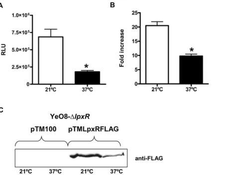
To confirm our predictions, we constructed LpxR mutants by site-directed mutagenesis (see Material and Methods). In addition to the amino acids corresponding to the active site amino acids in StLpxR, we wanted to study the effect of the D31G mutation for YeLpxR as the modelling and docking studies suggested that D31 has an important role in the YeLpxR specificity for the Kdo2-lipid
A species. The constructs were introduced intoE. coli MG1655 and the lipid A from the transformants grown at 37uC was Figure 1. Lipid A analysis fromY. enterocolitica lpxRmutant.(A) Negative ion MALDI-TOF mass spectrometry spectra of lipid A isolated from YeO8 grown at 21uC and 37uC. (B) Negative ion MALDI-TOF mass spectrometry spectra of lipid A isolated from YeO8-DlpxRKm (DlpxR) grown at 21uC and 37uC. (C) Negative ion MALDI-TOF mass spectrometry spectra of lipid A isolated from YeO8-DlpxRKm carrying pTMLpxR grown at 21uC and 37uC. The results in all panels are representative of three independent lipid A extractions.
analyzed by MALDI-TOF mass spectrometry. Most of the constructs containing LpxR mutants did trigger the deacylation of E. coli lipid A, detected by the presence of speciesm/z 1360, (Table 2). In contrast, constructs containing LpxR mutants, LpxR(N9A), LpxR(D10A), LpxR(S34A), and LpxR(H122A) did not deacylate E. coli lipid A. These results were expected since Rutten and co-workers have reported that these residues are located in the StLpxR active site and all of them are conserved in LpxR homologues [21]. Next, only those constructs triggering deacylation ofE. colilipid A were introduced into YeO8. When the YeO8 strains were grown at 37uC, all LpxR mutants restored the presence of the tetra-acyl species (m/z 1388) in the lipid A of YeO8-DlpxRKm (Table 2). Additionally, the mass spectrometry analysis revealed that LpxR(D31G) mutant did trigger the deacylation of lipid A in bacteria grown at 21uC as it was detected the presence of lipid A speciesm/z1414 andm/z1545 (Figure 7B). The latter is consistent with the deacylation of the lipid A species modified with aminoarabinose (m/z1954).
In summary, our results further confirmed the amino acids important for the catalytic activity of YeLpxR. Moreover, our results confirmed the molecular modelling predictions, thereby demonstrating that the presence of D31 in the active site pocket of YeLpxR causes steric hindrance for the binding and deacylation of lipid A species modified with aminoarabinose.
Regulation of lpxRexpression
In YeO8 the expression of the loci responsible for the lipid A modification with aminoarabinose, ugd and pmrF operon, is temperature regulated, being higher at 21uC than at 37uC [15]. Mechanistically, this is so because the expression of the positive regulatorsphoPQandpmrAB, which control the expression ofugd and thepmrFoperon, is also higher at 21uC than at 37uC [15]. In
turn, the temperature-dependent regulation ofphoPQandpmrABis explained by H-NS-dependent negative regulation alleviated by RovA, another major regulator ofYersinia [22,23], at 21uC [15]. Moreover, there is cross-talk between the regulators in such way that PhoPQ and PmrAB regulate positively the expression ofrovA and the effect of PhoPQ is more important [15].
The inverse correlation between the substitution of the lipid A with aminoarabinose and lipid A deacylation, prompted us to evaluate whetherphoPQandpmrABmight negatively regulatelpxR. Results shown in figure 8 revealed that the expression of lpxR::lucFFwas significantly up-regulated in thephoPQandpmrAB mutants at 21uC and 37uC (Figure 8A). However, the expression oflpxRreached wild-type levels in the doublephoPQ-pmrABmutant regardless the bacteria growth temperature (Figure 8A). RT-qPCR experiments showed that the levels oflpxRmRNA were higher in thephoPQand pmrABmutants than in the wild type and double phoPQ-pmrAB mutants, which were not significantly different (Figure S2).
Recently, we have shown thatrovAexpression is downregulated in thephoPQ and pmrABsingle mutants, being the lowest in the phoPQmutant, whereas in the phoPQ-pmrAB double mutant rovA expression is not significantly different to that in the wild type [15]. Therefore, the fact thatlpxRexpression follows the opposite trend in these mutants led us to analyze whether rovA negatively regulates the expression of lpxR. Indeed, luciferase activity was higher in therovAmutant than in the wild type and the levels were not significantly different that those observed in thephoPQmutant when bacteria were grown either at 21uC or 37uC (Figure 8A). Similar results were obtained when thelpxRmRNA levels were analyzed by RT-qPCR (Figure S2). The increasedlpxRexpression observed inrovAandphoPQsingle mutants at 21uC was no longer found in the double mutantrovA-phoPQ(Figure 8A and Figure S2). Figure 2. Temperature regulates the expression ofY. enterocolitica lpxR.(A) Analysis of the expression oflpxRby measuring luciferase activity of YeO8 carryinglpxR::lucFFtranscriptional fusion, which was grown at 21uC (white bars) or 37uC (black bars). Data are presented as mean6SD (n = 3). *, results are significantly different (p,0.05; two-tailedttest) from the results for bacteria grown at 21uC. (B) Analysis oflpxRmRNA levels by RT-qPCR. Total RNA was extracted from bacteria grown at 21uC (white bar) or 37uC (black bar). Data are presented as mean6SD (n = 3). *, results are significantly different (p,0.05; two-tailedttest) from the results for bacteria grown at 21uC. (C) Western blot analysis of LpxR FLAG tagged levels. Cell envelopes were purified from YeO8-DlpxRKm mutant carrying pTM100 or pTMLpxRFLAG plasmids. 80mg of proteins were run in SDS-12%
polyacrylamide gel, electrotransferred onto a nitrocellulose membrane, and developed by using anti-Flag antibodies. doi:10.1371/journal.ppat.1002978.g002
When bacteria were grown at 37uC,lpxRexpression in the rovA-phoPQmutant was significantly lower than those observed in the rovAandphoPQsingle mutants (p,0.05 for each comparison versus rovA-phoPQmutant) although still higher than that in the wild type (Figure 8A and Figure S2). Of note, the expression oflpxRwas no longer temperature regulated in therovA-phoPQmutant (Figure 8B). The fact that the expression of lpxR::lucFF in the triple mutant rovA-phoPQ-pmrAB at 21uC was less than in the wild-type strain may support the notion that, in the absence of the negative regulator RovA, PmrAB and/or a PmrAB-modulated regulator positively regulates lpxR. At 37uC, lpxRexpression in the triple mutant was not significantly different than those found in the double mutant phoPQ-pmrAB and the wild type (Figure 8A and Figure S2).
Collectively, our data revealed that the expression of lpxR is negatively controlled by the same regulators that activate the loci necessary for the substitution of the phosphate at the 49end of the glucosamine disaccharide with aminoarabinose.
Flagellar regulon and lipid A acylation
In a previous study, we observed the down regulation of YeO8 virulence factors in mutants lacking the lipid A late acyltrans-ferases LpxM, LpxL or LpxP [14]. These results raised the possibility that lipid A acylation may act as a regulatory signal by acting on a transduction pathway(s) [14]. In this context, we
sought to determine the impact of LpxR to the expression/ function of YeO8 virulence factors.
the mutant proteins were expressed (Figure S3).When the strains were grown at 37uC, YeO8 and YeO8-DlpxRKm produced the same luminescence (Figure 9B).
One virulence gene that is regulated as part of the flagellar regulon is yplA and hence its expression is regulated by flhDC [24,27,28]. Considering thatflhDCexpression was downregulated in thelpxRmutant, we speculated that yplAexpression could be affected in this mutant. The transcriptional fusion yplA::lacZYA [29] was introduced into the chromosome of the wild type and the lpxR mutant and theirb-galactosidase activities were measured. Indeed, theb-galactosidase activity was lower in YeO8-DlpxRKm than in the wild type (Figure 9C). Plasmids pTMYeLpxR, pTMLpxR(N9A) and pTMLpxR(S34A) complemented the phe-notype (Figure 9C).
In summary, these results indicate that the flagellar regulon is downregulated in thelpxRmutant with a concomitant decrease in motility and downregulation ofyplAexpression.
Invasin and lipid A acylation
Inv is an outer membrane protein ofY. enterocoliticaresponsible for invasion of the host [30,31]. Since YeO8 lipid A mutations affect inv expression [14], we asked whether inv expression is altered in thelpxRmutant. Aninv::phoAtranslational fusion [32] was introduced into the genome of YeO8 and YeO8-DlpxRKm and inv expression was monitored as alkaline phosphatase (AP) activity (Figure 10A). AP activity was significantly lower in thelpxR mutant than in the wild type. Plasmids pTMYeLpxR, pTMLpxR(N9A) and pTMLpxR(S34A) restored AP activity to wild-type levels. These differences ininvexpression prompted us to
study the ability of YeO8-DlpxRKm to invade HeLa cells by using a gentamicin protection assay. The amount of intracellular bacteria was 55% lower when cells were infected with the lpxR mutant than with the wild type (Figure 10B).
RovA is required for inv expression in Y. enterocolitica [33]. Therefore, among other possibilities, the lowinvexpression found in thelpxR mutant could be caused by downregulation of rovA expression. To address this, therovA::lucFFtranscriptional fusion [14] was introduced into the genome of the wild type and thelpxR mutant and the luminescence was determined. Results shown in figure 10C demonstrate thatrovAexpression was dowregulated in YeO8-DlpxRKm. This phenotype was complemented with plas-mids pTMYeLpxR, pTMLpxR(N9A) and pTMLpxR(S34A).
Together, our data show that the down-regulation of inv expression found in the lpxR mutant is most likely caused by downregulation of rovA expression, the positive transcriptional regulator ofinv.
Impact of lipid A acylation on pYV-encoded virulence factors
Y. enterocolitica harbours a plasmid (pYV)-encoded type III secretion system which is required for virulence. A set of virulence factors, called Yops, are secreted by this system and enable Y. enterocoliticato multiply extracellularly in lymphoid tissues [34–36]. In several pathogens, LPS polysaccharide status affects the expression of the type III secretion systems [37–39]. Therefore, we asked whether the production of theYersiniapYV-encoded type III secretion system is altered in thelpxRmutant. At 37uC and under low calcium concentrations, this system secretes the Yops to Figure 4. Lipid A analysis fromY. enterocoliticalipid A mutants.Negative ion MALDI-TOF mass spectrometry spectra of lipid A isolated from the indicatedY. enterocoliticastrains grown at 21uC (A,C,E) and 37uC (B,D,F). The results in all panels are representative of three independent lipid A extractions. (G) Analysis of the expression of lpxRby measuring luciferase activity of YeO8 (white bars) and YeO8-DpmrF(gray bars) carrying lpxR::lucFFtranscriptional fusion, which were grown at 21uC or 37uC. Data are presented as mean6SD (n = 3).
doi:10.1371/journal.ppat.1002978.g004
Figure 5. Modelling ofY. enterocoliticaO:8 LpxR.(A) The model of YeLpxR based on the StLpxR crystal structure. Theb-barrel is colored as rainbow, the helices are green and loops are wheat. (B) Close-up view of the active site. The amino acids mutated in this study are shown as sticks in yellow and pink.
the culture supernatant [40]. Analysis of Yop secretion revealed that the wild type and thelpxRmutant secreted similar levels of Yops (Figure 11A). We sought to determine whether the translocation of Yops to the cytosol of eukaryotic cells is affected in thelpxRmutant. Detection of cytoskeleton disturbances upon infection of epithelial cells is one of the most sensitive assays to establish Yop translocation [41]. The injection of YopE into the cytosol of A549 cells by wild-type bacteria induced disruption and condensation of the actin microfilament structure of the cells whereas this was not the case when cells were infected with YeO8-DyopEmutant (Figure 11B). YopE translocation to A549 cells was not affected in the lpxR mutant background (Figure 11C). As
expected, A549 cells infected with YeO8-DlpxRKm displayed similar cytoskeleton disturbances than those cells infected with the wild type (Figure 11B).
yadAis another pYV-encoded virulence gene whose expression is only induced at 37uC [42]. YadA is an outer membrane protein mediating bacterial adhesion, bacterial binding to proteins of the extracellular matrix and complement resistance (for a review see [43]). Analysis of YadA expression by SDS-PAGE demonstrated that YeO8-DlpxRKm and YeO8 produced the same amount of the protein (Figure 11D). To assess YadA functionality, we asked whether the YadA-dependent binding to collagen is altered in the lpxR mutant. To this end, we analyzed the binding of YadA-Figure 6. Docking of Kdo2-lipid A to LpxR.(A) The differences in surface cavities between YeLpxR (pink) and StLpxR (green) with K67 as sticks
with pink carbon atoms for YeLpxR and green carbon atoms for StLpxR and YeLpxR D31 as sticks with pink carbon atoms. G31 in StLpxR is hidden behind D31 in YeLpxR. (B) The YeLpxR model with Kdo2-lipid A (sticks with yellow carbon atoms) in the active site. The surface cavity of YeLpxR is shown in pink and K67 and D31 as sticks with pink carbon atoms. (C) The StLpxR model with Kdo2-lipid A (yellow sticks) in the active site. The surface cavity of StLpxR is shown in green and K67 and G31 as sticks with green carbon atoms. (D) The StLpxR model with Kdo2-lipid A including aminoarabinose in the active site. The surface cavity of StLpxR is shown in green along with K67 and G31 (green sticks).
doi:10.1371/journal.ppat.1002978.g006
expressing whole bacteria to collagen type I by immunofluores-cence (see Material and Methods). In contrast to the negative control, a pYV-cured strain (YeO8c), YeO8 and YeO8-DlpxRKm bound to collagen without differences between them (Figure 11E– F).
Taken together, these results suggest that the production and function of the pYV-encoded virulence factors Yops and YadA are not altered in thelpxRmutant.
Lipid A acylation and innate immunity
Cationic antimicrobial peptides (CAMPs) belong to the arsenal of weapons of the innate immune system against infections. In the case of Gram-negative bacteria, CAMPs interact with the lipid A moiety of the LPS [44–47] and lipid A modification is one of the strategies employed by Gram-negative bacteria to counteract the action of CAMPs. We and others have used polymyxin B as a model CAMP since it also binds to lipid A. Furthermore, resistance to this peptide reflects well the resistance to other mammalian peptides and correlates with virulence [48–51]. Therefore we evaluated the resistance of thelpxR mutant to polymyxin B. Results shown in figure 12A demonstrate that the mutant was as resistant as the wild type to the peptide when grown either at 21uC or at 37uC. Of note both strains were more susceptible to polymyxin B when grown at 37uC than at 21uC (Figure 12A).
Table 2.Effect oflpxRmutations on lipid A deacylation.
Lipid A deacylation
Mutations
E. coli(376C)
m/z1360 Y. enterocolitica(216C)m/z1414
N9A No No
D10A No n.a.
S34A No n.a.
H122A No n.a.
Q118A No No
Q57A Yes No
Y130A Yes No
G36A Yes No
F79A Yes No
P62A Yes No
W133A Yes No
D31G Yes Yes
n.a.; Not analyzed.
doi:10.1371/journal.ppat.1002978.t002
Figure 7. Presence of D31 in the active site pocket ofY. enterocoliticaO:8 LpxR affects the deacylation activity of the enzyme.
Negative ion MALDI-TOF mass spectrometry spectra of lipid A isolated from: (A)E. coliMG1655 (E. coli) carrying pTMLpxR(D31G) grown at 37uC. (B) YeO8-DlpxRKm (DlpxR) carrying pTMLpxR(D31G) grown at 21uC. (C) YeO8-DlpxRKm (DlpxR) carrying pTMLpxR(D31G) grown at 37uC. The results in all panels are representative of three independent lipid A extractions.
Figure 8. Y. enterocoliticaPhoPQ, PmrAB two-component systems and RovA control the expression of lpxR. (A) Analysis of the expression oflpxRby YeO8 (white bar), and mutants (grays bars) YeO8-DphoPQ(DphoPQ), YeO8-DpmrAB(DpmrABand YeO8-DphoPQ-DpmrAB (DphoPQ-pmrAB), Yvm927 (DrovAYvm927-DphoPQ-DpmrAB(DrovADphoPQ-DpmrAB) carrying the transcriptional fusionlpxR::lucFFgrown at 21uC or 37uC. Data are presented as mean6SD (n = 3). *, results are significantly different (p,0.05; two-tailedttest) from the results for YeO8 grown at the same temperature. (B) This panel displays the same results shown in panel A for YeO8 and the double mutant Yvm927-DphoPQ(DrovA-DphoPQ) and it is included for the sake of clarity.
doi:10.1371/journal.ppat.1002978.g008
Figure 9. Flagellar regulon is downregulated in theY. enterocoliticaO:8lpxRmutant.(A) Motility assays were performed with YeO8, and YeO8-DlpxRKm (DlpxR) in a semisolid agar plate (3% agar and 1% tryptone). Plates were incubated at 22uC for 24 h. (B) Analysis offlhDCexpression by YeO8, YeO8-DlpxRKm (DlpxR), and YeO8-DlpxRKm with the plasmids pTMYeLpxR (DlpxR/pTMYeLpxR); pTMYeLpxR(N9A) (DlpxR/pTMYeLpxR(N9A), and pTMYeLpxR(S34A) (DlpxR/pTMYeLpxR(S34A) carrying the transcriptional fusionflhDC::lucFFgrown at 21uC and 37uC. (C)b-galactosidase activity production by yplA’::lacZYA present in YeO8, YeO8-DlpxRKm (DlpxR), and YeO8-DlpxRKm with the plasmids pTMYeLpxR (DlpxR/pTMYeLpxR); pTMYeLpxR(N9A) (DlpxR/pTMYeLpxR(N9A), and pTMYeLpxR(S34A) (DlpxR/pTMYeLpxR(S34A) [b-galactosidase values given in Miller units, mean6SD (n = 3)]. *, results are significantly different (p,0.05; two-tailedttest) from the results for YeO8.
doi:10.1371/journal.ppat.1002978.g009
The mammalian immune system recognizes and responds toE. coli LPS via the TLR4 complex, resulting in the synthesis and secretion of pro-inflammatory cytokines that recruit immune cells to the site of infection. The ability of LPSs to evoke inflammatory responses and the potency of them are directly related to the structure of the molecule. It has been reported that underacylated LPSs are less inflammatory than hexa-acylated ones, being theE colilipid A (m/z1797) the prototype of hexa-acylated LPSs [52]. Therefore, the dramatic changes in lipid A acylation displayed by thelpxRmutant at 37uC led us to evaluate the immunostimulatory properties of YeO8 and YeO8-DlpxRKm. As cellular read-out, we determined TNFalevels secreted by macrophages infected either with the wild type or thelpxRmutant grown at 21uC and 37uC. YeO8 and YeO8-DlpxRinduced similar levels of TNFaalthough the levels induced by bacteria grown at 37uC were significantly lower than those triggered by bacteria grown at 21uC (p,0.05 for comparison of TNFa levels between temperatures for a given strain) (Figure 12B). This was dependent on the well known anti-inflammatory action of the pYV-encoded YopP [53,54], since a yopPmutant grown at 37uC induced similar levels of TNFathan those induced by wild-type bacteria grown at 21uC (Figure 12B). Therefore we sought to determine whether YopP could be counteracting the inflammatory response induced by YeO8-DlpxRKm. Indeed, YeO8-DyopP-DlpxRKm induced the highest levels of TNFa(Figure 12B). Further sustaining this notion, the TNFa levels induced by the lpxR mutant cured of the pYV virulence plasmid grown at 37uC were significantly higher than those induced by the virulence plasmid negative wild-type strain but not different than the YeO8-DyopP-DlpxRKm-triggered TNFa levels (Figure 12B). Of note, the TNFa levels induced by the virulence plasmid negative wild-type strain grown at 37uC were significantly lower than those triggered by bacteria grown at 21uC hence further highlighting the importance of lipid A acylation on the immunostimulatory properties of YeO8.
Discussion
Pathogenic yersiniae show a temperature-dependent variation in lipid A acylation [9–14]. At 21uC, Y. enterocolitica synthesizes hexa-acylated lipid A containing four 3-OH-C14, one C12 and
either one C16:1 or one C14. At 37uC, Y. enterocolitica lipid A
presents a tetra-acylated species (m/z1388) and a hexa-acylated one containing four 3-OH-C14, one C12and C14. In a previous
work, we identified and characterized the acyltransfreases,lpxM, lpxLandlpxP, responsible for the addition of C12, C14and C16:1,
respectively, to lipid A [14]. Moreover, we demonstrated that the expressions of these enzymes are temperature regulated [14]. However, the unique tetra-acyl lipid A found in the wild type grown at 37uC (m/z 1388) remained to be explained at the molecular level. We and others have established that this species is consistent with 39-O-deacylation of lipid A [12,14,17]. In this work by combining biochemistry, genetics and molecular modelling we present evidence that LpxR is the lipid A 39-O-deacylase of Y. enterocolitica.
YeLpxR is one of the closest homologues to StLpxR. Despite the presence of StLpxR in the Salmonella outer membrane, the bacterium does not produce 39-O-deacylated lipid A species under any growth conditions tested to date [18]. This has been termed as enzyme latency and similar findings have been reported for the Salmonella lipid A 3-O-deacylase PagL and E. coli PagP [55,56]. Our data revealed that YeLpxR is also latent in the membrane of YeO8 grown at 21uC. However, this is not a general feature of lipid A deacylases sinceH. pyloriLpxR is constitutively active [19]. Several explanations could underlie YeLpxR latency at 21uC. Firstly, we explored whether low temperature may affect the function of the enzyme. The fact that YeLpxR did deacylateE. coli lipid A when grown at 21uC does not support that low temperatures grossly inhibit the enzyme activity. Nevertheless, we do not by any means completely rule out that temperature may affect YeLpxR activity, and thorough biochemical analyses are warranted to rigorously define the functional parameters of YeLpxR activity. This will be the subject of future studies. We next hypothesized that specific features of YeO8 lipid A, which do not exist in the E. colilipid A, may be responsible for YeLpxR latency. The first conspicuous difference is the type of secondary fatty attached to the lipid IVA. InE. colithe late acyltransferases
LpxL and LpxM add laureate (C12) and myristate (C14)
respectively [1] whereas in YeO8 these enzymes transfer myristate (C14) and laureate (C12) respectively [14]. However, this cannot Figure 10.Inv expression is altered inY. enterocoliticaO:8lpxRmutant.(A) Alkaline phosphatase (AP) activities exhibited byinv::phoA translational fusion present in YeO8, YeO8-DlpxRKm (DlpxR), and YeO8-DlpxRwith the plasmids pTMYeLpxR (DlpxR/pTMYeLpxR); pTMYeLpxR(N9A) (DlpxR/pTMYeLpxR(N9A), and pTMYeLpxR(S34A) (DlpxR/pTMYeLpxR(S34A) [AP is expressed in enzyme units per OD600unit; mean6SD (n = 3)]. (B) Invasion of HeLa cells by YeO8, and YeO8-DlpxRKm (DlpxR). Invasion assays were done in triplicate without centrifugation (n = 3). (C) Analysis ofrovA expression by YeO8, YeO8-DlpxRKm (DlpxR), and YeO8-DlpxR with the plasmids pTMYeLpxR (DlpxR/pTMYeLpxR); pTMYeLpxR(N9A) (DlpxR/ pTMYeLpxR(N9A), and pTMYeLpxR(S34A) (DlpxR/pTMYeLpxR(S34A) carrying the transcriptional fusionrovA::lucFF. *, results are significantly different (p,0.05; one-tailedttest) from the results for YeO8.
account for the reduced LpxR activity since the enzyme did deacylate E. colilipid A. The presence of palmitoleate in YeO8 lipid A at 21uC but not at 37uC cannot be the reason since YeLpxR deacylatedE. colilipid A containing palmitoleate, found inE. coligrown at 21uC. Instead, our results revealed that the lipid A substitution with aminoarabinose is associated with YeLpxR latency since LpxR-dependent lipid A deacylation was clearly observed in thepmrFmutant grown at 21uC. Notably, the lack of aminoarabinose also releases Salmonella PagL from latency [56], hence suggesting a key role for the lipid A modification with aminoarabinose in LPS remodelling.
The molecular modelling and docking experiments further highlighted the importance of lipid A substitution with aminoar-abinose for YeLpxR function. D31 in YeLpxR forces the conserved K67 to adopt a different conformation compared to StLpxR. According to the docking results, the resulting loss of
cavity space in the vicinity of K67 in YeLpxR, causes the phosphate at the 49 end of Kdo2-lipidA to bind somewhat
differently to YeLpxR than to StLpxR. In the latter, the phosphate binds in the cavity near K67, while in YeLpxR it is forced to bind more outwards from the enzyme. The docking of Kdo2-lipidA
with aminoarabinose to StLpxR showed that aminoarabinose occupies the cavity space, which corresponds to a narrow connection between two larger cavities in YeLpxR. The large reduction in cavity volume at this particular site causes this space to be too small for the accommodation of aminoarabinose. Hence, D31 seems to cause steric hindrance for the binding of aminoarabinose-containing Kdo2-lipidA to YeLpxR. Therefore,
we predicted that D31 could have an important role for the YeLpxR substrate specificity. Indeed, the site-directed mutagenesis experiments validated that the presence of D31 in the active site pocket of YeLpxR causes a steric hindrance for the binding and Figure 11. The productions of Yops and YadA are not affected inY. enterocoliticaO:8lpxRmutant.(A) SDS-PAGE (the acrylamide concentration was 4% in the stacking gel and 12% in the separation one) and Coomasie brilliant blue staining of proteins from the supernatants of Ca2+- deprived cultures from YeO8 and YeO8-DlpxRKm. Result is representative of four independent experiments. (B) Actin disruption byYersinia
infection. A549 cells (monolayer of 70% confluence) were infected with YeO8, YeO8-DlpxRKm or YeO8-DyopE(MOI 25:1) for 1 h. After fixing and permeabilization of cells actin was stained with OregonGreen 514-phalloidin (1:100) and cells were analyzed by fluorescence microscopy. Result is representative of four independent experiments. (C) Translocation of YopE into A549 cells by YeO8, or YeO8-DlpxRKm (DlpxR) (MOI 25:1 and 1 h of infection). After digitonin extraction, aliquots corresponding to approximately 66104infected A549 cells were analysed by SDS-polyacrylamide gel electrophoresis and Western blotting using rabbit polyclonal antiserum raised against YopE (1:2000 dilution). Result is representative of four independent experiments. (D) SDS-PAGE (the acrylamide concentration was 4% in the stacking gel and 10% in the separation one) followed by Coomasie brilliant blue staining of cell extracts from strains grown in RPMI 1640 at 37uC. White arrow marks YadA protein. Result is representative of four independent experiments. (E)Y. enterocoliticastrains were allowed to adhere to collagen-coated coverslips. Weakly-bound bacteria were washed off and adherent bacteria stained with Hoechst 33342. YeO8c, pYV-cured derivative of YeO8 (Table 1). (F) Adhering bacteria to collagen-coated coverslips were counted. Wild-type bacteria (YeO8) adherence was set to 100%. Bars represent mean6SD (n = 4). *, results are significantly different (p,0.05; two-tailedttest) from the results for YeO8.
doi:10.1371/journal.ppat.1002978.g011
deacylation of lipid A species modified with aminoarabinose. Nevertheless, at present we do not rule out that other residues of YeLpxR also contribute to its latency. In this regard, Salmonella PagL is released from latency when specific amino acid residues located at extracellular loops of the enzyme are mutated and it has been postulated that these residues are involved in the recognition of aminoarabinose-modified lipid A [56–58]. Studies are going to explore whether residues located at extracellular loops of LpxR also contribute to enzyme latency.
The inverse correlation between the aminoarabinose content in the LPS and the LpxR-dependent lipid A deacylation prompted us to evaluate whether the same regulatory network governing the expression of the pmrF operon and ugd could regulate lpxR. Recently, we have shown that the global regulators RovA, PhoPQ, and PmrAB positively control the expression of the loci necessary for aminoarabinose biosynthesis at 21uC [15]. Furthermore, there is a cross-talk between these regulators since the expressions of phoPQandpmrABare downregulated in therovAmutant whereas rovA expression is downregulated in phoPQ and pmrAB single mutants [15]. Our findings support the notion that RovA and PhoPQ are negative regulators of lpxR since its expression was higher inphoPQand rovAsingle mutant backgrounds than in the wild type. In turn, the two-component system PmrAB and/or a PmrAB-regulated system may act as a positive regulator because lpxR expression was similar in the wild-type and rovA-phoPQ backgrounds.
One striking finding of our study is that motility and invasion of eukaryotic cells were reduced in thelpxRmutant grown at 21uC. Mechanistically, our data revealed that the expressions offlhDCand
rovA, the key regulators controlling the flagellar regulon and invasin respectively [22,25,33], were down-regulated in thelpxRmutant. Although we have reported that lipid A acylation status affects motility and invasion [14], the phenotypes were found in mutants lacking the late-acyltransferases and hence displaying major changes in the lipid A structure at 21uC [14]. This is in contrast to the lpxRmutant grown at 21uC, where the LpxR-dependent deacylation was hardly observed. The fact that YeLpxR is in latent stage at this growth temperature may suggest that, in the lpxR mutant background, the absence of the enzyme in the outer membrane, not the lipid A deacylation, acts as the regulatory signal underlying the reduced expressions of flhDC and rovA. Given experimental support to this hypothesis, the catalytically inactive mutants LpxR(N9A) and LpxR(S34A) restored the expressions of flhDC,ylpA,invandrovAto wild-type levels. These results are in good agreement with the notion that membrane-intrinsinc b-barrel proteins, such as LpxR, may launch transmembrane signal transduction pathways upon sensing outer membrane perturbations [59], in our case, the absence of the protein itself. Therefore, it can be speculated that those systems sensing extracytoplasmatic stresses could underlie the regulatory connection between the absence of LpxR and the expression ofY. enterocoliticavirulence factors. Giving indirect support to our speculation, it has been reported that lipid A deacylation inducessE-dependent responses inE. coli[60], the Cpx system senses changes in LPS O-polysaccharide [61]. Experiments are underway to test whether the activation status of the Cpx and/ orsE
systems is altered in thelpxRmutant background and whether any of these systems is responsible for the reduced expression of flhDCandrovAfound in the mutant.
Figure 12. Impact oflpxRonY. enterocoliticaO:8 interplay with the innate immune system.(A) YeO8 (black circle) or YeO8-DlpxRKm (white circle) grown at 21uC or 37uC were exposed to different concentrations of polymyxin B. Each point represents the mean and standard deviation of eight samples from four independently grown batches of bacteria. (B) TNFasecretion by infected macrophages with YeO8 (WT), YeO8-DlpxRKm (DlpxR), YeO8-DyoP(DyopP), YeO8-DyopP-DlpxRKm (DyopP-DlpxR).‘‘c’’ denotes bacteria without the virulence plasmid. Strains were grown at 21uC (denoted as 21) and 37uC (denoted as 37). The data are means and s.e.m. *, p,0.05 (for the indicated comparisons).
The LPS contains a molecular pattern recognized by the innate immune system thereby arousing several host defence responses. On one hand, CAMPs target this LPS pattern to bind to the bacterial surface, which is necessary for their microbicidal action. On the other hand, recognition of the LPS by the LPS receptor complex triggers the activation of host defence responses, chiefly the production of inflammatory markers. Not surprisingly, the modification of the LPS pattern is a virulence strategy of several pathogens to evade the innate immune system, andY. enterocolitica is not an exception. Recently, we have demonstrated that the temperature-dependent lipid A modifications with aminoarabi-nose and palmitate help Y. enterocolitica to avoid the bactericidal action of CAMPs [15]. In this context, it was not totally unexpected to find out that the lpxRmutant was as susceptible as the wild type to polymyxin B, a model CAMP, since the mass spectrometry analysis indicated that the aforementioned lipid A modifications were not affected in thelpxR mutant background. Concerning the activation of inflammatory responses, several studies highlight the critical role of pYV-encoded Yops, chiefly YopP, to prevent the activation of inflammatory responses in a variety of cells, including macrophages. Nevertheless, Rebeil and co-workers [12] conclusively demonstrated that purified LPS from Y. enterocolitcagrown at 37uC is less inflammatory than that purified from bacteria grown at 21uC. This is in agreement with the concept that underacylated LPSs are less inflammatory than hexa-acylated ones [52]. Therefore, it was plausible to speculate that the LpxR-dependent deacylation of LPS at 37uC was responsible for the reduced stimulatory potential of the LPS described by Rebeil and co-workers. To confirm this speculation we chose to challenge macrophages with alive bacteria instead of using purified LPS since there might be differences between the cellular recognition of purified LPS and the LPS expressed in the complex lipid environment of the bacterial outer membrane. To our initial surprise, we observed that the lpxR mutant elicited similar inflammatory response than the wild type when both strains were grown at 37uC. The fact that these responses were significantly lower than those elicited by bacteria grown at 21uC suggested that pYV-encoded factors were attenuating the inflammatory response. Therefore, we hypothesized that the arsenal of Yops injected to the cell were efficiently counteracting the activation of inflammatory responses evoked by the lpxR mutant LPS. In fact, our data demonstrated that the production and function of the pYV-encoded virulence factors were not affected in the lpxRmutant. Giving support to our hypothesis, the inflammatory response elicited by the lpxRmutant cured of the pYV virulence plasmid grown at 37uC was significantly higher than that induced by the virulence plasmid negative wild-type strain. Moreover, our findings suggest that, among all Yops, YopP plays a major role in counteracting the inflammation elicited by the lpxR mutant since the TNFa levels induced by the lpxRmutant cured of the pYV virulence plasmid grown at 37uC were not different than those triggered by YeO8-DyopP-DlpxR. On the whole, our results and those reported by Rebeil and co-workers [12] are consistent with a model in which the characteristic low inflammatory response associated toY. enterocoliticainfections might be the sum of the anti-inflammatory action exerted by YopP and the reduced activation of the LPS receptor complex due to the expression of a LpxR-dependent deacylated LPS. In this scenario, the latency of LpxR may facilitate a quick bacterial response upon entering the host to reduce the initial recognition of the pathogen by the LPS receptor complex. This will allow the pathogen to activate other host countermeasures, among others the pYV-encoded type III secretion system, which is a time consuming process.
Materials and Methods
Bacterial strains and growth conditions
Bacterial strains and plasmids used in this study are listed in Table 1. Unless otherwise indicated,Yersiniastrains were grown in lysogeny broth (LB) medium at either 21uC or 37uC. When appropriate, antibiotics were added to the growth medium at the following concentrations: ampicillin (Amp), 100mg/ml for Y. enterocoliticaand 50mg/ml forE. coli; kanamycin (Km), 100mg/ml in agar plates forY. enterocolitica, 50mg/ml in agar plates forE. coli, and 20mg/ml in broth; chloramphenicol (Cm), 20mg/ml; trimethoprim (Tp), 100mg/ml; tetracycline (Tet) 12.5mg/ml; and streptomycin (Str), 100mg/ml.
Y. enterocoliticamutant construction
the wild-type alleles by the mutant ones was done as described above and confirmed by PCR (data not shown).
To cure the pYV plasmid from YeO8-DlpxRKm, bacteria were grown at 37uC in Congo Red Magnesium oxalate agar plates [65]. Colony size and lack of uptake of Congo Red were used to detect loss of the virulence plasmid. This was further confirmed by testing the YadA-dependent autoagglutination ability [66].
Construction oflpxR::lucFFreporter fusion
A 443 bp DNA fragment containing the promoter region oflpxR was amplified by PCR using Ventpolymerase (see Table S1 for primers used), EcoRI digested, gel purified and cloned into EcoRI-SmaI digested pGPL01Tp suicide vector [15]. This vector contains a promoterless firefly luciferase gene (lucFF) and a R6K origin of replication. A plasmid in whichlucFFwas under the control of the lpxRpromoter was identified by restriction digestion analysis and named pGPL01TpYelpxR. This plasmid was introduced intoE. coli DH5a-lpir from which it was mobilized into Y. enterocolitica by triparental conjugation using the helper strain E. coli HB101/ pRK2013. Strains in which the suicide vectors were integrated into the genome by homologous recombination were selected. This was confirmed by PCR (data not shown).
Complementation oflpxRmutant
To complement the lpxRmutant, a DNA fragment of 1.5 kb was PCR-amplified using TaKaRa polymerase (see Table S1 for primers used) gel purified, and cloned into pGEMT-Easy (Promega) to obtain pGEMTComlpxR. A fragment, containing the putative promoter and coding region of the deacylase, was obtained by PvuII digestion of pGEMTComlpxR, gel purified and cloned into the ScaI site of the medium copy plasmid pTM100 [40] to obtain pTMLpxR. For the construction of plasmid pTMLpxRFLAG, thelpxRcoding region with its own promoter and a FLAG epitope sequence right before the stop codon was PCR amplified usingVentpolymerase, primers LpxRtagging and LpxrFLAG (Table S1) and genomic DNA as template. The fragment was phosphorylated, gel purified and cloned into ScaI-digested pTM100. pTMLpxR and pTMLpxRFLAG were intro-duced into E. coli DH5a-lpir and then mobilized into Y. enterocolitica strains by triparental conjugation using the helper strainE. coliHB101/pRK2013.
Isolation and analysis of lipidA
Lipid As were extracted using an ammonium hydroxide/ isobutyric acid method and subjected to negative ion matrix-assisted laser desorption ionization time-of-flight (MALDI-TOF) mass spectrometry analysis [14,67]. Analyses were performed on a Bruker Autoflex II MALDI-TOF mass spectrometer (Bruker Daltonics, Incorporated) in negative reflective mode with delayed extraction. Each spectrum was an average of 300 shots. The ion-accelerating voltage was set at 20 kV. Dihydroxybenzoic acid (Sigma Chemical Co., St. Louis, MO) was used as a matrix. Further calibration for lipid A analysis was performed externally using lipid A extracted fromE. colistrain MG1655 grown in LB at 37uC. Interpretation of the negative-ion spectra is based on earlier studies showing that ions with masses higher than 1000 gave signals proportional to the corresponding lipid A species present in the preparation [9,12,17,68]. Important theoretical masses for the interpretation of peaks found in this study are: lipid IVA, 1405; C12, 182, C14, 210;
C16:1, 236.2; aminoarabinose (AraNH), 131.1; C16, 239.
Site-directed mutagenesis
Site-directed mutagenesis of the lpxRgene was performed by PCR [69]. Plasmid pTMLpxR, obtained with a minipreparation kit
(Macherey-Nagel), was used as template and the desired mutations were introduced by the primer pairs described in Table S1. Amplifications were carried out in 50ml reaction mixture usingVent DNA polymerase (New England BioLabs.). The PCR was started with initial 70 sec incubation at 95uC and then steps (95uC 50 sec, 60uC 75 sec and 72uC 6 min) were repeated 20 times followed by a 10 min extension time at 72uC. The obtained PCR products were gel purified, phosphorylated with T4 polynucleotide kinase, ligated, and digested with DpnI to break down any remaining template plasmid. The ligated PCR-product was transformed into E. coli C600. Plasmid DNA was isolated from transformants and thelpxR gene was completely sequenced to confirm the generated mutations and to ensure that no other changes were introduced. The name of each mutant construct includes the wild-type residue (single-letter amino acid designation) followed by the codon number and mutant residue (typically alanine).
For the construction of plasmids pTMLpxR(N9A)FLAG and pTMLpxR(S34A)FLAG, the lpxR alleles encoded into pTMLpxR(N9A) and pTMLpxR(S34A) were PCR amplified using Ventpolymerase, and primers LpxRtagging and LpxrFLAG (Table S1). The fragments were phosphorylated, gel purified and cloned into ScaI-digested pTM100 [40]. Plasmids were introduced into E. coli DH5a-lpirand then mobilized intoY. enterocoliticastrains by triparental conjugation using the helper strainE. coliHB101/pRK2013.
Purification of membrane proteins and Western blot analysis of LpxR FLAG tagged levels
Overnight 5-ml cultures of Y. enterocolitica strains were diluted 1:21 into 100 ml of LB in a 250-ml flask. Cultures were incubated with aeration at 21uC or 37uC until OD600 0.8. Bacteria were
recovered by centrifugation (65006g; 10 min, RT) and they were resuspended in 2 ml of 10 mM Tris/HCl (pH 7.4)-5 mM MgSO4 containing 2% Triton X-100 (v/v). Cells were broken by sonication (Branson digital sonifier; microtip 1/80 diameter, amplitude 10%) for 1561 min cycles, each cycle comprised 1 min sonication step separated by 1 min intervals. Unbroken cells were eliminated by centrifugation (20006g, 20 min), and cell envelopes were recovered by ultracentrifugation (Beckman 70.1 Ti rotor; 45 0006g; 1 h, 4uC). The cell envelopes were resuspended in 500ml of distilled water. The protein concentration was determined using the BCA Protein Assay Kit (Thermo Scientifc). 80mg of proteins were separated on 4–12% SDS-PAGE, and semi-dry electrotransferred onto a nitrocellulose membrane using as transfer buffer SDS-PAGE-urea lysis buffer [a freshly prepared 1:1 mix of 16SDS running buffer (12 mM Tris, 96 mM glycine, 0.1% SDS] and urea lysis buffer (10 mM Na2HPO4, 1% b
-mercapto-ethanol, 1%SDS, 6 M urea)] [70]. Membrane was blocked with 4% skim milk in PBS. Membranes were stained using anti-Flag antibody (1:2000; Sigma) following the instructions of the supplier.
program VERTAA (Johnson & Lehtonen, 2004) in BODIL. Different rotamers for D10 and D31 were searched with the program Jackal (http://wiki.c2b2.columbia.edu/honiglab_public/ index.php/Software:Jackal). D10 was changed to the same rotamer as in the crystal structure of StLpxR, while the rotamer used for D31 was the one with the lowest energy according to Jackal. SURFNET [75] was used to detect surface cavities, while PyMOL (Version 1.4, Schro¨dinger, LLC) was used for preparing pictures. For the SURFNET calculations, the minimum radius for gap spheres was set to 1.5 A˚ and the maximum radius was 4.0 A˚. For the docking studies, a Kdo2-lipid A, both with and without
aminoarabinose, was modified from the coordinates for the LPS molecule in the crystal structure of FhuA [76]. The fatty acyl chains were removed from the Kdo2-lipid A molecule in order to
reduce the number of rotatable bonds and make the docking more reliable. Aminoarabinose was added to the modified Kdo2-lipid A
molecule with SYBYL (Version 8.0, Tripos Associates, Inc., St Louis, MO, USA), and the structure was minimized with the conjugate gradient method and Tripos force field. The modified Kdo2-lipid A, both with and without aminoarabinose, was docked
to the YeLpxR model and the StLpxR crystal structure (PDB code 3FID) with GOLD via Discovery Studio (CSC IT Center for Science Ltd, Espoo, Finland), with default docking parameters and the receptor cavity defined to D10, Q16, T/S34, K67, and Y130.
Luciferase activity
The reporter strains were grown at 21uC or at 37uC on an orbital incubator shaker (180 r.p.m.) until OD5401.6. The cultures
were harvested (25006g, 20 min, 24uC) and resuspended to an OD540of 1.0 in PBS. A 100ml aliquot of the bacterial suspension
was mixed with 100ml of luciferase assay reagent (1 mM D-luciferin [Synchem] in 100 mM citrate buffer pH 5). Lumines-cence was immediately measured with a Luminometer LB9507 (Berthold) and expressed as relative light units (RLU). All measurements were carried out in quintuplicate on at least three separate occasions.
Analysis of motility andflhDCexpression
Phenotypic assays for swimming motility were initiated by stabbing 2ml of an overnight culture at the centre of agar plates containing 0.3% agar and 1% tryptone [25,26]. Plates were analyzed after 24 h of incubation at RT and the diameters of the halos migrated by the strain from the inoculation point were compared. Experiments were run in quadruplicate in three independent occasions.
To measure flhDC expression, plasmid pRSFlhDC08 [26] encoding the transcriptional fusion flhDC::lucFF was integrated into the genomes of the strains by homologous recombination. This was confirmed by Southern blot (data not shown). Luminescence was determined as previously described.
b-galactosidase and alkaline phosphatase activities b-galactosidase activity was determined as previously described with bacteria grown in 1% tryptone at RT [77]. Alkaline phosphatase activity was determined in permeabilized cells and the results are expressed in enzyme units per OD600as previously
described [78]. Experiments were run in duplicate in three independent occasions.
Real time-quantitative PCR (RT-qPCR)
Bacteria were grown at 21uC or at 37uC in 5 ml of LB medium on an orbital incubator shaker (180 r.p.m.) until an OD600of 0.3.
0.5 ml of ice-cold solution EtOH/phenol [19:1 v/v (pH 4.3)] were
added to the culture and the mixture was incubated on ice for 30 min to prevent RNA degradation. Total RNA was extracted using a commercial NucleoSpin RNA II kit as recommended by the manufacturer (Macherey-Nagel).
cDNA was obtained by retrotranscription of 2mg of total RNA using a commercial M-MLV Reverse Transcriptase (Sigma), and random primers mixture (SABiosciences, Quiagen). 50 ng of cDNA were used as a template in a 25-ml reaction. RT-PCR analyses were performed with a Smart Cycler real-time PCR instrument (Cepheid, Sunnyvale, CA) and using a KapaSYBR Fast qPCR Kit as recommended by the manufacturer (Cultek). The thermocycling protocol was as follows; 95uC for 3 min for hot-start polymerase activation, followed by 45 cycles of 95uC for 15 s, and 60uC for 30 s. SYBR green dye fluorescence was measured at 521 nm.
cDNAs were obtained from three independent extractions of mRNA and each one amplified by RT-qPCR in two independent occasions. Relative quantities oflpxRmRNAs were obtained using the comparative threshold cycle (DDCT) method by normalizing to
rpoBandtonBgenes (Table S1).
Analysis of Yop secretion
Overnight cultures of Y. enterocolitica strains were diluted 1:50 into 25 ml of TSB supplemented with 20 mM MgCl2and 20 mM
sodium oxalate in a 100-ml flask. Cultures were incubated with aeration at 21uC for 2.5 h, and then transferred at 37uC for 3 h. The optical density at 540 nm of the culture was measured and the bacterial cells were collected by centrifugation at 15006g for 30 min. Ammonium sulphate (final concentration 47.5% w/v) was used to precipitate proteins from 20 ml of the supernatant. After overnight incubation at 4uC, proteins were collected by centrifu-gation (30006g, 30 min, 4uC) and washed twice with 1.5 ml of water. Dried protein pellets were resuspended in 50 to 80ml of sample buffer and normalized according to the cell count. Samples were analyzed by sodium dodecyl sulfate-polyacrylamide gel electrophoresis (SDS-PAGE) using 12% polyacrylamide gels and proteins visualized by Coomassie brilliant blue staining.
Control experiments revealed that the secretion of Yops was not affected inyopEandyopPmutants except that each mutant did not produce either YopE or YopP, respectively (data not shown).
Analysis of YadA production
Bacteria were grown overnight in 2 ml RPMI 1640 medium lacking phenol red at 37uC without shaking. The OD540of the
culture was measured and CFUs were determined by plating serial dilutions. Bacteria from 1-ml aliquot were recovered by centrifu-gation (16 0006g, 10 min, 4uC) and resuspended in 200ml of SDS-sample buffer. Samples were incubated for 4 h at 37uC and kept frozen at 220uC. Samples were analyzed by SDS-PAGE using 10% polyacrylamide gels and proteins visualized by Coomassie brilliant blue staining. Samples were normalized according to the cell count and they were not boiled before loading the gel.
Binding assay to collagen-coated slides
Overnight cultures ofY. enterocoliticastrains grown at 37uC were diluted 1:10 into 5 ml of LB and grown with aeration at 37uC for 2.5 h. bacteria were pelleted, washed once with PBS and resuspended to an OD540of 0.3 in PBS.