Associated with Chronic Atrophic Gastritis and Gastric
Cancer
Jun Chen1, Wei Wang2,3, Tao Zhang4, Jiajia Ji1, Qirong Qian5, Lungeng Lu6, Hualin Fu1, Weilin Jin1,7, Daxiang Cui1*
1Department of Bio-Nano Science and Engineering, Key Lab of Thin Film and Microfabrication Technology of Department of Education, Institute of Micro/Nano Science and Technology, Shanghai Jiao Tong University, Shanghai, People’s Republic of China,2State Key Laboratory of Oncology in South China, Sun Yat-sen University Cancer Center, Guangzhou, Guangdong, People’s Republic of China, 3Department of Gastric & Pancreatic Surgery, Sun Yat-sen University Cancer Center, Guangzhou, Guangdong, People’s Republic of China,4Department of Immunology, Fujian Academy of Medical Science, Fuzhou, People’s Republic of China,5Department of Surgery, Changzheng Hospital affiliated to Second Military Medical University, Shanghai, People’s Republic of China,6Institute of Digestive Disease, The First Affiliated Hospital of Shanghai Jiao Tong University, Shanghai, People’s Republic of China,7School of Life Sciences and Biotechnology, Shanghai Jiao Tong University, Shanghai, People’s Republic of China
Abstract
Background:Chronic inflammation plays a causal role in gastric tumor initiation. The identification of predictive biomarkers from gastric inflammation to tumorigenesis will help us to distinguish gastric cancer from atrophic gastritis and establish the diagnosis of early-stage gastric cancer. Phospholipase C epsilon 1 (PLCe1) is reported to play a vital role in inflammation and tumorigenesis. This study was aimed to investigate the clinical significance of PLCe1 in the initiation and progression of gastric cancer.
Methodology/Principal Findings: Firstly, the mRNA and protein expression of PLCe1 were analyzed by reverse transcription-PCR and Western blotting in normal gastric mucous epithelial cell line GES-1 and gastric cancer cell lines AGS, SGC7901, and MGC803. The results showed both mRNA and protein levels of PLCe1 were up-regulated in gastric cancer cells compared with normal gastric mucous epithelial cells. Secondly, this result was confirmed by immunohistochemical detection in a tissue microarray including 74 paired gastric cancer and adjacent normal tissues. Thirdly, an independence immunohistochemical analysis of 799 chronic atrophic gastritis tissue specimens demonstrated that PLCe1 expression in atrophic gastritis tissues were down-regulated since PLCe1 expression was negative in 524 (65.6%) atrophic gastritis. In addition, matched clinical tissues from atrophic severe gastritis and gastric cancer patients were used to further confirm the previous results by analyzing mRNA and protein levels expression of PLCe1 in clinical samples.
Conclusions/Significances:Our results suggested that PLCe1 protein may be a potential biomarker to distinguish gastric cancer from inflammation lesion, and could have great potential in applications such as diagnosis and pre-warning of early-stage gastric cancer.
Citation:Chen J, Wang W, Zhang T, Ji J, Qian Q, et al. (2012) Differential Expression of Phospholipase C Epsilon 1 Is Associated with Chronic Atrophic Gastritis and Gastric Cancer. PLoS ONE 7(10): e47563. doi:10.1371/journal.pone.0047563
Editor:Javier S. Castresana, University of Navarra, Spain
ReceivedJune 22, 2012;AcceptedSeptember 18, 2012;PublishedOctober 15, 2012
Copyright:ß2012 Chen et al. This is an open-access article distributed under the terms of the Creative Commons Attribution License, which permits unrestricted use, distribution, and reproduction in any medium, provided the original author and source are credited.
Funding:This work was supported by National Key Basic Research Program of China (973 Project) (2010CB933900), New Century Excellent Talent of Ministry of Education of China (NCET-08-0350), Special Infection Diseases Key Project of China (2009ZX10004-311), National Natural Science Foundation of China (31170961 and 31100717), Shanghai Science and Technology Fund (12ZR1415800) and Shanghai Jiao Tong University Innovation Fund For Postgraduates (Z-340-007). The funders had no role in study design, data collection and analysis, decision to publish, or preparation of the manuscript.
Competing Interests:The authors have declared that no competing interests exist. * E-mail: [email protected]
Introduction
Gastric cancer is the fourth common tumors in world. The incidence of gastric cancer is steadily increasing because of recent changes of lifestyle and diets habits in China [1]. As for gastric cancer, survival rates depend on the early diagnosis of the disease. Typically, the earlier a cancer is detected and diagnosed, the more successful the treatment will be. Therefore, it is very important to improve the rates of early diagnosis and early treatment for gastric cancer. We have tried to establish an early gastric cancer pre-warning system based on gene expression microarray analysis
since 2005 [2]. Meanwhile, we also hoped to find the early gastric cancer cellsin vivoby multi-mode targeted imaging techniques [3– 7]. However, lacking of biomarker for gastric cancer is still the major obstacle for early diagnosis.
molecular and biological changes, including gene amplification and activations, which occur from chronic gastritis to gastric cancer, can provide some new insights into the pathology of this disease and new prognostic markers. However, up to date, there are few reports on addressing special biomarkers that can distinguish gastric inflammation from gastric tumorigenesis.
The Phosphoinositide-specific phospholipase C (PLC) repre-sents a large family which catalyzes the hydrolysis of phosphati-dylinositol 4,5-bisphosphate into two vital second messengers, diacylglycerol and inositol 1,4,5-trisphosphate, which include six families of mammalian PLC isoforms (b,c,d,e,fandg) [11–13]. Among them, PLCeis a newly identified member of PLC family [12]. It is a key downstream effecter of Ras family small GTPases: Ras, Rap1, and Rap2 [12,14,15].
Recently, much works reported that PLCe1 played a crucial role in tumorigenesis and inflammation. PLCe1 expression has been found to correlate with human bladder cancer, and the knockdown of PLCe1in vitroandin vivoinhibited bladder tumor growth [16,17]. Furthermore, It has been reported that PLCe1 is over-expressed in skin cancer [18]. In PLCe2/2mice, it exhibited
marked resistance to tumor formation and to the 12-O-tetra-decanoylphorbol-13-acetate (TPA)-induce skin inflammation [18,19]. Subsequent studies showed that PLCe played crucial roles in spontaneous intestinal tumorigenesis with augmentation of angiogenesis and inflammation by using the adenomatous polyposis coli (Apc)Min/+mouse model [20].
Moreover, PLCe was required in activation of cytokine production in non-immune skin cells in a variety of inflammatory reactions, and this important function of PLCe was further confirmed in transgenic mice over-expressing PLCe[21,22]. On the other hand, PLCe was required for tumor necrosis factor (TNFa)-induced chemokine (C–C motif) ligand 2 (CCL2) expres-sion in human keratinocytes and cooperates with nuclear factor kappa B (NF-kB) pathway [23]. Such a function of PLCe in inflammation is unique among the PLC isozymes [13].
Recently, two single nucleotide polymorphism (SNP) rs2274223 and rs11187870 in PLCe1 loci have been identified as novel susceptibility locus for gastric cancer in Chinese population by genome wide gene-association analysis (GWAS) [24–26]. These results confirmed the association between PLCe1 and risk of gastric cancers.
In this study, we examined the expression pattern of PLCe1 in human gastric tissues including normal, atrophic gastritis, and tumor tissues, and made a hypothesis on the role of PLCe1 in chronic inflammation and tumorigenesis of gastric cancer. Compared with normal tissues, we found that PLCe1 protein was highly expressed in gastric cancer tissues, whereas it was down-regulated in atrophic gastric tissues. Our results strongly suggest that PLCe1 can be a potentially promising biomarker for distinguishing gastric cancer from atrophic gastritis and might be a potential biomarker for diagnosing early-stage gastric cancer.
Materials and Methods
Ethic Statements
The study was approved by the Ethics Committee of Sun Yat-sen University Cancer Center and the Ethics Committee of the First Affiliated Hospital of Shanghai Jiao Tong University, and written informed consent was obtained from each subject.
Cell lines
The SV40-transformed immortal gastric epithelial cell GES-1 were preserved in our institute and maintained as recommended [27]. Three gastric cancer cell lines AGS, SGC7901 and MGC803
were obtained from Chinese Academy of Sciences Cell Bank of Type Culture Collection and kept in our laboratory. All cell lines were cultured in Dulbecco’s Modified Eagle Medium (DMEM) supplemented with 10% fetal calf serum (FCS) (Sigma Chemical, St. Louis, MO), 100 units/ml penicillin and 0.1 mg/ml strepto-mycin. The cells were maintained at 37uC in a humidified chamber with 5% CO2.
RNA Extraction and Reverse Transcription-PCR
Total RNA from cells and tissues samples was extracted using the Trizol reagent (Invitrogen) according to the manufacturer’s instruction. The extracted RNA was pretreated with RNase-free DNase, and 2mg RNA from each sample was used for cDNA synthesis primed with random hexamers. For PCR-mediated amplification of PLCe1 cDNA, an initial amplification using PLCe1-specific primers was done with a denaturation step at 95uC for 10 min followed by 30 denaturation cycles at 95uC for 60 s, primer annealing at 55uC for 30 s, and primer extension at 72uC for 30 s. On completion of the cycling steps, a final extension at 72uC for 5 min was carried out before the reaction was stopped and stored at 4uC. Expression data were normalized to the geometric mean of the housekeeping glyceraldehyde-3-phosphate dehydrogenase (GAPDH) gene to control the variability in expression levels. Reverse transcription-PCR was designed using the Primer Express Software version 3.0 (Applied Biosystems). The sequences of the sense and antisense primers were as follows: 59 -GCAAGAAGTGGCCTTCTCAG-39 (F) and 59 -GAACTT-GAAGGGAGGGCATT -39 (R) for PLCe1, 59 -GCTCCTCCTGAGCGCAAG-39 (F) and 59 -CATCTGCTG-GAAGGTGGACA - 39(R) for GAPDH.
Western Blotting
Cells were harvested in sampling buffer [62.5 mmol/L Tris-HCl (pH 6.8), 2% SDS, 10% glycerol, and 5% 2-h-mercaptoeth-anol]. All fresh tissues were grounded to powder in liquid nitrogen and then lysed with the sampling buffer. Protein concentration was determined by Bradford assay (Bio-Rad Laboratories). Equal amounts of proteins were applied to 9% polyacrylamide SDS gels (SDS-PAGE), separated electrophoretically, and transferred onto polyvinylidene fluoride membranes (Amersham Pharmacia Bio-tech). The membrane was incubated with anti- PLCe1 rabbit antibody (1:250; Sigma). PLCe1 expression was detected with horseradish peroxidase-conjugated goat anti-rabbit IgG (1:2,000; Amersham Pharmacia Biotech) and an enhanced chemilumines-cence kit (Amersham Pharmacia Biotech) according to the manufacturer’s instructions.b-actin detected with an anti-b-actin rabbit antibody (1:1,000 dilution; Sigma) was used as the loading control.
Patients, Tissue Specimens and Tissue Microarray
Patients with atrophic gastritis and cancer were selected from patients who were scheduled for upper gastrointestinal endoscopy for routine screening for gastric cancer at the First Affiliated Hospital of Shanghai Jiao Tong University and Sun Yat-sen University Cancer Center from January 2003 to December 2009. We excluded patients who had received anti-ulcer agents or antibiotics for up to two months before the examination and those who had histories of gastric cancer, gastric or duodenal ulcer, or gastric surgery. Hematoxylin and eosin (H&E) staining on patient tissues was done to determine histopathologic features.
moderate (clearly transparent blood vessels and yellow-grayish discoloration up to the middle and upper body, with thinned and narrowed mucosal folds), and severe (clearly transparent large blood vessels and graygreenish discoloration up to the upper body, with disappearance of mucosal folds on air insufflation). The use of these criteria is justified by the previous reports on the consistency between endoscopy and histological findings for atrophic gastritis [28,29]. When both endoscopy and histological findings were confirmed, 799 atrophic gastritis patients including 281 mild, 265 moderate and 253 severe, respectively, were enrolled into our study. The 799 patients were included 439 male and 320 female, with a media age of 35.6 years (range, 18–55 years).
Each tumor sample was assigned a histological grade based on the World Health Organization (WHO) classification criteria. All gastric cancer patients were staged using the 7th edition of the International Union Against Cancer (UICC) Tumor-Node-Metastasis (TNM) staging system. Patients with cancer were enrolled into our study when their diagnosis was histologically confirmed. The normal tissues were eligible if their endoscopic diagnosis was normal. After screening H&E-stained slides for optimal tumor tissue and normal tissue adjacent to tumor with a distance of 10 cm from the tumor, we constructed TMA slides with 74 matched pairs of tumor tissues and adjacent to the tumor tissues (collaborating with Shanghai Biochip, Shanghai, Chi-na).The 74 gastric cancer patients (age range = 40–84 years; average age = 63.77 years; 23 female and 35 male) included 36 early cases (pTNM stage I = 11, pTNM stage II = 25) and 36 advanced cases (pTNM stage III = 32, pTNM stage IV = 6). Among these, 6 (0.81%) were classified as well differentiated adenocarcinoma, 40 (54.1%) as moderately differentiated, and 25 (33.8%) as poorly differentiated adenocarcinoma.
Immunohistochemistry
Immunohistochemical analysis was used to determine the expression pattern of PLCe1 protein in 74 matched human gastric cancer tissues and 799 atrophic gastritis samples. In brief, paraffin embedded specimens were cut into 4mm sections and baked at 60uC for 2 h followed by deparaffinization with xylene and rehydrated. The sections were submerged into EDTA antigenic retrieval buffer and microwaved for antigenic retrieval, after which they were treated with 3% hydrogen peroxide in methanol to quench endogenous peroxidase activity, followed by incubation with 1% bovine serum albumin to block nonspecific binding. Sections were incubated with rabbit anti- PLCe1 (1:50; Sigma) overnight at 4uC. Normal goat serum was used as a negative control. After washing, tissue sections were treated with biotinylated anti-rabbit secondary antibody (Zymed) followed by further incubation with streptavidin-horseradish peroxidase com-plex (Zymed). Tissue sections were then immersed in 3, 39 -diaminobenzidine and counterstained with 10% Mayer’s hema-toxylin, dehydrated, and mounted.
Scoring of PLCe1 Immunohistochemistry
PLCe1 immunoreactivity was evaluated independently by 3 pathologists (W.W., Q.Q. and L.L.) who were blinded to patient outcomes. The percentage of positively stained cells and intensity of staining cells was determined by each observer, and the average of 3 scores was calculated. We randomly selected 10 high-power fields (magnification,6400; 100 cells per high-power field) and counted 1000 cells. When the mean of percentage of PLCe 1-positive cells is close to 0% or 100%, the standard deviation (SD) is close to 0; and, when the mean is approximately 50%, the SD is approximately 5%. Thus, the SD does not increase with the mean. In this study, PLCe1 expression was graded as follows: tissue with
positive cytoplasmic staining in #25% of cells was graded negative, and tissue with positive cytoplasmic staining in.25% of cells was graded positive [30].
Statistical Analysis
All statistical analyses were carried out using the SPSS 17.0 statistical software package. The wilcoxon test method was used to analyze those matched gastric tumors tissues in the tissue microarray. Thex2 test and Fisher’s exact tests used to analyze
those gastric cancer and severe atrophic gastritis, and also were performed to determine the significance of the relationship between PLCe1 expression and clinicopathologic characteristics. Each experiment was performed independently at least twice with similar results. p,0.05 in all cases was considered statistically significant.
Results
Up-regulated Expression of PLCe1 in Gastric Cancer Cell Lines
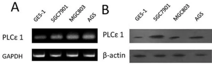
Firstly, we detected the mRNA and protein expression of PLCe1 in different gastric cell lines (Figure 1). Reverse transcrip-tion-PCR analysis and Western blotting analysis were conducted on these samples derived from normal gastric mucous cells GES-1 and three gastric cancer cell lines AGS, SGC7901 and MGC803. We found that all gastric cancer cell lines revealed higher PLCe1 expression than that in normal gastric cell controls at both mRNA and protein levels.
PLCe1 is up-regulated in Gastric Cancer Lesions but down-regulated in Atrophic Gastritis Lesions
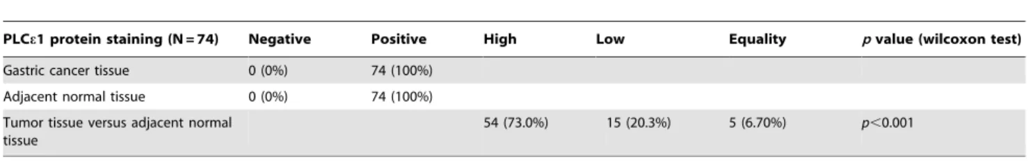
Then, we investigated the role of PLCe1 in both tumor and normal tissues in a gastric cancer tissue microarray. As shown in Figure 2, we observed that both tumor and normal tissues demonstrated the positive expression of PLCe1. However, as shown in Table 1, tissue microarray data analysis had shown that the percentage of PLCe1 positive expression in tumor tissues is significantly higher than that in adjacent normal tissues by wilcoxon test analysis (73.0% versus 20.3%,p,0.01).
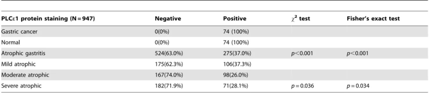
To determine PLCe1 expression in inflammation environment, we extended PLCe1 immunohistochemical analysis using another independent set of paraffin-embedded tissue sections of 799 atrophic gastritis tissue specimens. The immunohistochemical results showed that PLCe1 expression was positive in 275 (37.0%) atrophic gastritis including 106 (37.7%) mild atrophic gastritis, 98 (26.0%) moderate atrophic gastritis and 71 (28.1%) in severe atrophic, respectively. Moreover, the PLCe1 expression was correlated with the degree of atrophy (p= 0.036) (see Table 2).
Figure 1. Expression analysis of PLCe1 protein and mRNA in
gastric normal and cancer cell lines.A, expression of PLCe1 mRNA in gastric normal cell line GES-1and cancer cell lines SGC7901, MGC803 and AGS by reverse transcription-PCR. B, expression of PLCe1 protein gastric normal cell lines GES-1 and cancer cell lines SGC7901, MGC803 and AGS by Western blotting.
Therefore, compared with tumor and normal tissues, atrophic gastritis samples had decreased PLCe1 expression (p,0.001) (see Table 2).
We then confirmed these immunohistochemical tissue micro-array results by reverse-transcription-PCR and Western blotting analysis of three paired of gastric cancer and adjacent normal tissues and three paired of severe atrophic gastritis and adjacent normal tissues, which were taken from the same patient. Compared with adjacent normal tissues, three examined gastric tumors displayed up-regulation of PLCe1 expression at both mRNA and protein levels, but all of severe atrophic gastritis tissues
had lower expression of PLCe1(see Figure.3). In agreement with the results of immunohistochemical tissue microarray analysis, the paired analysis also showed PLCe1 was over-expressed in tumor, but down-regulated in severe atrophic gastritis in comparison with the matched adjacent normal tissues.
These observations suggested that PLCe1 expression status was very different in the context of inflammation versus tumorigenesis. Our results highly suggest that our results highly suggest that the PLCe1 protein could be a potential biomarker of gastric cancer.
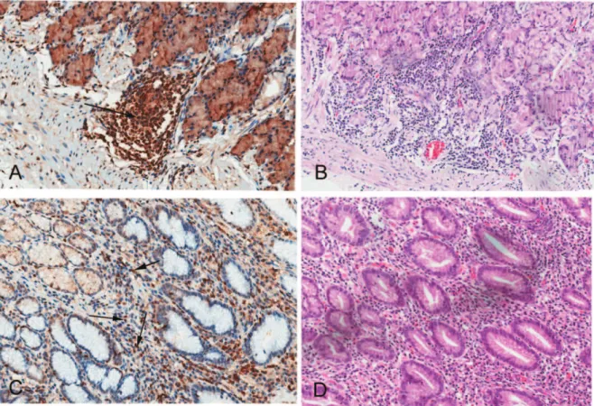
Figure 2. Immunohistochemical detection of PLCe1 protein in gastric cancer and atrophic gastritis patients.Representative images from Immunohistochemical analysis of 74 archived gastric cancer and 799 atrophic gastritis cases. A, overview of the tissue microarray by immunostaining of antibody PLCe1. Boxed biopsies are shown in detail in (B and C). B and C are matched tumor and adjacent normal tissues from same patient. The patient is pTNM stage III with poorly differentiated adenocarcinoma. Both tumor (B) and adjacent normal (C) cells are positive for PLCe1. Representative immunohistochemical (D)/histological (G) observations in a atrophic gastritis tissue, Representative immunohistochemical (E)/ histological (H)observations in a gastric normal tissue, Representative immunohistochemical (F)/histological (I) observations in a gastric cancer tissue. (Original magnification,62 for B and C,6200 for D, E, F, G, H and I).
doi:10.1371/journal.pone.0047563.g002
Table 1.The expression of PLCe1 protein in gastric cancer tissue and adjacent normal tissue.
PLCe1 protein staining (N = 74) Negative Positive High Low Equality pvalue (wilcoxon test)
Gastric cancer tissue 0 (0%) 74 (100%) Adjacent normal tissue 0 (0%) 74 (100%) Tumor tissue versus adjacent normal
tissue
54 (73.0%) 15 (20.3%) 5 (6.70%) p,0.001
Correlations between PLCe1 and Clinicopathologic Features
There is no statistically significant difference of PLCe1 expression in clinicopathologic data, such as age, sex, tumor size, classification, clinical stage, between patients at different stages of gastric cancer in the tissue microarray (shown in Table S1).
Particularly, as shown in Figure 2, PLCe1 protein expression in cytoplasm was frequently detected in all tumor tissues, normal tissues but less frequently in atrophic gastritis tissues.
However, as shown in Figure 4, in the tissue microarray, positive PLCe1 expression were observed in inflammatory cells and lymphocytes in 13 (48.1%) adjacent normal tissue samples from lymphnode metastases (N0) gastric cancer patients (shown in Figure 4A). Moreover, in severe atrophic gastritis tissues, 6 (2.37%) samples showed positive PLCe1 expression in inflammatory cells or lymphatic tissues (Figure 4B). Thus, these results imply that the expression of PLCe1 may be associated with inflammatory cells invasion during tumorigenesis and tumor metastases.
Discussion
Here, our study firstly demonstrated that PLCe1 was up-regulated in human gastric tumor tissues, but was down-up-regulated in inflammation environment, compared with normal tissues. The
differential expression of PLCe1 in different tissues highly suggested that the expression of PLCe1 could play a key role in inflammation and tumorigenesis.
Inflammation is recognized as a vital constituent of the local environment sustaining tumor development, including initiation, promotion, malignant conversion, invasion, and metastasis [31]. A causal relationship between gastric tumor promotion and inflam-mation has been supported from the observations that the inflammation caused by Helicobacter pylori infection can increase the risk of tumor promotion [8–10]. Recently, numerous reports have shown that PLCe1 expression levels are highly associated with inflammation and tumorigenesis [19–21,23]. However, the molecular mechanism underlying the biological significance of PLCe1 in gastric cancer development and progression remains obscure. Our study has provided evidence that PLCe1 up-regulation might be important in the progression of gastric cancer. Particularly, its down-regulation in inflammation environment, highly suggested that PLCe1 may be an important biomarker, and may be used to distinguish gastric cancer status from atrophic lesion stage.
Moreover, in our work, PLCe1 protein can be detected in inflammatory cells and lymphocyte tissues in some adjacent normal tissues at N0 degree and some severe atrophic gastritis tissues. In mammals, PLCe1 is reported to be expressed in
non-Table 2.The expression of PLCe1 protein in gastric cancer, normal and atrophic gastritis patients.
PLCe1 protein staining (N = 947) Negative Positive x2test Fisher’s exact test
Gastric cancer 0(0%) 74 (100%)
Normal 0(0%) 74 (100%)
Atrophic gastritis 524(63.0%) 275(37.0%) p,0.001 p,0.001
Mild atrophic 175(62.3%) 106(37.3%)
Moderate atrophic 167(74.0%) 98(26.0%)
Severe atrophic 182(71.9%) 71(28.1%) p= 0.036 p= 0.034
doi:10.1371/journal.pone.0047563.t002
Figure 3. Expression analysis of PLCe1 mRNA and protein in gastric cancer and atrophic gastritis patients.A and C, expression of PLCe1 mRNA and protein in each of the gastric tumors (T) and gastric adjacent normal tissues (N) paired from the same patient by reverse transcription-PCR and western blot. B and D, expression of PLCe1mRNA and protein in each of the severe atrophic gastritis (S) and gastric adjacent normal tissues (N) paired from the same patient by reverse transcription-PCR and Western blotting.
immune cells such as epidermal keratinocytes, dermal fibroblasts, and epithelial cells, but not in immune cells such as lymphocytes, granulocytes, macrophages, and dendritic cells [21,32,33]. Our results suggest that the function of PLCe1 protein in gastric cancer may be linked to its expression in the immune system.
According to these available results, we proposed a possible mechanism of PLCe1 protein functioning in tumorigenesis of gastric cancer: PLCe1 protein as an effecter of Ras and Rap small GTPases [12,14,15] is at first expressed in normal gastric mucous tissues. In the course of progression of atrophic gastric lesion, PLCe1 protein expression is decreased to prevent early inflam-mation infiltration, according to PLCe2/2mice exhibits marked resistance to the TPA-induce skin inflammation [18,19]. With increasing time and degree of inflammation of atrophic gastric lesion, PLCe1 protein is gradually expressed in immune cells because PLCewas required in a variety of inflammatory reactions [21,22], which may finally induce the formation of gastric cancer. Simultaneously, PLCe1 protein gets higher expression in gastric cancer cells because PLCeover-expression can promote intestinal tumorigenesis in ApcMin/+mice [20].
In summary, up-regulation of PLCe1 expression in gastric cancer but down-regulation in atrophic gastritis was observed by
our study. The importance role of PLCe1 in gastric cancer is further highlighted by our finding of its inverse correlation with chronic atrophic gastritis. These results not only suggest that PLCe1 may be used as a prognostic indicator to distinguish gastric cancer from atrophic gastritis, but also warrant further studies to establish a possible link between the biological function of PLCe1 and the pathogenesis of gastric cancer. Our studies also show that PLCe1 may have an important role in tumorigenesis of gastric cancer, especially at early stage, and might represent a novel marker for diagnosing early-stage gastric cancer.
Supporting Information
Table S1 The relationship between PLCe1 expression and clinicopathologic characteristics.
(DOC)
Author Contributions
Conceived and designed the experiments: JC WW TZ. Performed the experiments: JC WW J-jJ Q-rQ L-gL. Analyzed the data: JC TZ D-xC. Contributed reagents/materials/analysis tools: WW L-gL H-lF W-lJ. Wrote the paper: JC D-xC.
References
1. Parkin DM, Bray F, Ferlay J, Pisani P (2005) Global Cancer Statistics, 2002. CA: A Cancer Journal for Clinicians 55: 74–108.
2. Cui DX, Zhang L, Yan XJ, Zhang LX, Xu JR, et al. (2005) A microarray-based gastric carcinoma prewarning system. World Journal of Gastroenterology 11: 1273–1282.
3. Li Z, Huang P, Zhang X, Lin J, Yang S, et al. (2009) RGD-Conjugated Dendrimer-Modified Gold Nanorods for in Vivo Tumor Targeting and Photothermal Therapy{. Molecular Pharmaceutics 7: 94–104.
Figure 4. PLCe1 is positively expressed in lymphocytes in normal and atrophic gastritis tissues, respectively. Representative immunohistochemical (A)/histological (B) observations in a gastric normal tissue, Representative immunohistochemical (C)/histological (D) observations in a atrophic gastritis tissue. Arrows indicate positively stained immune cells (Original magnification,6200).
4. Kong Y, Chen J, Gao F, Li W, Xu X, et al. (2010) A Multifunctional Ribonuclease-A-Conjugated CdTe Quantum Dot Cluster Nanosystem for Synchronous Cancer Imaging and Therapy. Small 6: 2367–2373.
5. Chen L, Bao C-C, Yang H, Li D, Lei C, et al. (2011) A prototype of giant magnetoimpedance-based biosensing system for targeted detection of gastric cancer cells. Biosensors and Bioelectronics 26: 3246–3253.
6. He M, Huang P, Zhang C, Hu H, Bao C, et al. (2011) Dual Phase-Controlled Synthesis of Uniform Lanthanide-Doped NaGdF4 Upconversion Nanocrystals Via an OA/Ionic Liquid Two-Phase System for In Vivo Dual-Modality Imaging. Advanced Functional Materials 21: 4470–4477.
7. Huang P, Li Z, Lin J, Yang D, Gao G, et al. (2011) Photosensitizer-conjugated magnetic nanoparticles for in vivo simultaneous magnetofluorescent imaging and targeting therapy. Biomaterials 32: 3447–3458.
8. Parsonnet J, Friedman GD, Vandersteen DP, Chang Y, Vogelman JH, et al. (1991) Helicobacter pylori Infection and the Risk of Gastric Carcinoma. New England Journal of Medicine 325: 1127–1131.
9. Humans IWGotEoCRt (1994) Schistosomes, liver flukes and Helicobacter pylori.
10. Uemura N, Okamoto S, Yamamoto S, Matsumura N, Yamaguchi S, et al. (2001) Helicobacter pylori Infection and the Development of Gastric Cancer. New England Journal of Medicine 345: 784–789.
11. Rhee SG, Bae YS (1997) Regulation of Phosphoinositide-specific Phospholipase C Isozymes. Journal of biological chemistry 272: 15045–15048.
12. Song C, Hu C-D, Masago M, Kariya K-i, Yamawaki-Kataoka Y, et al. (2001) Regulation of a Novel Human Phospholipase C, PLCepsilon, through Membrane Targeting by Ras. J Biol Chem 276: 2752–2757.
13. Suh P-G, Park J-I, Manzoli L, Cocco L, Peak J, et al. (2008) Multiple roles of phosphoinositide-specific phospholipase C isozymes. BMB Reports 41: 415–434. 14. Bunney TD, Harris R, Gandarillas NL, Josephs MB, Roe SM, et al. (2006) Structural and Mechanistic Insights into Ras Association Domains of Phospholipase C Epsilon. Molecular cell 21: 495–507.
15. Bunney TD, Katan M (2006) Phospholipase C epsilon: linking second messengers and small GTPases. Trends in cell biology 16: 640–648. 16. Cheng H, Luo C, Wu X, Zhang Y, He Y, et al. (2011) shRNA Targeting PLCe1
Inhibits Bladder Cancer Cell Growth In Vitro and In Vivo. Urology 78: 474.e477–474.e411.
17. Ou L, Guo Y, Luo C, Wu X, Zhao Y, et al. (2010) RNA interference suppressing PLCE1 gene expression decreases invasive power of human bladder cancer T24 cell line. Cancer genetics and cytogenetics 200: 110–119. 18. Bai Y, Edamatsu H, Maeda S, Saito H, Suzuki N, et al. (2004) Crucial Role of
Phospholipase Ce in Chemical Carcinogen-Induced Skin Tumor Development. Cancer research 64: 8808–8810.
19. Ikuta S, Edamatsu H, Li M, Hu L, Kataoka T (2008) Crucial Role of Phospholipase Cein Skin Inflammation Induced by Tumor-Promoting Phorbol Ester. Cancer research 68: 64–72.
20. Li M, Edamatsu H, Kitazawa R, Kitazawa S, Kataoka T (2009) Phospholipase Cepromotes intestinal tumorigenesis of ApcMin/+mice through augmentation of inflammation and angiogenesis. Carcinogenesis 30: 1424–1432.
21. Hu L, Edamatsu H, Takenaka N, Ikuta S, Kataoka T (2010) Crucial Role of Phospholipase Cein Induction of Local Skin Inflammatory Reactions in the Elicitation Stage of Allergic Contact Hypersensitivity. The journal of immunology 184: 993–1002.
22. Takenaka N, Edamatsu H, Suzuki N, Saito H, Inoue Y, et al. (2011) Overexpression of phospholipase Ce in keratinocytes upregulates cytokine expression and causes dermatitis with acanthosis and T-cell infiltration. European Journal of Immunology 41: 202–213.
23. Harada Y, Edamatsu H, Kataoka T (2011) PLCecooperates with the NF-kB pathway to augment TNFa-stimulated CCL2/MCP1 expression in human keratinocyte. Biochemical and Biophysical Research Communications 414: 106– 111.
24. Wang M, Zhang R, He J, Qiu L, Li J, et al. (2012) Potentially Functional Variants of ,italic.PLCE1,/italic. Identified by GWASs Contribute to Gastric Adenocarcinoma Susceptibility in an Eastern Chinese Population. Plos One 7: e31932.
25. Abnet CC, Freedman ND, Hu N, Wang Z, Yu K, et al. (2010) A shared susceptibility locus in PLCE1 at 10q23 for gastric adenocarcinoma and esophageal squamous cell carcinoma. Nat Genet 42: 764–767.
26. Wang L-D, Zhou F-Y, Li X-M, Sun L-D, Song X, et al. (2010) Genome-wide association study of esophageal squamous cell carcinoma in Chinese subjects identifies susceptibility loci at PLCE1 and C20orf54. Nat Genet 42: 759–763. 27. Zhang Z, Li Z, Gao C, Chen P, Chen J, et al. (2008) miR-21 plays a pivotal role
in gastric cancer pathogenesis and progression. Lab Invest 88: 1358–1366. 28. Inoue M, Kobayashi S, Matsuura A, Hamajima N, Tajima K, et al. (1998)
Agreement of endoscopic findings and serum pepsinogen levels as an indicator of atrophic gastritis. Cancer epidemiology, biomarkers & prevention : a publication of the American Association for Cancer Research, cosponsored by the American Society of Preventive Oncology 7: 261–263.
29. Inoue M, Tajima K, Matsuura A, Suzuki T, Nakamura T, et al. (2000) Severity of chronic atrophic gastritis and subsequent gastric cancer occurrence: a 10-year prospective cohort study in Japan. Cancer Letters 161: 105–112.
30. Qian Y-B, Zhang J-B, Wu W-Z, Fang H-B, Jia W-D, et al. (2006) P48 is a predictive marker for outcome of postoperative interferon-a treatment in patients with hepatitis B virus infection-related hepatocellular carcinoma. Cancer 107: 1562–1569.
31. Grivennikov SI, Greten FR, Karin M (2010) Immunity, Inflammation, and Cancer. Cell 140: 883–899.
32. Boyer O, Benoit G, Gribouval O, Nevo F, Pawtowski A, et al. (2010) Mutational analysis of the PLCE1 gene in steroid resistant nephrotic syndrome. Journal of Medical Genetics 47: 445–452.