via Upregulation of the Aryl Hydrocarbon Receptor
Udi Gluschnaider1, Guy Hidas1,2, Gady Cojocaru1, Vladimir Yutkin1,2, Yinon Ben-Neriah1, Eli Pikarsky1*
1Department of Pathology and the Lautenberg Center for Immunology, Hebrew University Hadassah Medical School, Jerusalem, Israel,2Department of Urology, Hadassah-Hebrew University Medical Center, Jerusalem, Israel
Abstract
Background:Prostate cancer is a common and heterogeneous disease, where androgen receptor (AR) signaling plays a pivotal role in development and progression. The initial treatment for advanced prostate cancer is suppression of androgen signaling. Later on, essentially all patients develop an androgen independent stage which does not respond to anti hormonal treatment. Thus, alternative strategies targeting novel molecular mechanisms are required.b-TrCP is an E3 ligase that targets various substrates essential for many aspects of tumorigenesis.
Methodology/Principal Findings:Here we show thatb-TrCP depletion suppresses prostate cancer and identify a relevant growth control mechanism. shRNA targeted againstb-TrCP reduced prostate cancer cell growth and cooperated with androgen ablationin vitroand in vivo. We found that b-TrCP inhibition leads to upregulation of the aryl hydrocarbon receptor (AhR) mediating the therapeutic effect. This phenomenon could be ligand independent, as the AhR ligand 2,3,7,8-Tetrachlorodibenzo-p-Dioxin (TCDD) did not alter prostate cancer cell growth. We detected high AhR expression and activation in basal cells and atrophic epithelial cells of human cancer bearing prostates. AhR expression and activation is also significantly higher in tumor cells compared to benign glandular epithelium.
Conclusions/Significance: Together these observations suggest that AhR activation may be a cancer counteracting mechanism in the prostate. We maintain that combiningb-TrCP inhibition with androgen ablation could benefit advanced prostate cancer patients.
Citation:Gluschnaider U, Hidas G, Cojocaru G, Yutkin V, Ben-Neriah Y, et al. (2010)b-TrCP Inhibition Reduces Prostate Cancer Cell Growth via Upregulation of the Aryl Hydrocarbon Receptor. PLoS ONE 5(2): e9060. doi:10.1371/journal.pone.0009060
Editor:Chad Creighton, Baylor College of Medicine, United States of America
ReceivedOctober 21, 2009;AcceptedJanuary 16, 2010;PublishedFebruary 5, 2010
Copyright:ß2010 Gluschnaider et al. This is an open-access article distributed under the terms of the Creative Commons Attribution License, which permits unrestricted use, distribution, and reproduction in any medium, provided the original author and source are credited.
Funding:This work was supported by grants from the Dr. Miriam and Sheldon G. Adelson Medical Research Foundation, Prostate Cancer Foundation - Israel and the Prostate Cancer Research Foundation. The funders had no role in study design, data collection and analysis, decision to publish, or preparation of the manuscript.
Competing Interests:The authors have declared that no competing interests exist. * E-mail: peli@hadassah.org.il
Introduction
Prostate cancer is the sixth most common cancer in the world and the third leading cause of cancer in men. In developed countries, prostate cancer develops in one of every nine men older than 65 years. This is a heterogeneous disease in terms of its clinical course and outcome but androgen signaling seems to be a common feature in its development and progression [1]. AR signaling is known to regulate normal, benign and cancerous prostate cells in various processes (e.g. differentiation and proliferation). In addition, there are observations indicating that AR expression is oncogenic in prostate epithelium and promotes progression to androgen independence [2–8]. The initial treat-ment of advanced stage and metastatic prostate cancer is suppression of androgen production by specific drugs or surgical castration. Later on, essentially all patients develop an androgen independent stage which does not respond to anti hormonal treatment. The latter phase is always lethal; thus, alternative strategies targeting novel molecular mechanisms are required.
The ubiquitin–proteasome system is a crucial determinant of virtually all biological processes in eukaryotes. In this pathway, proteins are targeted for degradation by covalent ligation to
ubiquitin, a highly conserved small protein. Protein ubiquitination involves the concerted action of the E1 ubiquitin-activating enzyme, an E2 ubiquitin-conjugating enzyme and an E3 ubiquitin-protein ligase, the last of which delivers multiple ubiquitin molecules to the target protein [9–11]. Inhibiting the entire ubiquitin proteasome pathway has therapeutic value in cancer (e.g. Bortezomib,[12]). However, since proteasome inhib-itors have pleiotropic effects, inhibition of a single key E3 may offer a more specific treatment option with fewer side effects.
phosphorylated, directly recognized byb-TrCP and degraded by the proteasome[24]. Thus, b-TrCP has opposing effects on two key oncogenic pathways. While the role of b-catenin in prostate cancer is controversial [28,29], there is evidence suggesting that NF-kB plays a pro-tumorigenic role in prostate cancer [30–32]. The opposed effect on these two oncogenic proteins exemplifies the versatility of this particular E3 ligase. Furthermore,b-TrCP is directly implicated in several cancer types [33–36], thus it is plausible to further inquire its role in prostate cancer.
In this work, we show thatb-TrCP inhibition inhibits prostate cancer growth showing additive effect with androgen ablation,in vitroandin vivo. This effect is largely mediated via activation of the aryl hydrocarbon receptor (AhR) as knocking down this protein abolishes the effect ofb-TrCP inhibition.
Results
NF-kB Activation Correlates with Prostate Cancer Patients’ Outcome
b-TrCP is an important E3 ligase known to regulate many different substrates. NF-kB is a well characterized transcription factor negatively regulated by IkB, thus indirectly positively modulated by b-TrCP. The involvement of NF-kB in prostate cancer is well investigated and previous studies showed its tumorogenic role in the prostate. There is also recent indication for NF-kB involvement in the progression of prostate cancer to androgen independent stage [37]. In order to corroborate the relevance of this factor to prostate cancer progression, we tested its
activation state in primary human prostate cancer samples using immunohistochemical staining with antibodies against p65 (Figure 1A). 131 prostate cancer specimens, spotted on a single tissue microarray slide from patients with different Gleason grades were analyzed. 39% of the samples showed at least some p65 positive nuclei (Figure 1B). We also analyzed 14 prostate cancer metastases, 64% of which were positive (Figure 1A, B). We set to evaluate the correlation between prostate cancer Gleason score and NF-kB activation. Indeed, we found an association between the two (p = 0.014, Fisher’s exact test). It appeared that NF-kB nuclear staining was stronger in patients with higher Gleason score or in metastases raising the possibility that NF-kB activation could be correlated with prognosis. To test this possibility, we immunstained 80 primary tumors for p65, from patients that underwent radical prostatectomy and for which we had clinical follow-up. 77% of the patients whose primary tumor samples negatively stained for nuclear p65, showed no recurrence up to 8 years post radical prostatectomy. In contrast, the disease recurred in 65% of the NF-kB positive patients (Figure 1C). Another important pro-oncogenicb-TrCP substrate isb-catenin. Sinceb -TrCP inhibition would enhanceb-catenin stability and promote tumorigenesis, it was necessary to monitor also b-catening correlation to prostate cancer patients’ outcome. Nuclear localization ofb-catenin activation did not correlate with disease severity in the same cohort of patients (Figure S1). Together, these results imply a clinical advantage for inhibitingb-TrCP in prostate cancer. Yet, the multitude ofb-TrCP substrates (37, with possibly more to be identified) precludes a comprehensive analysis of all of
Figure 1. NF-kB activation correlates with prostate cancer patients’ outcome.Prostate cancer sections were immunostained using p65 antibodies. A. Representative photomicrographs of a primary tumor (left) and lymph node metastasis (right) from the same patient. B. Percent of negative, positive and strongly positive cases spotted on a tissue microarray from the indicated Gleason scores (p = 0.0014, Fisher’s exact test). C. primary prostate tumors (n = 131) were immunostained for p65 and assessed for NF-kB activity. Kaplan-Meier plot revealed a correlation between
them in the same way, and necessitates a direct approach to study the utility ofb-TrCP inhibition in prostate cancer.
b-TrCP Inhibition Reduces Prostate Cancer Cell Growth We next examined the effect ofb-TrCP inhibition on prostate cancer cell growth. To this end, we constructed a doxycycline inducible shRNA lentiviral vector targeting bothb-TrCP1 andb -TrCP2 with which we transduced the two androgen-sensitive human prostate cancer cell lines, LNCaP and LAPC4. Doxycy-cline treatment of the trasnduced LNCaP cells resulted in efficient knock down of bothb-TrCP genes (85% and 75% forb-TrCP1 andb-TrCP2, respectively) as determined using quantitative real time PCR (qRT PCR -Figure 2A). This was accompanied by IkBa stabilization both before and following TNF stimulation, demonstrating efficient induction of protein down regulation (Figure 2B). To evaluate the consequence ofb-TrCP knock down on prostate cancer cells, we monitored doxycycline-treated and untreated LNCaP cells for nine days and measured cell growthin vitro.Figure 2C shows diminished cell growth after eitherb-TrCP inhibition or androgen ablation treatment (lower left and upper right panels, respectively). Moreover, combining b-TrCP inhibi-tion with androgen ablainhibi-tion shows an additive effect (Figure 2C lower right panel andFigure 2D). We next quantified cell growth using the XTT method and confirmed our morphologic analysis (Figure 2D). Similar results were obtained with LAPC4 cells (Figure S2).
To rule out shRNA off target effects, we used a previously describedb-TrCP dominant negative construct lacking the F-box
domain [33]. This variant stabilizesb-TrCP substrates as it binds the phosphorylated targets, but fails to recruit the additional components of the SCF complex which are critical for E3 ligase catalytic activity. The b-TrCP inhibition effects using the dominant negative form, were essentially similar to the shRNA results (Figure S3A and B).
b-TrCP Inhibition Cooperates with Androgen AblationIn Vivo
To further analyze the effect ofb-TrCP inhibition on human prostate cancer tumor growth, we used a xenograft model. We injected LNCaP cells carrying an inducible b-TrCP shRNA construct into the subcutis of 39 immunosuppressed Balb/c Rag12/2male mice. 25 of the mice (64%) developed visible and measurable tumors 30 days post injection. Since initial tumor development was somewhat heterogeneous in size and kinetics, we randomly divided these 25 mice into four treatment groups: 1. Intact untreated mice (NT, n = 4; mean tumor volume 123.1670.7); 2. Mice given tetracycline in the drinking water resulting inb-TrCP inhibition (Tet, n = 5; mean tumor volume 80.3698.2); 3. Castrated mice resulting in androgen ablation (Cast, n = 7; mean tumor volume 121.6695.0); 4. Castration plus tetracycline (Cast + Tet, n = 8; mean tumor volume 180.36143.9). There was no significant difference in average tumor size at initiation of treatment between the four groups. We monitored tumor growth once a week, and sacrificed the mice 30 days post the initial treatment. RNA extracted from tumors from the Tet treated mice showedb-TrCP knock down
Figure 2.b-TrCP inhibition reduces prostate cancer cells growth.LNCaP cells were infected with a lentiviral vector bearing an inducible doxycycline dependentb-TrCP shRNA. A. Cells were treated for 72 hours with 1mg/ml doxycycline and RNA levels ofb-TrCP1 andb-TrCP2 were measured using qRT PCR. B. Western blot analysis of protein extracts from cells treated with TNF, doxycycline or MG-132 were immunoblotted with either IkB or tubulin. C. Representative photomicrograph of cells treated as indicated and stained with hematoxylin and eosin. D. XTT assay used to quantify cell growth at different time points. DOX, doxycycline; NT, no treatment; FCS, conditioned medium containing fetal calf serum; CSS, conditioned medium containing androgen depleted serum (charcoal stripped serum). Error bars, SD. * Significantly different from non treated cells and from each other, p-value,0.01, t-test.
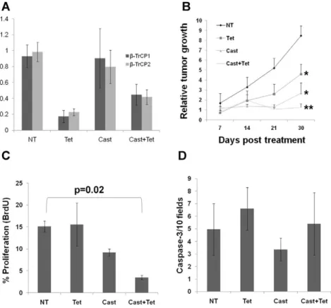
ranging from 50% to 80% (Figure 3A). Similar to ourin vitro findings, inhibiting b-TrCP reduced tumor growth with or without androgen ablation (Figure 3B). Analysis of tumor proliferation using BrdU immunostaining revealed that the mice treated with both androgen ablation and b-TrCP inhibition showed the lowest proliferation rates (Figure 3C and Figure S4). Tumor growth suppression could not be explained via apoptosis, since anti cleaved caspase-3 immunostaining did not reveal any differences between treatment groups (Figure 3D). Similar results were obtained with AT2.1 rat prostate cancer cells stably transfected with an inducible dominant negativeb -TrCP transgene (Figure S2B). In conclusion, our results indicate thatb-TrCP inhibition suppresses prostate cancer growth both in vitroand in vivo and shows an additive effect with androgen ablation.
Aryl Hydrocarbon Receptor (AhR) Is Upregulated upon
b-TrCP Inhibition and Androgen Ablation
To elucidate the molecular pathways that are responsible for the growth inhibitory effect ofb-TrCP depletion in combination with androgen ablation we conducted a wide range microarray analysis. LAPC4 cells infected with the shb-TrCP lentiviral vector, were either left untreated or treated for 72 hours with doxycycline, charcoal stripped serum or both. cDNA samples were subjected to microarray analysis using U133 Affimetrix chips probing,30,000 probe sets. We then sought to identify genes which are cooperatively affected by both androgen ablation and b-TrCP inhibition. Among the upregulated genes, were potential anti
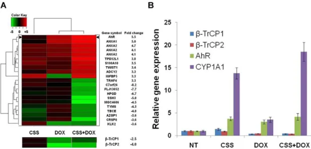
inflammatory genes (e.g. ANXA1) and among the prominently downregulated were prostate specific genes (e.g. KLK2). Yet, the most dramatic increase was the expression of the aryl hydrocarbon (dioxin) receptor (AhR). This gene was upregulated upon either androgen ablation orb-TrCP inhibition and was the most highly changed gene due to the combined treatment (Figure 4). We could attribute AhR level upregulation tob-TrCP depletion, since doxycycline alone did not alter the receptor’s mRNA (Figure S5). The AhR is a ligand activated transcription factor involved in organogenesis, in detoxification of endo- and xenobiotics and in mediating diverse organ-specific toxic responses of dioxins. This receptor belongs to the basic helix- loop-helix (bHLH)/PAS (Period -Aryl hydrocarbon receptor nuclear translocator-Single minded) family of heterodimeric transcriptional regulators. bHLH/PAS proteins are involved in the control of diverse physiological processes such as circadian rhythms, organ develop-ment, neurogenesis, metabolism and the stress response to hypoxia [38–40]. Recent studies revealed a connection between the AhR pathway and prostate cancer bothin vitroandin vivo, showing that the AhR interacts and inhibits the AR. Moreover, the AhR acts as an E3 ligase of the AR [41] and AhRnull TRAMP mice show increased prostate tumorigenesis [42]. We used qRT PCR to validate the cDNA array analysis. We found that while each treatment alone increased AhR RNA levels more than 2 folds, the combined treatment resulted in more than 4 fold upregulation in both LAPC4 (Figure 4B) and LNCaP cells (Figure S6). To test the functional activity of the AhR pathway we measured the mRNA levels of its canonical target cytochrome p450 1A1 (CYP1A1). Our
Figure 3.b-TrCP inhibition cooperates with androgen ablation treatmentin vivo.LNCaP cells bearing a tetracycline inducedb-TrCP shRNA construct were injected subcutaneously to immunosuppressedRag12/2mice. Mice (n$4 in each group) were either untreated (NT), treated with tetracycline in their drinking water (Tet), physically castrated (Cast) or both (Cast+Tet) for 30 days. Tumor volumes were measured weekly. (A). qRT PCR for bothb-TrCP isoforms was performed on RNA extracted from the tumors harvested at day 30. (B) Tumor growth kinetics. Tissue sections were stained for BrdU (C) or activated caspase 3 (D) and the proliferation and apoptosis scores, respectively, were determined for each tumor. Shown are mean6standard deviation (A) or6S.E.M (B, C and D).* p-value,0.05, ** p-value = 0.0002, t-test; p-value in C refers to t-test.
analyses show that CYP1A1 levels were increased in correlation with AhR levels in the 4 treatment groups (Figure 4B). It should be noted that this upregulation occurred without addition of an
exogenous AhR ligand. Addition of the potent AhR ligand TCDD further augmented CYP1A1 upregulation (Figure 5C and data not shown).
Figure 4. Aryl hydrocarbon receptor (AhR) expression is increased after androgen ablation andb-TrCP treatments.A. LAPC4 cells infected with inducibleb-TrCP shRNA were treated for 72 hours with the indicated treatment, subjected to RNA extraction and cDNA microarray analysis (Affymetrix). After data normalization, gene expression profiles were compared between treatment and the untreated control samples. A. Heat map dendrogram showing the ten most highly up (red) and down (green) regulated genes due to the combined treatment. Fold change refers to the combined treatment probes values relative to control. Expression ofb-TrCP isoforms is presented below. B. LAPC4 cells infected with the same lentiviral vector and treated as indicated were subjected to RNA extraction and qRT PCR analysis with the listed primers. CSS, charcoal stripped serum; DOX, doxycycline; Error bars, SD.
doi:10.1371/journal.pone.0009060.g004
The Growth Suppression Effect ofb-TrCP Inhibition Is Mediated via Upregulation of the Aryl Hydrocarbon Receptor
To investigate the significance of the AhR pathway activation following b-TrCP depletion, we first infected LNCaP cells with inducible AhR shRNA. While doxycycline treatment resulted in reduced AhR mRNA levels we could not detect any effect on cell growth (Figure 5A). Next we co-infected LNCaP cells bearing inducibleb-TrCP shRNA with the inducible AhR shRNA vector. Addition of doxycycline to the medium of double knockdown cells resulted in reduced b-TrCP mRNA levels, similar to the single knockdown cells; yet as expected, in these cells AhR levels were decreased rather than increased both at the mRNA and protein levels (Figure 5C, D). Thus, the double knockdown cells allow us to test whether the growth inhibitory effect ofb-TrCP modulation is mediated via AhR upregulation. Indeed, in the double knockdown LNCaP cells,b-TrCP depletion failed to reduce cell growth either with or without androgen ablation (Figure 5B). Similar results were obtained with a different shRNA targeted against the AhR (data not shown). Interestingly, addition of the potent exogenous ligand TCDD did not reduce cell growth alone; nor had it an effect with any of the different treatments (Figure S7). This suggests that this AhR effect is ligand independent. Western blot analysis confirmed b-catenin stabilization and the AhR mRNA upregulation uponb-TrCP inhibition (Figure 5C). Thus, the double knockdown results indicate that most of the effect ofb -TrCP knockdown is mediated by upregulation of the AhR.
AhR Expression in Prostate Cancer Patients
Observing AhR upregulation upon b-TrCP inhibition in prostate cancer cells prompted us to inspect AhR status in various
stages of prostate cancer. To address this aim, we collected 39 specimens of primary prostate cancer tumors from the Hadassah Medical Center. Out of this cohort, 17 patients suffered from disease recurrence. We performed immunohitochemical anti AhR staining and monitored cytoplasmatic and nuclear AhR expres-sion. First, we detected high AhR in basal cells located in the benign gland perimeter (Figure 6A). Interestingly, proliferative inflammatory atrophy (PIA), considered as a precursor lesion to prostate cancer showed very strong cytoplasmic and nuclear staining (Figure 6B). We used a subjective score from 0 to 3 to quantitate staining intensity in normal and malignant epithelial cells in each specimen. Our analysis demonstrates upregulation of AhR in both the cytoplasmic and nuclear locations in the malignant epithelium (Figure 6C, D, p,0.001, Mann-Whitney test). However, we did not observe a correlation between AhR expression in the tumor cells and disease recurrence (data not shown).
Discussion
Prostate cancer is a heterogeneous disease comprising many genetic and phenotypic features. One important hallmark of prostate cancer is progression to an androgen independent stage (AI) after hormonal treatment. The causes for this transition are not fully understood and adjuvant treatments fortifying hormonal manipulation are required. One factor that is often implicated in tumor progression in many types of cancer is NF-kB. Here we confirm that NF-kB is often activated in advanced prostate cancer patients (Figure 1). Moreover, NF-kB activation correlates with prostate cancer recurrence (Figure 1C). One of the major regulators of NF-kB is the E3 ubiquitin ligase SCFb-TrCP, which targets IkB as well as many other substrates for ubiquitination and
Figure 6. AhR is upregulated in malignant prostate cells.Prostate sections were immunostained with AhR antibody. Photomicrographs show strong AhR expression in basal cells (arrows in A) and proliferative inflammatory atrophy (B). C. Higher AhR expression is noted in malignant glands (T) compared with normal glands (N). D. Normal and malignant glands were scored using a 0–3 scale. Mean values6S.E.M. are shown (p-value,0.001, Mann-Whitney test).
degradation, [27].b-TrCP is thought to play a pro-tumorigenic role in certain types of cancers [43]. Based on observed upregulation of NF-kB, it was important to follow another common pro-oncogenicb-TrCP substrate,b-catenin, which was also implicated in prostate cancer [28,29,44]. However, we could not detect a correlation betweenb-catenin activation and prostate cancer recurrence (Figure S1), suggesting that only someb-TrCP targets are of relevance to the disease progression. To evaluate the full scope of b-TrCP effects, we set out to inhibit b-TrCP in prostate cancer cells and to monitor its effect on prostate cancer cell growth. Ourin vitroandin vivostudies revealed that uponb -TrCP inhibition prostate cancer cell growth is reduced. We could also detect an additive effect when combiningb-TrCP inhibition with androgen ablation (Figure 2 and 3). To elucidate the mechanisms of growth suppression by b-TrCP inhibition, we carried a cDNA array analysis and observed an additive upregulation of the AhR (Figure 4). AhR was previously demonstrated to act as an E3 ligase of the AR [41], providing us a possible link to AR signaling. Moreover, we noted that AhR expression is higher upon combining androgen ablation and b -TrCP inhibition. We therefore hypothesized that the upregulation of the AhR could be mediating the growth inhibitory effect ofb -TrCP knockdown in prostate cancer cells. This notion was supported by previous reports demonstrating an inhibitory role for the AhR pathway in prostate cancer. Morrowet alshowed an AhR ligand dependent growth inhibition in LNCaP cells. Likewise, other studies have also implicated the androgen receptor in AhR growth inhibition [45,46]. In our study, knocking down the AhR per se in LNCaP cells did not alter prostate cancer cell growth (Figure 5A), suggesting that the basal levels of AhR do not exert an inhibitory effect unless stimulated by ligand. On the other hand boosting AhR expression via b-TrCP depletion was associated with considerable ligand-independent growth suppression. AhR depletion reversed the growth suppression effect of b-TrCP knockdown, even under androgen ablation, proving that AhR upregulation, which we also noted in other stress conditions (e.g. atrophy, inflammation and androgen ablation) accounts for theb -TrCP inhibitory effect (Figure 5). Interestingly, ligand adminis-tration did not affect cell growth even though it did upregulate expression of the classic AhR pathway target CYP1A1. These results implicate a ligand and CYP1A1 independent AhR pathway in prostate cancer cells.
Chesireet alidentified the AhR as a putativeb-catenin target in LNCaP cells [47]. As we show thatb-TrCP depletion stabilizesb -catenin along with the upregulation of AhR (Figure 5D), it is possible that the cause of AhR elevation following b-TrCP depletion isb-catenin stabilization.
Our studies indicate a novel ligand independent strategy of boosting AhR expression as means of suppressing prostate cancer growth. This strategy may also be echoed in the natural history of prostate cancer. We found that AhR is normally expressed at low levels in the prostatic epithelium basal cells (cytoplasmic and nuclear staining,Figure 6A) and is substantially upregulated in areas of proliferative inflammatory atrophy (PIA), considered a precursor lesion to cancer (Figure 6B). AhR activation in basal and atrophic cells may therefore be viewed as an anticancer mechanism. It is possible that microenvironmental inflammatory signals are responsible for such stress induced signaling. A mutation in one allele of b-TrCP1 was identified in one human prostate tumor in a systematic screen of Wnt pathway mutations [48]. However this mutation is unlikely to have an effect on the NF-kB pathway as the other allele and possibly both b-TrCP2 alleles were wild type. Similarly, to our knowledge, activating mutations in the NF-kB pathway were so far not reported in
human prostate tumors. Nevertheless, NF-kB known to mediate malignant transformation is constitutively upregulated in this tumor type via multiple mechanisms. We conclude that different stress signals, including inflammation, atrophy and androgen ablation upregulate AhR expression in both normal and malignant prostate cells and conduct a protective mechanism. Inhibitingb -TrCP at advanced disease stages may be relevant in developing strategies for enhancing the efficacy of prostate cancer treatments.
Materials and Methods
Ethics Statement
Experiments with human tissues were approved by Institutional Review Board, Hadassah-Hebrew University Medical Center. Due to the retrospective nature of this study and according to the declaration of Helsinki, participants were not obtained constantly informed. In addition, our IRB waived the need for written informed consent. All mice experiments were approved by the IACUC.
Dominant Negativeb-TrCP (WD)
Dominant negativeb-TrCP (WD)was cloned using E3RS excluded from pCDNA3-EE-hE3RS plasmids with the primers 59 to 39
forward: GCGGCCGCTATGGACCC-GGCCGAG (with NotI site in its 59); reverse: TTATCTGGAGATGTAGGTGT; the product was cloned into TA vector (Invitrogen). The last vector was cut with AvrII/ASP718, filled in and blunt ligated. The relevant fragment was cut and inserted into pFLAG-CMVTM-2 expression vector (Sigma-Aldrich) withNotI/BamHI. This proce-dure produced a WD construct lacking part of the F-box and conjugated to FLAG. The WD-FLAG was inserted under bidirectional teracycline promoter expressing GFP. The resulting plasmid was transfected into AT2.1 Rat prostate cancer cells expressing the tetracycline trans-activator, using FUGENE reagent (Roche Applied Science). The transfected cells were selected using hygromycin and neomycin (Sigma-Aldrich) con-taining media to produce a stable clone which expresses a dominant negativeb-TrCP upon addition of tetracycline or its derivate doxycycline.
Inducibleb-TrCP and AhR shRNA
The human shRNA 59- GUGGAAUUUGUGGAACAUC 39
targeted against b-TrCP1 and b-TrCP2 was constructed into pTER plasmid and inserted into a modified pRRL.sin.PPT. tetO7.MCS.PRE lentiviral vector. The vector consists of an HI promoter, tet operator, the shRNA coding sequence and eF1a
promoter driving the tet repressor fused to eGFP. Virus production and infection were carried out as previously described [49]. To target the AhR we used the following shRNA sequences: 1. 59CAGCUGAAUUAAAUAACAU 39; 2. 59 CAGACAGUA-GUCUGUUAUA 39. Both of which proved to be efficient in knocking down the receptor (the presented data represent those obtain with the latter sequence). shRNA expression is induced only after Tetracycline or Doxycycline (Sigma-Aldrich) administration. The vector alone was used as control. Double knocked down LNCaP cells were designed by co-infecting shb-TrCP cells with lentiviral vectors carrying either of the above described sequence.
Cell Culture
Israel); AT2.1, LNCaP and 293T were furnished to us by Dr. Rachel Bar-Shavit (Department of Oncology, Hadassah Medical Center, Jerusalem, Israel). All cell lines were incubated at 37uC 5% CO2in appropriate medium containing 10% FCS or CSS as indicated.100 pM Methyltrienolone (R1881, Perkin-Elmer, New England Nuclear) was added to LAPC4 full media.
Cell Proliferation Assay
MTT (3-(4,5-Dimethylthiazol-2-yl)-2,5-diphenylte-trazolium bromide) and XTT (2,3-Bis(2-methoxy-4-nitro-5- sulfophenyl)-2H-tetrazolium-5-carboxanilide) was used as the protocol indicates (Biological Industries, Kibbutz Beit Haemek, Israel). All experi-ments were carried out in 96 well plates with eight repeats of at least 3 independent infections.
Western Blotting
Whole-cell lysates were prepared from transfected or infected cells by extraction in lysis buffer containing 50 mM Tris (pH 8), 150 mM NaCl, 1% NP-40, 0.1% SDS, 10 mM NaF, 1 mM Na3VO4, 1 mM phenylmethylsulfonyl fluoride, 1 mg/ml leupep-tin, 1 mg/ml aprotinin and 1 mM dithiothreitol. Proteins were resolved by 10% SDS-PAGE, transferred onto nitrocellulose membranes, probed with appropriate antibodies, incubated with Peroxidase-conjugated Goat anti mouse or Rabbit IgG (Jackson Laboratories) and developed using the ECL kit (Pierce). Primary antibodies used were: anti-Flag (Sigma); anti-IkB, anti-phpspho-IkB (Cell Signaling); Aryl Hydrocarbon Receptor, CYP1A1 (Santa-Cruz); phospho-b-catenin (BD Biosciences).
Xenografts
AT2.1 or LNCaP cells were harvested washed and reconstituted in PBS. 106 51B cells per 200ml volume were injected subcutaneously to 6–7 weeks old atymic (Nude) male mice. 106 LNCaP cells were injected to 6–7 weeks oldrag12/2male mice together with Matrigel (BD bioscience). Tumors were measured in two dimensions with caliper, and tumor volume (mm3) was calculated with the formula V = (lengthXwidth2)/2. Half of the mice received Doxycycline (0.2 mg/ml, AT2.1) or tetracycline (1.5 mg/ml, LNCaP) supplemented with 5% sucrose in their drinking water. Half of the mice were surgically castrated: mice were anesthetized using Ketamine/2% Xylazine at 5.7:1 ratio (0.1 ml per 25–30 gram mouse). Surgical castration was performed via a midline scrotal incision allowing bilateral access to the hemiscrotal contents. After exposing each testicle, a 3-0 Vicryl suture was used to ligate the spermatic cord and then remove the testicle. Mice were treated with Carprofen (Rimadyl) as analgesics after surgery. Two hours before sacrifice, mice were injected with BrdU intraperitoneally 100ml per 10 grams of body weight (RPN201, Amersham Pharmacia Biotech Inc). For AT2.1 xenografts, NUDE mice were treated pre-injection and tumors were weighted at the end of the experiment. LNCaP xenografted mice were treated 30 days post injection when measurable tumors were established. Tumor relative growth was calculated individ-ually for each mouse, comparing each week’s measurement to the treatments’ day 0 (30 days post injection). All mice experiments were approved by the IACUC.
Immunohistochimestry
mouse tumor specimens were fixed in 4% neutral-buffered formalin and embedded in paraffin. Patients paraffin embedded samples were collected from the archives of the Department of Pathology at the Hadassah-Hebrew University Medical Center. Experiments with human tissues were approved by the
institu-tional review board. 5mM sections were dewaxed and hydrated
through graded ethanol dilutions, then cooked in appropriate buffer (pH 7.4) in a pressure cooker at 115uC for 3 minutes. Endogenous peroxidase activity was blocked with 3% hydrogen peroxide followed by washing. The sections were then incubated with the indicated antibodies: p65 (Neomarkers; 1:100),
anti-b-catenin (Santa Cruz; 1:300) and anti-AhR (Santa Cruz; 1:200). All sections were counterstained with hematoxylin.
RNA, cDNA Micrroarray and Real-Time PCR
Total RNA was extracted from LAPC4 or LNCaP cells infected withb-TrCP shRNA lentiviral vector with TRI Reagent (Sigma). For cDNA microarray RNA was extracted from LAPC4 infected cells using TRIzolH(Invitrogen). cRNA preparation and hybrid-ization was performed using standard manufacture protocol; Biotin-labeled target synthesis reactions were performed using standard protocols supplied by the manufacturer (Affymetrix, Santa Clara CA, USA). From each RNA sample, 5mg were converted into double-stranded cDNA by reverse transcription with SuperScriptTMII Reverse Transcriptase (Life Technologies, Helgerman CT, USA), using T7-oligo-dT as a primer. Expression value (signal) was calculated using Affymetrix Genechip software MicroArray Suite 5.0. Only probe sets that had at least an intensity of 20 and a present call at one of the microarrays were selected. Next quantile normalization was applied to the log2 transformed expression values (Bolstad BM Bioinformatics 19: 185–193). Forb-TrCP knockdown determination and validation studies 2mg of RNA were used as template for synthesis of cDNA
using SuperScriptTM II Reverse Transcriptase. The cDNA was subsequently used as Real Time PCR template. All Real Time PCR reactions were carried out using Absolute Blue QPCR SYBR Green Low ROX Mix (ABgene) with the following primers (59to 39): b-TrCP1 Forward: ATCGGATTCCACGGTCAGAG, Re-verse: AATCAACGTGTTTAGCATT-TCACCT;b-TrCP2 For-ward: CCATCAAAGTCTGGAGCACGA, Reverse: CGCT-TGTGCCCATTGAGAGTA; AhR Forward: ACATCACC-TACGCCAGTCG, Reverse: CTCTATGCCGCTTGGAAG-GAT; CYP1A1 forward: TGAATGCCTTCAAGGAC-CTG, Reverse: TCAGGCTGTCTGTGATGTCC.
All microarray data is MIAME compliant. The raw data has been deposited in GEO (accession number GSE19141).
Supporting Information
Figure S1 b-catenin activation does not correlate with prostate cancer patients’ outcome. Primary prostate cancer tumors were immunostained using b-catenin antibodies. A. Representative photomicrographs of samples from negative (left) and positive (right)b-catenin stained tumors. B. Kaplan Meier curves plotting
b-catenin positive (blue) vs. negative (green) patients’ recurrence free interval. Scale bars in A, 50mM.
Found at: doi:10.1371/journal.pone.0009060.s001 (1.02 MB TIF)
Figure S2 b-TrCP shRNA cooperates with androgen ablation to reduce LAPC4 cell growth. LAPC4 cell infected with the mentioned inducible lentiviral vector containing b-TrCP shRNA. A. qRT-PCR demonstrating efficientb-TrCP1 andb-TrCP2 knockdown. B. XTT assay was used to quantify cells proliferation rates (means6
S.E.M.). Error bars in A, SD. NT, no treatment; DOX, doxycycline; CSS, charcoal stripped serum. * All treatments were statistically different from control (p-value,0.05, t-test).
Found at: doi:10.1371/journal.pone.0009060.s002 (0.19 MB TIF)
stably transfected with an inducible dominant negative b-TrCP were treated as indicated for 72 hours and subjected to MTT cell proliferation assay. B. LNCaP cells infected with lentiviral vector expressing eGFP (GFP) or dominant negative b-TrCP (DF-box) and assayed using the XTT reagent. C. Athymic 6–8 weeks male NUDE mice were divided into the 4 indicated groups (n$4) and subcutaneously grafted with AT2.1 cells bearing the doxycycline dependent dominant negativeb-TrCP construct. Tumor volumes were measured two weeks post injection. Shown are means 6
S.E.M for A and C and means 6 SD for B. * Significantly different from control group (p,0.05, t-test); ** Significantly different from all treatment groups (p,0.01, t-test). NT, no treatment; DOX, doxycycline; CSS, charcoal stripped serum; Cast, castrated mice.
Found at: doi:10.1371/journal.pone.0009060.s003 (0.20 MB TIF)
Figure S4 b-TrCP inhibition cooperates with androgen ablation treatment to reduce prostate cancer cells proliferation in vivo. LNCaP xenografts from treated Rag12/2 mice were immuno-stained with anti BrdU antibodies. Representative photomicro-graphs for each of the four treatment groups are shown. NT, no treatment; cast, castrated mice; Tet, tetracycline.
Found at: doi:10.1371/journal.pone.0009060.s004 (2.87 MB TIF)
Figure S5 Doxycycline does not upregulate AhR. LNCaP cells infected with a GFP expressing lentiviral vector were subjected to qRT PCR analysis with the indicated primers. Means6S.E.M of the relative genes expressions are shown. NT, no treatment; DOX, doxycycline.
Found at: doi:10.1371/journal.pone.0009060.s005 (0.12 MB TIF)
Figure S6 b-TrCP inhibition upregulates the AhR in LNCaP cells. LNCaP cells infected with an inducible shb-TrCP lentiviral vector and treated as indicated were subjected to RNA extraction and qRT PCR analysis with the listed primers. CSS, charcoal stripped serum; DOX, doxycycline; Error bars, SD.
Found at: doi:10.1371/journal.pone.0009060.s006 (0.33 MB TIF)
Figure S7 TCDD does not alter LNCaP cell growth in vitro. LNCaP cells were infected with a lentiviral vector harboring an inducible doxycycline dependent b-TrCP shRNA. Cells were treated with 1mg/ml doxycycline, 10 nM TCDD or both and XTT assay was used to quantify cell growth at different time points. Shown are means 6 SEM. NT, no treatment; DOX, doxycycline.
Found at: doi:10.1371/journal.pone.0009060.s007 (0.07 MB TIF)
Acknowledgments
We are grateful to Shafika Alkawasmy, Moran Ephraim and Dr. Shay Porat for technical assistance, to Dr. Yoav Smith for bioinformatic analyses, to Nir Sharon for statistical analyses and to Drs. Rinnat Porat and Ilan Stein for stimulating discussions and constructive critical reading of the manuscript.
Author Contributions
Conceived and designed the experiments: UG EP. Performed the experiments: UG. Analyzed the data: UG EP. Wrote the paper: UG EP. Patient samples and data collection, immunohistochemistry: GH. Bioinfor-matics analysis: GC. Animal surgery technical support: VY. Conceived the rationale and hypothesis: YB-N.
References
1. Deutsch E, Maggiorella L, Eschwege P, Bourhis J, Soria JC, et al. (2004) Environmental, genetic, and molecular features of prostate cancer. Lancet Oncol 5: 303–313.
2. Chen CD, Welsbie DS, Tran C, Baek SH, Chen R, et al. (2004) Molecular determinants of resistance to antiandrogen therapy. Nat Med 10: 33–39. 3. Coffey DS, Isaacs JT (1981) Control of prostate growth. Urology 17: 17–24. 4. Gao S, Lee P, Wang H, Gerald W, Adler M, et al. (2005) The androgen receptor
directly targets the cellular Fas/FasL-associated death domain protein-like inhibitory protein gene to promote the androgen-independent growth of prostate cancer cells. Mol Endocrinol 19: 1792–1802.
5. Heinlein CA, Chang C (2004) Androgen receptor in prostate cancer. Endocr Rev 25: 276–308.
6. Rahman M, Miyamoto H, Chang C (2004) Androgen receptor coregulators in prostate cancer: mechanisms and clinical implications. Clin Cancer Res 10: 2208–2219.
7. Rowland JG, Robson JL, Simon WJ, Leung HY, Slabas AR (2007) Evaluation of an in vitro model of androgen ablation and identification of the androgen responsive proteome in LNCaP cells. Proteomics 7: 47–63.
8. Setlur SR, Rubin MA (2005) Current thoughts on the role of the androgen receptor and prostate cancer progression. Adv Anat Pathol 12: 265–270. 9. Zhou P (2005) Targeted protein degradation. Curr Opin Chem Biol 9: 51–55. 10. Hershko A, Ciechanover A (1998) The ubiquitin system. Annu Rev Biochem 67:
425–479.
11. Maniatis T (1999) A ubiquitin ligase complex essential for the NF-kappaB, Wnt/ Wingless, and Hedgehog signaling pathways. Genes Dev 13: 505–510. 12. Adams J (2001) Proteasome inhibition in cancer: development of PS-341. Semin
Oncol 28: 613–619.
13. Fuchs SY, Spiegelman VS, Kumar KG (2004) The many faces of beta-TrCP E3 ubiquitin ligases: reflections in the magic mirror of cancer. Oncogene 23: 2028–2036.
14. Bhatia N, Thiyagarajan S, Elcheva I, Saleem M, Dlugosz A, et al. (2006) Gli2 is targeted for ubiquitination and degradation by beta-TrCP ubiquitin ligase. J Biol Chem 281: 19320–19326.
15. Ding Q, He X, Hsu JM, Xia W, Chen CT, et al. (2007) Degradation of Mcl-1 by beta-TrCP mediates glycogen synthase kinase 3-induced tumor suppression and chemosensitization. Mol Cell Biol 27: 4006–4017.
16. Dorrello NV, Peschiaroli A, Guardavaccaro D, Colburn NH, Sherman NE, et al. (2006) S6K1- and betaTRCP-mediated degradation of PDCD4 promotes protein translation and cell growth. Science 314: 467–471.
17. Guardavaccaro D, Kudo Y, Boulaire J, Barchi M, Busino L, et al. (2003) Control of meiotic and mitotic progression by the F box protein beta-Trcp1 in vivo. Dev Cell 4: 799–812.
18. Kanemori Y, Uto K, Sagata N (2005) Beta-TrCP recognizes a previously undescribed nonphosphorylated destruction motif in Cdc25A and Cdc25B phosphatases. Proc Natl Acad Sci U S A 102: 6279–6284.
19. Li Y, Kumar KG, Tang W, Spiegelman VS, Fuchs SY (2004) Negative regulation of prolactin receptor stability and signaling mediated by SCF(beta-TrCP) E3 ubiquitin ligase. Mol Cell Biol 24: 4038–4048.
20. Liang C, Zhang M, Sun SC (2006) beta-TrCP binding and processing of NF-kappaB2/p100 involve its phosphorylation at serines 866 and 870. Cell Signal 18: 1309–1317.
21. Nakayama KI, Nakayama K (2005) Regulation of the cell cycle by SCF-type ubiquitin ligases. Semin Cell Dev Biol 16: 323–333.
22. Ray D, Osmundson EC, Kiyokawa H (2006) Constitutive and UV-induced fibronectin degradation is a ubiquitination-dependent process controlled by beta-TrCP. J Biol Chem 281: 23060–23065.
23. Tan M, Gallegos JR, Gu Q, Huang Y, Li J, et al. (2006) SAG/ROC-SCF beta-TrCP E3 ubiquitin ligase promotes pro-caspase-3 degradation as a mechanism of apoptosis protection. Neoplasia 8: 1042–1054.
24. Hart M, Concordet JP, Lassot I, Albert I, del los Santos R, et al. (1999) The F-box protein beta-TrCP associates with phosphorylated beta-catenin and regulates its activity in the cell. Curr Biol 9: 207–210.
25. Ougolkov A, Zhang B, Yamashita K, Bilim V, Mai M, et al. (2004) Associations among beta-TrCP, an E3 ubiquitin ligase receptor, beta-catenin, and NF-kappaB in colorectal cancer. J Natl Cancer Inst 96: 1161–1170.
26. Sadot E, Simcha I, Iwai K, Ciechanover A, Geiger B, et al. (2000) Differential interaction of plakoglobin and beta-catenin with the ubiquitin-proteasome system. Oncogene 19: 1992–2001.
27. Yaron A, Hatzubai A, Davis M, Lavon I, Amit S, et al. (1998) Identification of the receptor component of the IkappaBalpha-ubiquitin ligase. Nature 396: 590–594.
28. Chesire DR, Ewing CM, Gage WR, Isaacs WB (2002) In vitro evidence for complex modes of nuclear beta-catenin signaling during prostate growth and tumorigenesis. Oncogene 21: 2679–2694.
29. Pearson HB, Phesse TJ, Clarke AR (2009) K-ras and Wnt signaling synergize to accelerate prostate tumorigenesis in the mouse. Cancer Res 69: 94–101. 30. Cai Y, Wang J, Li R, Ayala G, Ittmann M, et al. (2009) GGAP2/PIKE-a
directly activates both the Akt and nuclear factor-kappaB pathways and promotes prostate cancer progression. Cancer Res 69: 819–827.
31. Nadiminty N, Chun JY, Lou W, Lin X, Gao AC (2008) NF-kappaB2/p52 enhances androgen-independent growth of human LNCaP cells via protection from apoptotic cell death and cell cycle arrest induced by androgen-deprivation. Prostate 68: 1725–1733.
33. Belaidouni N, Peuchmaur M, Perret C, Florentin A, Benarous R, et al. (2005) Overexpression of human beta TrCP1 deleted of its F box induces tumorigenesis in transgenic mice. Oncogene 24: 2271–2276.
34. Kudo Y, Guardavaccaro D, Santamaria PG, Koyama-Nasu R, Latres E, et al. (2004) Role of F-box protein betaTrcp1 in mammary gland development and tumorigenesis. Mol Cell Biol 24: 8184–8194.
35. Soldatenkov VA, Dritschilo A, Ronai Z, Fuchs SY (1999) Inhibition of homologue of Slimb (HOS) function sensitizes human melanoma cells for apoptosis. Cancer Res 59: 5085–5088.
36. Tang W, Li Y, Yu D, Thomas-Tikhonenko A, Spiegelman VS, et al. (2005) Targeting beta-transducin repeat-containing protein E3 ubiquitin ligase augments the effects of antitumor drugs on breast cancer cells. Cancer Res 65: 1904–1908.
37. Jin RJ, Lho Y, Connelly L, Wang Y, Yu X, et al. (2008) The nuclear factor-kappaB pathway controls the progression of prostate cancer to androgen-independent growth. Cancer Res 68: 6762–6769.
38. Barouki R, Coumoul X, Fernandez-Salguero PM (2007) The aryl hydrocarbon receptor, more than a xenobiotic-interacting protein. FEBS Lett 581: 3608–3615.
39. Marlowe JL, Puga A (2005) Aryl hydrocarbon receptor, cell cycle regulation, toxicity, and tumorigenesis. J Cell Biochem 96: 1174–1184.
40. Bock KW, Kohle C (2006) Ah receptor: dioxin-mediated toxic responses as hints to deregulated physiologic functions. Biochem Pharmacol 72: 393–404. 41. Ohtake F, Baba A, Takada I, Okada M, Iwasaki K, et al. (2007) Dioxin receptor
is a ligand-dependent E3 ubiquitin ligase. Nature 446: 562–566.
42. Fritz WA, Lin TM, Cardiff RD, Peterson RE (2007) The aryl hydrocarbon receptor inhibits prostate carcinogenesis in TRAMP mice. Carcinogenesis 28: 497–505.
43. Frescas D, Pagano M (2008) Deregulated proteolysis by the F-box proteins SKP2 and beta-TrCP: tipping the scales of cancer. Nat Rev Cancer 8: 438–449. 44. Yang X, Chen MW, Terry S, Vacherot F, Bemis DL, et al. (2006) Complex regulation of human androgen receptor expression by Wnt signaling in prostate cancer cells. Oncogene 25: 3436–3444.
45. Kizu R, Okamura K, Toriba A, Kakishima H, Mizokami A, et al. (2003) A role of aryl hydrocarbon receptor in the antiandrogenic effects of polycyclic aromatic hydrocarbons in LNCaP human prostate carcinoma cells. Arch Toxicol 77: 335–343.
46. Morrow D, Qin C, Smith R 3rd, Safe S (2004) Aryl hydrocarbon receptor-mediated inhibition of LNCaP prostate cancer cell growth and hormone-induced transactivation. J Steroid Biochem Mol Biol 88: 27–36.
47. Chesire DR, Dunn TA, Ewing CM, Luo J, Isaacs WB (2004) Identification of aryl hydrocarbon receptor as a putative Wnt/beta-catenin pathway target gene in prostate cancer cells. Cancer Res 64: 2523–2533.
48. Gerstein AV, Almeida TA, Zhao G, Chess E, Shih Ie M, et al. (2002) APC/ CTNNB1 (beta-catenin) pathway alterations in human prostate cancers. Genes Chromosomes Cancer 34: 9–16.