The Ciliopathy Protein CC2D2A Associates
with NINL and Functions in
RAB8-MICAL3-Regulated Vesicle Trafficking
Ruxandra Bachmann-Gagescu1,2☯
*, Margo Dona3,4☯, Lisette Hetterschijt3,4,
Edith Tonnaer3, Theo Peters3, Erik de Vrieze3,4, Dorus A. Mans4,5, Sylvia E. C. van Beersum4,5, Ian G. Phelps6, Heleen H. Arts4,5,7, Jan E. Keunen8, Marius Ueffing9, Ronald Roepman4,5, Karsten Boldt9, Dan Doherty6, Cecilia B. Moens10, Stephan C. F. Neuhauss1, Hannie Kremer3,4,5, Erwin van Wijk3,4*
1Institute for Molecular Life Sciences, University of Zurich, Zurich, Switzerland,2Institute of Medical Genetics, University of Zurich, Zurich, Switzerland,3Department of Otorhinolaryngology, Radboud University Medical Centre, Nijmegen, the Netherlands,4Radboud Institute for Molecular Life Sciences, Radboud University Nijmegen, the Netherlands,5Department of Human Genetics, Radboud University Medical Centre, Nijmegen, the Netherlands,6Department of Pediatrics, University of Washington, Seattle, Washington, United States of America,7Department of Biochemistry, University of Western Ontario, London, Ontario, Canada,8Department of Ophthalmology, Radboud University Medical Centre, Nijmegen, the Netherlands,9Division of Experimental Ophthalmology and Medical Proteome Center, Centre for Ophthalmology, Eberhard Karls University Tuebingen, Germany,10Fred Hutchinson Cancer Research Center, Seattle, Washington, United States of America
☯These authors contributed equally to this work.
*[email protected](RBG);[email protected](EvW)
Abstract
Ciliopathies are a group of human disorders caused by dysfunction of primary cilia, ubiqui-tous microtubule-based organelles involved in transduction of extra-cellular signals to the cell. This function requires the concentration of receptors and channels in the ciliary mem-brane, which is achieved by complex trafficking mechanisms, in part controlled by the small GTPase RAB8, and by sorting at the transition zone located at the entrance of the ciliary compartment. Mutations in the transition zone geneCC2D2Acause the related Joubert and Meckel syndromes, two typical ciliopathies characterized by central nervous system malfor-mations, and result in loss of ciliary localization of multiple proteins in various models. The precise mechanisms by which CC2D2A and other transition zone proteins control protein entrance into the cilium and how they are linked to vesicular trafficking of incoming cargo remain largely unknown. In this work, we identify the centrosomal protein NINL as a physical interaction partner of CC2D2A. NINL partially co-localizes with CC2D2A at the base of cilia andninlknockdown in zebrafish leads to photoreceptor outer segment loss, mislocalization of opsins and vesicle accumulation, similar tocc2d2a-/- phenotypes. Moreover, partialninl
knockdown incc2d2a-/- embryos enhances the retinal phenotype of the mutants, indicating a genetic interaction in vivo, for which an illustration is found in patients from a Joubert Syn-drome cohort. Similar to zebrafishcc2d2amutants,ninlmorphants display altered Rab8a localization. Further exploration of the NINL-associated interactome identifies MICAL3, a protein known to interact with Rab8 and to play an important role in vesicle docking and a11111
OPEN ACCESS
Citation:Bachmann-Gagescu R, Dona M, Hetterschijt L, Tonnaer E, Peters T, de Vrieze E, et al. (2015) The Ciliopathy Protein CC2D2A Associates with NINL and Functions in RAB8-MICAL3-Regulated Vesicle Trafficking. PLoS Genet 11(10): e1005575. doi:10.1371/journal.pgen.1005575
Editor:Susan K. Dutcher, Washington University School of Medicine, UNITED STATES
Received:February 26, 2015
Accepted:September 16, 2015
Published:October 20, 2015
Copyright:© 2015 Bachmann-Gagescu et al. This is an open access article distributed under the terms of theCreative Commons Attribution License, which permits unrestricted use, distribution, and reproduction in any medium, provided the original author and source are credited.
Data Availability Statement:All relevant data are within the paper and its Supporting Information files.
Funding:This study was financially supported by the Swiss National Science Foundation
Ambizione-SCORE grant PZ00P3_142404/1 to RBG;‘Stichting
fusion. Together, these data support a model where CC2D2A associates with NINL to pro-vide a docking point for cilia-directed cargo vesicles, suggesting a mechanism by which transition zone proteins can control the protein content of the ciliary compartment.
Author Summary
Ciliopathies are a group of disorders caused by dysfunction of primary cilia, ubiquitous organelles involved in signal transduction. Mutations inCC2D2Acause two ciliopathies, Joubert and Meckel syndromes, and result in loss of ciliary protein localization. The mech-anism by which CC2D2A, located at the ciliary transition zone, controls ciliary protein composition and its link to vesicular trafficking of incoming cargo remain largely unknown. Here, we identify a series of physical interactions linking CC2D2A to vesicular trafficking controlled by the small GTPase RAB8, suggesting a new model, whereby CC2D2A provides a specific docking point for ciliary-bound vesicles at the entrance to the ciliary compartment. We first identify NINL as a physical and genetic interaction partner of CC2D2A, show that both proteins co-localize at the entrance to the cilium and demon-strate that absence of Ninl or Cc2d2a result in similar retinal phenotypes in zebrafish, including mislocalization of Rab8. We further identify MICAL3, a protein known to bind RAB8, as another NINL interaction partner, thus linking CC2D2A to RAB8A-controlled trafficking. Finally, we describe an individual with Joubert syndrome, in whom combined
CC2D2AandNINLmutations result in an enhanced phenotype, illustrating the impact of the detected interaction on the disease.
Introduction
Primary cilia are microtubule-based organelles protruding from the apical surface of most dif-ferentiated vertebrate cell types where they play a crucial role in transduction of extra-cellular signals to the cell [1]. Cilia achieve this function by concentrating and regulating receptors and channels that are required for sensing these signals in their membrane domain. Consequently, the ciliary membrane has a distinct composition from that of the adjacent plasma membrane, despite them being continuous with each other [2]. The tight regulation required to maintain the specificity of the ciliary membrane composition is achieved by complex trafficking and sorting mechanisms at the entry point to the ciliary compartment, as well as by a diffusion bar-rier present at the base of the cilium [3,4]. The transition zone, at the base of the ciliary axo-neme, plays a crucial role in this sorting mechanism [5,6]. Indeed, dysfunction of proteins normally localized at the transition zone leads to both abnormal access to the ciliary compart-ment for proteins that should not localize there and loss of normal localization for ciliary pro-teins [5,7]. The actual mechanism, by which these transition zone proteins contribute to this sorting of ciliary proteins, remains however largely unknown.
Mutations in transition zone proteins in humans lead to several ciliopathies such as Joubert syndrome. Ciliopathies are a group of human disorders caused by dysfunction of primary cilia and characterized by overlapping genetics and phenotypes [8]. As cilia are present on most ver-tebrate cells, their dysfunction can manifest as a wide array of phenotypic features affecting most organs systems [9]. Retinal dystrophy is a common finding in ciliopathies given that reti-nal photoreceptor outer segments, which are the site of the phototransduction cascade, are highly specialized primary cilia [10]. Joubert syndrome (JBTS) (OMIM 213300) is a
(grants Vici-865.12.005 to RR, Veni-91613008 to HHA and Veni-016.136.091 to EvW); the Netherlands Organisation for Health Research and Development (ZonMW E-rare grant 40-42900-98-1006 to EvW); the Dutch Kidney Foundation (CP11.18 to HHA); the
European Community’s Seventh Framework
Programme FP7/2009 (grant agreement 241955 SYSCILIA to HK, MU and RR); the National Institute of Neurological Disorders and Stroke (NINDS) R01NS064077, to DD and the Eunice Kennedy Shriver National Institute of Child Health and Human Development (NICHD) University of Washington Intellectual and Developmental Disabilities Research Center Genetics Core P30HD002274 to DD. The funders had no role in study design, data collection and analysis, decision to publish, or preparation of the manuscript.
prototypical ciliopathy with a phenotypic spectrum that can encompass most of the typical ciliopathy phenotypes [11,12]. It is characterized by a specific hindbrain malformation termed the molar tooth sign (MTS), in addition to which affected individuals may have retinal dystro-phy, tubulo-interstitial kidney disease, liver fibrosis, skeletal dysplasia and polydactyly [13–15]. To date, mutations in over 27 different genes have been reported as an underlying cause for JBTS [12,16–20]. Most of these genes encode proteins associated in multi-protein complexes localized at the transition zone of the primary cilium [7,21].
Mutations inCC2D2A(Coiled-coil and C2-domains containing protein 2A) are the second most common genetic cause for JBTS, accounting for almost 9% of affected individuals [12,22]. Moreover, mutations inCC2D2Acan also result in the genetically related and more severe Meckel syndrome, which is a perinatal-lethal disorder characterized by encephalocele, polydac-tyly, cystic kidneys and liver fibrosis [23]. CC2D2A is part of one of the ciliary transition zone complexes with several other JBTS proteins [7,21]. TwoCc2d2amouse mutants have been described, presenting with severe brain malformation (holoprosencephaly), microphthalmia, curved body axis and randomized left-right axis, all typical ciliopathy-associated phenotypes [7,24]. Interestingly, mouse embryonic fibroblasts from one of the reportedCc2d2a-/-mice appear to lack cilia entirely [24] whereas disruption of CC2D2A function in the other reported mutant does not compromise ciliogenesis in mouse embryonic fibroblasts [7]. Instead, the ciliary localiza-tion of several proteins (including ARL13B, Adenylyl Cyclase III, Smoothened and Polycystin2) is lost, suggesting that presence of CC2D2A at the transition zone is required for appropriate tar-geting of proteins to the ciliary compartment [7]. The zebrafishcc2d2amutantsentinel demon-strates a curved body axis, pronephric cysts and a striking retinal phenotype with short and dysmorphic photoreceptor outer segments [25]. In addition, the photoreceptors ofcc2d2a
mutants also show mislocalization of opsins in the cell body and cytoplasmic accumulation of vesicles in the apical portion of the cells and around the connecting cilium (equivalent of the tran-sition zone in photoreceptors), suggesting a defect in opsin trafficking. Opsins are the photosensi-tive pigment molecules concentrated at high levels in the outer segments and required for sensing the light signal. Trafficking of opsins from the cell body towards the ciliary compartment is (at least in part) controlled by the small GTPase Rab8, which coats rhodopsin-carrier vesicles allow-ing their dockallow-ing and fusion at the ciliary base [26]. Expression of a dominant-negative form of Rab8a leads to accumulation of rhodopsin-containing vesicles in photoreceptors [27]. In addi-tion, RAB8A is also involved in ciliary membrane biogenesis in other cell types and thus appears to play a general role in orchestrating trafficking towards the ciliary compartment [28,29]. The trafficking defect observed incc2d2a-/-photoreceptors appears to be mediated by loss of normal Rab8 localization [25] but the precise mechanism by which loss of this transition zone protein affects the localization of Rab8 and the trafficking of ciliary-directed opsins remains unclear.
In the current work, we identify a chain of physical interactions linking CC2D2A to RAB8A through NINL and MICAL3. Using a zebrafish model, we demonstrate that loss of Ninl func-tion leads to a similar retinal phenotype as loss of Cc2d2a, including short outer segments, mis-localization of opsins and accumulation of vesicles. Based on the physical and genetic
interactions that we identify, we propose a model in which CC2D2A provides a docking point at the photoreceptor ciliary base, allowing RAB8A-positive vesicles to bind through a series of interactions involving CC2D2A-NINL-MICAL3-RAB8A.
Results
CC2D2A associates with NINL
most of the ciliopathy-associated proteins [30]. A direct binary interaction between CC2D2A and both isoforms (A and B) of the centrosome- and basal body-associated protein NINL (Ninein-like protein) was identified (Fig 1a). NINL isoforms A and B are distinguished by the fact that isoform B is 349 amino acids shorter due to skipping of the large exon 17 (S1a Fig). Both isoforms share predicted EF-hand domains in the N-terminal region as well as coiled-coil domains in the more C-terminal portion. Both isoforms display similar broad expression pat-terns, with the strongest expression patterns in cochlea, brain, testis, kidney and retina [31].
By generating deletion constructs for CC2D2A and subsequent evaluation of the interaction with NINL, we could pinpoint the interaction to the two predicted coiled-coil domains (433-637aa) present in CC2D2A (Fig 1a). Since CC2D2A and NINL isoform B demonstrated the strongest interaction (Fig 1a), we focused on NINL isoform B (NINLisoB) for confirmation and further investigation of this interaction. Co-immunoprecipitation assays performed using full-length tagged-constructs for NINLisoBand CC2D2A, showed co-precipitation of the two pro-teins. FLAG-tagged LRRK2 that was used as a negative control did not co-precipitate with HA-tagged NINLisoB, which confirmed the specificity of the interaction between NINLisoBand CC2D2A in this assay (Fig 1b). A reciprocal co-immunoprecipitation experiment confirmed the interaction between NINLisoBand CC2D2A (Fig 1c).
CC2D2A and NINL co-localize at the base of cilia independently of each
other
To further validate the interaction between CC2D2A and NINLisoBin ciliated mammalian cells, we transfected hTERT-RPE1 cells (human telomerase reverse transcriptase retinal pig-ment epithelium cells) with expression-constructs of wild-type mRFP-tagged NINLisoB, eCFP-tagged CC2D2A or a combination of both. When expressed alone, eCFP-eCFP-tagged CC2D2A localizes to the ciliary base (basal body, accessory centriole) and also (partly) to the ciliary tran-sition zone, which was visualized using anti-RPGRIP1L as a marker (Fig 2a and 2b). mRFP-tagged NINL isoform B was localized at the ciliary base adjacent to the ciliary transition zone (Fig 2c and 2d). When co-expressed, NINLisoBand CC2D2A co-localized at the base of the pri-mary cilium (Fig 2e-e”).
To investigate the localization and function of endogenous Ninl, we turned to the zebrafish model. The zebrafish genome harbors a singleninlorthologue (Genbank NP_001268727) that has 45% similarity with humanNINL. Conserved domains include the predicted EF-hand domains and multiple coiled-coil domains. Cloning of zebrafishninlfrom whole embryo mRNA at 5dpf revealed that all identified zebrafish transcripts lack the large exon 17 which is present only in human NINL isoform A but not in isoform B (S1A Fig). Therefore, zebrafish
Fig 1. CC2D2A associates with NINL.(a) Yeast two-hybrid interaction assays were performed with different fragments of CC2D2A fused to the GAL4 DNA binding domain (BD) and full length NINL isoform A and B, fused to the GAL4 activation domain (AD). Activation of the reporter genes, which indicates a physical interaction, was dependent on coiled-coil (CC) domains 1 and 2 of CC2D2A and either NINL isoform A or B. (b) The top panel of the immunoblot (IB) shows that FLAG-tagged CC2D2A, but not the FLAG-tagged LRRK2 that was included as a negative control, was co-precipitated with HA-tagged NINL isoform B using a rat monoclonal antibody directed against the HA-epitope. Protein input is shown in the lower panel; anti-HA precipitates are shown in the middle panel. (c) In a reciprocal experiment, HA-tagged NINLisoBwas co-precipitated with FLAG-tagged CC2D2A, but not with FLAG-tagged LRRK2. Protein input is shown in the lower panel; anti-FLAG precipitates are shown in the middle panel.
In order to determine whether Cc2d2a localization is dependent on the presence of Ninl, we performed morpholino-induced knockdown studies in zebrafish. Injection of 2 ng/nl ofninl
translation-blocking morpholino (atgMO) led to efficient knockdown of Ninl, as demonstrated by substantially decreased antibody staining in cryosections through morphant retina (S2a and S2b”Fig). On Western blots of whole 5dpf larval extracts, a single strong band of 80 kDa is present in wild-type fish (S2d Fig), which is consistent with results from immunoprecipitation from retinal bovine extracts with a previously published antibody against humanNINL(S2e Fig[31]). This band is strongly reduced inninlatgMO injected larvae (S2d Fig), supporting the specificity of the morpholino and of the antibody.
Ninl knockdown led to typical ciliopathy-associated phenotypes, including curved body shape, enlarged brain ventricle and pronephric cysts (S3a–S3g Fig). The specificity of the observed phenotype was confirmed by rescue experiments, in which co-injection of 2 ng/nl
ninlMO with capped MO-resistant humanNINL-mRNA reduced the prevalence of the curved body phenotype in a dose-dependent manner (curved body shape in 71% ofninlatgMO injected larvae (n = 207) versus 36% inninlatgMO +ninlmRNA injected larvae (n = 203), data pooled from 2 biological replicates,P<0.0001, two-tailed Fisher’s exact test;S4a–S4d Fig).
Finally, the specificity of the observed phenotypes was further confirmed by a second morpho-lino againstninltargeting the splice site at the intron14/exon15 junction and thus causing aber-rant splicing with premature truncation (S5c Fig). This splice morpholino led to similar phenotypes as the atgMO, including ventriculomegaly and abnormal photoreceptor outer seg-ments (S5a and S5b Fig). The body curvature phenotype was absent in the splice morphants, which may be explained either by rescue of this early phenotype by maternalninlmRNA, which remains unaffected by splice morpholinos (as seen in some ciliopathy zebrafish mutants such astalpid3where only the maternal zygotic mutants have a curved body shape [32]), or by less efficient gene knockdown with this morpholino, as normal transcript persists in addition to the aberrant transcript (S5c Fig). Indeed, using the anti-NINL antibody, we observed a milder decrease of Ninl protein on Western blots and on immuno-histochemistry of retinal cryosections at 5dpf for theninlex15 spMO as compared to the atgMO (S2c and S2d Fig).
Localization of Cc2d2a at the connecting cilium, shown by anti-Cc2d2a immunostaining, was unaffected by Ninl knockdown (Fig 2h). Conversely, immunostainings using anti-Ninl antibodies revealed no clear mislocalization of Ninl in the retina ofcc2d2a-/-larvae (Fig 2i). Taken together, these data indicate that Cc2d2a and Ninl co-localize at the ciliary base inde-pendently of each other.
NINL knockdown in zebrafish leads to outer segment loss, opsin
mislocalization and vesicle accumulation
Sincecc2d2a-/-zebrafish have prominent retinal abnormalities [25], we focused our phenotypic analysis on the retina ofninlmorphants. Retinal lamination was unaffected inninlmorphants
(Fig 3a and 3b). In contrast, photoreceptors demonstrated shortened axonemes and abnormal
Co-expression of mRFP-tagged NINL isoform B (red signal) and eCFP-tagged CC2D2A showed co-localization of both proteins around the ciliary base (yellow signal). (f) In wild-type larval zebrafish retina (4 dpf), Cc2d2a marked by anti-Cc2d2a antibodies (red signal) is localized apically to the photoreceptor basal body (marked by anti-centrin antibodies, green signal). (g) Ninl, stained with anti-Ninl antibodies, (red signal) is localized at the zebrafish photoreceptor ciliary base, partially overlapping with and apical to the green centrin signal. (h) Cc2d2a localization is unaffected byninlknockdown and (i) Ninl localization is normal in cc2d2a-/-larvae. (j) Schematic representation of the localization of Ninl and Cc2d2a in zebrafish
photoreceptor cells. (f-i) are immunostainings on cryosections from 4 dpf larvae. Nuclei were stained with DAPI (blue signal) in all panels. Scale bars are 10μm in a-e, and 4μm in f-i.
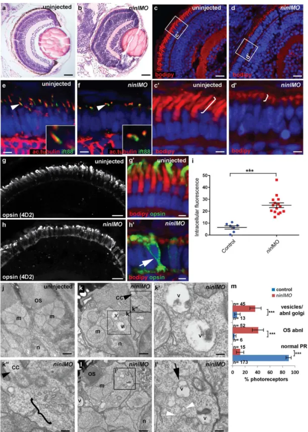
Fig 3. Zebrafishninlknockdown causes loss of axonemes and outer segments, opsin mislocalization and vesicle/vacuole accumulation.(a-b) Paraffin sections stained with Hematoxylin/Eosin of control (a) andninlknockdown larvae (b) demonstrating shortened outer segments and grossly
outer segments, as seen on retinal cryosections at 4 dpf stained with boron-dipyrromethene (bodipy) to mark the outer segment membrane disks (Fig 3c and 3d’) and anti-acetylated alpha-tubulin and anti-Ift88 antibodies to mark the axoneme (Fig 3e and 3f). Measurement of outer segment (OS) length of early 4dpf larvae, performed in a blinded manner as to injection status, revealed a significant shortening (mean OS length 1.6 +/- 0.26μm inninlatgMO
mor-phants compared to 3.9 +/- 0.32μm in wild-type,P<0.0001, unpaired Student’s t-test, n>10
larvae from each group in each of 2 biological replicates;S4e Fig). This retinal phenotype was observed with both theninltranslation-blocking and the splice-blocking morpholinos (S5b’ Fig). Little to no photoreceptor cell death was observed with TUNEL assay on cryosections of 4dpf morphant larvae (S3i and S3j Fig) compared toift88-/- retinas that are known to display prominent photoreceptor cell death at the same stage (and were thus used as positive controls;
S3k Fig). In general, minimal cell death was observed at 4dpf throughout the embryo, including in brain of larvae with overt ventriculomegaly (S3l Fig). Co-injection of 150pg of capped humanNINLmRNA with 2ng/nlninlatgMO restored normal outer segment length (mean OS length in rescued larvae 3.8 +/- 0.25μm,P<0.0001, unpaired Student’st-test, n = 10 larvae; S4a’–S4c’and S4e Fig).
Immuno-staining with anti-opsin antibodies (4D2 antibody) demonstrated significant accu-mulation of opsins in the inner segment and throughout the cell body ofninl-depleted photore-ceptors (Fig 3g–3i; mean intracellular fluorescence was significantly increased inninl
morphants compared to controls,P<0.0001, unpaired Student’st-test, n = 15 morphant larvae
and 7 control larvae, 2 replicate experiments). At the ultra-structural level, two types of abnor-mal membrane-bound structures were observed by transmission electron microscopy inninl
morphants: large vacuole-like structures were present in the cell body and small vesicular struc-tures accumulated around the Golgi complex and below the connecting cilium (Fig 3j–3m; vac-uolar and/or vesicular structures were present in 45/112 photoreceptors from 6 morphant eyes compared to 13/192 photoreceptors from 4 uninjected and 4 Control Oligo injected eyes;
P<0.0001, Fisher’s exact test). These phenotypes are partially reminiscent of those observed in
cc2d2a-/-embryos [25], supporting a common or coordinated function for Cc2d2a and Ninl in the process of vesicular trafficking towards the ciliary compartment.
Ninl
genetically interacts with
cc2d2a
and may act as a genetic modifier
for
CC2D2A
-associated Joubert Syndrome
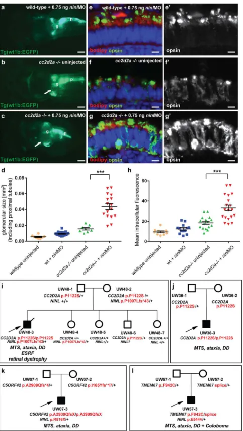
To further delineate the relationship betweencc2d2aandninl, we tested whether a synergistic effect was detectable between the two genes by using partialninlknockdown in thecc2d2a
mutant background. We observed that injection of a sub-phenotypic dose ofninlMO (0.75 ng/ nl), which causes no discernible phenotype in wild-type larvae, significantly increased the pen-etrance and severity of pronephric cysts incc2d2amutants: 89% ofninlMO-cc2d2a-/-zebrafish developed cysts compared to 40% of uninjectedcc2d2a-/-larvae (p<0.0001, Fisher’s exact test)
and the size of these cysts was significantly increased (as measured by the area of the dilated
compared to controls (g) where opsins are restricted to the outer segment. (i) Quantification of the intracellular opsin accumulation inninlmorphant
photoreceptors compared to control: each single datapoint in the scatter graph displays the averaged mean grey value from one larva. The mean value and the Standard Error of the Mean (SEM) are displayed as bars. The difference is statistically significant (***= p<0.0001, Student’st-test). (j-l’) Transmission electron microscopy of control (j) andninlknockdown larvae (k-l’) demonstrates absent or shortened and dysmorphic outer segments (OS) and
accumulation of large vacuoles (v, arrow inl’) and smaller vesicular structures (bracket ink”and white arrowheads inl’) in morphants. Black arrowheads point to the connecting cilium in k and k”. k’and k”are the boxed areas in k and l’is the boxed area in l. (m) Quantification of the % of photoreceptors
displaying these phenotypes. Absolute numbers of photoreceptors are also indicated. Error bars indicate 95% Confidence Intervals. The differences between morphant (red bars) and controls (blue bars) are statistically significant (***= p<0.0001, Fisher’s exact test). Larvae in all panels are 4 dpf old. Scale bars are 30μm ina-b, 15μm inc-d and g-h, 3μm inc’-d’and g’-h’, 4μm ine-f, 0.5μm inj-k and land 150nm ink’-k”andl’. OS outer segment, CC connecting cilium, m mitochondria, n nucleus, v vacuole.
glomerulus and proximal tubules: 0.044 +/-0.004 mm2forcc2d2a-/- +ninlMO (n = 16) as compared to 0.016 +/-0.002 mm2for uninjectedcc2d2a-/- (n = 8,P<0.0001, unpaired
Stu-dent’st-test) (Fig 4a–4d). Importantly, thecc2d2a+/- andcc2d2a+/+ siblings from the same injection clutch did not develop pronephric cysts at these sub-phenotypicninlMO doses (Fig 4a and 4d). In the retina, the opsin mislocalization phenotype incc2d2a-/-larvae (4 dpf) was enhanced by the addition of the same sub-phenotypic dose ofninlMO (Fig 4e–4h;P<0.0001,
Student’st-test, n = 19cc2d2a-/- +ninlMO and n = 16cc2d2a-/- uninjected, 2 replicates). These findings support a genetic interaction betweencc2d2aandninland suggest thatNINL
could be a genetic modifier forCC2D2A-caused disorders or even contribute to the genetic spectrum underlying Joubert/Meckel syndrome. Following this rationale, we sequencedNINL
in a cohort of 346 individuals with Joubert syndrome (from 291 families) using a molecular inversion probes (MIPs) capture method followed by next-generation sequencing [33] but did not identify any individuals carrying bi-allelic rare deleteriousNINLvariants. We did however find 3 individuals with heterozygousNINLmutations predicted to be deleterious. Individual UW48-3 carried the homozygous missenseCC2D2Amutation c. 3364C>T (p.P1122S),
previ-ously shown to be causal for Joubert syndrome, and a heterozygousNINLframeshift mutation leading to a stop codon after 43 amino acids (c.3020delC, p.P1007Lfs43) (Fig 4i) (and no other rare deleterious variant in any of the known JS genes). Phenotypically, this subject had a severe form of JBTS with retinal dystrophy, hearing loss, ventriculomegaly in addition to the MTS and renal failure leading to death at age 7 years. In comparison, subject UW 36–3 carried the same homozygousCC2D2Ac.3364C>T (p.P1122S) mutation but no additionalNINL
vari-ants (or rare deleterious varivari-ants in other JBTS genes) and presented with the“pure JBTS” phe-notype, consisting only of the MTS with associated ataxia, developmental delay and respiratory rhythm disturbance (Fig 4j). Subject UW07-3 carried a heterozygousNINLnonsense mutation (c.2446 G>A, p.R816X) in addition to causal, compound heterozygousC5ORF42frameshift
mutations (c.8726delG; p.A2909Qfs
4 and c.493delA, p.I165Yfs
17). This subject presented a classical Joubert phenotype without extra-neuronal manifestations, suggesting that the addi-tionalNINLframeshift had no effect on the clinical manifestations (Fig 4k). Finally, subject UW57-3 carried a heterozygousNINLmissense mutation (c.1631A>T, p.E544V), predicted to
be deleterious by Polyphen2, along with bi-allelic causalTMEM67mutations (c.2825T>G, p.
F942C and c.978+3 A>G). This individual had Joubert syndrome with coloboma but no
reti-nal, renal or hepatic involvement (Fig 4l). Given the known association betweenTMEM67
mutations and coloboma [12,34], this additional feature is most likely explained by the causal gene mutations, while the additionalNINLvariant appears to have no obvious effect on the phenotype in individual UW57-3. While it remains possible that additional sequence variants in non-JBTS genes also contributed to the enhanced phenotype in individual UW48-3 and while our findings from a large human cohort remain of anecdotal nature given the rarity of this highly heterogeneous genetic disorder, taken together with the zebrafish experiments, they suggest thatNINLmay act as a genetic modifier specifically forCC2D2A-caused Joubert syndrome.
Ninl is required for correct Rab8a localization
Fig 4. Genetic interaction betweenninlandcc2d2a.(a-d) Partialninlknockdown enhances the cystic kidney phenotype ofcc2d2amutants. (a-c) Glomerulus and proximal pronephric tubules highlighted in the transgenic line Tg(wt1b-EGFP). (a) Injection of a low dose ofninlatgMO (0.75 ng/nl) causes no cysts in
zebrafish photoreceptors in a punctate manner [25]. When expressed inninlmorphants (atgMO), mCherry-tagged Rab8a localized in significantly fewer puncta than when expressed in controls (42% of expressing photoreceptors displayed Rab8 puncta inninlmorphants (n = 38/87 from 14 larvae) compared to 73% in uninjected controls (n = 48/66 from 13 larvae),
p= 0.0005, two-tailed Fisher’s exact test;Fig 5a–5c). Instead, expression of the transgene was mostly diffuse throughout the photoreceptor cell body ofninlmorphants. A similar result was obtained using an anti-Rab8a antibody that recognizes endogenous small Rab8a puncta, which are found throughout the cell body, concentrated at the synapse and in the inner and outer seg-ments in controls (Fig 5d-d’). Inninl-knockdown larvae, the number of endogenous Rab8a puncta was significantly reduced (Fig 5e-e’and quantification in f: the average number of puncta perμm2was reduced to 0.04 +/- 0.01 (or 1 puncta per 25μm2) inninlmorphants as
compared to 0.09 +/- 0.01 (or 1 puncta per 11μm2) in uninjected wild-type,P= 0.01, unpaired
Student’s t-test), supporting a role for Ninl in Rab8 localization.
MICAL3 associates with NINL and is mislocalized in NINL and
CC2D2A-depleted cells
In order to unravel the underlying molecular cause of the observed vesicle accumulation and to identify proteins that interact with NINL, we next generated N-terminal Strep/FLAG-tagged fusion proteins of NINL isoA and isoB. A single-step affinity purification combined with quan-tification by stable isotope labeling of amino acids in cell culture (SILAC) and tandem affinity purification (TAP) [37] were applied to isolate the protein complexes in their native functional states from human embryonic kidney 293T (HEK293T) cells. The complexes were subse-quently analyzed by liquid chromatography coupled with tandem mass spectrometry (LC-MS/ MS). The identified interactome consisted of 174 unique proteins (Fig 6a,S1 table). An impor-tant association was found with multiple subunits of the cytoplasmic dynein 1-dynactin motor complex (DYNC1H1, DYNC1LI1, DYNC1LI2, DYNCI2, DYNLRB1, DCTN1-4, and DCTN6) which is involved in minus end–directed microtubule-associated transport. In addition, six actin-binding proteins (ARP1, ARP1B, ARP10, CAPZA1, CAPZA2 and CAPZB) and three subunits of Ca2+/calmodulin-dependent protein kinase II (CaMKII) (CAMK2A, CAMK2D, and CAMK2G), involved in non-canonical Wnt5a signaling, synaptic plasticity and kidney development [38], were found to associate with NINL. An additional relevant NINL interaction
glomerular + proximal tubular area displayed as a scatter plot, demonstrating a significant increase in proximal pronephric area incc2d2a-/- larvae injected with low-doseninlatgMO. The bars represent the mean
and standard error of the mean (SEM) for each treatment group and each datapoint is an individual fish. (e-g’) Immunohistochemistry with anti-opsin antibody (4D2, green) on retinal cryosections of 4dpfcc2d2a -/-uninjected larvae (f-f”) andcc2d2a-/- larvae injected with subphenotypic doses ofninlMO (g’g”’), that cause no mislocalization in wild-type fish (e-e’), demonstrates that partialninlknockdown increases the
mislocalization of opsins (e’-g’). (h) Quantification of the mean intracellular fluorescence displayed as a scatter plot shows significant increase in intracellular fluorescence incc2d2a-/- larvae injected with low dose ofninlatgMO. The bars represent the mean and standard error of the mean (SEM) for each treatment group
and each datapoint represents the mean intracellular fluorescence from 10 photoreceptors in one individual fish. Cell membrane and outer segments are stained with bodipy (red ine-g). Nuclei are counterstained with DAPI. Scale bars are 100μm in (a-c) and 4μm in (e-g’). (i) Pedigree of a consanguineous family with one affected boy (UW48-3) and 4 unaffected siblings. UW48-3 carried a homozygous missenseCC2D2A
mutation as well as a frameshift mutation inNINLleading to premature truncation. (j) Pedigree of a family
where the affected individual (UW36-3) carries the same homozygousCC2D2Amutation as in (i) but no additional rare deleterious variants. (k) Pedigree of a family where the affected individual (UW07-3) carries compound heterozygousC5ORF42frameshift mutations and a nonsense mutation inNINL. (l) Pedigree of a
family where the affected individual (UW57-3) carries compound heterozygousTMEM67mutations and a
missenseNINLmutation. The phenotype of the affected individuals is detailed initalicon each pedigree under the corresponding mutations.MTSMolar Tooth Sign,DDDevelopmental Delay,ESRFEnd-Stage Renal Failure.
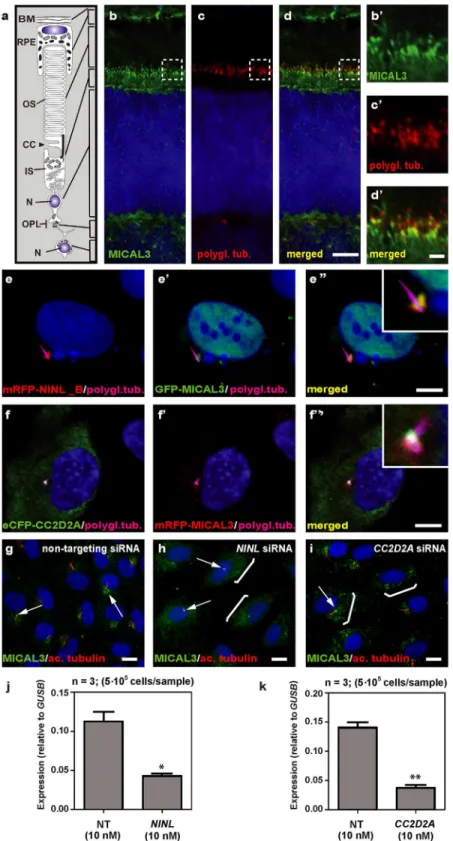
partner identified was MICAL3 (Microtubule-associated Monooxygenase, Calponin and LIM domain containing 3 protein), which is known to participate in a protein complex with RAB6 and RAB8 that is involved in the fusion of exocytotic vesicles [39], a process that appears to be deficient in the retina ofcc2d2amutants andninlmorphants. We validated the interaction between NINLisoBand MICAL3 by reciprocal co-immunoprecipitations (Fig 6b) and con-firmed that endogenously expressed MICAL3 is present at the photoreceptor connecting
Fig 5. Ninl is required for correct Rab8A localization.(a-a’) Expression of a rhodopsin-promoter driven cherry-tagged Rab8a in wild-type photoreceptors is mostly concentrated in one or several puncta (arrowsa-a’) whereas it is diffuse in the majority ofninlmorphant photoreceptors (b-b’). (c) Proportion of Rab8a-cherry expressing photoreceptors with punctate expression versus diffuse expression (bars represent 95% confidence interval;**P<0.001,Fisher’s
exact test). (d-d’) Endogenous Rab8a localization as seen by immunohistochemistry using an anti-Rab8a antibody (green) displays similar puncta
(arrowheads) in wild-type photoreceptors, while the number of puncta is decreased inninlmorphant photoreceptors (e-e’). (f) Quantification of the number of Rab8a puncta displayed in the form of a scatter plot indicating that significantly fewer endogenous Rab8 puncta perμm2are present inninlmorphants compared to uninjected controls (*P = 0.01, unpaired Student’st-test; bars represent standard error of the mean). Scoring was performed blinded as to
injection status for (c) and (f). Outer segments are counterstained with bodipy in (d-e). Nuclei are counterstained with DAPI. All images are cryosections of 4 dpf larvae. Scale bars are 4μm in all panels.
cilium in rat retina (P20), partially overlapping with the cilium and basal body marker polyglu-tamylated tubulin (Fig 7b–7d’). In hTERT-RPE1 cells, mRFP-tagged NINLisoB(Fig 7e-e”) and eCFP-tagged CC2D2A (Fig 7f-f”) partially overlapped with tagged MICAL3.
To evaluate the role of NINL and CC2D2A in MICAL3 localization, we silenced the expres-sion ofNINLandCC2D2Ain ciliated hTERT-RPE1 cells using siRNA, which was quantified by qPCR analysis (Fig 7j and 7k). Subsequent immunohistochemical stainings showed pre-dominant MICAL3 localization at the ciliary base in non-targeting siRNA-treated cells (Fig 7g) whereas silencing ofNINLexpression resulted in a dispersed distribution of MICAL3 through-out the cell body (Fig 7h). Downregulation ofCC2D2Aexpression in hTERT-RPE1 cells had a less pronounced effect on MICAL3 localization, resulting in partial mislocalization to the cell body (Fig 7i). These findings support a link between CC2D2A and MICAL3-RAB8-mediated vesicle trafficking/fusion through NINL.
Discussion
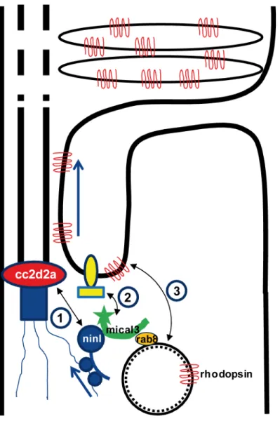
Dysfunction of transition zone proteins causes several ciliopathies such as Joubert syndrome, Meckel syndrome, nephronophthisis or Usher syndrome [16,21,40–43]. Previous work sug-gests that transition zone proteins in general, and CC2D2A in particular, are required for cor-rect localization of transmembrane proteins to the ciliary membrane [7,25],. The mechanism by which transition zone proteins exert this function and the link to upstream ciliary-directed vesicular trafficking mechanisms remain however largely unknown. In this work, we identify NINL as a novel physical interaction partner for the transition zone protein CC2D2A and pro-pose a model linking CC2D2A to RAB8A-controlled vesicle trafficking through a dual role for NINL in microtubule-based vesicle transport (Fig 8). The association of NINL with both the cytoplasmic dynein 1-dynactin motor complex (Dona et al, co-submitted manuscript) and MICAL3 supports a role for NINL in the initial transport of trans-Golgi network-derived RAB8A-MICAL3 coated vesicles towards the base of the photoreceptor cilium, while the asso-ciation of NINL with CC2D2A provides a docking point for these incoming vesicles at the entrance of the ciliary compartment.
Ciliary transmembrane proteins are synthesized in the cell body and travel from the Golgi towards the cilium in vesicles which move along microtubules using a cytoplasmic dynein motor [44]. Once at the entrance of the ciliary compartment, these vesicles must dock and fuse with the periciliary membrane to deliver their cargo into the ciliary membrane [45]. This path has been particularly well studied in photoreceptors, where large quantities of opsins and mem-brane continuously have to replenish the disks which constitute this photo-sensitive structure [26,46,35]. Opsin trafficking is severely affected in both zebrafishcc2d2amutants andninl
morphants, suggesting that both proteins play an important role in this transport which is cru-cial for the correct morphogenesis and homeostasis of the outer segments. Their co-localization at the base of the photoreceptor cilium could suggest that Cc2d2a and Ninl play a similar or combined role in opsin transport or that one protein is required to localize the other. However, since we found that each protein localizes independently of the other, and thatninlknockdown enhances thecc2d2anull mutant phenotype, the relationship between them is likely more com-plex than a simple linear pathway. At the ultrastructural level, the loss of function phenotypes of these two proteins also slightly diverge from each other: although vesicles accumulate in both cases in the affected photoreceptors, small vesiculo-tubular structures accumulate mostly apically around the connecting cilium incc2d2amutants [25], while this work shows that small
using untagged eGFP as a negative control (right panel) showed that eGFP-tagged MICAL3 immunoprecipitates with FLAG-tagged NINLisoBbut not with untagged eGFP.
MICAL3 (green signal,e’) at the ciliary base when co-expressed (yellow signal,e”). Co-expression of eCFP-CC2D2A (green signal,f) and mRFP-MICAL3 (red signal, f’) resulted in partial overlap at the base of the cilia (yellow signal,f”). (g) Endogenous MICAL3 (green signal) detected by immunostaining clusters at the ciliary base (white arrows; cilium marked with anti-acetylated tubulin in red) of hTERT-RPE1 cells treated with non-targeting siRNA. (h, i) Knockdown ofNINL(h) orCC2D2A(i) expression by siRNA results in
dispersed distribution of MICAL3 throughout the cell body (brackets) with retention of some MICAL3 puncta at the ciliary base (arrows). qPCR analysis ofNINL(j) andCC2D2A(k) siRNA treated hTERT-RPE1 cells. Cells were transfected with 10nM siRNA and all qPCR data were normalized againstGUSBlevels. Bar and error bars refer to mean and standard deviation, respectively (n = 3, on two biologicial replicates).*: P<0.05;
**: P<0.01 versus non targeting siRNA (NT) (student’st-test). Nuclei are counter stained with DAPI in all
panels (blue signal). Scale bars: are 5μm in d, 1μm in d’and 10μm in e-i.
doi:10.1371/journal.pgen.1005575.g007
Fig 8. Proposed model for CC2D2A and NINL function in trafficking, docking and fusion of rhodopsin-carrier vesicles.1) CC2D2A binds NINL and thus provides a docking point at the base of the connecting cilium for incoming vesicles. 2) NINL binds MICAL3 which in turn binds RAB8 that is coating the rhodopsin-carrier vesicles. Since NINL also associates with the cytoplasmic dynein1 motor complex, it provides a link between the carrier vesicles and the motor generating the movement along the microtubules. 3). MICAL3 subsequently interacts with ELKS and its redox activity promotes remodeling of the docking complex resulting in fusion of the vesicle at the periciliary region.
vesicles and larger vacuoles are also present more basally and closer to an abnormal Golgi apparatus inninlmorphants. This suggests that both proteins are important for vesicular traf-ficking but play different roles in this process.
NINL has been previously shown to bind several other ciliopathy proteins present at the base of cilia, specifically LCA5 and USH2A [31], suggesting that it may play a more pivotal role in vesicular trafficking in photoreceptors than CC2D2A. Given that the zebrafishninl mor-phant phenotype is more severe than thecc2d2amutant phenotype, this further suggests a more central role for Ninl than for Cc2d2a in cilium-directed trafficking. This hypothesis is also supported by the lack of bi-allelicNINLmutations in a large human cohort of Joubert syn-drome. Indeed, this may be interpreted as lack of tolerance to loss-of-function mutations in
NINL, as these would lead to more severe phenotypes or early embryonic lethality. The direct interaction between NINL and the dynein 1-dynactin complex [47] which we confirmed and expanded in the associated study by Dona et al, suggests that NINL might be involved in minus end–directed microtubule-associated transport of organelles and cargo towards the base of the cilium. An appealing model would thus propose that NINL functions both more upstream in ciliary-directed vesicular trafficking than CC2D2A as well as at the base of the cilium where it interacts with several different proteins including CC2D2A.
While no bi-allelic rare deleteriousNINLvariants were identified in our JBTS cohort, we did find heterozygousNINLmutations in individuals with Joubert syndrome. Interestingly, only the individual with causal bi-allelicCC2D2Amutations and a heterozygous truncatingNINL
mutation had a severe phenotype with retinal and terminal renal disease. In comparison, the individuals with causal mutations in other JBTS genes and a heterozygous deleteriousNINL
mutation (or with the same causalCC2D2Amutation alone) had the classical“pure Joubert”
phenotype without retinal or renal involvement. While bi-allelicCC2D2Amutations can result in a wide range of JBTS-associated phenotypes, the majority of individuals with causal
CC2D2Amutations and JBTS display the“pure JBTS”phenotype [22]. The more severe pheno-type only of the individual carrying causalCC2D2Amutations and an additionalNINL trun-cating variant suggests that deleterious variants inNINLmay act as genetic modifiers
specifically ofCC2D2A-caused ciliopathies such as Joubert syndrome. Unfortunately, the rarity of this disorder and its prominent genetic heterogeneity with over 27 associated genes prevent identification of multiple individuals sharing the same combination of causal and additional genetic variants, precluding identification of a statistically significant effect of rare variants as genetic modifiers using human genetics alone. Our findings from a large Joubert cohort there-fore remain of anecdotal nature. However, the physical and genetic interaction in zebrafish identified in this work substantially strengthen the significance of this finding and suggest that deleterious variants inNINLmay indeed enhance the retinal and renal phenotype in individu-als withCC2D2A-associated Joubert syndrome. The effect on the retinal phenotype may be explained by the importance of NINL function in photoreceptors as highlighted in the present study. Enhancement of the renal phenotype by the additionalNINLmutation may be explained by the association identified in this study between NINL and the PKD2-target CaMKII, which is important for renal development [38].
proteins directly including CEP290 and RGPR [48,49], no direct interaction has been demon-strated between CC2D2A and RAB8A, despite a functional interaction in zebrafish photore-ceptors and a requirement for CC2D2A in RAB8A localization in mouse embryonic fibroblasts [24,25]. Our findings now provide a model explaining the link between CC2D2A and RAB8A
(Fig 8): RAB8A-coated vesicles destined to the ciliary compartment are bound by MICAL3
which in turn binds NINL that is associated to the cytoplasmic dynein 1 motor complex (Dona et al, companion manuscript), allowing the movement along the microtubules. Once at the base of the cilium, NINL interacts with CC2D2A, providing the specificity of the docking point at the entrance to the ciliary compartment. Finally, the redox activity of MICAL3 promotes remodeling of the complex allowing fusion of the vesicle and release of cargo into the peri-cili-ary membrane.
A role for CC2D2A in promoting the assembly of ciliary subdistal appendages was recently suggested whereby CC2D2A would be required for docking of transport vesicles [24]. This is compatible with our model which also provides a possible mechanism to explain how transi-tion zone proteins may regulate ciliary protein compositransi-tion by providing specific docking points at the entry to the ciliary compartment. Dysfunction of transition zone proteins can lead to a variety of ciliopathies and it is likely that abnormal ciliary protein composition is at least in part responsible for the observed disease phenotypes even in the absence of ciliogenesis defects. This provides an opportunity for the development of pathway-specific therapies aiming at modulating trafficking routes and restoring normal ciliary protein content. In this perspective, unraveling the cell biological function of disease genes such asCC2D2Aas presented in the cur-rent study is a prerequisite for the future development of pharmacological treatments for patients with ciliopathies.
Materials and Methods
Ethics statement
All animal protocols were in compliance with Swiss legal ethical guidelines and the European Union Regulatory Agency guidelines for the use of fish in biomedical research and experiments and were approved by the local authorities (Veterinäramt Zürich TV4206). Human Subject Research Procedures were approved by the Institutional Review Boards at the University of Washington and Seattle Children’s Hospital (IRB-UW # 28853), and all participants or their legal representatives provided written informed consent.
Zebrafish
instructions. The cherry-Rab8a construct was previously described [25]. All quantifications were performed blinded as to injection status. All animal protocols were in compliance with internationally recognized guidelines for the use of fish in biomedical research and experiments and were approved by the local authorities (Veterinäramt Zürich TV4206).
Plasmids
pDONR201 vectors containing cDNA encoding human NINL isoform A and B as well as aa 1–998, aa 433–637, aa 992–1177 of humanCC2D2Awere previously described [31,52]. Using Gateway cloning technology, cDNA fragments encoding aa 992–1620 and aa 1171–1620 of humanCC2D2A(NM_001080522) were cloned in pDONR201 according to manufacturer’s instructions. pEGFP-C1-MICAL3 was kindly provided by Dr. A. Akhmanova (Utrecht Univer-sity, The Netherlands).
Yeast two-hybrid interaction assay
The direct interaction between CC2D2A and other ciliary proteins was tested using a GAL4--based yeast two-hybrid system (Hybrizap, Stratagene, USA) as previously described [30]. The DNA binding domain (GAL4-BD) fused to full length CC2D2A was used as a bait to test the interaction with previously described ciliopathy and cilium-associated proteins fused to an activation domain (GAL4-AD). Constructs encoding GAL4-BD and GAL4-AD fusion proteins were co-transformed in yeast strain PJ69-4A. The direct interaction between baits and preys induced the activation of the reporter genes, resulting in the growth of yeast colonies on selec-tive media (deficient of histidine and adenine) and induction ofα-galactosidase andβ -galacto-sidase colorimetric reactions [54].
Affinity purification of protein complexes
HEK293T cells transiently expressing the SF-TAP tagged NINLisoBwere grown in SILAC DMEM (PAA) supplemented with 3 mM l-glutamine (PAA), 10% dialyzed fetal bovine serum (PAA), 0.55 mM lysine, and 0.4 mM arginine. Light SILAC medium was supplemented with
12C
6,14N2lysine and12C6,14N4arginine. Heavy SILAC medium was supplemented with either 13C
6lysine and13C6,15N4arginine or13C6,15N2lysine and13C6,15N4arginine. 0.5 mM proline
twice with two or more peptides (peptide probability>80%) in three experiments. The protein
probability threshold was set to 99%.
Knockdown of
NINL
and
CC2D2A
in cultured hTERT-RPE1 cells by
RNAi
Three Silencer Select siRNAs targetingNINLandCC2D2Awere purchased from Life Technol-ogies (targeting sequences are listed inS2 Table). For transfection, a pool of three siRNAs per gene (45 nM final concentration) were plated in MW12 plates with or without glass slides. Lipofectamine RNAiMax (LifeTechnologies) and Opti-MEM (LifeTechnologies) were added to the duplexes and incubated for 10–20 minutes according to manufacturer’s protocol to allow the formation of transfection complexes. Human telomerase reverse transcriptase-trans-formed retinal pigment epithelium (hTERT-RPE1) cells from American Type Culture Collec-tion (ATCC) were then plated in MW12 plates. Per plate, non-targeting Silencer Select duplexes (LifeTechnologies) were included as negative controls. After 24 hours of transfection, cells were serum-starved to induce ciliogenesis. After 72 hours of transfection, knockdown-effi-ciency was determined by isolating total RNA from one 12-well with Trizol (Invitrogen, USA), followed by first-strand cDNA synthesis (iScript; Bio-Rad, USA). Quantitative PCRs using GoTaq (Promega), with validatedNINL-,CC2D2A-andGUSB-specific primers (sequences are listed inS3 Table), were performed as previously described [31]. The second 12-well of cells were fixed with 2% paraformaldehyde, permeabilized with 1% Triton-X-100/PBS and stained with anti-MICAL3 antibodies (kindly provided by Dr. A. Akhmanova). Images were taken with an Axioplan2 Imaging fluorescence microscope (Zeiss, Germany) equipped with a DC350FX camera (Zeiss, Germany).
Co-immunoprecipitation in HEK293T cells
HA-tagged NINL isoform B was expressed by using the mammalian expression vector pcDNA3-HA/DEST, FLAG-tagged CC2D2A, LRRK2 and STRAD by using p3xFLAG-CMV/ DEST and strep-FLAG-tagged NINL isoform B by using pNTAPe5/DEST from the Gateway cloning system (Invitrogen, USA). eGFP and eGFP-tagged MICAL3 were expressed from pEGFP-C1 (Clontech, USA). All plasmids contain a CMV promoter. HEK293T cells were co-transfected using Effectene (Qiagen, USA) according to manufacturer’s instructions. Twenty-four hours after transfection cells were washed with PBS and subsequently lysed on ice in lysis buffer (50 mM Tris-HCl pH 7.5, 150 mM NaCl, 1% Triton-X-100 supplemented with complete protease inhibitor cocktail (Roche, Germany)). HA-tagged NINL isoform B was immunopre-cipitated from cleared lysates overnight at 4°C by using rat monoclonal anti-HA-beads (Roche, Germany), while FLAG-tagged CC2D2A, LRRK2, STRAD and NINL isoform B were immuno-precipitated by using monoclonal anti-FLAG M2 Agarose beads (Sigma, Germany) and eGFP-tagged MICAL3 was immunoprecipitated using anti-GFP polyclonal antibodies (Abcam) cou-pled to ProtA/G beads (Santa Cruz, USA). After 4 washes in lysis buffer, the protein complexes were analyzed on immunoblots using the Odyssey Infrared Imaging System (LI-COR, USA). Tagged molecules were detected by anti-HA, anti-FLAG or anti-GFP mono- or polyclonal antibodies. As secondary antibody IRDye800 goat-anti-mouse IgG (Rockland Antibodies and Assays) and Alexa Fluor 680 goat-anti-rabbit IgG (Life Technologies) were used.
Immunohisto- and immunocytochemistry
before incubation with primary antibodies overnight. Primary antibodies were mouse mono-clonal acetylated alpha tubulin (1:500, clone 6-11B-1, Sigma), mouse monomono-clonal anti-polyglutamylated tubulin GT335 (1:500, gift from C. Janke, Institut Curie, France), mouse anti-zebrafish Cc2d2a (1:20, [25]), rabbit anti-NINL (1:100; LSBio Cat# LS-C201509), mouse anti-pancentrin 20H5 (1:200, clone 20H5 Millipore), mouse anti-Rab8a (1:100, clone 3G1 Novus Biologicals), mouse anti-opsin 4D2 (1:100, gift from R. Molday, University of British Columbia) and rabbit anti-Ift88 (gift from B. Perkins [58], Cleveland Clinic), mouse monoclo-nal anti-FLAG (1:1000, Sigma), rabbit polyclomonoclo-nal anti-human MICAL3 [39]. Secondary anti-bodies were Alexa Fluor goat anti-rabbit or goat anti-mouse IgG (Life Technologies) used at 1:300. Bodipy (1:300, Invitrogen) was applied for 20 minutes after the secondary antibodies and nuclei were counterstained with DAPI. Rab8 puncta detected by immuno-staining using the mouse anti-Rab8 antibody were analyzed blinded as to injection status in ImageJ. A region of interest was manually determined on single confocal sections and was thresholded (allways with the same parameters); the“analyze particles”function of ImageJ was then used to deter-mine the number of puncta perμm2. For quantification of intracellular fluorescence after 4D2
(opsin) immuno-staining, a region of interest including 10–15 photoreceptor cell bodies was determined on single confocal sections using ImageJ and the mean grey value was measured. For quantification of the proximal pronephric area, the fluorescent region corresponding to the glomerulus and the proximal tubules up to the curved part of the tubule was outlined man-ually in ImageJ and the“measure”function was used to determine the area of the outlined region. All quantifications were performed blinded as to injection status. Confocal imaging was performed on a Leica HCS LSI.
Paraffin sections and Transmission Electron Microscopy
For paraffin sections, 4 dpf oldninlmorphant larvae were fixed in 4% PFA overnight at 4°C, embedded in paraffin and sectioned following standard protocols. For Transmission Electron Microsopy,ninlmorphant and control larvae were fixed overnight at 4°C in a freshly prepared mixture of 2,5% glutaraldehyde and 2% paraformaldehyde in 0.1 M sodiumcacodylate buffer (pH 7.4). After rinsing in buffer, specimens were post-fixed in a freshly prepared mixture, con-taining 1% osmiumtetroxide and 1% potassiumferrocyanide in 0.1 M sodiumcacodylate buffer (pH 7.4), during 2 h at room temperature. After rinsing, tissues were dehydrated through a graded series of ethanol and embedded in epon. Ultrathin (rostrocaudally) sections (70nm), comprising zebrafish eyes at the optic nerve level, were collected on formvar coated grids, sub-sequently stained with 2% uranyl acetate and Reynold’s lead citrate, and examined with a Jeol1010 electron microscope.
Sequencing of
NINL
in a cohort of Joubert syndrome patients
SeattleSeq (http://snp.gs.washington.edu/SeattleSeqAnnotation138/). Minimal quality criteria for analyzed variants were DP (Depth)8, QD (Quality by Depth)>5, and ABHet
(Hetero-zygous Allele Balance)<0.8. The variant list was then filtered for rare and deleterious variants.
Only variants with minor allele frequency of<1% were considered given the rarity of JBTS
(estimated prevalence 1/80’000 [11]). All non-sense, frameshift and canonical splice-site vari-ants, as well as missense variants with Polyphen2 scores>0.8 were considered deleterious.
Selected variants were Sanger confirmed.
Statistical analyses
For all quantifications of zebrafish experiments, the Graphpad Prism6 software (http://www. graphpad.com/scientific-software/prism/) was employed to generate scatter plots, calculate mean values and SEM values, and perform statistical tests. Continuous data was analyzed using two-tailed, unpaired Student’s t-test and categorical data was analyzed using Fisher’s exact test.
Supporting Information
S1 Fig. Cloning and characterization of zebrafishninl.(A) Schematic representation of the
protein structure of human NINLisoAand NINLisoB(H.s. NINLisoAandH.s. NINLisoB) and zeb-rafish ninl (D.r. ninl) as predicted by using the Pfam homepage (http://pfam.xfam.org/). (B)
Ninlexpression during zebrafish development by whole mount RNAin situhybridization. Spe-cific expression was found in the following structures as indicated by numbers and arrows: (a) 14 somite stage: otic placode (1); developing eye (2); neural tube (spinal cord) (3); (b) 18 somite stage: neural tube (3); pronephros (4); (c) 18 somite stage: inner ear (1); optic nerve (5). (d) At 6 dpf, expression was observed in the tectum (6), the heart (7) and in the eye (2), predomi-nantly in the photoreceptor cell layer.H.s.:homo sapiens;D.r.:danio rerio; CC: coiled-coil; IF: intermediate filament domain; som: somites; dpf: days post-fertilization.
(TIF)
S2 Fig. Specificity of the anti-Ninl antibody.(a) Indirect immunohistochemical staining
using anti-Ninl antibody on 4 dpf retinal cryosections of control MO-injected larvae (green sig-nal) shows punctate staining, partially overlapping with the ciliary marker anti-polyglutamy-lated tubulin (a’, a”, red signal). (b) Indirect immunohistochemical staining using anti-Ninl antibody on 4 dpf retinal cryosections ofninlatgMO-injected larvae (b, green signal) along with the ciliary marker anti-polyglutamylated tubulin (b’, b”, red signal). Specific Ninl-immu-nofluorescence is largely abolished inninlmorphants, whereas the polyglutamylated tubulin signal is still detected. (c) Indirect immunohistochemical staining of anti-Ninl on 4 dpf retinal cryosections ofninlex15 spMO-injected larvae (green signal) along with the ciliary marker anti-polyglutamylated tubulin (c’, c”, red signal). Specific Ninl-immunofluorescence was still detected but at a diminished level inninlmorphants whereas the polyglutamylated tubulin sig-nal was usig-naltered. Nuclei are stained with DAPI (blue sigsig-nal). Scale bars: 4μm. (d) Western
blot analysis using protein extracts obtained from 100 zebrafish larvae injected with either con-trol MO (6ng),ninlatgMO (2ng) orninlex15 spMO (4ng). A specific product was detected with a molecular weight of ~80kDa in control MO-injected larvae (left panel). This band was almost completely abolished in theninlatgMO-treated larvae, but was still detected inninl
spMO-injected larvae although with a slightly diminished intensity. Anti-actin antibodies were used as a loading control (right panel). (e) Immunoprecipitation from bovine retinal extracts with anti-human NINL antibody detects 3 bands, the strongest being of the same size as the band found on Western blot of zebrafish lysates (~80 kDa).
S3 Fig. Phenotypes of theninlatgMO.(a) Representative clutch of zebrafish larvae at 2dpf injected with the phenotypic dose ofninlatgMO (2ng/nl). (b) Titration curve for theninl
atgMO illustrating the distribution of phenotypes in 2dpf larvae at two different concentra-tions: at 1ng/nl, a minority of injected larvae present a curved body shape (10%) and/or ventri-culomegaly (20%) (n = 19). At ~2ng/nl, on average 66% of injected larvae present a curved body shape and ~40% present ventriculomegaly and/or pronephric cysts (n = 84). 95% Confi-dence Interval bars are shown. (c) Representative 2dpf-oldninlatg-morphant displaying curved body shape, ventriculomegaly and pronephric cyst (arrow). (d-e) Dorsal view of 2dpf larvae showing the normal morphology of the brain folds in wild-type (d) and the enlarged ventricle in morphants (e). (f-g) Transgenic Tg(wt1b:EGFP) zebrafish line used to highlight the larval pronephros, shows the morphology of the fused glomerulus and proximal tubules in wild-type (f) and the dilatation of the region inninlmorphants (“kidney cysts”, white arrow in
g). (h) A clutch ofninlmorphants at 4dpf. (i-j) TUNEL assay on 4dpf cryosections through ret-inas fromninlmorphants shows the range of cell death detected (curved larvae in (h) were sec-tioned for the TUNEL assay). Note the absence of TUNEL-positive cells in the photoreceptor (PR) cell layer in the morphants. (k) Theift88-/- retina is used as a positive control, given the known death of photoreceptors in this mutant at 4dpf. (l) Cryosection through a 4dpf brain in a morphant larva displaying a dilated brain ventricle (v) shows no significant neuronal cell death. Scale bars represent 500μm in (a) and (h), 100μm in (c-g), 10μm in (i-k) and 30μm in
(l). PR PhotoReceptors, INL Inner Nuclear Layer. (TIF)
S4 Fig. Rescue of the morphant phenotype supports its specificity.(a-d) Co-injection of 2 ng
ninlatgMO with 150 pg capped MO-resistant mRNA encoding humanNINLisoform B reduced the incidence of body curvature defects from 71% inninlatgMO injected larvae (n = 207) to 36% inninlatgMO +NINLmRNA injected larvae (n = 203) (P<0.0001, two-tailed
Fisher’s exact). A subset of these larvae were sectioned and a perfect correlation was observed in rescue between body curvature defects and defects in photoreceptor outer segment forma-tion (a’-c’). (e) Quantification of the rescue of retinal outer segment length showing that mean OS length was rescued from 1.6 +/- 0.26μm inninlmorphants to 3.8 +/- 0.25μm with
co-injection ofNINLmRNA (P<0.0001, unpaired Student’st-test).
(TIF)
S5 Fig. Recapitulation of phenotype by aninlex15 spMO (4 dpf).(a-b) Injection of 4ngninl
ex15 spMO (n>100) results in heart edema and small eyes. No defects in body curvature were
observed in comparison to control MO-injected larvae. (a’, b’) Analyses of bodipy-stained reti-nas ofninlex15 spMO-injected larvae (n = 10) revealed defects in photoreceptor outer segment formation (10 of 10) similar to those observed inninlatgMO-treated larvae, whereas stained retinas of control MO-injected larvae (n = 10) appeared normal (10 of 10). Scale bars represent 500μm (a-b) and 5μm (a’-b’). (c) RT-PCR analysis on RNA isolated from 25 larvae that were
either uninjected, injected with control MO (6ng) or injected with various amounts ofninl
ex15 spMO (2, 4, 6ng), collected at two different time points after injection (2 dpf and 4 dpf). One PCR product of the expected length (~500bp) was obtained from RNA from uninjected and control MO-injected larvae. Sequence analysis revealed that this was the predicted tran-script including exons 13–16. RT-PCR analysis on RNA obtained from the morphant larvae resulted in two products: Sequence analysis of both fragments revealed that the shorter product is the predicted wild-type transcript (ex13-16) and that the longer transcript in addition includes the entire intron 14 (85 bp), resulting in premature termination of translation already after two codons in intron 14. This aberrant splicing persists at 4dpf.
S1 Table. TAP-data and SILAC data. SF-TAP analysis with over-expressed N-terminally
SF-TAP-tagged NINL in HEK293T cells.Shown are the number of unique identified peptides
as well as the sequence coverage for each protein detected by mass spectrometry. Proteins iden-tified in the SF-TAP analysis of empty vector control experiments were removed.SILAC
anal-ysis with over-expressed N-terminally SF-TAP-tagged NINL in HEK293T cells. Shown are
the ratios and significance value for WT/SF-control experiments. (XLSX)
S2 Table. siRNA sequences.
(DOCX)
S3 Table. Primer sequences.
(DOCX)
Acknowledgments
We would like to thank the participating patients and their families.
Author Contributions
Conceived and designed the experiments: RBG MD EdV DD CBM SCFN JEK HK RR EvW. Performed the experiments: RBG MD EdV LH ET IGP TP DAM HHA SECvB MU KB EvW. Analyzed the data: RBG MD EdV IGP DD DAM HHA SECvB MU KB EvW. Contributed reagents/materials/analysis tools: RBG DD CBM SCFN RR KB MU EvW. Wrote the paper: RBG EvW MD.
References
1. Goetz SC, Anderson KV. The primary cilium: a signalling centre during vertebrate development. Nat Rev Genet. 2010; 11 (5): 331–344. doi:10.1038/nrg2774PMID:20395968
2. Tyler KM, Fridberg A, Toriello KM, Olson CL, Cieslak JA et al. Flagellar membrane localization via asso-ciation with lipid rafts. J Cell Sci. 2009; 122 (6): 859–866.
3. Hu Q, Milenkovic L, Jin H, Scott MP, Nachury MV et al. A Septin Diffusion Barrier at the Base of the Pri-mary Cilium Maintains Ciliary Membrane Protein Distribution. Science. 2010; 329 (5990): 436–439. doi:10.1126/science.1191054PMID:20558667
4. Nachury MV, Seeley ES, Jin H. Trafficking to the Ciliary Membrane: How to Get Across the Periciliary Diffusion Barrier. Annu Rev Cell Dev Biol. 2010; 26 (1): 59–87.
5. Williams CL, Li C, Kida K, Inglis PN, Mohan S et al. MKS and NPHP modules cooperate to establish basal body/transition zone membrane associations and ciliary gate function during ciliogenesis. J Cell Biol. 2011; 192 (6): 1023–1041. doi:10.1083/jcb.201012116PMID:21422230
6. Reiter JF, Blacque OE, Leroux MR. The base of the cilium: roles for transition fibres and the transition zone in ciliary formation, maintenance and compartmentalization. EMBO reports. 2012; 13 (7): 608–
618. doi:10.1038/embor.2012.73PMID:22653444
7. Garcia-Gonzalo FR, Corbit KC, Sirerol-Piquer MS, Ramaswami G, Otto EA et al. A transition zone com-plex regulates mammalian ciliogenesis and ciliary membrane composition. Nat Genet. 2011; 43 (8): 776–784. doi:10.1038/ng.891PMID:21725307
8. Hildebrandt F, Benzing T, Katsanis N. Ciliopathies. N Engl J Med. 2011; 364 (16): 1533–1543. doi:10. 1056/NEJMra1010172PMID:21506742
9. Badano JL, Mitsuma N, Beales PL, Katsanis N. The Ciliopathies: An Emerging Class of Human Genetic Disorders. Annu Rev Genom Human Genet. 2006; 7 (1): 125–148.
10. Insinna C, Besharse JC.Intraflagellar transport and the sensory outer segment of vertebrate photore-ceptors. Dev Dyn. 2008; 237 (8): 1982–1992. doi:10.1002/dvdy.21554PMID:18489002
12. Bachmann-Gagescu R, Dempsey JC, Phelps IG, O'Roak BJ, Knutzen DM et al. Joubert syndrome: a model for untangling recessive disorders with extreme genetic heterogeneity. J Med Genet. 2015; 52 (8): 514–22. doi:10.1136/jmedgenet-2015-103087PMID:26092869
13. Maria BL, Hoang KBN, Tusa RJ, Mancuso AA, Hamed LM et al. "Joubert Syndrome" Revisited: Key Ocular Motor Signs With Magnetic Resonance Imaging Correlation. J Child Neurol. 1997; 12 (7): 423–
430. PMID:9373798
14. Romani M, Micalizzi A, Valente EM. Joubert syndrome: congenital cerebellar ataxia with the molar tooth. Lancet Neurol. 2013; 12 (9): 894–905. doi:10.1016/S1474-4422(13)70136-4PMID:23870701 15. Doherty D. Joubert Syndrome: Insights Into Brain Development, Cilium Biology, and Complex Disease.
Semin Pediatr Neurol. 2009; 16 (3): 143–154. doi:10.1016/j.spen.2009.06.002PMID:19778711 16. Romani M, Micalizzi A, Valente EM Joubert syndrome: congenital cerebellar ataxia with the molar
tooth. Lancet Neurol. 2013; 12 (9): 894–905. doi:10.1016/S1474-4422(13)70136-4PMID:23870701 17. Romani M, Micalizzi A, Kraoua I, Dotti M, Cavallin M et al. Mutations in B9D1 and MKS1 cause mild
Joubert syndrome: expanding the genetic overlap with the lethal ciliopathy Meckel syndrome. Orphanet J Rare Dis. 2014; 9 (1): 72.
18. Tuz K, Bachmann-Gagescu R, O’Day DR, Hua K, Isabella CR et al. Mutations in CSPP1 Cause Pri-mary Cilia Abnormalities and Joubert Syndrome with or without Jeune Asphyxiating Thoracic Dystro-phy. Am J Hum Genet. 2014; 94 (1): 62–72. doi:10.1016/j.ajhg.2013.11.019PMID:24360808 19. Bachmann-Gagescu R, Phelps IG, Dempsey JC, Sharma VA, Ishak GE, et al. KIAA0586 is mutated in
Joubert Syndrome.Hum Mutat. 2015; 36 (9): 831–5. doi:10.1002/humu.22821PMID:26096313 20. Thomas S, Wright KJ, Le Corre S, Micalizzi A, Romani M et al. A Homozygous PDE6D Mutation in
Jou-bert Syndrome Impairs Targeting of Farnesylated INPP5E Protein to the Primary Cilium. Hum Mutat. 2014; 35 (1): 137–146. PMID:24166846
21. Sang L, Miller JJ, Corbit KC, Giles RH, Brauer MJ et al. Mapping the NPHP-JBTS-MKS Protein Net-work Reveals Ciliopathy Disease Genes and Pathways. Cell. 2011; 145 (4): 513–528. doi:10.1016/j. cell.2011.04.019PMID:21565611
22. Bachmann-Gagescu R, Ishak GE, Dempsey JC, Adkins J, O'Day D et al. Genotype–phenotype correla-tion in CC2D2A-related Joubert syndrome reveals an associacorrela-tion with ventriculomegaly and seizures. J Med Genet. 2012; 49 (2): 126–137. doi:10.1136/jmedgenet-2011-100552PMID:22241855 23. Mougou-Zerelli S, Thomas S, Szenker E, Audollent S, Elkhartoufi N et al. CC2D2A mutations in Meckel
and Joubert syndromes indicate a genotype–phenotype correlation. Hum Mutat. 2009; 30 (11): 1574–
1582. doi:10.1002/humu.21116PMID:19777577
24. Veleri S, Manjunath SH, Fariss RN, May-Simera H, Brooks M et al. Ciliopathy-associated gene Cc2d2a promotes assembly of subdistal appendages on the mother centriole during cilia biogenesis. Nat Com-mun. 2014; 20 (5):4207.
25. Bachmann-Gagescu R, Phelps IG, Stearns G, Link BA, Brockerhoff SE et al. The ciliopathy gene cc2d2a controls zebrafish photoreceptor outer segment development through a role in Rab8-depen-dent vesicle trafficking. Hum Mol Genet. 2011; 20 (20): 4041–4055. doi:10.1093/hmg/ddr332PMID: 21816947
26. Deretic D, Wang J. Molecular assemblies that control rhodopsin transport to the cilia. Vision Res. 2012; 75 (0): 5–10.
27. Moritz OL, Tam BM, Hurd LL, Peränen J, Deretic D et al. Mutant rab8 Impairs Docking and Fusion of Rhodopsin-bearing Post-Golgi Membranes and Causes Cell Death of TransgenicXenopus Rods. Mol Biol Cell. 2001; 12 (8): 2341–2351. PMID:11514620
28. Nachury MV, Loktev AV, Zhang Q, Westlake CJ, Peränen J et al. A Core Complex of BBS Proteins Cooperates with the GTPase Rab8 to Promote Ciliary Membrane Biogenesis. Cell. 2007; 129 (6): 1201–1213. PMID:17574030
29. Westlake CJ, Baye LM, Nachury MV, Wright KJ, Ervin KE et al. Primary cilia membrane assembly is ini-tiated by Rab11 and transport protein particle II (TRAPPII) complex-dependent trafficking of Rabin8 to the centrosome. PNAS. 2011; 108 (7): 2759–2764. doi:10.1073/pnas.1018823108PMID:21273506 30. Cevik S, Sanders AAWM, van Wijk E, Boldt K, Clarke L et al. Active Transport and Diffusion Barriers
Restrict Joubert Syndrome-Associated ARL13B/ARL-13 to an Inv-like Ciliary Membrane Subdomain. PLoS Genet. 2013; 9 (12): e1003977 EP. doi:10.1371/journal.pgen.1003977PMID:24339792 31. van Wijk E, Kersten FFJ, Kartono A, Mans DA, Brandwijk K et al. Usher syndrome and Leber congenital
amaurosis are molecularly linked via a novel isoform of the centrosomal ninein-like protein. Hum Mol Genet. 2009; 18 (1): 51–64. doi:10.1093/hmg/ddn312PMID:18826961