ABSTRACT
Aim The objective of this study was to assess the capacity of beta-tricalcium phosphate to facilitate bone formation in the socket and prevent post-extraction alveolar resorption.
Materials and methods after premolar extraction in 16 patients, the sockets were filled with beta-tricalcium phosphate. six months later, during the implant placement surgery, a trephine was used to harvest the bone samples which were processed for histological and histomorphometrical analyses. data were gathered on patient, clinical, histological and histomorphometric variables at the extraction and implant placement sessions, using data collection forms and pathological reports.
Results Clinical outcomes were satisfactory, the biomaterial was radio-opaque on X-ray. Histological study showed: partial filling with alveolar bone of appropriate maturation and mineralization for the healing time, osteoblastic activity and bone lacunae containing osteocytes. The biomaterial was not completely resorbed at six months.
Conclusion Beta-tricalcium phosphate is a material capable of achieving preservation of the alveolar bone when it is positioned in the immediate post-extraction socket followed by suture; it also helps the formation of new bone in the socket. Further studies are needed comparing this technique with other available biomaterials, with growth factors and with sites where no alveolar preservation techniques are performed.
Post-extraction application of beta-tricalcium phosphate
in alveolar socket
To CiTe THis arTiCle
Muñoz-Corcuera M, Bascones-Martínez a, ripollés-de ramón J. Post-extraction application of beta tricalcium phosphate in alveolar socket. J osseointegr 2015;7(1):8-14.
inTRoduCTion
In normal conditions, healthy bone is under continuous remodelling and has an effective self-repair capacity. Bone remodelling maintains a continuous balance of
bone formation and resorption in a dynamic process that adapts the bone to local forces (1). Above a critical defect size, however, bone cannot be repaired by its own osteogenic activity, and some type of bone grafts must be used (2).
Jaw bone defects can be caused by surgical resection, traumatic loss, ossification impairment (in the elderly), periodontal and peri-implant diseases and congenital disorders. These defects may complicate the surgical phase of implant supported rehabilitation treatment due to insufficient bone volume for an adequate implantation (3, 4). Jaw bone loss is frequently caused by post-extraction alveolar resorption, a physiological phenomenon which leads to a reduction of the original height and width of the alveolar ridge to a degree that varies among localizations and patients (5). Alveolar ridge preservation techniques have been developed to address the ensuing clinical problem, especially in aesthetic areas (5). They are conducted during or after extraction and are designed to minimize external ridge resorption and maximize bone formation inside the socket (5). Measures include autologous bone grafts, allografts, bone of animal origin (xenografts) and synthetic bone substitutes (alloplastic grafts), as well as the application of growth factors and gene therapies (3, 4, 6).
Beta-tricalcium phosphate (beta-TCP) is widely used as a biocompatible, resorbable and osteoconductive ceramic substitute to repair bone defects. Thanks to its physicochemical characteristics, it has been successfully used to fill spaces in multiple settings, including biology, veterinary medicine, human medicine and dentistry (7-12). It has also been proposed as a vehicle for growth factors that stimulate bone formation (12, 13). Various authors have reported on its capacity as a biomaterial for bone regeneration in animals and humans (4, 14-21). The study is aimed at evaluating granular beta-TCP in post-extraction sockets in order to measure its bone regenerative potential and its capacity to preserve the original height and width of the alveolar bone for subsequent implant placement.
Specifically, the study objectives were the following. 1. To analyze the clinical and radiological results
department of oral Medicine and surgery, school of dentistry, Madrid Complutense university, Madrid, spain
1 Professor at dental school
obtained after placement of the biomaterial in the post-extraction socket and at the subsequent insertion of dental implants.
2. To assess the effectiveness of beta-TCP as bone filling material in the post-extraction socket.
3. To perform histological analysis of the amount and quality of bone formed in the dental socket six months after the placement of the biomaterial. 4. To determine the percentage of biomaterial particles
in contact with patient bone.
MATeRiAlS And MeTHodS
Study design
This prospective longitudinal observational clinical study complied with the principles of the Helsinki Declaration and was approved by the clinical research ethics committee of the San Carlos Clinical Hospital, Madrid (Spain).
All patients in the study were aged over 18 years and scheduled for ≥ 1 premolar extraction due to periodontal disease, caries or fractures and for subsequent replacement with dental implant(s) up to a maximum of four premolar extractions (one per quadrant) per patient. Exclusion criteria were: failure to sign informed consent or commit to compliance with the study appointment schedule; the presence of endocrine-metabolic disease or chronic, general or local disease; the presence of disease that may be affected by the surgery or by the intraoperative or postoperative medication; alveolar socket wall defects; smoking habit of ≥10 cigarettes/ day, due to its relationship with implant failure; and treatment with bisphosphonates or antibiotics during the previous month. Patients were recruited from the School of Dentistry clinic (Complutense University of Madrid, Spain) and private clinics. A non-probabilistic sampling of consecutive cases was conducted and only patients who met the above criteria were included. Sixteen patients were enrolled in the study between March 2008 and July 2010, with a mean age of 44.3 years (standard deviation: 10.74); seven were male (44%) with mean age of 39.7 years and nine were female (56%) with mean age of 48 years. No participant (0%) was a daily drinker of alcohol, and two (12%) were daily smokers (of 1-9 cigarettes). A total of 19 upper and 2 lower teeth were extracted (lower teeth were excluded from the analysis because of this small number). After a baseline clinical assessment, all patients received basic periodontal therapy before the surgery and were instructed to maintain good oral hygiene throughout the study.
Surgical procedure
After applying local anaesthesia and performing full- thickness buccal and lingual flap elevation, the premolar was extracted; a full-thickness flap was elevated to
enable a subsequent suture to keep the granules of the material in place. Any granulation tissue present in the socket was removed by surgical curettage, and the socket was filled with 0.5 g beta-TCP KeraOs® (Keramat, La Coruña, Spain) mixed with physiological saline solution or blood from the same patient (Fig. 1). The socket was then closed by suture using a coronally repositioned flap (Fig. 2). Patients were instructed to rinse daily for two weeks with 0.12% chlorhexidine digluconate. Sutures were removed at 7-10 days post-extraction.
During the implant placement surgery (about 6 months after biomaterial placement), a bone biopsy was harvested using a trephine (inner diameter of 2.2 mm, outer diameter of 3 mm), placed in a 10% buffered formalin and sent to the Ceramic Institute of Galicia (Santiago de Compostela, Spain) laboratory for processing.
Histological processing
The specimens were processed to obtain thin undecalcified sections following Donath’s method and using the EXACT system.
Briefly, specimens were fixed in buffered 10% formalin, progressively dehydrated in alcohol and then embedded in photopolymerizable methacrylate resin (Technovit 7200®, VLC-Heraus Kulzer GMBH, Werheim, Germany). After polymerization, the specimens were cut with a diamond saw and then ground with silicon carbide papers to a width of about 70 microns. After thinning, samples were stained with Levai Laczko stain and chromotrope 2R/Harris haematoxylin. A motorized Olympus BX51 microscope with Olympus DP71 camera was used to image the specimens, with Olympus D-cell capture software and Photoshop CS3 image processing software, employing a Wacom Intuos 4 pen tablet and
Fig. 1socket filled with a mixture of beta-TCP and patient blood.
applying the Olympus MicroImage 4.0 program to obtain histomorphometric measurements.
Data were gathered on the following.
1. Patient variables, sex, age, and consumption of alcohol and cigarettes (smoker = 1-9, non-smoker = 0 cigarettes/day, to test whether a light tobacco habit affects socket healing).
2. Clinical variables, biomaterial stability within socket and primary implant stability.
3. Radiological findings.
4. Histological variables at 6 months, degree of bone neoformation in socket, amount and quality of newly formed bone, degree of contact between patient bone and beta-TCP and degree of beta-TCP resorption, all assessed by direct microscopic observation.
5. histomorphometric variables, areas of newly formed bone, immature bone, old bone, biomaterial and lamellar bone, bone-biomaterial contact index (perimeter of material in contact with bone / perimeter of whole material), remnant volume (surface of material present / [surface of material present + total bone surface]) and immature:mature bone ratio (mature bone surface / total bone surface). Specifically designed forms were used to collect data at the following time points: tooth extraction, gathering patient variables; suture withdrawal (7-10 days post-extraction), recording radiological findings; and implant placement (around 6 months post-extraction), gathering radiological findings and data on material retention in the socket and primary implant stability. Histological data were obtained from the pathology report on samples taken at implant placement.
Microsoft Excel and SPSS were used for the statistical analyses, which included: descriptive analysis of patient, clinical and histomorphometric variables; frequency histograms for histomorphometric variables; Shapiro-Wilks normality tests for histomorphometric variables, age and healing time; 95% confidence intervals for histomorphometric variables; use of the Pearson correlation coefficient to analyse associations of different histomorphometric variables with each other and with healing time and age; analysis of variance (ANOVA) to determine the effect of healing time on newly formed bone area, biomaterial area and bone-biomaterial contact index; and the Student’s t test to compare newly formed bone area, biomaterial area and bone-biomaterial contact index between shorter and longer healing times (5-6 months versus 7-8 months, respectively).
ReSulTS
Patient variables
One male patient abandoned the study before implant placement. Among the 15 remaining patients, 21 biopsies were obtained after a mean healing time of 6.2 months (standard deviation: ±1.05). Out of the 21 biopsies, 3 were impaired during grinding and could not be processed, and 2 were incorrectly sampled and excluded from the analyses. Hence histological and histomorphometric analyses were conducted in a final sample of 16 biopsies (Table 1).
Clinical results
None of the patients evidenced biomaterial loss at implant placement; in some cases, the most superficial area showed residual graft particles that had no effect on the surgical procedure or primary stability, which was obtained in all cases. X-ray images revealed no complications, and in all the films, high radiopacity and consequent prompt identification of the material was detected.
Histological and histomorphometric results
No biomaterial fragments or necrotic bone splinters were detected in any of the 16 biopsies analyzed. In three cases, the biomaterial was integrated in the bone and surrounded by fibrous tissue with rim of osteoblasts and osteoid matrix; in one case, the biomaterial was surrounded by lax conjunctive tissue; in five cases, it was surrounded by mature bone trabeculae with scant osteoid and osteoblastic rimming; in seven cases, modest to highly abundant immature bone trabeculae growth was observed with osteoid and osteoblasts rim. Ten of the biopsies showed the presence of medullary fibrosis, at a low level in most cases.
Evidence of vital bone growth was found in the sockets, with bone neoformation in close contact with graft particles. All samples showed residual particles of the material, with various degrees of material remodelling and resorption (Fig. 3, 4, 5).
The histological study at 6 months revealed that the degree of bone neoformation in the socket was generally moderate, that the newly formed bone was immature (consistent with the healing time) and surrounded by and in direct contact with biomaterial fragments and that the beta-TCP material showed initial signs of resorption. Table 2 exhibits the results of the histomorphometric
Females Males Mean Standard deviation
age (yrs) 48 39.7 44.3 10.74
Number 9 (56.25%) 7 (43.75%)
smokers (1-9 cigs/day) 2 (12%) 0 (0%)
drinkers 0 (0%) 0 (0%)
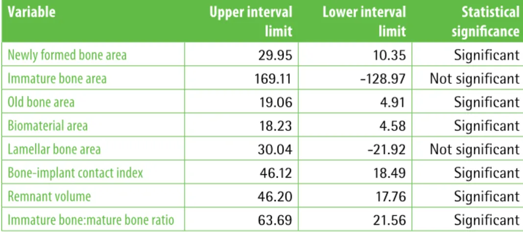
variables, which were found to follow a normal distribution (Shapiro-Wilk test). The frequency histograms showed that the mean contact between bone and biomaterial was <20% in 8 out of 15 biopsies and that the newly formed bone area was >20% in most of them; the biomaterial area was <20% in most of the biopsies. Calculation of 95% confidence intervals showed significance for all variables, except for the immature bone area and lamellar bone area, for which there were measurements in only two cases (Table 3). These two variables were excluded from analysis, using Pearson’s correlation coefficient, of the relationships of histomorphometric variables with each other and with healing time and age; a positive correlation was found between remnant volume and biomaterial area (p= 0.0056) and between old bone area and the immature bone:mature bone ratio (p= 0.015).
Although the healing period was established as 6 months for this study, this time was sometimes influenced by specific patient circumstances and ranged from 5 to 8 months. The results for newly formed bone area,
Fig. 3Newly formed bone around the biomaterial, (white arrows) with faint signs of resorption in the central area of the biopsy (red arrow). Most of the bone is newly formed, with traces of old bone at the periphery. levai-laczko stain. 100X.
Fig. 4Material integrated in the bone tissue; there is a predominance of lax connective tissue (red arrow), although with areas of denser connective tissue. The biomaterial is integrated in bone trabeculae (white arrows). levai-laczko stain. 100X.
Fig. 5Biomaterial surrounded by immature bone. Chromotrope 2r/Harris haematoxylin staining.100X.
TaBle 2results. descriptive statistics of histomorphometric variables. Table shows the minumim and maximum values for each variable; the mean and the median are also showed for each variable.
Variable (%) Minimum value Maximum value Mean Median Standard deviation
Newly formed bone area 0.30 45.33 20.15 13.64 15.42
immature bone area 8.34 31.80 20.07 20.07 16.58
old bone area 0.43 21.03 11.98 11.87 7.65
Biomaterial area 0.33 26.25 11.40 7.99 8.88
lamellar bone area 2.02 6.11 4.06 4.06 2.89
Bone-implant contact index 0 69.70 32.31 19.82 24.94
remnant volume 0 98.85 31.98 35.60 25.68
biomaterial area and bone-biomaterial contact index were analyzed in function of healing time, finding no significant differences. Then, newly formed bone area, biomaterial area and bone-biomaterial contact index were compared between healing times of 5-6 months and 7-8 months, finding no significant differences, altough borderline significance (p=0.08) was obtained for newly formed bone area.
diSCuSSion
In this study, post-extraction placement of beta-TCP in the socket did not cause any complications and achieved good clinical outcomes. There was histological evidence of bone neoformation at implant placement, with the presence of osteocytes and immature bone. The mean percentage of neoformed bone was 20.15%, in line with previous reports (22-25). The biomaterial area was less than 20% in most of the biopsies, confirming the resorbability of the biomaterial. The biomaterial was readily identifiable on X-ray, being much denser than the adjacent bone, as previously reported by Von Doernberg et al. (26). This characteristic is useful for the radiographic follow-up of healing, because the radiopacity changes as the material is resorbed and replaced by new bone.
Clinical studies on humans generally require the use of non-invasive techniques, e.g. radiology; but a biopsy study is the currently optimal method to assess the regeneration, quantity and quality of bone. A two-phase approach, inserting the graft in the first phase and the implant in the second, allows a histological sample to be obtained (20). This technique was applied in the present study.
We mixed beta-TCP with saline solution or blood from the patient, as in the study by Horowitz et al. (24), given the difficulty of managing this porous material in granular form (27).
Six months as bone healing time was selected, because most of the ceramic is resorbed, and the grafted tissue
can be considered sufficiently stable for functional implant loading (19, 24). A study in pigs (28) found beta-TCP degradation to be slow, with 80% of the material resorbed at 28 weeks and 97% at 86 weeks; therefore, the authors recommended an interval of 5-6 months before implant placement in grafted areas, concluding that the cell response to their simultaneous placement could damage implant osseointegration. Some authors suggested lengthening this healing time in order to increase implant stability (4), and it was found that the presence of residual particles at 9 months does not compromise implant placement (23). In contrast, as reported above, Ormianer et al. achieved a 97% success rate after the immediate placement of implants in augmented areas and their immediate implant loading (22).
With regard to the mechanism of beta-TCP degradation before its substitution by bone, it was attributed by Wiltfang et al. (28) to chemical hydrolysis (halisteresis) and the activity of phagocytic cells (multinucleated giant cells). Two degradation pathways have since been described: osteoclast-mediated resorption and dissolution in interstitial fluid (23). A study in 2005 detected no osteoclastic activity in biopsies from sinuses augmented with this biomaterial, but this finding does not rule out the participation of osteoclasts although it suggested that it is limited (29). Besides these two mechanisms, it has been postulated that beta-TCP resorption may also be mediated by cells other than osteoclasts (20). However, Martinez et al. (30) suggested that osteoclasts or macrophage cells may not play an important role in beta-TCP resorption, as they found in the bone-beta-TCP interface cells of the reticuloendothelial system.
Some data are available on the use of beta-TCP for alveolar preservation (22-25). Ormianer studied the use of beta-TCP alone in 338 patients, although alveolar preservation was not investigated in all of these, and the number of patients undergoing the different procedures was not specified; the mean follow-up was 19.2 months and the global implant survival rate was
TaBle 3results. 95% confidence intervals. Calculation of 95% confidence intervals showed significance for all variables, with the exception of immature bone area and lamellar bone area, for which there were measurements in only two cases.
Variable upper interval
limit
lower interval limit
Statistical significance
Newly formed bone area 29.95 10.35 Significant
immature bone area 169.11 -128.97 Not significant
old bone area 19.06 4.91 Significant
Biomaterial area 18.23 4.58 Significant
lamellar bone area 30.04 -21.92 Not significant
Bone-implant contact index 46.12 18.49 Significant
remnant volume 46.20 17.76 Significant
97.6%. In 2008, Brkovic reported on the use of beta-TCP with collagen alone in one patient, followed up for 9 months, reporting good clinical outcomes with bone formation activity.
In 2012, Horowitz used beta-TCP with a membrane in 30 patients, followed up for a mean of 6 months, also observing good outcomes with preservation of 91% of the socket width. Finally, in the same year, Brkovic studied 20 patients in two groups, one receiving beta-TCP with membrane and apically repositioned flap and the other beta-TCP alone, with a mean follow-up of 9 months, concluding that socket preservation was lower in the group without membrane. Our results are comparable to the findings of these four studies, because the implant survival was 100%, the clinical outcomes were good, bone neoformation was observed in the biopsies, and there was only a small volume of residual bone (11.98%) (Table 2).
There have also been reports on socket preservation with the use of other materials. Thus, Liasella et al. employed allografts with good results (31), while De Coster et al. (32) used biphasic ceramics but obtained poor outcomes that delayed implant placement.
After experiencing some problems in harvesting the specimens from the trephine, the protocol was modified and the samples were processed with the trephine as a block. Zerbo (33) also found it difficult to remove beta-TCP biopsies in a single piece from the trephine, and Suba (20) reported that biomaterial particles frequently broke during sample preparation.
In the present study three biopsies were lost in the polishing process, due to the complexity of sample processing, and one biopsy was taken from the incorrect area, a problem that some authors have resolved by using surgical guides (25).
In the study by Horowitz 2010 (34), two cases are discussed. In the first one an identical procedure to the one here described was followed, except for the use of a resorbable membrane after the placement of the biomaterial. The clinical outcome was excellent, allowing the placement of a dental implant 6 months after extraction. The biomaterial was replaced by new vital bone, just as in our work. Their second case is that of a smoker patient; the biomaterial was placed in the socket followed by a membrane. Healing time in this case was 10 months, after which an implant was placed. The clinical, radiological and histological results are comparable to those of our study, they observed the formation of osteon and Haversian systems in the biopsy due to increased healing time.
With the limitations of this study, especially regarding the small sample size, the histological and clinical results are in agreement with reports by various authors, evidencing problem-free healing, primary stability of implants placed in the augmented area, and an adequate substitution of beta-TCP particles by newly formed bone at 6 months.
ConCluSion
The clinical and radiographic outcomes of this procedure are satisfactory, with no associated complications. Beta-tricalcium phosphate seems to be a biomaterial capable of achieving preservation of the alveolar bone when it is positioned immediately in post-extraction socket followed by suture; also facilitating the formation of new bone in the socket in the first six months. This resorbable material allows predictable and reproducible bone regeneration. As advantages, it can be noted its unlimited availability, its easy handling and its great radiopacity, allowing radiographic follow-up of the area. Multiple publications have shown the suitability of this material for use in bone augmentation techniques. Further clinical studies and randomized clinical trials are needed, comparing this technique with other available biomaterials, with growth factors and with alveoli in which no alveolar preservation techniques are performed.
ACKnoWledGeMenTS
This study was supported by an FPU training grant for university professors from the Spanish Ministry of Education (nº AP2008-00011) and by a project of Universidad-Empresa Foundation (47/2009) signed with Keramat.
ReFeRenCeS
1. allegrini s Jr, Koening B Jr, allegrini Mr, yoshimoto M, gedrange T, Fanghaenel J et al. alveolar ridge sockets preservation with bone grafting-review. ann acad Med stetin 2008;54:70-81.
2. Khan y, yaszemski MJ, Mikos ag, laurencin CT. Tissue engineering of bone: material and matrix considerations. J Bone Joint surg am 2008;90(suppl 1):36-42.
3. Tripplet rg, schow sr, laskin dM. oral and maxillofacial surgery advances in implant dentistry. int J oral Maxillofac implants 2000;15:47-55. 4. Zijderveld sa, Zerbo ir, van der Bergh JPa, schulten eaJM, ten Bruggenkate
CM. Maxillary sinus floor augmentation using a beta-tricalcium phosphate (Cerasorb) alone compared to autogenous bone grafts. int J oral Maxillofac implants 2005;20:432-40.
5. darby i, Chen s, de Poi r. ridge preservation: what is it and when should it be considered. aust dent J 2008;53:11-21.
6. giannoudis PV, dinopoulos H, Tsiridis e. Bone substitutes: an update. injury 2005;36 suppl 3:s20-7.
7. deppe H, Horch H, Neff a. Conventional versus Co2 laser-assisted treatment of peri-implant defects with the concomitant use of pure-phase β-tricalcium phosphate: a 5-year clinical report. int J oral Maxillofac implants 2007;22:79-86.
8. Franch J, díaz-Bertrana C, lafuente P, Fontecha P, durall i. Beta-tricalcium phosphate as a synthetic cancellous bone graft in veterinary orthopaedics. a retrospective study of 13 clinical cases. Vet Comp orthop Traumatol 2006;19:196-204.
10. liu g, Zhao l, Cui l, liu w, Cao y. Tissue-engineered bone formation using human bone marrow stromal cells and novel beta-tricalcium phosphate. Biomed Mater 2007;2:78-86.
11. allabouch a, Colat-Parros J, salmon r, Naim s, Meunier JM. Biocompatibility of some materials used in dental implantology: histological study. Colloids surf B Biointerfaces 1993;1:323-9.
12. aybar B, Bilir a, akçakaya H, Ceyhan T. effects of tricalcium phosphate bone graft materials on primary cultures of osteoblast cells in vitro. Clin oral impl res 2004;15:119-25.
13. Byun H, wang H. sandwich bone augmentation using recombinant human platelet-derived growth factor and beta-tricalcium phosphate alloplast: case report. int J Periodontics restorative dent 2008;28:83-7.
14. alam s, ueki K, Marukawa K, ohara T, Hase T, Takazakura d et al. expression of bone morphogenetic protein 2 and fibroblast growth factor 2 during bone regeneration using different implant materials as onlay bone graft in rabbit mandibles. oral surg oral Med oral Pathol oral radiol endod 2007;103:16-26.
15. Boix d, weiss P, gauthier o, guicheux J, Bouler JM, Pilet P et al. injectable bone substitute to preserve alveolar ridge resorption after tooth extraction: a study in dog. J Mater sci Mater Med 2006;17:1145-52.
16. Fiorellini JP, Kim dM, Nakajima y, weber HP. osseointegration of titanium implants following guided bone regeneration using expanded polytetrafluoroethylene membrane and various bone fillers. int J Periodontics restorative dent 2007;27:287-94.
17. Masago H, shibuya y, Munemoto s, Takeuchi J, umeda M, Komori T et al. alveolar ridge augmentation using various bone substitutes--a web form of titanium fibers promotes rapid bone development. Kobe J Med sci 2007;53:257-63.
18. suba Z, Takács d, gyulai-gaäl s, Kovács K. Facilitation of beta-tricalcium phosphate-induced alveolar bone regeneration by platelet-rich plasma in beagle dogs: a histologic and histomorphometric study. int J oral Maxillofac implants 2004;19:832-8.
19. Horch HH, sader r, Pautke C, Neff a, deppe H, Kolk a. synthetic, pure-phase beta-tricalcium phosphate ceramic granules (Cerasorb) for bone regeneration in the reconstructive surgery of the jaws. int J oral Maxillofac surg 2006;35:708-13.
20. suba Z, Takács d, Matusovits d, Barabás J, Fazekas a, szabó g. Maxillary sinus floor grafting with beta-tricalcium phosphate in humans: density and microarchitecture of the newly formed bone. Clin oral impl res 2006;17:102-8.
21. szabó g, Huys l, Coulthard P, Maiorana C, garagiola u, Barabas J et al. a prospective multicenter randomized clinical trial of autogenous bone versus beta-tricalcium phosphate graft alone for bilateral sinus elevation: histologic and histomorphometric evaluation. int J oral Maxillofac implants 2005;20:371-81.
22. ormianer Z, Palti a, shifman a. survival of immediately loaded dental
implants in deficient alveolar bone sites augmented with beta-tricalcium phosphate. implant dent 2006;15:395-403.
23. Brkovic BM, Prasad Hs, Konandreas g, Milan r, antunovic d, sándor gK et al. simple preservation of a maxillary extraction socket using beta-tricalcium phosphate with type i collagen: preliminary clinical and histomorphometric observations. J Can dent assoc 2008;74:523-8.
24. Horowitz ra, Mazor Z, Miller rJ, Krauser J, Prasad Hs, rohrer Md. Clinical evaluation of alveolar ridge preservation with a beta-tricalcium phosphate socket graft. Compend Contin educ dent 2009;30:588-90, 592, 594 passim; quiz 604, 606
25. Brkovic BM, Prasad Hs, rohrer Md, Konandreas g, agrogiannis g, antunovic d, sándor gK. Beta-tricalcium phosphate/type i collagen cones with or without a barrier membrane in human extraction socket healing: clinical, histologic, histomorphometric, and immunohistochemical evaluation. Clin oral investig 2012;16:581-90.
26. von doernberg MC, von rechenberg B, Bohner M, grünenfelder s, van lenthe gH, Müller r et al. in vivo behavior of calcium phosphate scaffolds with four different pore sizes. Biomaterials 2006;27:5186-98.
27. walsh wr, Vizesi F, Michael d, auld J, langdown a, oliver r et al. Beta-TCP bone graft substitutes in a bilateral rabbit tibial defect model. Biomaterials 2008;29:266-71.
28. wiltfang J, Merten Ha, schlegel Ka, schultze-Mosgau s, Kloss Fr, rupprecht s et al. degradation characteristics of alpha and beta tri-calcium-phosphate (TCP) in minipigs. J Biomed Mater res 2002;63:115-21.
29. Zerbo ir, Bronckers al, de lange g, Burger eH. localisation of osteogenic and osteoclastic cells in porous beta-tricalcium phosphate particles used for human maxillary sinus floor elevation. Biomaterials 2005;26:1445-51. 30. Martinez a, Franco J, saiz e, guitian F. Maxillary sinus floor augmentation
on humans: Packing simulations and 8 months histomorphometric comparative study of anorganic bone matrix and β-tricalcium phosphate particles as grafting materials. Mater sci eng C Mater Biol appl 201015;30:763-9.
31. iasella JM, greenwell H, Miller rl, Hill M, drisko C, Bohra aa et al. ridge preservation with freeze-dried bone allograft and a collagen membrane compared to extraction alone for implant site development: a clinical and histologic study in humans. J Periodontol 2003;74:990-9.
32. de Coster P, Browaeys H, de Bruyn H. Healing of extraction sockets filled with BoneCeramic® prior to implant placement: preliminary histological findings. Clin implant dent relat res 2011;13:34-45.
33. Zerbo ir, Zijderveld sa, de Boer a, Bronckers al, de lange g, ten Bruggenkate CM et al. Histomorphometry of human sinus floor augmentation using a porous beta-tricalcium phosphate: a prospective study. Clin oral implants res 2004;15:724-32.