TOLL-LIKE RECEPCTORS (TLRs) AND RETINOIC ACID
INDUCIBLE GENE
–
I (RIG-I) ACTIVATION BY VIRAL
ANALOGS IN BOVINE ENDOMETRIAL CELLS
Luisa Cunha Carneiro
Médica Veterinária
TOLL-LIKE RECEPCTORS (TLRs) AND RETINOIC ACID
INDUCIBLE GENE
–
I (RIG-I) ACTIVATION BY VIRAL
ANALOGS IN BOVINE ENDOMETRIAL CELLS
Luisa Cunha Carneiro
Orientadora: Profa. Dra. Vera F.M.Hossepian de Lima
Coorientador: Prof. Dr. João Paulo Elsen Saut
Tese apresentada à Faculdade de Ciências Agrárias e Veterinárias – Unesp, Câmpus de Jaboticabal, como obtenção do título de Doutor em Medicina Veterinária área de Reprodução Animal.
Carneiro, Luisa Cunha
C289t Toll-Like Receptors (TLRs) and Retinoic Acid Inducible Gene – I (RIG-I) activation by viral analogs in bovine endometrial cells / Luisa Cunha Carneiro. –– Jaboticabal, 2016
xxii, 103 p. ; il. ; 28 cm
Tese (doutorado) - Universidade Estadual Paulista, Faculdade de Ciências Agrárias e Veterinárias, 2016
Orientadora: Vera Fernanda Martins Hossepian de Lima Coorientador: João Paulo Elsen Saut
Banca examinadora: Ricarda Maria dos Santos, Lindsay Unno Gimenes, Maria Emilia Franco Oliveira, Erica Azevedo Costa
Bibliografia
1. Citocinas pró-inflamatórias. 2. Imunidade. 3. Útero. 4. Vacas. I. Título. II. Jaboticabal-Faculdade de Ciências Agrárias e Veterinárias.
CDU 619:612.6:636.2
“The two most important days in your life are the day you are born and the
I thank God for giving me strength and wisdom to win and never give up my dreams.
A special thank to my father Carlos Fernando, mother Eliana, and brother Pedro that supported me in every single moment during my hole PhD and gave me the most sincere love I could have.
A lovely thank to a person that has been part of my life not for too long but has changed everything. Thanks Bernardo Nogueira for being the best boyfriend, supporting me during this period, being so patience, for helping me in a way that nobody is able to and for making me laugh in the hardest moments.
Thanks to my grandmother Sonia Carneiro for telling me the best words on the hardest moments.
Thanks to my grandmother Solange, grandfather Edson and all my aunts, uncles and Cunha’s and Carneiro’s cousins for being the best family I could ever ask for. Thanks to my lovely sister in law Lais Carvalho for being able to help me any time.
Thanks for my supervisor Vera Hossepian de Lima for accepting me as her PhD student and agreeing in all my crazy ideas.
Thanks for João Paulo Elsen Saut for accepting to co-ordinate this project and to introduce me to Swansea University.
A special thanks to my welsh supervisor Prof. Martin Sheldon and Dr. James Cronin for believing on me to develop this project and also for being more than professors, for being friends during all my stay at Swansea University.
A friendly thank do Sholeem Griffin, for being the best friend I could ever ask for to divide the lab and our lovely office. Also for being so patience and for being able to let me understand all the hard things that I never had practice before.
completely and showed me a special love friend that I never felt before.
Thanks to all my friends that I made in Swansea, specially to Flavia, Teg, Nazar, Tina, Isaias, Danilo, Marg. We had the best time together.
A special thanks to Pablo Noleto, for sharing with me my last days in Swansea University and coming to work with the best smile and making my day better.
More than special thanks to Tamara and Talita Ribeiro for being my roommates in Jaboticabal, for supporting my crazy ideias, being so friendly and proving me that a true friendship can last forever.
Thanks to all those sincere friends that I meet in Jaboticabal during my PhD time. Specially to those that I meet in the last year and made my days unforgettable.
Thanks to Manuela Vila Boas for being my best friend since I was a vet student and for telling me most sincere words and having the most beautiful heart ever.
Thanks to Camila Arabe for being like a sister and showing me that a true friendship can last even at some kilometers away.
Thanks to CNPq for the financial of my PhD during two years.
Thanks to CAPES Foundation to financial my research one year at Swansea University.
Thanks to all the Professors involved in my thesis, giving me suggestions and correcting my thesis. Specially Professor Lindsay Uno Gimenes, for being so friendly and helping with great ideas. Professor José Carlos Barbosa for helping on the statistical analyses and Professor Bernardo Nogueira for correcting the worst mistakes.
Thanks for all the people that work in Unesp University that were always able to aswer my doubts, specially to Diego, Márcia and Gabi.
SUMMARY
Page
ABSTRACT……… xi
RESUMO……….. xii
LIST OF ABBREVIATIONS……… xiii
LIST OF TABLES………. xvii
LIST OF FIGURES……… xviii
CHAPTER 1- GENERAL CONSIDERATIONS………. 23
1- Introduction……… 24
2-Immune system defense……….. 25
3- Cellular response against invading pathogens………. 27
4 -Toll-like receptors in immune responses………. 28
4.1-Definition of Toll-like receptors……… 28
4.2- Toll-like receptors structure………... 29
4.3 - Most important Toll-like receptors ligand: PAMPs………….. 30
4.4 -Toll-like receptors types…..………... 30
4.5 - Toll-like receptor signaling pathways……… 32
4.5.1- MyD88-dependent signaling……… 32
4.5.2 - MyD88-independent signaling……… 32
4.6 - Expression of Toll-like receptors in cows……… 34
5 - RIG-I signaling in immune system………. 35
5.1- Definition and function of Retinoic Acid Inducible Gene I , RIG-I ……….……….. 35
5.2-RIG-I like receptors family structure………... 35
5.3- RLRs signaling………. 36
5.4- RNA Polymerase III pathway in RIG-I activation…………... 36
6 - Viral infection against immune response……… 37
7- Proinflammatory cytokines (focus on IL-6 and IL-8)………….. 38
8 -What to expect in next studies………. 40
CHAPTER 2 - BOVINE ENDOMETRIAL CELLS ACTIVATE IMUNE
RESPONSES AFTER A VIRAL dsRNA ANALOG
INDUCTION………... 53
Abstract……… 54
Introduction………. 55
Material and Methods……… 56
Results……….. 62
Discussion ……….. 68
Conclusions………. 73
REFERENCES……… 74
CHAPTER 3- BOVINE ENDOMETRIAL CELLS DETECT dsRNA ANALOG MEDIATED BY RIG-I CYTOSOLIC PATHWAY ………... 79
Abstract ………... 81
Introduction……….. 82
Material and Methods ………... 82
Results ………. 87
Discussion ……….. 91
Conclusions………. 95
REFERENCES ……….. 96
TOLL-LIKE RECEPCTORS (TLRs) AND RETINOIC ACID INDUCIBLE GENE – I (RIG-I) ACTIVATION BY VIRAL ANALOGS IN BOVINE ENDOMETRIAL CELLS
ABSTRACT- In general, the objective of this study was to determine if bovine endometrial cells replied to virus analogs of pathogen associated molecular pattern (PAMPs) by production of proinflammatory cytokines after Toll-Like Receptor (TLR) activation in the cell endosome and after retinoic acid inducible gene – I (RIG-I) stimulation in the cell cytoplasm. In the first experiment, uterine samples from post pubertal cross-breed beef cows were dissected using a protocol to obtain epithelial and stromal cells. A negative control and four different PAMPs: LPS, ssRNA, Poly I:C (LMW), Poly (I:C) HMW were used. Two treatments (transfected and non-transfected) groups were investigated during 24 hours. In the other experiment, endometrial cells were treated with only Poly (I:C) LMW and a negative Control group. All incubated at 0, 2, 6, 12, 24, 36, 48 and 72 hours. Supernatants were collected to develop Elisa for IL-6 and IL-8.Epithelial cells produced IL-6 in response do Poly I:C (HMW) compared to Control (P< 0.05), otherwise, LPS induced IL-6 and IL-8 in stromal (P< 0.05). The transfection Reagent differ between cells and treatments (P> 0.05). Still, in stromal cells treated by Poly I:C (LMW) the production of IL-6 was higher at 48 and 72 hours (P< 0.05), and for IL-8 at 6, 12, 24, 36, 48 and 72 hours when compared to the Control (P< 0.05). In the second experiment, uterine samples from others post pubertal mixed-breed beef cows were used. To obtain stromal and epithelial cells, uterine samples were dissected with the same protocol as the first experiment. The PAMP Poly (I:C) LMW and a negative control were used. Proteins for RIG-I and p65 were collected after 12, 24, 48 and 72 hours.In response to Poly (I:C) LMW induction, stromal cells activated RIG-I at 48 hours (P< 0.05) were compared to the Control group. On the other hand, epithelial cells were not sufficient stimulated Poly (I:C) LMW to activate RIG-I at any time point evaluated (P> 0.05). The protein p65 after stimulated by Poly (I:C) LMW was activated at 12 hours by stromal (P< 0.05) and at 24 hours by epithelial cells (P< 0.05). In conclusion, bovine endometrial cells were elemental factors in the activation of both TLR and RIG-I pathway in order to start an immune defense against viral infection.
ATIVAÇÃO DE RECEPTOTRES “TOLL-LIKE” (TLRS) E DE GENES INDUTORES DE ÁCIDO RETINÓICO – I (RIG-I) POR ANÁLOGOS VIRAIS EM CÉLULAS
ENDOMETRIAIS BOVINAS
RESUMO- De modo geral, o objetivo deste estudo foi determinar se as células endometriais bovinas responderam a análogos virais de padrões moleculares associados a patógenos (PAMPs) mediante a produção de citocinas pró-inflamatórias após ativadas pelos receptores “Toll-Like” (TLRs) no endossoma celular e no citoplasma celular pelo genes indutores de ácido retinóico tipo I (RIG-I). No primeiro experimento, amostras uterinas de vacas de corte mestiças pós-púberes foram dissectadas para obtenção de células endometriais epiteliais e estromais. Um controle negativo e quatro PAMPs: LPS, ssRNA, Poly I:C (LMW), Poly (I:C) HMW foram utilizados. Dois grupos de tratamentos (transfectados e não transfectados) foram analisados durante 24 horas. Em outro experimento, células endometriais foram tratadas apenas com o PAMP Poly (I:C) LMW e um grupo Controle Negativo. Neste, os grupos foram incubados às 0, 2, 6, 12, 24, 36, 48 e 72 horas. Sobrenadantes foram colhidos para desenvolver o teste de ELISA para IL-6 e IL-8. Células epiteliais produziram IL-6 em resposta ao Poly I:C (HMW) quando comparadas com o Controle (Grupo DOTAP positivo; P< 0.05), enquanto que o LPS induziu produção de IL-6 e IL-8 em células estromais (P< 0.05). O uso de um reagente de transfecção entre as células e tratamentos demonstrou efeito (P> 0.05). Ainda, células estromais tratadas por Poly I:C (LMW) demonstraram uma maior produção de IL-6 às 48 e 72 horas (P< 0.05), e para o IL-8 às 6, 12, 24, 36, 48 e72 horas quando comparadas com o grupo Controle (P< 0.05). No segundo experimento, outras amostras uterinas de vacas de corte pós-púberes foram utilizadas. A obtenção de células endometriais estromais e epiteliais foram isoladas pelo mesmo protocolo do primeiro experimento. O PAMP Poly (I:C) LMW e um controle negativo foram utilizados. Proteínas para o RIG-I e p65 foram colhidas após 12, 24, 48 e 72 horas de tratamento. Em resposta a Poly (I:C) LMW, células estromais ativaram o RIG-I às 48 hours (P< 0.05) quando comparadas com o grupo controle. Enquanto que, as células epiteliais não foram suficientemente estimuladas pelo Poly (I:C) LMW na ativação do RIG-I em nenhum momento testado (P> 0.05). A proteína p65 depois de estimulada pela Poly (I:C) LMW foi ativada às 12 horas pelas células estromais (P< 0.05) e às 24 horas pelas células epiteliais (P< 0.05). Conclui-se que, células endometriais bovinas foram esConclui-senciais na ativação das vias exercidas tanto pelos TLR como RIG-I com função de iniciar uma defesa imunológica contra infecções virais.
LIST OF ABBREVIATIONS
A Ampere
AMPs Activated proteins kinase AP-1 Activator protein 1
APP Acute phase proteins BSA Bovine Serum Albumin
CARD Caspase activation and recruitment domain
cm Centimeter
CO2 Carbon Dioxide
CpG ODN CpG oligodeoxynucleotides
DExD/H Box proteins with the domain DEAD or DEAH box helicases
DMSO Dimetil Sulfoxide DNA Deoxyribonucleic acid dsRNA Double-stranded RNA
ELISA Enzyme-Linked Immunosorbent Assay
FBS Fetal Bovine Serum
g Gravitational force
GPI Glycosylphosphatidylinositol HBSS Hanks Balanced Solution IFN I Type I Interferon
IFNs Interferons IFN-α Interferon alpha IFN-β Interferon- beta
IgG Immunoglobulin G
IKKα/β Inhibitor of nuclear factor kappa-B kinase subunit alpha/beta
IL-1A Interleukin 1A IL-1B Interleukin 1B IL-24 Interleukin 24 IL-6 Interleukin 6 IL-8 Interleukin 8
ILs Interleukins
IRF Interferons regulated factors IRF7 Interferon regulatory factor 7
Km Kilometers
LBP LPS-binding protein
LGP2 Laboratory of genetics and physiology 2
LPS Lipopolysaccharide
LRR Leucine rich repeat
MAPK Mitogen-activated protein kinase
MAVS Mitochondrial antiviral-signaling protein
MDA5 Melanoma differentiation-associated protein 5
µL Microliter
μm Micrometer
mg Milligram
ml Milliliter
mm Millimeter
MTT 3-(4,5-dimethylthiazol-2-yl)-2,5-diphenyltetrazolium bromide test
MyD88 Myeloid differentiation primary response gene 88
ng Nanogram
NLRs NOD-like receptors
nm Nanometer
NF-κB Nuclear Factor Kappa B
p65 Protein 65
PAMPS Pathogen-associated molecular patterns PBS Phosphate Buffer Saline
Pol III RNA polymerase III
Poly (I:C) HMW Polyinosinic-polycytidylic acid High Molecular Weight Poly (I:C) LMW Polyinosinic-polycytidylic acid Low Molecular Weight Poly (U) Polyuridylic Acid
Poly (I:C) Polyinosinic-polycytidylic acid PKR dsRNA-dependent protein kinase R PRRs Pattern recognition receptors
PVDF Polyvinylidene fluoride
RIG-I Retinoic acid inducible gene – I RLHs RIG-I-like helicases
RLRs RIG-I-like receptors family
RNA Ribonucleic acid
SA-HRP Streptavidin-horseradish peroxidase ssDNA Single-stranded DNA
ssRNA Single-stranded RNA
TBK 1 Serine/threonine-protein kinase 1 TBST Tris Buffered Saline Tween TIR Toll/interleukin-1 receptor TLR1 Toll-Like Receptor 1 TLR2 Toll-Like Receptor 2 TLR3 Toll-Like Receptor 3 TLR4 Toll- Like Receptor 4 TLR5 Toll-Like Receptor 5 TLR6 Toll-Like Receptor 6 TLR7 Toll-Like Receptor 7 TLR8 Toll-Like Receptor 8 TLR9 Toll-Like Receptor 9 TLR11 Toll-Like Receptor 11 TLRs Toll-like receptors
TMB 3,3',5,5'-Tetramethylbenzidine TNF Tumor necrosis factor
TRAF Tumor necrosis receptor-associated factor TRAF3 Tumor necrosis receptor-associated factor 3 TRAM Translocation associated membrane protein
TRIF TIR-domain-containing adapter-inducing interferon-β
UK United Kingdom
USA United States of America
v Volts
LIST OF TABLES
Table Page
Chapter 2
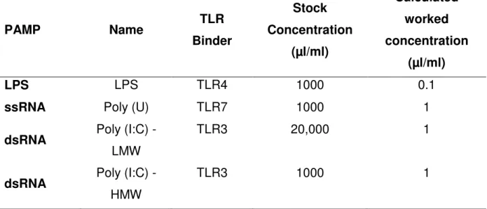
Table 1 Toll-like receptor binder, stock and empirically concentrations of LPS, ssRNA, Poly I:C (LMW) and Poly I:C (HMW) used in bovine endometrial stromal and epithelial cells.
59
Chapter 3
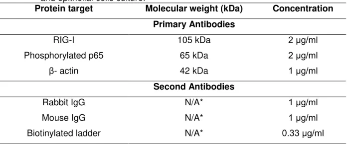
Table 1 RIG-I, phosphorylated p65 and β-actin molecular weight, worked concentrations and second antibodies treated in bovine endometrial stromal and epithelial cells culture.
LIST OF FIGURES
Figure Page
Chapter 1
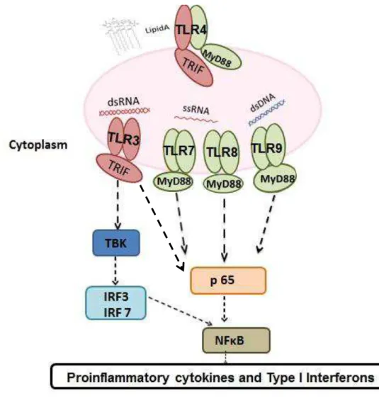
Figure 1 Toll-like receptors 3,4,7,8 and 9 pathways summarized: MyD88 independent pathway (TRIF pathway): when a TLR3 analog (dsRNA) activated this pathway, the adaptor protein TRIF is recruited to phosphorylate TBK and p65 to end up producing proinflamatory cytokines and IFNs mediated by activation of IRF3/7 and NF-κB respectively.
MyD88 dependent pathway: when TLR 7, 8 or 9 analogs (ssRNA, dsDNA) activate this pathway, the adaptor protein MyD88 is recruited to phosphorylate p65 resulting in production of proinflammatory cytokines and IFNs mediated by activation NF-κB.TLR4 analog (LIPD A) is able to
activate both pathways by recruiting both MyD88/TRIF adaptor proteins to result in the production of proinflammatory cytokines.
34
Chapter 2
Figure 1 (A, B)
ELISA IL-6 (pg/ml) results from bovine endometrial epithelial and stromal cells transfected (DOTAP positive) or not (DOTAP negative) treated with different PAMPs and a Control. The P values were calculated by a two way ANOVA and means were compared by Tukey test. Means followed by the same letter on each DOTAP Positive and Negative
were not different at 5%; ns Non-significant; * Signifcantly diferent at 5%* (n=3 for epithelial; n=5 for stromal).
Figure 2 (A,B)
ELISA IL-8 (pg/ml) results from bovine endometrial epithelial and stromal cells transfected (DOTAP positive) or not (DOTAP negative) treated with different PAMPs and a Control. The P values were calculated by a two way ANOVA and means were compared by Tukey test. Means followed by the same letter on each DOTAP Positive and Negative were not differen at 5%. ns Non-significant; *Significantly different when P<0.05; ** Significantly different when P< 0.01; (n=3 for epithelial; n=5 for stromal).
64
Figure 3 Cows endometrial epithelial cells cytokine production treated with the TLR3 ligand Poly I:C (LMW). Culture medium was collected following 0, 2, 6, 12, 24, 36, 48 and 72 hours of Poly I:C (LMW) stimulation and analyzed for IL-6 by ELISA. Cells were treated with Poly I:C (LMW) at a final concentration of 1µL/ml. The P values were calculated by a two way ANOVA and means were compared by Tukey test. (n=3 cows)
65
Figure 4 Cows endometrial stromal cells cytokine production treated with the TLR3 ligand Poly I:C (LMW). Culture medium was collected following 0, 2, 6, 12, 24, 36, 48 and 72 hours of Poly I:C (LMW) stimulation and analyzed for IL-6 by ELISA. Cells
were treated with Poly I:C (LMW) at a final concentration of 1µL/ml. The P values were calculated by a two way ANOVA and means were compared by Tukey test. Significantly different from control: *P< 0.05; ***P< 0.001. (n=4 cows)
Figure 5 Cows endometrial epithelial cells cytokine production treated with the TLR3 ligand Poly I:C (LMW). Culture medium was collected following 0, 2, 6, 12, 24, 36, 48 and 72 hours of Poly I:C (LMW) stimulation and analyzed for IL-8 by ELISA. Cells were treated with Poly I:C (LMW) at a final concentration of 1µL/ml. The P values were calculated by a two way ANOVA and means were compared by Tukey test. (n= 3 cows)
67
Figure 6 Cows endometrial stromal cells cytokine production treated with the TLR3 ligand Poly I:C (LMW). Culture medium was collected following 0, 2, 6, 12, 24, 36, 48 and 72 hours of Poly I:C (LMW) stimulation and analyzed for IL-8 by ELISA. Cells were treated with Poly I:C (LMW) at a final concentration of 1µL/ml. The P values were calculated by a two way ANOVA and means were compared by Tukey test. Significantly different from control: *P< 0.05; **P< 0.01; ***P< 0.001; (n=5 cows)
68
Chapter 3
Figure 1 Stimulation of cultured bovine endometrial stromal cells with Poly (I:C) induced by RIG-I at 12, 24, 48
and 72 hours of treatment and by a Control group (no Poly (I:C)). Proteins levels were compared with
the corresponding β-actin levels of each cell performed by a Western Blot test. Cells were treated with Poly (I:C) at a final concentration of 1µL/ml. Statistical significances were calculated using a two-way ANOVA with Tukey test. *P <0.05; **P <0.01.
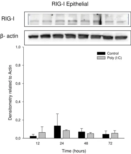
Figure 2 Stimulation of cultured bovine endometrial epithelial cells with Poly (I:C) induced by RIG-I at 12, 24, 48 and 72 hours of treatment and by a Control group (no Poly (I:C)). Proteins levels were
compared with the corresponding β-actin levels of each cell performed by a Western Blot test. Cells were treated with Poly (I:C) at a final concentration of 1µL/ml. Statistical significances were calculated using a two-way ANOVA with Tukey test. *P <0.05; **P <0.01.
89
Figure 3 Stimulation of cultured bovine endometrial stromal cells with Poly (I:C) induced by p65 (NF-κB) at 12,
24, 48 and 72 hours of treatment and by a Control group (no Poly (I:C)). Proteins levels were compared with the corresponding β-actin levels of each cell performed by a Western Blot test. Cells were treated with Poly (I:C) at a final concentration of 1µL/ml. Statistical significances were calculated using a two-way ANOVA with Tukey test. *P <0.05; **P <0.01.
Figure 4 Stimulation of cultured bovine endometrial epithelial cells with Poly (I:C) induced by p65
(NF-κB) at 12, 24, 48 and 72 hours of treatment and by
a Control group (no Poly (I:C)). Proteins levels
were compared with the corresponding β-actin levels of each cell performed by a Western Blot test. Cells were treated with Poly (I:C) at a final concentration of 1µL/ml. Statistical significances were calculated using a two-way ANOVA with Tukey test. *P <0.05; **P <0.01.
1.Introduction
Reproductive and immune systems might seem to be functionally separated but a complex interlinks exists. This way, reproductive system encounters various challenges including sexually transmitted diseases, systemic infections that affect reproductive organs and chronic inflammatory conditions, which can be overcome with the help of the immune system (KANNAKIA; SHANMUGAM; VERMA, 2011).
The immune system gives protection against a variety of pathogens and is based on a limited repertoire of germline-encoded receptors called pattern recognition receptors (PRRs). This, receptors have the ability to recognize conserved microbial components, known as pathogen-associated molecular patterns (PAMPs) (ALBIGER et al., 2007).
Several classes of PRRs have been implicated in innate immune defense. These include Toll-like receptors (TLRs) (TAKAEDA; AKIRA, 2005), lectin receptors (HUYSAMEN; BROWN, 2009), retinoic acid inducible gene I (RIG-I) (YONEYAMA; FUJITA, 2007), NOD-like receptors (NLRs) (MARTINON; MAYOR; TSCHOPP, 2006) and cytosolic deoxyribonucleic acid (DNA) sensors (TAKAOKA; TANIGUCHI, 2008; HORNUNG et al., 2009).
According to Kawai and Akira (2010), the science community has given an increasing attention to the TLRs. Studies had focused on the post-transcriptional modifications in molecules regulation that are involved in this receptors signaling and characterization of target genes. As well, additional information on the organization of these genes in different species could help to understand the evolution of these types of PRRs (McGUIRE et al., 2006).
In uterine inflammation, an effort needs to be made to understand the risk factors for biological mechanisms and how the uterus is able to detect infection, respond to microorganisms and how infection modulates normal function (SHELDON et al., 2008). In this context, the endometrium defense against infections seems to rely heavily on innate immunity, which when activated leads to an inflammatory response by producing cytokines (WIRA et al., 2005; MOR; CARDENAS, 2010).
The recognizing of bacteria and viruses in the endometrium is dependent on innate immunity. Whereas much is known about bacteria and their recognition by TLRs on endometrial cells, little is known about how viruses are detected. Based on that, the main hypothesis of this study was that bovine endometrial (epithelial/stromal) cells sense and respond to viral PAMPs via TLRs, and cytosolic stimulus induced by RIG-I at different times of stimulation.
In general, the objective of this study was to determine if bovine endometrial cells replied to PAMPs virus analogs by producing proinflammatory cytokines after TLR activation in the cell endosome and after RIG-I activation in the cell cytoplasm.
2. Immune uterine defense system
The immune system can be divided in two; namely innate and adaptive. Although the adaptive immune is more specific and robust, activation of antigen specific effective response stays behind compared to the innate immune (KANNAKIA; SHANMUGAM; VERMA, 2011). While sometimes it is considered that
innate immunity is “primitive” or “crude” compared to the adaptive, the opposite is
true, as in fact, the innate has been refined for a longer period of time than the adaptive, and is more efficient in almost every way (BEUTLER, et al., 2004).
expansion of a random repertoire of antigen receptors on lymphocytes (ALBIGER et al., 2007).
Innate immunity in the genital tract is highly dependent on the expression of PRRs that will detect PAMPs. These PRRs, such as TLRs, are highly conserved across phyla and are able to detect a range of different PAMPs associated with fungi, viruses and bacteria (BEUTLER, 2004; AKIRA et al., 2006).
A rapid progress was made in understanding innate immune recognition of microbial components and its critical role in host defense against infection (KAWAI; AKIRA, 2010). Although this recognition plays a major role in uterine defense mechanisms, how it occurs is not fully understood (ESPOSITO et al., 2014).
When studying uterine immune defense, some anatomical barriers work against invading pathogens, as the vulva, vagina and cervix. Other physical barriers in the genital tract include the stratified squamous epithelium of the vagina, the columnar epithelium of the endometrium, the basement membrane of ovarian follicles, and the zona pellucida of the oocyte (SHELDON et al., 2014).
According to Yunhea et al. (2013), the uterus is able to block and eliminate invading pathogens via its mechanical protective barrier and immune functions, thereby maintaining its normal physiological activities. Still, damage of these two defense systems (immune and adaptive) is likely to result in uterine inflammation and lead to disease, infertility or even death.
During cows postpartum period, an elevated expression of genes encoding TLRs, like Toll- Like Receptor 4 (TLR4), inflammatory mediators as Nuclear Factor Kappa b (NF-ΚB), Interleukin 8 (IL-8), Interleukin 1A (IL-1A), Interleukin 6 (IL-6), Interleukin 12A (IL-12A) and effector molecules, such as acute phase proteins (APP) and activated proteins kinase (AMPs), are characteristic of local uterine innate immune response (CHAPWANYA et al., 2009).
3. Cellular response against invading pathogens
Some responses against invading pathogens are observed specially at the cellular level. Uterine leukocytes and polymorphonuclear (PMN) cells are responsible for phagocytizing and cleaning contamination (GILBERT et al., 2007). When innate immune cells, such as macrophages and dendritic cells are activated to recognize different pathogens they initiate an entire defense (CARGILL; WOMACK, 2007).
Macrophages are the major population of tissue-resident mononuclear phagocytes and the predominant targets for infection by intracellular pathogens including mycobacteria. They play a dual role in anti-mycobacterial host defense that, currently, is not very clear. Also, it contributes to cell-mediated immunity and bacterial elimination and, is able to provide an essential niche for intracellular survival and escape from host defense mechanisms (VERRECK et al., 2004).
Dendritic cells are closely related to macrophages, but they are more potent antigen-presenting cells (PLAKS et al., 2008). As related by Schulke et al. (2008), this type of cells need to pass by a maturation process, because of that, they require migration to secondary lymphoid organs. During an infection, microbial factors will trigger dendritic cells maturation, with amplification of the response as a result of the subsequent release of endogenous activators (SKOBERNE; BEIGNON; BHARDWAJ, 2004).
To recognize pathogens, dendritic cells have some molecular sensors and antigen-processing machinery (COLLIN; MCGOVERN; HANIFFA, 2013). Also, they can control an additional checkpoint in the efferent phase of the immunity response (BENNETT; CHAKRAVERTY, 2012).
The uterine endometrium is a mucosa comprising of a layer of single columnar epithelial cells overlying a stromal that contains blood vessels and immune cells as well as endometrial stromal cells (DAVIES et al., 2008). Furthermore, the endometrial epithelial and stromal cells appear to have an immunological responsiveness as they express PRRs for the detection of microbes and also to produce a classical inflammatory response to bacteria and their PAMPs (HERATH et al., 2006).
According to Sheldon et al. (2008), in the uterine lumen the first line of defense against microbes are the epithelial cells. Considering that stromal are much more abundant than epithelial cells in the endometrium and closer in proximity to the vasculature and mononuclear cells, this type of cells may have equal importance in the immune response during an inflammatory process (CRONIN et al., 2011).
4. Toll-like receptors in immune responses
4.1. Definition of Toll-like receptors
Toll-like receptors are defined as membrane-bound proteins usually present at the cell surface, but they can also be membrane bound internally such as in endosomes (MEYLAN; TSCHOOP, 2006). In response to virus infections, viral components such as RNA and DNA are recognized by TLRs and (RIG-I)-like helicases (RLHs), and cells are activated to produce type I IFNs and proinflammatory cytokines (HONDA; TAKAOK; TANIGUCHI, 2006).
The founding member of the TLRs was a Toll in Drosophila melanogaster, which was first found to instruct dorsal ventral patterning in early embryos, and regulate anti-fungal innate immunity in adult flies. Sequence homology search in mammalian genomes has subsequently identified at least 11 members of TLRs (DU, et al., 2000).
regulating TLR gene expression in response to inflammatory mediators remain not fully characterized (MOGENSEN, 2009).
4.2. Toll-like receptors structure
As a conserved innate immune receptors, the Toll-like family belong to a type I transmembrane proteins with an amino terminus (KANNAKIA; SHANMUGAM; VERMA, 2011). In addition to a single transmembrane domain, TLRs are characterized by an extracellular leucine rich repeat (LRR) and a cytoplasmic toll/interleukin-1 receptor (TIR) domain (WERLING; JUNGI, 2003).
The LRR domain in TLRs occurs in proteins ranging from viruses to eukaryotes and appears to provide a structural framework for the formation of protein–protein interactions (CARGILL; WOMACK, 2007). Also, the central part of the LRRs possesses more irregular or longer motifs and varies among different TLRs, implying the functional importance of the central part in ligand recognition (MATSUSHIMA et al., 2007).
The TIR domain usually is involved in downstream signal transduction (KANNAKIA; SHANMUGAM; VERMA, 2011). When this domain is activated, it recruits cytoplasmic adaptor proteins like myeloid differentiation primary response gene 88 (MyD88) and TOLLIP, which in turn associate with various kinases to set off signaling cascades (CARGILL; WOMACK, 2007) (More details were exposed in section 4.5).
Upon ligand recognition, when the protein MyD88 is recruited an association with the cytoplasmic domain of the TLRs via interaction between the TIR domains will be constructed (ALBIGER et al., 2007).
It’s important to notice that the over activation of TLRs may break out immune
4.3. Most important Toll-like receptor ligand: PAMPs
The molecules PAMPs are characterized by being invariant among entire classes of pathogens, essential for their survival and distinguishable from being “self”
(JANEWAY, 1989).
These PAMPs are evolutionarily conserved in pathogens and usually critical for infection and pathogenesis Toll-like receptors recognize PAMPs such as peptidoglycan, lipopolysaccharides (LPS), nucleic acids and combinations, in an efficient non-self-reactive manner allowing initiation of a complex signaling cascade to activate various transcription factors and pro-inflammatory cytokines (KANNAKIA; SHANMUGAM; VERMA, 2011).
According to Janeway (1989), this recognition of microbial pathogens is an essential element for the initiation of innate immune responses such as inflammation and is mediated by PRRs that recognize molecular structures that are broadly shared by PAMPs. Different PRRs may recognize the same PAMP; hence TLRs in concert with other PRRs orchestrate both pathogen-specific and cell type-specific host immune responses to fight infections (KAWAI; AKIRA, 2011).
Binding of PAMPs to PRRs activates signal transduction pathways by mitogen-activated protein kinase (MAPK) and NF-κB transcription factors, leading to secretion of prostaglandins, cytokines and chemokines (GHOSH et al., 1998; LI; VERMA, 2002; AKIRA; TAKEDA, 2004).
4.4. Toll-like receptors types
endoplasmic reticulum, endosomes, lysosomes and endolysosomes, where they recognize microbial nucleic acids (KAWAI; AKIRA, 2010).
Signaling of TLR3 and TLR4 has been most extensively characterized, whereas much less is known about how TLR7, TLR8, and TLR9 act in inflammatory responses. Understanding the molecular mechanisms regulating the induction of type I IFNs by these TLRs is likely to reveal novel therapeutic and immune-modulation strategies in order to eliminate acute and chronic viral infections (SCHOENEMEYER et al., 2005).
The TLR3 was originally identified as recognized a synthetic analog of double-stranded RNA (dsRNA) called polyinosinic-polycytidylic acid (Poly(I:C)), it mimics viral infection and induces antiviral immune responses by promoting the production of both type I interferon and inflammatory cytokines, which suggests that TLR3 has an essential role in preventing virus infection (KAWAI; AKIRA, 2010).
The TLR4 is the most extensively studied PRR and it recognizes a variety of ligands (host heat shock proteins, fibrinogen and proteins from virus, pneumolysin, a cytotoxin from Streptococcus pneumoniae) but is mostly known to recognize LPS. Still, this is proved by the fact that TLR4 knock-out mice were shown to be unresponsive to LPS (HOSHINO et al., 1999).
The fact is that LPS is recognized by TLR4 but this TLR alone is not sufficient for signaling (MIYAKE, 2006). Some accessory proteins are required for this recognition. Because of this, LPS binds first LPS-binding protein (LBP), which is an acute phase protein that circulates in the bloodstream and binds to glycosylphosphatidylinositol (GPI) linked co-receptor CD14, which is expressed on the cell surface (ALBIGER et al., 2007).
4.5. Toll-like receptor signaling pathways
An improved understanding of the inflammatory pathways at the molecular level which play an important role in normal immune function, metabolism and reproduction, may improve the ability to predict and prevent cows disorders (ESPOSITO et al., 2014).
According to Kawai and Akira (2006), the process of TLR ligand activation induces two signaling pathways, one is dependent and the other is MyD88-independent, both ending up with the production of proinflammatory cytokines and type I interferons.
4.5.1. MyD88-dependent signaling
All TLRs, except TLR3, signal through the MyD88 dependent pathway still, only the TLR4 is able to activate MyD88 dependent and independent pathways (ADACHI et al., 1998). Basically, the MyD88 dependent pathway requires a signal transduction intermediates specially by protein kinases that transforms growth factor-activated kinase, to final activate NF-κB in order to induce production of pro-inflammatory cytokines (MEDZHITOV; KAGAN, 2006).
A practical example was showed in the study of Yunhea et al. (2013), when TLR4 recognized LPS and recruited an adaptor protein MyD88 to initiate the MyD88-dependent pathway which had included a series of signal transduction intermediates and activated the NF-κB that started the production of proinflammatory cytokines such as tumor necrosis factor (TNF), Interleukin 1 1), IL-6 and Interleukin 12 (IL-12).
In case of an over activation of TLR, the protein TOLLIP will be recruited to block the MyD88 dependent signaling pathway by preventing the phosphorylation of associated protein kinases (BULUT et al., 2001; ZHANG; GHOSH, 2002).
4.5.2. MyD88-independent signaling
(IFN-α) and IFN inducible genes. Whilst TLR3-mediated signaling only requires the adaptor molecule TIR-domain-containing adapter-inducing interferon-β (TRIF), while on the other hand, TLR4-mediated signaling needs in addition to TRIF another adaptor protein known as translocation associated membrane protein (TRAM), both will activate NF-κB to induce expression of proinflammatory cytokines (CUSSON-HERMANCE et al., 2005).
Figure 1. Toll-like receptors 3,4,7,8 and 9 pathways summarized: MyD88 independent pathway (TRIF pathway): when a TLR3 analog (dsRNA) activated this pathway, the adaptor protein TRIF is recruited to phosphorylate TBK and p65 to end up producing proinflamatory cytokines and IFNs mediated by activation of IRF3/7 and NF-κB
respectively. MyD88 dependent pathway: when TLR 7, 8 or 9 analogs (ssRNA, dsDNA) activate this pathway, the adaptor protein MyD88 is recruited to phosphorylate p65 resulting in production of proinflammatory cytokines and IFNs mediated by activation NF-κB.TLR4 analog (LIPD A) is able to activate both pathways by recruiting both MyD88/TRIF adaptor proteins to result in the production of proinflammatory cytokines.
4.6. Expression of Toll-like receptors in bovine endometrium
different species of farm and companion (KANNAKIA; SHANMUGAM; VERMA, 2011).
Different authors have been studied the expression profile of TLRs in selected tissues and cell subsets of cattle and buffalo (MENZIES; INGHAM, 2006; WERLING et al., 2006; VAHANAN et al., 2008); The full-length coding sequence of TLR3 has been well characterized in Water Buffalo (Bubalis bubalis) and Nilgai (Boselaphus
tragocamelus) showing a 98% of homology to that as in cattle (DHARA et al., 2007). Ten different types (1-10) of TLRs have been identified and physically mapped in cattle having 95% nucleotide sequence identical with human (McGUIRE et al., 2006; MENZIES; INGHAM, 2006).
As related by Herath et al. (2006) and Davies et al. (2008), studies proved that the endometrium of cows express TLRs 1–10, whereas purified populations of endometrial epithelial cells express TLRs 1–7 and 9, and stromal cells express TLRs 1–4, 6, 7, 9 and 10. Apart from that, TLR4 in cows appear to be functional in endometrial epithelial cells as they secreted prostaglandin E2 in response to bacterial PAMPs (KANNAKIA; SHANMUGAM; VERMA, 2011).
5. RIG-I signaling in immune system
5.1. Definition and function of Retinoic Acid Inducible Gene I, RIG-I
In the cytosol, studies have identified NLRs and RIG-I as two additional families of innate immunity receptors that recognize PAMPs (INOHARA et al., 2005; MEYLAN; TSCHOPP, 2006).
The RIG-I with its helicase domain has been demonstrated to be an essential regulator for dsRNA signaling that results in the activation of the transcriptional factors NF-κB as well as IRF-3 (SUMPTER et al., 2005; YONEYAMA et al.; 2004). Moreover, the relationship between RIG-I and TLRs in the recognition of viruses need to be more expolored (KATO el al., 2005).
The RLRs consists of three helicases known as: RIG-I, MDA5 and by a protein called laboratory of genetics and physiology 2 (LGP2) (YONEYAMA, et al., 2004; ROTHENFUSSER et al., 2005).
A RNA helicase domain that recognizes viral dsRNA is present in the RLRs family. RIG-I and LGP2 contain a C-terminal regulatory domain that recognizes
ssRNA containing 5′-triphosphate, which distinguishes foreign RNAs from self-RNAs
that normally contain 5′-modification. Still, RIG-I and MDA5 contain an N-terminal and
a caspase activation and recruitment domain (CARD), which interact with the CARD domain of mitochondrial antiviral-signaling protein (MAVS), also known as virus-induced signaling adapter (VISA), which is located out of the mitochondrial membrane (KAWAI et al., 2005; MEYLAN et al., 2005; SETH et al., 2005; XU et al., 2005).
5.3. RLRs signaling
Two CARDs and a protein with the domain DEAD or DEAH box helicases (DExD/H-box) belong to RIG-I and MDA5, the helicase domains of RIG-I and MDA5 recognizes viral RNAs, and their CARDs are responsible for signaling through interaction with a CARD-containing adaptor, MAVS (KAWAI et al., 2005; KUMAR et al., 2006).
Both RIG-I and MDA5 engage the mitochondrial adapter protein MAVS, this protein subsequently triggers downstream signaling and activation of the inhibitor of nuclear factor kappa-B kinase subunit alpha/beta (IKKα/β)- NF-κB pathway or the TBK1-IRF3 pathway and transcriptional regulation of proinflammatory cytokines and type I IFN genes, respectively (ABLASSER et al., 2009).
On the other hand, LGP2 does not possess a CARD, but only a DExD/H-box helicase domain, and has been reported to act as a negative regulator factor (ROTHENFUSSER et al; 2005; YONEYAMA et al.; 2005) ), specially at the RIG- I mediated pathway (SAITO et al.; 2007; VENKATARAMAN et al., 2007).
According to Ishii et al. (2006) and Sun et al. (2006), some genetic studies have shown that cytosolic DNA can induce IFN production in mice dendritic cells lacking RIG-I or MAVS, suggesting that the DNA signaling pathway is distinct from the RIG-I pathway.
The RNA polymerase III (Pol III)-RIG-I pathway appears to be functional in both human and mouse cells, but in this last species seems to be redundant with additional DNA sensing mechanisms (ABLASSER et al., 2009).
Studies from Chiu, McMillan, Chen (2009) demonstrated that RIG-I binds to RNA but not to DNA; so the question of how DNA might activate the RIG-I pathway need to be raised.This RNA species contains 5′-triphosphate and forms a dsRNA. The conversion of DNA to RNA can be recapitulated in vitro using cytosolic extracts. Due to that, Pol-III seems to be the enzyme responsible for transcribing the DNA template into an RNA ligand that activates RIG-I. These results suggest that Pol-III is a cytosolic DNA sensor that triggers type-I interferon production through the RIG-I pathway
6. Viral infection against immune response
Viruses are obligatory intracellular pathogens; therefore, their replication (and the pathogenic consequences of infection) depends critically on the ability to transmit their genomes from infected to non-infected host organisms and from infected to uninfected cells (MARSH; HELENIUS, 2006). They are also restricted in using metabolic and biosynthetic pathways of the cells that they infect. These pathways vary between cell types, lineage, and stage of differentiation and with the state of cell activation (DONOFRIO et al., 2007).
The innate immune system has evolved several distinct viral recognition systems that integrate complex networks of signaling pathways, which can lead to activate some pathway-specific transcription factors and the induction of immune response genes (SCHOENEMEYER et al., 2005).
functional consequences of the interaction, that usually are highly specific, and the presence of receptors determines in a large degree which cell types and species can be infected (MARSH; HELENIUS, 2006).
Studies of Seth, Sun and Chen (2006), certified that two basic events are required to trigger an effective anti-viral response: first, a detection of the invading virus by immune system receptors; and second an initiation of protein signaling cascades that regulate the synthesis of IFNs. In viral infections, the induction of IFN I have shown to be primarily due to the recognition of dsRNA that is a sign of replicating viruses(SUMPTER et al., 2005; YONEYAMA et al.; 2004).
Based on Hiscott et al. (2006) comments, the probing of the immune response for millions of years was caused by viruses, also, this investigation of this response is yielding important clues about which pathways must be compromised in order for virus infection perpetuate. Utilization of this knowledge will be a cornerstone in the understanding of molecular aspects of viral pathogenesis and the improvement of strategies for the development of vaccines and antiviral agents.
7. Proinflamatory cytokines (focus on IL-6 and IL-8)
In general, cytokines are considered to have different functions, they are primarily involved in host responses to disease or infection, and their participation in homeostatic mechanisms has been less explored (DINARELLO, 2000). Moreover, cells have mechanisms for regulating both signaling pathways to avoid injurious effects due to overproduction of inflammatory cytokines during microbial infection (KUMAR; KAWAI; AKIRA, 2011).
Binding to specific receptors on the neutrophil surface, cytokines can influence different cell functions, for example the ability to localize at the site of inflammation, phagocytic activity, production of oxygen metabolites, and the release of lysosomal enzymes (SEMMANI; KABBUR, JAIN; 1993).
Another important proinflammatory cytokine group is composed by the Interleukins (ILs). They are secreted proteins that bind to their specific receptors and play a role in the communication among leukocytes. Also, investigations of the mechanisms of immune and inflammatory cell functions have identified a growing list of ILs and interactions among different cell types that contribute to their effector and suppressive functions (AKDIS et al., 2011).
According to Fischer et al. (2010), in cows, it is important to study and understand the defense made by some proinflammatory cytokines specially 6, IL-8, and TNF, as they accelerate PMN infiltration into the bovine endometrium following infection.
A balance between the effects of proinflammatory and anti-inflammatory cytokines is thought to determine the outcome of disease, whether in the short or long term. In fact, some studies suggested that the susceptibility to disease is genetically determined by the balance or expression of either proinflammatory or anti-inflammatory cytokines (DINARELLO, 2000).
As one of the most important members of the cytokines, IL-6 has a helix bundle structure consisting of four long α-helices. Also, it is a multifunctional, pleiotropic cytokine involved in regulation of immune responses, acute-phase responses, hematopoiesis, and inflammation (AKDIS et al., 2011).
A more detailed study conducted by Hurst et al. (2001), revealed that in innate immunity, IL-6 directs leukocyte trafficking and activation and induces production of acute-phase proteins by hepatocytes cells.
Chemokine is a group of cytokines, and it’s well known that chemokine IL-8 is one of the most studied cytokines during immune inflammatory response. Still, the major effector functions of IL-8 are activation and recruitment of neutrophils to the site of infection or injury (MATSUSHIMA et al., 1988).
The chemokine IL-8, is produced by a variety of cells, such as monocytes and macrophages, neutrophils, lymphocytes, and endothelial and epithelial cells after stimulation with IL-1A, interleukin 1B (IL-1B), interleukin 17 (IL-17), tumor necrosis factor alpha (TNF-α), or TLRs (COELHO et al., 2005).The presence of recombinant IL-8 has been shown to increase the influx of PMN leucocytes into the bovine uterus (ZERBE et al., 2003).
In a more specific study developed by Yoshimura et al. (1987), IL-8 was identified as a neutrophil-specific chemotactic factor and later classified as a member of the chemokine family. Recent studies have shown that IL-8 promoter contains binding sites for the transcription factor NF-κB andactivator protein 1(AP-1) (AKDIS et al., 2011). Others studies developed by Turner et al. (2012), found that endometrial epithelial and stromal cells mounted cellular responses to bacterial lipopeptides typical of innate immunity with secreting both IL-6 and IL-8.
8. What to expect in the next studies
As a result of the researches from Kannakia, Shanmugam and Verma (2011), they assumed that one particular area to focus on the PRRs studies should be the analysis of potential TLR agonists for development of new vaccine/adjuvant to prevent reproductive infection. Moreover, polymorphisms in TLR genes and their association with diseases of economic importance in cattle such as mastitis have been established. In the future these facts could be used as molecular markers in order to select animals in the development of immunogenetically superior stocks.
by which primary pathogens can modulate or suppress innate immune responses by interfering at different levels with TLR signaling, and how opportunistic pathogens may take advantage of, for example, host responses to viral infections to gain access to deep tissue from local sites should be the focus (ALBIGER et al., 2007).
It is clear to observe that the importance of PRRs signaling for both immune homeostasis and for defense mechanisms against pathogens has emerged in the past decade (KONDO; KAWAI; AKIRA, 2012). An advancement has been made not only in the understanding of the structure of TLRs but also in revealing the complexity of TLR-mediated signaling and in the identification of PAMPs derived from microbial pathogens such as mycobacteria, bacteria, viruses, fungi and parasites (KUMAR; KAWAI; AKIRA, 2011).
The virus infection models tested to date support roles for TLRs, rather than RLRs, in instructing the adaptive immune system. However, investigations are required, since these two PRR systems provide different contributions depending on the viruses involved and also may depend on the route of infection (TAKEUCHI; AKIRA, 2008).
Related to bovine immune function, future work should focus on determining which pathogen, bacteria or virus can cause endometritis, and understanding how the host response to infection is regulated in the endometrium. New knowledge of uterine diseases will provide a platform for new therapeutics and vaccines (CARNEIRO; CRONIN; SHELDON, 2015).
REFERENCES
ABLASSER, A.; BAUERNFEIND, G.; HARTMANN, G.; LATZ, E.; FITZGERALD, K.A.; HORNUNG, V. RIG-I dependent sensing of poly(dA-dT) via the induction of an RNA polymerase III transcribed RNA intermediate. Nature Immunology, v. 10, p. 1065-1072, 2009.
ADACHI, O.; KAWAI, T.; TAKEDA, K.; MATSUMOTO, M.; TSUTSUI, H.; SAKAGAMI, M.; NAKANISHI, K.; AKIRA, S. Targeted disruption of the MyD88 gene results in loss of IL-1- and IL-18-mediated function. Immunity, v. 9, p. 143–150, 1998.
AKDIS, M.; BURGLER, S.; CRAMERI, R.; EIWEGGER, T.; MD, FUJITA, H.;
GOMEZ, E.; KLUNKER, S.; MEYER, N.; O’MAHONY, L.; PALOMARES, O.;
RHYNER, C.; QUAKED, N.; SCHAFFARTZIK, A.; VAN DE VEEN, W.; ZELLER, S.; ZIMMERMANN, M.; AKDIS, A.C. Interleukins, from 1 to 37, and interferon-g: Receptors, functions, and roles in diseases. Journal of Allergy and Clinical Immunology, v. 127, p. 702-792, 2011.
AKIRA, S.; TAKEDA, K. Toll-like receptor signaling. Nature Reviews Immunology, v. 4, p. 499–511, 2004.
AKIRA, S.; UEMATSU, S.; TAKEUCHI, O. Pathogen recognition and innate immunity. Cell, v. 124, p. 783–801, 2006.
ALBIGER, B.; DAHLBERG, S.; HENRIQUES-NORMARK, B.; NORMARK, S. Role of the innate immune system in host defence against bacterial infections: focus on the Toll-like receptors. Journal of Internacional Medicine, v. 261, p. 511-610, 2007.
BENNETT, C. L.; CHAKRAVERTY, R. Dendritic cells in tissues: in situ stimulation of immunity and immunopathology. Trends in Immunology, v. 33, p. 7-13, 2012.
BEUTLER, B. Innate immunity: an overview. Molecular Immunology, v. 40, p. 845–
859, 2004.
BULUT, Y.; FAURE, E.; THOMAS, L.; EQUILS, O.; ARDITI, M. Cooperation of Toll-like receptor 2 and 6 for cellular activation by soluble tuberculosis factor and Borrelia burgdorferi outer surface protein A lipoprotein: role of Toll-interacting protein and IL-1 receptor signaling molecules in Toll-like receptor 2 signaling. Journal of Immunology, v. 167, p. 987–994, 2001.
CARGILL E. J.; WOMACK, J. E. Detection of polymorphisms in bovine toll-like receptors 3, 7, 8, and 9. Genomics, v. 89, p. 745–755, 2007.
CARNEIRO, L. C.; CRONIN, J. G.; SHELDON, I.M. Mechanism linking bacterial infections of the bovine endometrium to disease and infertility. Reproductive Biology (2015), http://dx.doi.org/10.1016/j.repbio.2015.12.002.
CHAPWANYA, A.; MEADE, K. G.; DOHERTY, M. L.; CALLANAN, J. J.; MEE, J. F.;
O’FARRELLY, C. Histopathological and molecular evaluation of Holstein-Friesian
cows postpartum: Toward an improved understanding of uterine innate immunity.
Theriogenology, v. 71, p. 1396–1407, 2009.
CHIU, Y.; MACMILLAN, J.B.; CHEN, Z.J. RNA Polymerase III Detects Cytosolic DNA and Induces Type-I Interferons Through the RIG-I Pathway. Cell, v. 138, p. 576–591, 2009.
COELHO, A. L.; HOGABOAM, C. M.; KUNKEL, S.L. Chemokines provide the sustained inflammatory bridge between innate and acquired immunity. Cytokine and Growth Factor Revies, v. 16, p. 553-560, 2005.
COLLIN, M.; MCGOVERN, N.; HANIFFA, M. Human dendritic cell subsets.
Immunology, v. 140, p. 22–30, 2013.
CRONIN, J. G.; TURNER, M. L.; GOETZE, L.; BRYANT, C. E.; SHELDON, I. M. Toll-like receptor 4 and MYD88-dependent signaling mechanisms of the innate immune system are essential for the response to lipopolysaccharide by epithelial and stromal cells of the bovine endometrium. Biology of Reproduction, v. 86, p. 1-9, 2011.
DAVIES, D.; MEADE, K. G.; HERATH, S.; ECKERSALL, P. D.; GONZALEZ, D.; WHITE, J. O.; CONLAN, R. S.; O’FARRELLY, C.; SHELDON, I. M. Toll-like receptor and antimicrobial peptide expression in the bovine endometrium. Reproductive Biology and Endocrinology, v. 6, p. 1-12, 2008.
DHARA, A.; SAINI, M.; DAS, D.K.; SWARUP, D.; SHARMA, B.; KUMAR, S.; GUPTA, P. K. Molecular characterization of coding sequences and analysis of Toll-like receptor 3 mRNA expression in water buffalo (Bubalus bubalis) and nilgai (Boselaphus tragocamelus). Immunogenetics , v. 59, p. 69–76, 2007.
DINARELLO, C. A. Proinflammatory cytokines. Chest, v. 118, p. 503-508, 2000.
DONOFRIO, G.; HERATH, S.; SARTORI, C.; CAVIRANI, S.; FLAMMINI, C. F.; SHELDON, I. M. Bovine herpesvirus 4 is tropic for bovine endometrial cells and modulates endocrine function. Reproduction, v. 134, p. 183-197, 2007.
DU, X.; POLTORAK, A.; WEI, Y.; BEUTLER, B Three novel mammalian toll-like receptors: gene structure, expression., and evolution. European Cytokine Network, v. 11, p. 362-371, 2000.
ESPOSITO, G.; IRONSA, P. C.; WEB. E. C.; CHAPWANYA, A. Interactions between negative energy balance, metabolic diseases, uterine health and immune response in transition dairy cows. Animal Reproduction Science, v. 144, p. 60– 71, 2014.
FISCHER, C.; DRILLICH, M.; ODAU, S.; HEUWIESER, W.; EINSPANIER, R.; GABLER, C. Selected pro-inflammatory factor transcripts in bovine endometrial epithelial cells are regulated during the oestrous cycle and elevated in case of subclinical or clinical endometritis. Reproduction, Fertility and Development, v. 22, p. 818–829, 2010.
GHOSH, S.; MAY, M. J.; KOPP, E. B. NF-kappa B and Rel proteins: evolutionarily conserved mediators of immune responses. Annual Reviews in Immunology, v. 16, p. 225–260, 1998.
GILBERT, R.; SANTOS, N.; GALVÃO, K.; BRITTIN, S.; ROMAN, H.The relationship between postpartum uterine bacterial infection (BI) and subclinical endometritis (SE).
HERATH, S.; DOBSON, H.; BRYANT, C. E.; SHELDON, I. M. Use of the cow as a large animal model of uterine infection and immunity. Journal of Reproductive Immunology, v. 69, p. 13-22, 2006.
HISCOTT, J.; NGUYEN, T. L.; ARGUELLO, M.; NAKHAEI, P.; PAZ, S. Manipulation of the nuclear factor-kappaB pathway and the innate immune response by viruses.
Oncogene, v. 25, p. 6844-6867, 2006.
HONDA, K.; TAKAOKA, A.; TANIGUCHI, T. Type I interferon [corrected] gene induction by the interferon regulatory factor family of transcription factors. Immunity, v. 25, p. 349-360, 2006.
HORNUNG, V.; ABLASSER, A.; CHARREL-DENNIS, M.; BAUERNFEIND, F.; HORVATH, G.; CAFFREY, D. R.; LATZ, E.; FITZGERALD, K. A. AIM2 recognizes cytosolic dsDNA and forms a caspase-1-activating inflammasome with ASC. Nature, v. 458, p, 514–518, 2009.
HOSHINO, K.; TAKEUCHI, O.; KAWAI, T.; SANJO, H.; OGAWA, T.; TAKEDA, Y.; TAKEDA, K.; AKIRA, S. Cutting edge: Toll-like receptor 4 (TLR4)-deficient mice are hyporesponsive to lipopolysaccharide: evidence for TLR4 as the LPS gene product.
Journal of Immunology, v. 162, p. 3749–3752, 1999.
HURST, S. M.; WILKINSON, T. S.; MCLOUGHLIN, R.M.; JONES, S.; HORIUCHI, S.; YAMAMOTO, N.; ROSE-JOHN, S.; FULLER, G.M.; TOPLEY, N.; JONES, S.A. Il-6 and its soluble receptor orchestrate a temporal switch in the pattern of leukocyte recruitment seen during acute inflammation. Immunity v. 14, p. 705-714, 2001.
HUYSAMEN, C.; BROWN, G. D. The fungal pattern recognition receptor, Dectin-1, and the associated cluster of C-type lectin-like receptors. FEMS Microbiol Lett, v. 290, p. 121–128, 2009.
INOHARA, N.; CHAMAILLARD, M.; McDONALD, C.; NUNEZ, G. NOD-LRR proteins: role in host-microbial interactions and inflammatory disease. Annual Review of Biochemistry, v. 74, p. 355–383, 2005.
ISHIKAWA, Y.; NAKADA, K.; HAGIWARA, K.; KIRISAWA, R.; IWAI, H.; MORIYOSHI, M.; SAWAKUKAI, Y. Changes in interleukin-6 concentration in peripheral blood of pre- and partum dairy cattle and its relationship to post-partum reproductive disease. The Journal of Veterinary Medical Science, v. 66, p. 1403–1408, 2004.
IWASAKI, A.; MEDZHITOV, R. Toll-like receptor control of the adaptive immune responses. Nature Immunology v.5, p.987–995, 2004.
JANEWAY, C.A. Approaching the asymptote? Evolution and revolution in immunology. Cold Spring Harbor Symposia on Quantitative Biology, v. 54, p.1–
13, 1989.
JANEWAY, C. A.; MEDZHITOV, R. Innate immune recognition. Annual Review of mmunololy, v. 20, p. 197–216, 2002.
KANNAKIA, T. R.; SHANMUGAM, M.; VERMA, P. C. Toll-like receptors and their role in animal reproduction. Animal Reproduction Science, v. 125, p.1– 12, 2011.
KATO, H.; SATO, S.; YONEYAMA, M.; YAMAMOTO,M.; UEMATSU, S.; MATSUI,K.; TSUJIMURA, T.; TAKEDA, K.; FUJITA, T.; TAKEUCHI, O.; AKIRA, S. Cell Type-Specific Involvement of RIG-I in Antiviral Response. Immunity, v.23, p.19–28, 2005.
KATO, H.; OSAMU, T.; MIKAMO-SATOH, E.; HIRAI,R.; KAWAI, T.; MATSUSHITA, K.; HIIRAGI, A.; DERMODY, T. S.; FUJITA, T.; AKIRA, S. Length-dependent recognition of double stranded ribonucleic acids by retinoic acid – inducible gene-I and melanoma differentiation – associated gene 5. Journal of Experimental Medicine, v. 205, p. 1601-1610, 2008.
KAWAI, T.; AKIRA, S. TLR signaling. Cell Death and Differentiation, v. 13, p.816–
825, 2006.
KAWAI, T.; AKIRA, S. The role of pattern-recognition receptors in innate immunity: update on Toll-like receptors. Nature Immunology, v. 11, p. 373-384, 2010.
KAWAI, T.; TAKAHASHI, K.; SATO, S.; COBAN, C.; KUMAR, H.; KATO, H.; ISHII, K. J.; TAKEUCHI, O.; AKIRA, S. IPS-1, an adaptor triggering RIG-I- and Mda5-mediated type I interferon induction. Nature Immunology, v.6, p. 981– 988, 2005.
KONDO, T.; KAWAI, T.; AKIRA, S. Dissecting negative regulation of Toll-like receptor signalling. Trends in Immunology, v. 33, p. 449-458, 2012.
KUMAR, H.; KAWAI, T.; AKIRA, S. Pathogen recognition by the innate immune system. International Reviews of Immunology, v. 30, p. 16-34, 2011.
KUMAR, H.; KAWAI, T.; KATO, H.; SATO, S.; TAKAHASHI, K.; COBAN, C.; YAMAMOTO, M.; UEMATSU, S.; ISHII, K. J.; TAKEUCHI, O.; AKIRA, S. Essential role of IPS-1 in innate immune responses against RNA viruses. Journal of Experimental Medicine, v. 203, p. 1795 – 1803, 2006 .
LI, Q.; VERMA, I. M. NF-kappa B regulation in the immune system. Nature Reviews
Immunology, v. 2, p. 725–734, 2002.
LIU, Y. J.. IPC: professional type 1 interferon-producing cells and plasmacytoid dendritic cell precursors. Annual Reviews of Immunology, v. 23, p. 275 – 306, 2005.
MARSH, M.; HELENIUS, A. Virus Entry: Open Sesame. Cell, v. 124, p. 729-740, 2006.
MARTINON, F.; MAYOR, A.; TSCHOPP, J. The inflammasomes: guardians of the body. Annual Review of Immunology, v. 27, p. 229–265, 2006.
MATSUSHIMA, K.; MORISHITA, K.; YOSHIMURA, T.; LAVU, S.; KOBAYASHI, Y.; LEW, W.; PAELLA, E.; KUNG, H. F.; LEORNARD, E. J.; OPPENHEIM, J. J. Molecular cloning of a human monocyte-derived neutrophil chemotactic factor (MDNCF) and the induction of MDNCF mRNA by interleukin 1 and tumor necrosis factor. The Journal of Experimental Medicine, v. 167, p. 1883-1893, 1988.
McGUIRE, K.; JONES, M.; WERLING, D.; WILLIAMS, J.L.; GLASS, E. J.; JANN, O. Radiation hybrid mapping of all 10 characterized bovine Toll-like receptors. Animal Genetics, v. 37, p. 47–50, 2006.
MEDZHITOV, R.; KAGAN, J. C. Phosphoinositide-mediated adaptor recruitment controls toll-like receptor signaling. Cell, v. 125, p. 943–955, 2006.
MENZIES, M.; INGHAM, A. Identification and expression of Toll-like receptors 1-10 in selected bovine and ovine tissues. Veterinary Immunology and Immunopathology, v. 109, p. 23–30, 2006.
MEYLAN, E.; CURRAN, J.; HOFMANN, K.; MORADPOUR, D.; BINDER, M.; BARTENSCHLAGER, R.; TSCHOPP, J. Cardif is an adaptor protein in the RIG-I antiviral pathway and is targeted by hepatitis C virus. Nature, v. 437, p.1167–1172, 2005.
MEYLAN, E.; TSCHOOP, J. Toll-like receptors and RNA helicases: two parallel ways to trigger antiviral responses, Molecular Cell, v. 22, p.561–569, 2006.
MIYAKE K. Roles for accessory molecules in microbial recognition by Toll-like receptors. Journal of Endotoxin Research, v.12, p.195– 204, 2006.
MOGENSEN, T.H. Pathogen Recognition and Inflammatory Signaling in Innate Immune Defenses. Clinical Microbiology Reviews, v.22, p.240-273, 2009.
MOR, G.; CARDENAS, I. The immune system in pregnancy: a unique complexity.
American Journal of Reproductive Immunology, v. 63, p. 425–433, 2010.
MUZIO, M.; BOSISIO, D.; POLENTARUTTI, N.; D’AMICO, G.; STOPPACCIARO, A.;
MANCINELLI, R.; VAN’T VEER, C.; PENTON-ROI, G.; RUCO, L. P.; ALLAVENA, P.;
MANTOVANI, A. Differential expression and regulation of Toll-like receptors (TLR) in human leukocytes: selective expression of TLR3 in dendritic cells. Journal of Immunology, v. 164, p. 5998–6004, 2000.
PLAKS, V.; BIRNBERG, T.; BERKUTZKI, T.; SELA, S.; BENYASHAR, A.; KALCHENKO, V.; MOR, G.; KESHET, E.; DEKEL, N.; NEEMAN, M.; JUNG, S. Uterine DCs are crucial for decidua formation during embryo implantation in mice.
ROTHENFUSSER, S.; GOUTAGNY, N.; DIPERNA, G.; GONG, M.; MONKS, B.G.; SCHOENEMEYER, A.; YAMAMOTO, M.; AKIRA, S.; FITZGERALD, K.A. The RNA helicase Lgp2 inhibits TLR-independent sensing of viral replication by retinoic acid-inducible gene-I. Journal of Immunology, v. 175, p. 5260 – 5268 , 2005 .
SAITO, T.; HIRAI, R.; LOO, Y. M.; OWEN, D.; JOHNSON, C. L.; SINHA, S. C.; AKIRA, S.; FUJITA, T.; GALE, M. Regulation of innate antiviral defenses through a shared repressor domain in RIG-I and LGP2. Proceedings of the National Academy of Science, v.104, p.582 – 587, 2007.
SATO, S.; SUGIYAMA, M.; YAMAMOTO, M.; WATANABE, Y.; KAWAI, T.; TAKAEDA, K.; AKIRA, S. Toll/IL-1 receptor domain-containing adaptor inducing IFN-beta (TRIF) associates with TNF receptor-associated factor 6 and TANK-binding kinase 1, and activates two distinct transcription factors, NFkappa B and IFN-regulatory factor-3, in the Toll-like receptor signaling. Journal of Immunology, v. 171, p. 4304–4310, 2003.
SCHOENEMEYER, A.; BARNES, B. J.; MANCL, M. E.; LATZ, E.; GOUTAGNY, N.; PITHA, P. M.; FITZGERALD, K. A.; GOLENBOCK, D. T. The Interferon Regulatory Factor, IRF5, is a central mediator of Toll-like Receptor 7 Signaling. The Journal of Biological Chemistry, v. 280, p. 17005-17012, 2005.
SCHULKE, L.; MANCONI, F.; MARKHAM, R.; FRASER, I. S. Endometrial dendritic cell populations during the normal menstrual cycle. Human Reproduction, v. 23: p. 1574–1580, 2008.
SEMNANI, M. J.; KABBUR, M. B.; JAIN, N. C. Activation of Bovine Neutrophil Functions by Interferon-gamma, Tumour Necrosis Factor-alpha, and Interleukin-1 alpha. Comparative Haematology International, v. 3, p. 81-88, 1993.
SETH, R. B.; SUN, L.; EA, C. K.; CHEN, Z. J. Identification and characterization of MAVS, a mitochondrial antiviral signaling protein that activates NF-kappaB and IRF 3. Cell, v. 122, p. 669–682, 2005.
SETH, R. B.; SUN, L.; CHEN, Z. J. Antiviral innate immunity pathways. Cell Research, v. 16, p. 141-147, 2006.
SHELDON, I. M.; WILLIAMS, E. J.; MILLER, A. N. A.; NASH, D. M.; HERATH, S. Uterine diseases in cattle after parturition. The Veterinary Journal, v. 176, p. 115–