Decline of FoxP3+ Regulatory CD4 T Cells in
Peripheral Blood of Children Heavily Exposed
to Malaria
Michelle J. Boyle1,2, Prasanna Jagannathan1, Lila A. Farrington1, Ijeoma Eccles-James1, Samuel Wamala3, Tara I McIntyre1, Hilary M. Vance1, Katherine Bowen1, Felistas Nankya3, Ann Auma3, Mayimuna Nalubega3, Esther Sikyomu3, Kate Naluwu3, John Rek3,
Agaba Katureebe3, Victor Bigira3, James Kapisi3, Jordan Tappero4, Mary K Muhindo3, Bryan Greenhouse1, Emmanuel Arinaitwe3, Grant Dorsey1, Moses R. Kamya5, Margaret E. Feeney1,6*
1Department of Medicine, University of California San Francisco, San Francisco, California, United States of America,2Center for Biomedical Research, The Burnet Institute, Melbourne, Australia,3Infectious Diseases Research Collaboration, Kampala, Uganda,4CDC, Atlanta, Georgia, United States of America,
5Department of Medicine, Makerere University College of Health Sciences, Kampala, Uganda,
6Department of Pediatrics, University of California San Francisco, San Francisco, California, United States of America
Abstract
FoxP3+ regulatory CD4 T cells (Tregs) help to maintain the delicate balance between patho-gen-specific immunity and immune-mediated pathology. Prior studies suggest that Tregsare induced byP.falciparumbothin vivoandin vitro; however, the factors influencing Treg homeostasis during acute and chronic infections, and their role in malaria immunopatho-genesis, remain unclear. We assessed the frequency and phenotype of Tregsin well-charac-terized cohorts of children residing in a region of high malaria endemicity in Uganda. We found that both the frequency and absolute numbers of FoxP3+ Tregsin peripheral blood declined markedly with increasing prior malaria incidence. Longitudinal measurements con-firmed that this decline occurred only among highly malaria-exposed children. The decline of Tregsfrom peripheral blood was accompanied by reducedin vitroinduction of Tregsby par-asite antigen and decreased expression of TNFR2 on Tregsamong children who had intense prior exposure to malaria. While Tregfrequencies were not associated with protection from malaria, there was a trend toward reduced risk of symptomatic malaria once infected withP. falciparumamong children with lower Tregfrequencies. These data demonstrate that chronic malaria exposure results in altered Treghomeostasis, which may impact the devel-opment of antimalarial immunity in naturally exposed populations.
Author Summary
In malaria endemic regions, immunity is slow to develop and does not provide substantial protection against reinfection. Rather, following repeated exposure, older children and
OPEN ACCESS
Citation:Boyle MJ, Jagannathan P, Farrington LA, Eccles-James I, Wamala S, McIntyre TI, et al. (2015) Decline of FoxP3+ Regulatory CD4 T Cells in Peripheral Blood of Children Heavily Exposed to Malaria. PLoS Pathog 11(7): e1005041. doi:10.1371/ journal.ppat.1005041
Editor:Aubrey J Cunnington, Imperial College, UNITED KINGDOM
Received:April 15, 2015
Accepted:June 23, 2015
Published:July 16, 2015
Copyright:This is an open access article, free of all copyright, and may be freely reproduced, distributed, transmitted, modified, built upon, or otherwise used by anyone for any lawful purpose. The work is made available under theCreative Commons CC0public domain dedication.
Data Availability Statement:All relevant data are within the paper and its Supporting Information files.
adults eventually develop protection from most symptomatic manifestations of the infec-tion. This may be due in part to the induction of immunoregulatory mechanisms by theP.
falciparumparasite, such as FoxP3+ regulatory T cells (Tregs). Prior human studies have shown that Tregsare induced by malaria parasites bothin vivoandin vitro, but the role of these cells in immunity in children who are chronically exposed to malaria remains unclear. In this study, we assessed the frequency and features of Tregsamong children from areas of high malaria transmission in Uganda. We found that this regulatory T cell popula-tion declined markedly with increasing malaria episodes. This loss was associated with decreased expression of TNFR2, which is a protein implicated in stability of Tregs. Addi-tionally, T cells from highly malaria exposed children demonstrated a reduced propensity to differentiate into Tregsfollowing parasite stimulation. Together our data suggest that repeated episodes of malaria alter Treghomeostasis, which may influence the development of immunity to malaria in children.
Introduction
FoxP3+ regulatory CD4 T cells (Tregs) play a central role in preventing autoimmunity and maintaining self-tolerance. In the setting of infection, Tregshelp to maintain the delicate bal-ance between pathogen-specific immunity and immune-mediated pathology. Preserving this equilibrium requires a complicated balance between regulatory and effector T cell activity. For instance, in the murine leishmania model, Treg-mediated suppression of effector immune responses interferes with complete parasite clearance—but paradoxically, the resulting patho-gen persistence fosters the long-term maintenance of effector immune responses that are required for protection from reinfection [1,2]. Given their central role in immunoregulation, the timing, magnitude, and duration of Tregactivity must be fine-tuned for promote resolution of the effector immune response only after control of the pathogen has been achieved. Malaria, like many other parasite infections, has been reported to induce an expansion of the Treg popu-lation [3]. However, the factors governing Treghomeostasis in the setting ofP.falciparum infection, which in high transmission regions is characterized by both recurrent symptomatic episodes in young children and persistent asymptomatic infection in older individuals, remain unclear, as does the role of Tregsin the immunopathogenesis of malaria.
P.falciparuminfection in humans induces multiple immunoregulatory pathways that likely evolved to protect the host from severe malaria by down-modulating the acute inflammatory response, perhaps at the cost of interfering with clearance of parasitemia and development of immunologic memory. Several lines of evidence suggest that Tregsare induced during human
P.falciparuminfection and play a role in modulating the host response. Following experimen-talP.falciparumsporozoite infection of naïve human subjects,FOXP3mRNA is upregulated and peripheral blood CD25+CD4+ T cells expand [4]. In rural Gambia, the percentage and absolute count of FoxP3+CD127lowCD4 T cells were shown to increase following the malaria transmission season, and are significantly higher among malaria-exposed rural Gambians than among ethnically matched urban Gambians with no malaria exposure [5]. Moreover, a number of studies have shown that peripheral Tregfrequencies correlate with parasite burden in infected individuals [6–8]. Together these data suggest that Tregsare induced byP.
falcipa-ruminfectionin vivo. This conclusion is further supported byin vitrostudies demonstrating that FoxP3+ Tregsare induced by co-culture of PBMC withP.falciparum-infected red blood cells or parasite schizont extracts [9–13].
University of California San Francisco Resource Allocation Global Health Policy Award Program (http://globalhealthsciences.ucsf.edu/) (MJB), the Burroughs Wellcome Fund/American Society of Tropical Medicine and Hygiene (http://www.astmh. org/) (PJ), the National Health and Medical Research Council Australia (https://www.nhmrc.gov.au/) Early-Career Fellowship (MJB). The Burnet Institute is supported by the National Health and Medical Research Council Australia Infrastructure for Research Institutes Support Scheme and by the Victorian State Government Operational Infrastructure Support. The funders had no role in study design, data collection and analysis, decision to publish, or preparation of the manuscript. The findings and conclusions in this paper are those of the authors and do not necessarily represent the views of the Centers for Disease Control and Prevention.
Induction of Tregsby parasite antigens may have implications for the development of a host-protective immune response.FOXP3mRNA levels in children with acute malaria have been shown to correlate inversely with the magnitude of the subsequent Th1 memory response toP.
falciparummeasured 28 days after infection [6]. Similarly,FOXP3expression among malaria-naive adults following experimental sporozoite vaccination correlates inversely with the subse-quent Th1 memory response [14]. It is possible thatP.falciparuminduction of Tregsmay con-tribute to the failure of the adaptive immune response to mediate parasite clearance, as has been demonstrated in other parasitic infections such as leishmania and filariasis [1,2,15]. How-ever, the role of Tregsin protection or risk from symptomatic malaria remains unclear. High frequencies of CD25highT cells (putatively regulatory T cells) were associated with increased risk of malaria in one prospective cohort study [16]. Consistent with this, among previously naïve adults experimentally infected with malaria, Treginduction was associated with increased parasite replication rates [4]. Further, a recent study in children and adults in Indonesian Papua found a trend towards lower proportions of activated Tregsin individuals who had asymptomatic infection compared to symptomatic malaria or healthy controls, suggesting dampened activation of Tregsmay be associated with decreased risk of disease [17]. However, it has also been suggested that Tregsmay serve a protective role in preventing immunopathology during infection [18,19]. Murine studies have failed to provide clear resolution of this issue, as different models have yielded conflicting data. Early reports described enhanced control of parasitemia and improved survival in mice experimentally depleted of Tregs[20], but subse-quent studies that used more precise definitions of Tregs, different depletion regimens, or different parasite strains have failed to demonstrate a consistent host-protective role (summa-rized in [19]).
To better understand the role of Tregsin the immunopathogenesis of malaria in the setting of chronic exposure, we assessed the frequencies and phenotypic features of Tregsin Ugandan children of varying ages and malaria exposure histories. Our results indicate that while Treg fre-quencies are expanded in a high compared to low transmission settings, in high transmission settings children with repeated malaria infection experience a marked and progressive decline in peripheral blood Tregs, accompanied by reducedin vitroinduction of Tregsby parasite anti-gen and decreased expression of TNFR2. This loss of circulating Tregsmay have implications for the development of protective immunity to malaria, and suggests that chronic antigen stim-ulation, such as that observed in areas of chronicPlasmodiuminfection, may result in patho-gen-driven alteration of Treghomeostasis.
Results
The frequency of FoxP3+ regulatory CD4 T cells in peripheral blood
declines with increasing prior malaria exposure
To investigate the relationship between Tregfrequencies and prior malaria exposure, we mea-sured peripheral blood Tregfrequencies in 2 separate cohorts of children in the high malaria transmission region of Tororo District, Uganda (annual entomological inoculation rate (aEIR) 310 bites ppy [21]). In both cohorts, participants were followed prospectively from enrollment at approximately 6 months of age, with comprehensive documentation of all malaria episodes at a dedicated study clinic, and at the time of analysis were either 2 years old (PROMOTE cohort, no chemoprevention control arm, n = 82) or 4 years old (TCC cohort, n = 75) (S1
Table). Tregfrequencies were measured as the percentage of CD4 T cells that were FoxP3
Fig 1. Regulatory T cells decline with increasing prior malaria incidence and mosquito exposure among children in a high transmission setting.
Regulatory T cell frequencies were analyzed as the percentage of FoxP3+CD25+ of CD4+ T cells from(A)fresh whole blood in 2-year old (PROMOTE-cohort, no chemoprevention control arm) and(B)and frozen PBMCs from 4-year olds (TCC) and the association with prior malaria incidence analyzed. In both 2 and 4 year olds, Tregfrequencies declined with increasing prior malaria incidence.(C/D)Regulatory T cell frequencies, analyzed as the percentage of
FoxP3+CD25+CD127dimof CD4+ T cells from 1 to 11 year old children (PRISM cohort, high transmission Nagongera, Tororo District), declined with
increasing mean daily household mosquito exposure (from monthly CDC light traps)(C)and age(D).(E)The relationship between Tregfrequencies and age
was analyzed in children from the low transmission Jinja District; there was no decline in Tregfrequencies with age in children from the low malaria
transmission settings. For all analyses, Spearman’s rho and p are indicated.
inverse relationship was strengthened by further gating on the CD127dimsubset, which more stringently defines suppressive Tregs(Spearman’s r = -0.36, p = 0.001; assessed in 4-year-old cohort only, for gating strategy seeS1B Fig). The frequency of Tregsamong children who had asymptomaticP.falciparuminfection at the time of assessment (determined by blood smear) did not differ from that of uninfected children (Wilcoxon ranksum p = 0.951). Furthermore, there was no relationship between the frequency of Tregsand the duration of time since the last malaria episode, which might be expected if Tregstransiently increase in response to acute malaria (Spearman’s r = 0.094, p = 0.422), similar to what has been shown in malaria-naïve adults [4]. We measured CD4 T cell responses toP.falciparum-infected red blood cells from blood samples obtained concurrently (TCC cohort, n = 56), but we observed no statistical rela-tionship between the frequency of Tregsand other effector or regulatory T cell populations, including cells producing IFNγ(p = 0.65), TNFα(p = 0.17), or the recently described IL10-pro-ducing“self-regulatory”CD4 T cells (p = 0.99) [22–25].
The cross-sectional data above are consistent with either a malaria-driven decline in periph-eral Tregfrequencies or an increased susceptibility to symptomatic malaria among children whose Tregfrequencies are inherently low. To distinguish between these possibilities, we mea-sured Tregfrequencies in a third cohort of children residing in the same high transmission Nagongera, Tororo District (PRISM cohort, age 1 to 11 years, n = 91 [21]), in whom mosquito exposure was directly measured using CDC light traps within the homes of individual cohort participants [26]. In this cohort, we observed an inverse relationship between Tregfrequencies and mean daily household mosquito exposure, consistent with a parasite-driven decline in Tregs(Spearman’s rho = -0.265, p = 0.014,Fig 1C). In contrast to the younger cohorts of chil-dren described above, we did not observe an inverse correlation between Tregsand the inci-dence of prior clinical malaria in this cohort (Spearman’s rho = 0.043, p = 0.685), likely because older children do not develop symptomatic clinical malaria with eachP.falciparum
infection, and thus malaria incidence is not a good measure of totalP.falciparumexposure beyond early childhood. There was, however, a strong inverse relationship between Tregsand age (Spearman’s rho = -0.385, p = 0.0002;Fig 1D), suggesting that Tregsprogressively decline with age in this high endemnicity setting. This decline was not attributable to age-related changes in total lymphocyte counts, as a similar relationship was observed when absolute num-bers of Tregs(perμl of blood) were calculated by normalization to absolute CD4 cell counts in a subset of children (r = -0.424, p = 0.025; n = 28,S2 Fig). To investigate whether the age-related decline in Tregfrequencies was unique to this high malaria transmission setting, we compared Tregfrequencies among children age 1.5 to 11 years who were enrolled in the observational malaria cohort (PRISM), but at the low transmission Jinja District (aEIR 2.8 bites ppy [21]). Among children at the lower transmission site, Tregfrequencies did not decline with age (r = -0.05, p = 0.83; n = 34;Fig 1E). Together these data suggest that exposure to malaria parasites may contribute to a loss of peripheral blood Tregsin this high transmission setting.
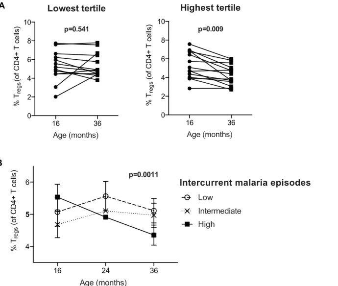
Longitudinal decline in T
regfrequencies within children correlates with
higher intercurrent malaria exposure
was no change in Tregfrequencies between 16 and 36 months (Wilcoxon signed rank test p = 0.54). Repeated-measures analysis using generalized estimating equations confirmed that changes in Tregfrequencies over time differed between the exposure groups (p = 0.0011,Fig
2B). Together, these data suggest that very high malaria exposure during childhood results in the loss of peripherally circulating Tregswithin individuals over time.
T
regdynamics during childhood differ between high and low malaria
transmission settings
Prior studies have shown that experimental and naturalP.falciparuminfection induces the expansion of regulatory T cellsin vivo[4,5]. To investigate whether and how Tregdynamics Fig 2. Regulatory T cells decrease over time in individuals with high but not low malaria incidence.Tregfrequencies of CD4+ T cells (FoxP3+CD25
+CD127dim) were measured at 16, 24 and 36 months of age (PROMOTE- SP arm).(A)Children were divided into tertiles based on incidence of malaria
between 16 and 36 months; lowest tertile median incidence 4.0 episodes ppy (IQR 3.0–5.5); intermediate tertile median incidence 8.2 episodes ppy (IQR 7.3–9.2); highest tertile median incidence 13.7 episodes ppy (IQR 10.3–14.6). The median duration since last malaria infection in these three tertiles was 8.5, 20, and 100 days, respectively. Wilcoxon matched pairs signed rank test p values indicated. Between 16 and 36 months, Tregfrequencies declined in
individuals in the highest but not lowest tertile of malaria incidence.(B)Changes in Tregfrequencies between 16, 24 and 36 months were compared between
children in the lowest, intermediate, and highest tertiles of malaria exposure by generalized estimate equations, accounting for repeated measures, age, duration since last malaria episode and parasite status at time of sampling.
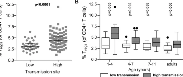
differ between settings of high and low exposure, we compared Tregfrequencies between chil-dren over a range of ages in the high transmission district of Tororo to chilchil-dren from the low transmission district of Jinja (PRISM cohort age 1 to 11 years). Children in the high transmis-sion Tororo District experienced a much higher malaria incidence (median 3.6 vs. 0 ppy) and a much shorter duration since last infection (median 62 vs. 230 days) than children in the low transmission Jinja District (full details inS1 Table). Overall, Tregfrequencies (FoxP3+CD25 +CD127dim) were higher in children from the high transmission district compared to the lower transmission district across all age groups (Wilcoxon ranksum p<0.0001,Fig 3A), and this
dif-ference was most marked in the youngest age group (Fig 3B), possibly reflecting an earlier expansion of Tregsin response to initial infections during early childhood or evenin utero[27–
30]. The difference in Tregsfrequencies between the low and high transmission study sites decreased with increasing age, and this trend extended to adulthood (Fig 3B). Thus, our data suggest that in areas of high malaria transmission malaria infections early in life induce Tregs, as has been previously described among naïve or comparatively low-exposure individuals [4,5,7,8]. However, in areas of intense and continual malaria exposure, parasite-driven induc-tion of Tregsis diminished, and instead there is a progressive decline of Tregswith repeated malaria episodes. This decline does not appear to be transient, as Tregfrequencies do not corre-late with the duration of time since last malaria episode or asymptomatic parasite infection. Instead, there appears to be sustained and progressive loss of Tregswith age among children heavily exposed to malaria.
Reduced TNFR2 expression on T
regsfollowing chronic malaria exposure
We next assessed expression of TNFR2 on Tregcells, as this receptor has been shown to be criti-cal for both proliferative expansion of Tregsand maintenance ofFOXP3expression in inflam-matory environments [31,32]. Furthermore, Tregsexpressing TNFR2 have been shown to have enhanced suppressive capacity [8,33,34], and are increased during malaria infection [8]. We found that the percentage of Tregsexpressing TNFR2 was significantly lower among PRISM cohort children from the high transmission Tororo District than among children of similar age from the low transmission Jinja district (p<0.0001,Fig 4A, seeS3 Figfor gating strategy).
Among Tororo children, expression of TNFR2 was inversely correlated with number of recent malaria episodes (Coef = -0.31, p = 0.032,Fig 4B), although expression was slightly higher on Tregsfrom children currently PCR-positive forP.falciparuminfection (p = 0.043). These data suggest that TNFR2 expression is transiently up-regulated during parasitemia but declines over time following repeated malaria episodes. This decrease in TNFR2 expression could contribute to the loss of FoxP3+ Tregsfrom peripheral blood by decreasing the stability ofFOXP3 expres-sion [31,32].
Altered homeostasis of T
regsfollowing intense malaria exposure
frequencies shown inS4 Fig). As previously reported, co-culture of PBMCs from naïve adults with PfSE resulted in consistent induction of Tregs(Fig 5AandS4C Fig). However, using PBMC from malaria-exposed children and adults, induction of Tregswas reduced compared to naïve adults (Fig 5A). Further, whereas all children with low prior malaria exposure (<2
episodes ppy) exhibited Treginduction (fold change>1), only 55% of children with high prior malaria exposure (>6 episodes ppy) induced Tregsfollowing PfSE stimulation (p = 0.03;Fig
5B). This suggests that heavy prior exposure to malaria may limit the propensity of CD4 cells to differentiate into Tregsupon re-encounter with parasite antigens.
We next investigated whether heavy prior malaria exposure increased the susceptibility of Tregsto apoptosis, as has been shown in chronic HIV-1 infection [36]. The percentage of Tregs Fig 3. FoxP3+ regulatory T cells are increased in high compared to low transmission settings, but decrease with age only in highly exposed children. (A)FoxP3+CD25+CD127dimregulatory T cell frequencies in children age 1 to 11 years (PRISM cohort) residing in the low transmission Jinja
District (n = 34) were compared to children in the high transmission Tororo District (n = 91). Wilcoxon ranksum p value indicated.(B) FoxP3+CD25 +CD127dimregulatory T cell frequencies were compared between children from low and high transmission areas at age 1
–4 (n = 11 and n = 18), 4–7 (n = 14 and n = 35) and 7–11 (n = 9 and n = 38) years of age and adults (n = 9 and n = 37). Wilcoxon ranksum for age group comparisons, p values indicated.
doi:10.1371/journal.ppat.1005041.g003
Fig 4. TNFR2 expression on FoxP3+ regulatory T cells declines with increasing prior malaria incidence. A.Frequencies of TNFR2 expressing Tregs
(FoxP3+CD25+CD127dim) were compared between children in the high malaria transmission area (Tororo District) and in the low transmission area (Jinja
district). TNFR2 expression on Tregswas higher in low compared to high transmission settings. Wilcoxon signed rank test indicated.B.The association
between frequencies of TNFR2 expressing Tregsand number of recent malaria episodes was analyzed among children from high malaria exposure area
(Tororo District). TNFR2 expression declined with increasing number of malaria episodes in last 90 days. Regression coefficient and p value are indicated.
Fig 5. Evidence for changed homeostasis of Tregsin children with high malaria exposure. A.PBMCs from 28 month old children (PROMOTE no
chemoprevention control arm), malaria-exposed adults (PRISM cohort, Nagongera, Tororo District) and naïve adults were incubated for 7 days with protein extract from mature stageP.falciparuminfected RBCs (PfSE) or uninfected RBCs (uE). Tregfrequencies were enumerated (FoxP3+CD25+CD127dim) and
induction factor was calculated based on frequency fold change between PfSE and uE stimulated PBMCs. Parasite induction of Tregswas reduced in
exposed compared to naïve individuals. Wilcoxon signed rank test indicated for comparison between naïve-adults and exposed infant and adult samples.B.
The proportion of children with any Treginduction by PfSE was compared between those with low (<2 episodes ppy, n = 10) and high (>6 episodes ppy,
n = 10) prior malaria incidence. There was a reduced proportion of infants with any Treginduction in those who had high compared to low prior malaria
incidence. Chi-square test indicated.C.Pro-apoptotic marker CD95 was measured on Tregsin children (PRISM cohort) from the high transmission area
(Tororo District). Association between the level of CD95 expression on Tregs, as measured by MFI of CD95+ Tregs, and age was analyzed. The level of CD95
expression increased with age.D.Pro-survival Bcl2 was measured on Tregsin children (PRISM cohort, Nagongera, Tororo) from the high transmission area.
Association between the expression of Bcl2 on the Tregpopulation and age was analyzed. The level of Bcl2 expression decreased with age. Spearman’s rho
expressing the pro-apoptotic marker CD95 increased with age among Tororo children (Rho = 0.175, p = 0.079), as did the level of CD95 expression (as calculated by MFI of CD95 on CD95+ Tregs, Spearman’s Rho = 0.44, p = 0.0001) (Fig 5C). Conversely, expression of the anti-apoptotic marker Bcl2 on Tregsdeclined with age (Rho = -0.266 p = 0.035) (Fig 5D). However there was no independent relationship between expression of these markers and prior malaria incidence, current parasite infection, nor time since last malaria episode, suggesting that age may independently affect the sensitivity of Tregsto apoptosis. To further investigate this, three distinct measures of apoptosis (YoPro, Annexin V and activated Caspase 3) were measured on Tregsbothex vivoand following stimulation with camptothecin (an activator of apoptosis) or
PfSE in 28-month infants with low or high prior malaria incidence (PROMOTE no-chemopre-vention control arm). There was no difference in sensitivity to apoptosis as measured by any of the markers eitherex vivoor following stimulation with camptothecin or parasite antigen; the frequencies of positively stained cellsex vivo, and the fold change of apoptosis staining, were the same regardless of prior malaria exposure (Fig 5EandS5 Fig). Together these data suggest that Treghomeostasis may be altered in the setting of heavy malaria exposure, in part due to reduced induction of peripheral Tregcells, with little evidence for increased susceptibility to antigen-driven apoptosis.
Lower T
regfrequencies may be associated with a decreased risk of
symptoms following parasite infection
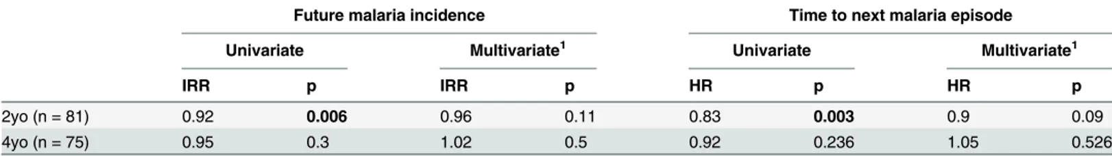
We finally asked whether the frequency of circulating Tregsinfluences susceptibility to malaria. We assessed the influence of Tregfrequencies on protection from malaria in both the 2-year-old PROMOTE no chemoprevention control arm and 4-year-2-year-old TCC cohorts using two methods; a time-to-event analysis (time to next malaria episode), and negative binomial regres-sion of the relationship of Tregfrequencies to malaria incidence in the year following assess-ment. Among 2-year-olds, we found that higher Tregfrequencies were associated with an increased time to next malaria episode and a lower future malaria incidence in univariate anal-ysis (Table 1). However, after adjusting for prior malaria incidence in a multivariate model to account for heterogeneity in environmental exposure to infected mosquitoes [22,37,38], this relationship was no longer significant, suggesting that differences in environmental exposure intensity may underlie this association [37]. Among 4-year-olds, Tregfrequencies were not associated with time to next malaria infection or malaria incidence during follow-up. Similarly, no relationships between Tregfrequencies and malaria incidence in follow-up or time to next
episodes ppy) prior malaria incidence was measured ex vivo and following stimulation with camptothecin (an activator of apoptosis) orP.falciparumantigen. Data from YoPro staining is representative of all measures of apoptosis (seeS5 Figadditional measures of apoptosis including activated Caspase 3 and AnnexinV). There was no difference in sensitivity to apoptosis between infants with low and high prior malaria incidence. Wilcoxon signed rank test indicated.
doi:10.1371/journal.ppat.1005041.g005
Table 1. Relationship between Tregfrequencies and prospective risk of malaria.
Future malaria incidence Time to next malaria episode
Univariate Multivariate1 Univariate Multivariate1
IRR p IRR p HR p HR p
2yo (n = 81) 0.92 0.006 0.96 0.11 0.83 0.003 0.9 0.09
4yo (n = 75) 0.95 0.3 1.02 0.5 0.92 0.236 1.05 0.526
1
–Multivariate is adjusted for prior malaria incidence
malaria episode were observed in the PRISM 1–11 year old cohorts, in either the low or the high transmission study sites, even after adjustment for household mosquito exposure. Thus we did not find clear evidence that Tregsare associated with the risk of clinical malaria.
Given their immunoregulatory role, is also possible that Tregsplay a role in protecting the host from the symptomatic manifestations of malaria onceP.falciparuminfection is estab-lished [1,2,15,19]. To assess whether Tregsmay influence the risk of clinical disease once infected, we analyzed the relationship between Tregfrequencies and the probability of symp-toms once parasitemic using generalized estimate equations with robust standard errors, accounting for repeated measures [16,39]. Comparing children with the lowest compared to highest tertiles of Tregs, lower Tregfrequencies were associated with an increased monthly prob-ability of infection, consistent with the exposure induced decline in Tregsdescribed. However, lower Tregfrequencies were also associated with an overall decreased probability of becoming symptomatic once infected (2 year old PROMOTE cohort; OR = 0.4, p = 0.039, 4 year old TCC cohort; OR = OR = 0.37, p = 0.06), suggesting that the decline in circulating Tregs may be asso-ciated with the acquisition of clinical immunity.
Discussion
Here, we have shown through both cross-sectional and longitudinal studies that the percentage and absolute number of FoxP3+ Tregsin peripheral blood are influenced by repeated exposure to malaria. While children in settings of intense exposure have higher Tregfrequencies during early childhood, frequencies decline throughout childhood in settings of high (but not low) exposure, and the extent of Tregloss correlates with the intensity ofP.falciparumexposure. We provide bothin vivoandin vitroevidence that among children in high exposure settings, there is a reduction of parasite induced Tregexpansion during infection. Further, we show a down-regulation of TNFR2, which is required for stabilization of the FoxP3+ regulatory phenotype in inflammatory environments [31]. These data demonstrate that chronic exposure to malaria results in altered Treghomeostasisin vivo, which may have a downstream impact on the acqui-sition of immunity and control of infection.
Our data indicate that the dynamics of Treginduction and homeostasis differ markedly between high and low malaria transmission settings. Although children residing in high trans-mission areas had higher Tregfrequencies overall, perhaps in response to a parasite-driven Treg expansion in early childhood orin utero[27–30], we observed a markeddeclinein Treg fre-quencies beginning at 1–2 years of age, which appeared to be driven by persistent parasite exposure. Further, among highly exposed children, we saw no association between Treg fre-quencies and current or recent infection, suggesting that Tregshave reducedin vivoinduction during infection of these chronically exposed children. Consistent with this, we demonstrated that induction of Tregsfollowing parasite stimulation of PBMC was diminished in heavily exposed adults and children, providingin vitroevidence that chronic antigen exposure may blunt the proliferative expansion of Tregsin response to malaria. This is in contrast to published studies suggesting that Tregsexpand in response to malariain vivoandin vitro[4,7–13,18,19]. The most likely explanation for the difference in our findings is that these earlier studies were performed largely on malaria-naïve volunteers or relatively low-exposed populations. Overall our data suggest that while Tregsmay be induced in initial encounters with parasites, induction capacity is diminished after repeated parasite exposure and instead Tregsundergo a steady decline in the periphery. The induction of TregsbyPlasmodiumis believe to occur through acti-vation of latent membrane-bound TGFβ[11,40], which can be blocked by antibodies to theP.
may represent an immune evasion strategy. The reduced capacity of parasite antigen to induce Tregsin heavily malaria exposed children suggests that the host may be able to circumvent para-site induction of Tregs, potentially enabling enhanced control of infection.
Another potential mechanism for the observed decline in Tregsamong children chronically exposed to malaria is via loss ofFOXP3expression by“unstable”Tregs,which has been reported to occur in highly inflammatory immune environments [41–45]. Sustained expression of the canonical transcription factorFOXP3by Tregsis critical for maintenance of regulatory function [46]. Several recent studies suggest that Tregscan become“unstable”and loseFOXP3 expres-sion in response to cues in the microenvironment, although the significance, extent, and trig-gers of this phenomenon remain subject to considerable debate [44,45,47–52]. Lineage tracking experiments have elegantly shown that antigen-driven activation and inflammation can drive a subset of FoxP3hiTregsto lose bothFOXP3expression and suppressor function [44], and even acquire an effector phenotype [53]. Further, repeated TCR stimulation leads to the loss ofFOXP3expression and the conversion to pro-inflammatory cytokine producing cells in natural Tregsin vitro[54]. Our data suggest a potential mechanism for such Treg destabi-lization in malaria infection, as recurrent exposure resulted in down-regulated Tregexpression of TNFR2, which has been shown to be critical for both the proliferative expansion of Tregsand stabilization of theirFOXP3expression in inflammatory environments [31,32,55]. In the set-ting of malaria, TNFR2+ Tregshave previously been shown to have higherFOXP3expression and enhanced suppressive function [8]. Thus our data are consistent with mounting evidence suggesting that peripherally induced Tregshave significant plasticity in response to inflamma-tory environments such as that observed in malaria infection, which may culminate in loss of
FOXP3expression and suppressive function.
Several additional processes might contribute to the observed loss of Tregsin peripheral blood. Because Tregstrack to sites of inflammation, it is possible that Tregsinduced byP.
falcipa-rumtraffic to the liver, spleen, or secondary lymphoid organs during infection. Invasive sam-pling was not possible in our study cohorts; therefore we were unable to exclude a preferential sequestration of Tregsin tissues or lymphoid organs. However, we did not observe any statistical relationship of Tregfrequencies with the presence of parasitemia, nor with the amount of time elapsed since the lastP.falciparuminfection, as might be expected if Tregsmigrate to sites of local inflammation during active infection. Alternatively, loss of Tregsthrough apoptosis might contribute to their decline following repeated malaria infections. However, we observed no relationship between prior malaria incidence and the expression of the pro-apoptotic molecule CD95 or the anti-apoptotic marker Bcl2 on Tregs(after controlling for age), nor did we observe a differential susceptibility towards apoptosisex vivo, or followingin vitrore-stimulation with parasite antigens.
CD4 effector responses. In prior studies,FOXP3mRNA levels measured during acute malaria were shown to correlate inversely with the magnitude of the subsequent Th1 memory response toP.falciparummeasured 28 days after infection [6]. Similarly,FOXP3expression among malaria-naive adults following experimental sporozoite vaccination was shown to correlate inversely with the Th1 memory response measured>100 days later [14]. Thus, while Tregsare
likely to influence the development of parasite-specific T cell memory responses, no relation-ship between these populations could be demonstrated through our concurrent measurements in peripheral blood, which maybe due in part to the chronicity of malaria exposure in these children and/or the substantial heterogeneity within the cohort with regard to time elapsed since the last infection. Furthermore, additional parameters of Tregfunction that cannot readily be measured in peripheral blood in large cohorts, such as suppression of T cell proliferation and modulation of APC function, are likely to influence the cellular immune response to malaria, but could not be assessed in the present study.
While our results clearly suggest that repeated malaria impacts peripherally circulating Tregs in children, the role of these cells in protection from malaria and the development of immunity remains unclear. We observed no association between Tregfrequencies and future malaria inci-dence or time to next malaria episodes in any of our cohorts. However, our data suggest that although children with the lowest Tregfrequencies had a higher monthly probability of infec-tion, they were less likely to become symptomatic once infected compared to children with the highest Tregfrequencies over the entire study period. While these data suggest that clinical immunity is acquired as Tregsdecline, the role of Tregsin mediating clinical immunity remains unclear, and may not be causal—rather, declining Tregfrequencies may coincide with other immune changes that mediate protection. Because all children in our study cohorts have easy access to dedicated study clinics and prompt antimalarial drug treatment, the incidence of severe malaria was extremely low, preventing assessment of the potential role of Tregsin protec-tion from severe disease. We were similarly unable to assess the impact of Tregactivity on path-ogen persistence following infection, because all cases of symptomatic malaria were promptly treated with potent artemisinin-based drugs, thus altering the natural course of infection. In other protozoal infections, such as leishmaniasis and toxoplasmosis, pathogen-induced Tregs have been reported to curb the inflammatory response, allowing long-term pathogen persis-tence [1,2,15]. Indeed, in murine models of leishmania, pathogen persistence resulting from Treg-mediated immune suppression has been shown to be a requirement for immunity to re-infection [1]. The long-term asymptomatic maintenance of low-burdenP.falciparuminfection that is commonly observed among adults in high-transmission areas [58] may represent a simi-lar phenomenon, but the role of Tregsin mediating this process is not known. Although we did not observe higher frequencies of peripheral blood Tregsamong children with asymptomaticP.
falciparuminfection, which is not routinely treated in Uganda, this does not exclude a role for Tregsin maintaining this state of host-parasite equilibrium.
Materials and Methods
Ethics approval
Written informed consent was obtained from the adult individual or parent/guardian of all study participants. Study protocols were approved by the Uganda National Council of Science and Technology and the institutional review boards of the University of California, San Fran-cisco, Makerere University and the Centers of Disease Control and Prevention.
Study participants
Samples for this study were obtained from children enrolled in 3 longitudinal childhood malaria cohort studies conducted in Tororo District and Jinja District of eastern Uganda. Cohort characteristics are described inS1 Table. For all cohorts, samples were selected on the bases of availability of PBMCs.
1. The PROMOTE-Chemoprevention Study was conducted from 2010–2013 and enrolled 400
children who were randomized to receive chemoprevention with monthly sulfadoxine-pyri-methamine (SP), daily trimethoprim-sulfamethoxazole (TS), monthly dihydroartemisinin-piperaquine (DP) or no chemoprevention (control arm) from 6–24 months of age, then fol-lowed for an additional year after the intervention ended. Results of this trial have been pub-lished [59]. In this report we only include data from children who were randomized to receive“no chemoprevention (control arm)”or SP, which was found to have no efficacy for prevention of malaria [59]. Samples from this cohort were taken at 16, 24, 28 and 36 months of age as indicated.
2. The Tororo Child Cohort Study (TCC) was conducted from 2007–2012 in Tororo district, and enrolled children at approximately 6 months of age and followed until age 5. Results of this study have previously been published [60]. Samples used here are from patients who were HIV-negative children born to HIV-negative mothers, taken at age 4 years.
3. The PRISM cohort was initiated in 2011 and is ongoing. This longitudinal observational cohort consists of 200 households across two study sites, the Nagongera sub-county in Tor-oro district and the Walukuba sub-county in Jinja district. Description of the study and results have been published in [21] In all households, one adult caregiver and all eligible children aged 6 months to 11 years were enrolled into the study. Samples used here were taken from cross-sectional bleeds of study participants taken between August 2013 and March 2014. Household-level mosquito exposure was calculated based on mosquito counts obtained from CDC light traps placed monthly within the household of each individual trial participant in the 2012 [26].
For the PROMOTE-Chemoprevention, TCC and PRISM Nagongera high transmission area, the estimated entomological inoculation rate (aEIR) is approximately 310 bites ppy. In contrast, at the PRISM Walukuba low transmissions site the aEIR is estimated at 2.8 [21].
Clinical management and measurement of malaria incidence
malaria were defined as all febrile episodes accompanied by any parasitemia requiring treat-ment, but not preceded by another treatment in the prior 14 days. The incidence of malaria was calculated as the number of episodes per person years (ppy) from the time of enrolment into the cohort. In a subset of PRISM cohort children used to assess TNFR2 expression on Tregs parasite infection was assessed via PCR from dried blood spots as previously described [61].
FoxP3+ Regulatory T cells measurements and
P. falciparum
specific
CD4 responses
Tregfrequencies were enumerated from whole blood and fresh and cryopreserved PBMCs as indicated below. For enumeration of Tregsfrom whole blood (PROMOTE-Chemoprevention, control arm 2-year-old samples), 100μl of fresh whole blood was stained with BD Pharmingen anti-CD3-FITC (UCHT1), anti-CD4-PE-CY7 (SK3), and CD25-APC (M-A251) for 20 min-utes and then lysed and permeabilized with eBioscience RBC lysis buffer. Cells were washed and then incubated with eBioscience anti-FoxP3-PE (PCH101). Samples were acquired on Accuri C6 Cytometer.
For analysis of Tregsfrom fresh PBMCs (PRISM Nagongera cohort), PBMCs were isolated by Ficoll density gradient centrifugation and rested over night in 10% fetal bovine serum. PBMCs were stained with BD Pharmingen anti-CD3 PerCP (SK7), anti-TNFR2-Alexa646 (hTNFR-M1), CD95-PECy7 (DX2) and Biolegend CD4-APC-Cy7 (OKT4), anti-CD25-BrillantViolet510 (M-A251), anti-CD127-PacificBlue (A019D5). Following surface staining, cells were fixed and permeabilized with eBioscience FoxP3 staining set and intracellu-lar stained with FoxP3-PE (PCH101) and BD Pharmingen anti-Bcl2-FITC (Bcl-2/100) as per manufacturers protocol. Samples were acquired on three laser BD FACsCantoII with FACS-Diva software.
For analysis of Tregsfrom frozen PBMCs (Tororo Child Cohort 4-year-olds, PROMOTE-Chemoprevention SP arm longitudinal samples at 16, 24, and 28 months of age, PRISM Walu-kuba cohort), cryopreserved PBMCs were thawed using standard methods, and immediately stained with the following panels of antibodies; BD Pharmingen anti-CD3-FITC (UCHT1), anti-CD4-PE-CY7 (SK3), CD25-APC (M-A251) and Biolegend anti-CD127 Pacific Blue (A019D5); or Biolegend CD3-BrilliantViolet650 (OKT3), CD4-PerCP (OKT4), anti-CD127-FITC (A019D5); or Biolegend anti-CD3-PerCP (OKT3), anti-CD4-APC-Cy7 (OKT4), anti-CD25-BrillantViolet510 (M-A251), anti-CD127-PacificBlue (A019D5), anti-TNFR2-APC (3G7A02). Live/dead aqua amine (Invitrogen) was included in all panels. Following surface staining, cells were fixed and permeabilised with eBioscience FoxP3 staining set and intracellu-lar stained with FoxP3-PE (PCH101) as per manufacturers protocol. Samples were acquired on LSR2 three laser flow cytometer (Becton Dickinson) with FACSDiva software.
For calculation of absolute Tregscounts (i.e. cells perμl, PRISM Nagongera cohort), periph-eral blood CD4 T cell concentrations were measured from whole blood using counting beads, and Tregfrequencies were calculated by normalization to total CD4 T cell numbers.
CD4 T cell responses to
P. falciparum
infected RBCs
Analysis of CD4+ T cell responses toP.falciparuminfected RBCs via intracellular cytokine staining was performed as previously described [22,56]. PBMCs were stimulated withP. falcip-aruminfected RBCs or uninfected RBCs and CD4 T cell production of IFNγ, IL10, and TNFα
Induction of Regulatory T cells by
P. falciparum
in vitro
PBMCs from PROMOTE subjects (28 months of age; no chemoprevention control arm) and adults from the high malaria transmission region of Tororo were thawed and washed in 10% Human serum (AB) media (Gemini), and 3–6X10⁶PBMC were labeled with 1 ml of 1.25 mM 5,6-carboxyfluorescein diacetate succinimidyl ester (CFSE; Molecular Probes) for seven min-utes. CFSE-labeled PBMC were incubated in 96-well, deep-well culture plates (Nunc, Roskilde, Denmark) at 106PBMC/ well in 1 ml for 7 days withP.falciparumschizont extract (PfSE) (W2 strain) or protein extract from uninfected RBCs (uE) at a effector to target ratio equivalent to 1:1 PBMC:infected RBC. PHA (1μg/ml) was used as a positive control. PfSE extracts were made from the W2 stain grown in standard culture conditions and confirmed to be free of mycoplasma contamination using MycoAlert (Lonza). Mature stage parasites were magnet purified from culture using MACs purification columns. Purified parasites or uninfected RBCs were freeze thawed 3+ times (via snap freezing on liquid nitrogen and then transfer to 37°C water bath) to produce PfSE and uE and stored at -20°C. Following culture of PBMCs with pro-tein extracts, cells were treated with 100 units of DNase I (Invitrogen) in culture media for 5 minutes and then surface stained with Biolegend CD3-BrilliantViolet650 (OKT3), anti-CD4-PerCP (OKT4), anti-CD25-PE-Cy7 (BC96), anti-CD127 Pacific Blue (A019D5), BD Pharmingen anti-CD8-ABC-H7 (SK1) and Live/dead aqua amine (Invitrogen cells). Following surface staining, cells were fixed and permeabilized with eBioscience FoxP3 staining set and stained with FoxP3-PE (PCH101). Proliferation with PHA was used to ensure cell viability, and cells incubated with uE were used as a background control. Of the infants tested, the prior median malaria incidence was 1.2 episodes ppy in the low exposed group and 8.5 episodes ppy in the high exposed group.
Susceptibility of T
regsto apoptosis
PBMCs from PROMOTE subjects (28 months of age; no chemoprevention control arm) were thawed and rested overnight either in standard media (untreated), 5uM camptothecin (Sigma) orP.falciparumschizont extract (PfSE) or protein extract from uninfected RBCs (uE) at an effector:target ratio of 3:1. To test for induction of apoptosis, stains for AnnexinV (Biolegend) or YoPro (Invitrogen), or activated Caspase 3 FITC (BD) were used according to the manufac-turer’s instructions in combination with the following antibodies: AnnexinV and YoPro
stain-ing—CD3 (OKT3) Brilliant Violet 650, CD4 (RPA-T4) APC-Cy7, CD127 (A019D5) APC,
CD25 (BC96) PE-Cy7 from Biolegend; for Caspase3—CD3 (OKT3) Brilliant Violet 650, CD4 (RPA-T4) PerCP, CD127 (A019D5) Pacific Blue, CD25 (BC96) PE-Cy7. Tregswere gated as CD3+CD4+CD25+CD127dim. Sensitivity to apoptosis was measuredex vivo(untreated con-trol), after induction with camptothecin (fold change compared to untreated), and after stimu-lation withP.falciparumschizont extract (fold change comparing PfSE to uE).
Flow cytometry data analysis
Statistical analysis
Data analysis was performed using Stata version 12 (Stata Corp, College Station, Tx) and PRISM version 6 (Graph Pad). Associations between Tregfrequencies and other continuous variables (prior malaria incidence, age, time since last malaria episode) were assessed using Spearman’s correlation. Changes in Tregfrequencies within an individual over time were assessed using the Wilcoxon signed rank test. All other two-group comparisons of continuous variables were performed using the Wilcoxon rank sum test. Repeated measures analysis of longitudinal changes in Tregfrequencies was performed using generalized estimating equations, with adjustment for concurrent parasitemia, age and duration since last malaria episode. Cate-gorical variables were compared using Chi sq test. Associations between Tregfrequencies and time to next malaria episode were evaluated using the Kaplan-Meier product limit formula, and a multivariate cox proportional hazards model was used to adjust for surrogates of malaria exposure (cumulative episodes since enrollment in study for TCC and PROMOTE cohorts, or age for PRISM cohorts). Negative binomial regression was used to estimate associations between Tregfrequencies and the prospective incidence of malaria in the following year (inci-dence rate ratios, IRR) and prevalence of asymptomatic parasitemia in the following year (prevalence rate ratios, PRR), adjusting for malaria exposure as above. Two-sided p-values were calculated for all test statistics and p<0.05 was considered significant. In the PRMOTE
and TCC cohorts, associations between the highest and lowest tertiles of Tregfrequencies and the monthly risk of parasitemia, probability of symptoms if parasitemic, and incidence of malaria, stratified by year of age, were evaluated using generalized estimating equations with robust SEs accounting for repeated measures in the same patient, for the period of the inter study (6 months to 3 or 5 years of age) [62].
Supporting Information
S1 Table. Cohort characteristics. (PDF)
S1 Fig. Gating strategy for regulatory T cells.Frequencies of regulatory T cells were enumer-ated by staining whole blood samples (2yo cohort) or frozen PBMC samples (4yo cohort).A. For 2yo, whole blood was stained and cells analyzed on four-color Accuri flow-cytometer.B. For 4yo samples, PBMCs were thawed and stained and analyzed on a LSRII. See alsoFig 1A
and 1B.
(PDF)
S2 Fig. Absolute count of regulatory T cells decline with increasing age among children in a high transmission setting.Absolute count of regulatory T cells, analyzed as the percent of FoxP3+CD25+CD127dimexpressing CD4+ T cells, normalized to CD4+ T cell absolute counts, from 1 to 11 year old children (PRISM cohort, high transmission Nagongera, Tororo District), declined with increasing age.
(PDF)
S3 Fig. Gating strategy for TNFRII expression.Frequencies of regulatory T cells expression TNFR2 were enumerated by staining PBMCs. Gating for FoxP3+CD25+CD127dim regulatory T cells was as forS1 Fig. TNFR2 staining was gated on FMO controls, as indicated.
(PDF)
S4 Fig. Regulatory T cell induction assays. A.PBMCs were incubated withP.falciparum
+CD25+CD127lowregulatory T cells in samples used for stimulation assays.C.In PBMCs from malaria-naïve adults, incubation with PfSE resulted in increased frequencies of Tregs com-pared to PBMCs cultured with uE. See alsoFig 5A and 5B.
(PDF)
S5 Fig. Additional measures of Tregapoptosis.Activated Caspase 3(A)and AnnexinV(B)
staining of Tregsfrom 28 month old children (PROMOTE, no chemoprevention control arm, with low (<2 episodes ppy) and high (>6 episodes ppy) prior malaria incidence was measured
ex vivo and following stimulation with camptothecin (an activator of apoptosis) orP. falcipa-rumantigen.
(PDF)
Acknowledgments
We are grateful to all the parents and guardians for kindly giving their consent and to the study participants for their cooperation. We thank all the members of the study teams for their dedica-tion and excellent work. We thank J. Legac and P. Rosenthal, for technical support and provid-ing parasite cultures and Mary Prahl and Mary Fontana for critical readprovid-ing of the manuscript. The findings and conclusions in this paper are those of the authors and do not necessarily repre-sent the views of the Centers for Disease Control and Prevention.
Author Contributions
Conceived and designed the experiments: MJB PJ LAF IEJ MEF. Performed the experiments: MJB PJ LAF IEJ SW TIM HMV KB FN AA MN ES KN. Analyzed the data: MJB PJ LAF BG GD MEF. Contributed reagents/materials/analysis tools: JR AK VB JK JT MKM EA MRK. Wrote the paper: MJB PJ LAF BG GD MEF.
References
1. Belkaid Y, Piccirillo CA, Mendez S, Shevach EM, Sacks DL (2002) CD4+CD25+ regulatory T cells con-trol Leishmania major persistence and immunity. Nature 420: 502–507. doi:10.1038/nature01152
PMID:12466842
2. Belkaid Y, Blank RB, Suffia I (2006) Natural regulatory T cells and parasites: a common quest for host homeostasis. Immunol Rev 212: 287–300. doi:10.1111/j.0105-2896.2006.00409.xPMID:16903921 3. Velavan TP, Ojurongbe O (2011) Regulatory T cells and parasites. J Biomed Biotechnol 2011:
520940. doi:10.1155/2011/520940PMID:22262943
4. Walther M, Tongren JE, Andrews L, Korbel D, King E, et al. (2005) Upregulation of TGF-beta, FOXP3, and CD4+CD25+ regulatory T cells correlates with more rapid parasite growth in human malaria infec-tion. Immunity 23: 287–296. doi:10.1016/j.immuni.2005.08.006PMID:16169501
5. Finney OC, Nwakanma D, Conway DJ, Walther M, Riley EM (2009) Homeostatic regulation of T effec-tor to Treg ratios in an area of seasonal malaria transmission. Eur J Immunol 39: 1288–1300. doi:10. 1002/eji.200839112PMID:19338000
6. Walther M, Jeffries D, Finney OC, Njie M, Ebonyi A, et al. (2009) Distinct roles for FOXP3 and FOXP3 CD4 T cells in regulating cellular immunity to uncomplicated and severe Plasmodium falciparum malaria. PLoS Pathog 5: e1000364. doi:10.1371/journal.ppat.1000364PMID:19343213 7. Bueno LL, Morais CG, Araújo FF, Gomes JAS, Corrêa-Oliveira R, et al. (2010) Plasmodium vivax:
induction of CD4+CD25+FoxP3+ regulatory T cells during infection are directly associated with level of circulating parasites. PLoS ONE 5: e9623. doi:10.1371/journal.pone.0009623PMID:20224778 8. Minigo G, Woodberry T, Piera KA, Salwati E, Tjitra E, et al. (2009) Parasite-dependent expansion of
TNF receptor II-positive regulatory T cells with enhanced suppressive activity in adults with severe malaria. PLoS Pathog 5: e1000402. doi:10.1371/journal.ppat.1000402PMID:19390618
10. Scholzen A, Mittag D, Rogerson SJ, Cooke BM, Plebanski M (2009) Plasmodium falciparum-mediated induction of human CD25Foxp3 CD4 T cells is independent of direct TCR stimulation and requires IL-2, IL-10 and TGFbeta. PLoS Pathog 5: e1000543. doi:10.1371/journal.ppat.1000543PMID:19680449 11. Clemente A, Caporale R, Sannella AR, Majori G, Severini C, et al. (2011) Plasmodium falciparum
solu-ble extracts potentiate the suppressive function of polyclonal T regulatory cells through activation of TGFβ-mediated signals. Cell Microbiol 13: 1328–1338. doi:10.1111/j.1462-5822.2011.01622.xPMID:
21699642
12. Finney OC, Lawrence E, Gray AP, Njie M, Riley EM, et al. (2012) Freeze-thaw lysates of Plasmodium falciparum-infected red blood cells induce differentiation of functionally competent regulatory T cells from memory T cells. Eur J Immunol 42: 1767–1777. doi:10.1002/eji.201142164PMID:22585585 13. Finney OC, Riley EM, Walther M (2010) Phenotypic analysis of human peripheral blood regulatory T
cells (CD4+FOXP3+CD127lo/-) ex vivo and after in vitro restimulation with malaria antigens. Eur J Immunol 40: 47–60. doi:10.1002/eji.200939708PMID:19877016
14. Todryk SM, Walther M, Bejon P, Hutchings C, Thompson FM, et al. (2009) Multiple functions of human T cells generated by experimental malaria challenge. Eur J Immunol 39: 3042–3051. doi:10.1002/eji. 200939434PMID:19658096
15. Taylor MD, van der Werf N, Maizels RM (2012) T cells in helminth infection: the regulators and the regu-lated. Trends Immunol 33: 181–189. doi:10.1016/j.it.2012.01.001PMID:22398370
16. Todryk SM, Bejon P, Mwangi T, Plebanski M, Urban B, et al. (2008) Correlation of memory T cell responses against TRAP with protection from clinical malaria, and CD4 CD25 high T cells with suscep-tibility in Kenyans. PLoS ONE 3: e2027. doi:10.1371/journal.pone.0002027PMID:18446217 17. Kho S, Marfurt J, Noviyanti R, Kusuma A (2015) Preserved dendritic cell HLA-DR expression and
reduced regulatory T cell activation in asymptomatic Plasmodium falciparum and P. vivax infection. Infection and. . .. doi:10.1128/IAI.00226-15
18. Scholzen A, Minigo G, Plebanski M (2010) Heroes or villains? T regulatory cells in malaria infection. Trends Parasitol 26: 16–25. doi:10.1016/j.pt.2009.10.004PMID:19914134
19. Finney OC, Riley EM, Walther M (2010) Regulatory T cells in malaria—friend or foe? Trends Immunol 31: 63–70. doi:10.1016/j.it.2009.12.002PMID:20056484
20. Hisaeda H, Maekawa Y, Iwakawa D, Okada H, Himeno K, et al. (2004) Escape of malaria parasites from host immunity requires CD4+ CD25+ regulatory T cells. Nat Med 10: 29–30. doi:10.1038/nm975
PMID:14702631
21. Kamya MR, Arinaitwe E, Wanzira H, Katureebe A, Barusya C, et al. (2015) Malaria transmission, infec-tion, and disease at three sites with varied transmission intensity in Uganda: implications for malaria control. Am J Trop Med Hyg 92: 903–912. doi:10.4269/ajtmh.14-0312PMID:25778501
22. Jagannathan P, Eccles-James I, Bowen K, Nankya F, Auma A, et al. (2014) IFNγ/IL-10 co-producing cells dominate the CD4 response to malaria in highly exposed children. PLoS Pathog 10: e1003864. doi:10.1371/journal.ppat.1003864PMID:24415936
23. Portugal S, Moebius J, Skinner J, Doumbo S, Doumtabe D, et al. (2014) Exposure-dependent control of malaria-induced inflammation in children. PLoS Pathog 10: e1004079. doi:10.1371/journal.ppat. 1004079PMID:24743880
24. Gitau EN, Tuju J, Karanja H, Stevenson L, Requena P, et al. (2014) CD4+ T cell responses to the Plas-modium falciparum erythrocyte membrane protein 1 in children with mild malaria. J Immunol 192: 1753–1761. doi:10.4049/jimmunol.1200547PMID:24453249
25. Gitau EN, Tuju J, Stevenson L, Kimani E, Karanja H, et al. (2012) T-cell responses to the DBLα-tag, a short semi-conserved region of the Plasmodium falciparum membrane erythrocyte protein 1. PLoS ONE 7: e30095. doi:10.1371/journal.pone.0030095PMID:22272280
26. Wanzirah H, Tusting LS, Arinaitwe E, Katureebe A, Maxwell K, et al. (2015) Mind the gap: house struc-ture and the risk of malaria in Uganda. PLoS ONE 10: e0117396. doi:10.1371/journal.pone.0117396
PMID:25635688
27. Bisseye C, van der Sande M, Morgan WD, Holder AA, Pinder M, et al. (2009) Plasmodium falciparum infection of the placenta impacts on the T helper type 1 (Th1)/Th2 balance of neonatal T cells through CD4(+)CD25(+) forkhead box P3(+) regulatory T cells and interleukin-10. Clin Exp Immunol 158: 287–
293. doi:10.1111/j.1365-2249.2009.04014.xPMID:19758375
28. Brustoski K, Moller U, Kramer M, Hartgers FC, Kremsner PG, et al. (2006) Reduced cord blood immune effector-cell responsiveness mediated by CD4+ cells induced in utero as a consequence of placental Plasmodium falciparum infection. J Infect Dis 193: 146–154. doi:10.1086/498578PMID:16323143 29. Mackroth MS, Malhotra I, Mungai P, Koech D, Muchiri E, et al. (2011) Human cord blood CD4+CD25hi
30. Flanagan KL, Halliday A, Burl S, Landgraf K, Jagne YJ, et al. (2010) The effect of placental malaria infection on cord blood and maternal immunoregulatory responses at birth. Eur J Immunol 40: 1062–
1072. doi:10.1002/eji.200939638PMID:20039298
31. Chen X, Wu X, Zhou Q, Howard OMZ, Netea MG, et al. (2013) TNFR2 is critical for the stabilization of the CD4+Foxp3+ regulatory T. cell phenotype in the inflammatory environment. J Immunol 190: 1076–
1084. doi:10.4049/jimmunol.1202659PMID:23277487
32. Okubo Y, Mera T, Wang L, Faustman DL (2013) Homogeneous expansion of human T-regulatory cells via tumor necrosis factor receptor 2. Sci Rep 3: 3153. doi:10.1038/srep03153PMID:24193319 33. Chen X, Subleski JJ, Hamano R, Howard OMZ, Wiltrout RH, et al. (2010) Co-expression of TNFR2 and
CD25 identifies more of the functional CD4+FOXP3+ regulatory T cells in human peripheral blood. Eur J Immunol 40: 1099–1106. doi:10.1002/eji.200940022PMID:20127680
34. Chen X, Subleski JJ, Kopf H, Howard OMZ, Männel DN, et al. (2008) Cutting edge: expression of TNFR2 defines a maximally suppressive subset of mouse CD4+CD25+FoxP3+ T regulatory cells: applicability to tumor-infiltrating T regulatory cells. J Immunol 180: 6467–6471. doi:10.4049/jimmunol. 180.10.6467PMID:18453563
35. Clemente AM, Severini C, Castronovo G, Tanturli M, Perissi E, et al. (2014) Effects of soluble extracts from Leishmania infantum promastigotes, Toxoplasma gondii tachyzoites on TGF-βmediated path-ways in activated CD4+ T lymphocytes. Microbes Infect 16: 778–787. doi:10.1016/j.micinf.2014.08. 002PMID:25130316
36. Xing S, Fu J, Zhang Z, Gao Y, Jiao Y, et al. (2010) Increased turnover of FoxP3high regulatory T cells is associated with hyperactivation and disease progression of chronic HIV-1 infection. J Acquir Immune Defic Syndr 54: 455–462. doi:10.1097/QAI.0b013e3181e453b9PMID:20585263
37. Greenhouse B, Ho B, Hubbard A, Njama-Meya D, Narum DL, et al. (2011) Antibodies to Plasmodium falciparum antigens predict a higher risk of malaria but protection from symptoms once parasitemic. Journal of Infectious Diseases 204: 19–26. doi:10.1093/infdis/jir223PMID:21628654
38. Jagannathan P, Nankya F, Stoyanov C, Eccles-James I, Sikyomu E, et al. (2015) IFNγResponses to Pre-erythrocytic and Blood-stage Malaria Antigens Exhibit Differential Associations With Past Expo-sure and Subsequent Protection. Journal of Infectious Diseases 211: 1987–1996. doi:10.1093/infdis/ jiu814PMID:25520427
39. Jagannathan P, Kim CC, Greenhouse B, Nankya F, Bowen K, et al. (2014) Loss and dysfunction of Vδ2⁺γδT cells are associated with clinical tolerance to malaria. Science Translational Medicine 6: 251ra117. doi:10.1126/scitranslmed.3009793PMID:25163477
40. Omer FM, de Souza JB, Corran PH, Sultan AA, Riley EM (2003) Activation of transforming growth fac-tor beta by malaria parasite-derived metalloproteinases and a thrombospondin-like molecule. J Exp Med 198: 1817–1827. doi:10.1084/jem.20030713PMID:14676296
41. Wan YY, Flavell RA (2007) Regulatory T-cell functions are subverted and converted owing to attenu-ated Foxp3 expression. Nature 445: 766–770. doi:10.1038/nature05479PMID:17220876
42. Zheng S-G, Wang J, Horwitz DA (2008) Cutting edge: Foxp3+CD4+CD25+ regulatory T cells induced by IL-2 and TGF-beta are resistant to Th17 conversion by IL-6. J Immunol 180: 7112–7116. PMID:
18490709
43. Koenen HJPM, Smeets RL, Vink PM, van Rijssen E, Boots AMH, et al. (2008) Human CD25highFoxp3-pos regulatory T cells differentiate into IL-17-producing cells. Blood 112: 2340–2352. doi:10.1182/ blood-2008-01-133967PMID:18617638
44. Bailey-Bucktrout SL, Martínez-Llordella M, Zhou X, Anthony B, Rosenthal W, et al. (2013) Self-antigen-driven activation induces instability of regulatory T cells during an inflammatory autoimmune response. Immunity 39: 949–962. doi:10.1016/j.immuni.2013.10.016PMID:24238343
45. Li Z, Li D, Tsun A, Li B (2015) FOXP3(+) regulatory T cells and their functional regulation. Cell Mol Immunol. doi:10.1038/cmi.2015.10
46. Williams LM, Rudensky AY (2007) Maintenance of the Foxp3-dependent developmental program in mature regulatory T cells requires continued expression of Foxp3. Nat Immunol 8: 277–284. doi:10. 1038/ni1437PMID:17220892
47. Sakaguchi S, Vignali DAA, Rudensky AY, Niec RE, Waldmann H (2013) The plasticity and stability of regulatory T cells. Nat Rev Immunol 13: 461–467. doi:10.1038/nri3464PMID:23681097
48. Miyao T, Floess S, Setoguchi R, Luche H, Fehling HJ, et al. (2012) Plasticity of Foxp3(+) T cells reflects promiscuous Foxp3 expression in conventional T cells but not reprogramming of regulatory T cells. Immunity 36: 262–275. doi:10.1016/j.immuni.2011.12.012PMID:22326580
50. Bailey-Bucktrout SL, Bluestone JA (2011) Regulatory T cells: stability revisited. Trends Immunol 32: 301–306. doi:10.1016/j.it.2011.04.002PMID:21620768
51. Zhou X, Bailey-Bucktrout SL, Jeker LT, Penaranda C, Martínez-Llordella M, et al. (2009) Instability of the transcription factor Foxp3 leads to the generation of pathogenic memory T cells in vivo. Nat Immu-nol 10: 1000–1007. doi:10.1038/ni.1774PMID:19633673
52. Komatsu N, Mariotti-Ferrandiz ME, Wang Y, Malissen B, Waldmann H, et al. (2009) Heterogeneity of natural Foxp3+ T cells: a committed regulatory T-cell lineage and an uncommitted minor population retaining plasticity. Proc Natl Acad Sci USA 106: 1903–1908. doi:10.1073/pnas.0811556106PMID:
19174509
53. Oldenhove G, Bouladoux N, Wohlfert EA, Hall JA, Chou D, et al. (2009) Decrease of Foxp3+ Treg cell number and acquisition of effector cell phenotype during lethal infection. Immunity 31: 772–786. doi:
10.1016/j.immuni.2009.10.001PMID:19896394
54. Hoffmann P, Boeld TJ, Eder R, Huehn J, Floess S, et al. (2009) Loss of FOXP3 expression in natural human CD4+CD25+ regulatory T cells upon repetitive in vitro stimulation. Eur J Immunol 39: 1088–
1097. doi:10.1002/eji.200838904PMID:19283780
55. Kleijwegt FS, Laban S, Duinkerken G, Joosten AM, Zaldumbide A, et al. (2010) Critical role for TNF in the induction of human antigen-specific regulatory T cells by tolerogenic dendritic cells. J Immunol 185: 1412–1418. doi:10.4049/jimmunol.1000560PMID:20574005
56. Boyle MJ, Jagannathan P, Bowen K, McIntyre TI, Vance HM, et al. (2015) Effector Phenotype of Plas-modium falciparum-Specific CD4+ T Cells Is Influenced by Both Age and Transmission Intensity in Nat-urally Exposed Populations. Journal of Infectious Diseases. doi:10.1093/infdis/jiv054
57. Jangpatarapongsa K, Chootong P, Sattabongkot J, Chotivanich K, Sirichaisinthop J, et al. (2008) Plas-modium vivax parasites alter the balance of myeloid and plasmacytoid dendritic cells and the induction of regulatory T cells. Eur J Immunol 38: 2697–2705. doi:10.1002/eji.200838186PMID:18825754 58. Doolan DL, Dobaño C, Baird JK (2009) Acquired immunity to malaria. Clinical Microbiology Reviews
22: 13–36–TableofContents. doi:10.1128/CMR.00025-08PMID:19136431
59. Bigira V, Kapisi J, Clark TD, Kinara S, Mwangwa F, et al. (2014) Protective efficacy and safety of three antimalarial regimens for the prevention of malaria in young ugandan children: a randomized controlled trial. PLoS Med 11: e1001689. doi:10.1371/journal.pmed.1001689PMID:25093754
60. Wanzira H, Kakuru A, Arinaitwe E, Bigira V, Muhindo MK, et al. (2014) Longitudinal outcomes in a cohort of Ugandan children randomized to artemether-lumefantrine versus dihydroartemisinin-pipera-quine for the treatment of malaria. Clin Infect Dis 59: 509–516. doi:10.1093/cid/ciu353PMID:
24825870
61. Schwartz A, Baidjoe A, Rosenthal PJ, Dorsey G, Bousema T, et al. (2015) The Effect of Storage and Extraction Methods on Amplification of Plasmodium falciparum DNA from Dried Blood Spots. Am J Trop Med Hyg 92: 922–925. doi:10.4269/ajtmh.14-0602PMID:25758652