34
Article received on October 30, 2014 and accepted for publishing on December 15 2014.
Para eoplastic Cushi g’s “y dro e i a patie t with
multiple tumors
–
case report
Adina Mazilu1; Mona Gheorghiu2,3; Mădăli a Mușat2,3; N. Tă ase1; R. Petrescu1; A. Ciuche1; A. Tudose1; Florina Vasilescu1
INTRODUCTION
Paraneoplastic Cushing syndrome represents 5-10% of all Cushing syndrome and has a severe prognosis due to severe metabolic imbalance, denutrition, associated infections and progression of tumoral underlying pathology. The death is a rule in more than 50% of cases. Some medication used to treat it – Metirapone – is not available in Romania. Ketoconazole was recently approved by CHMP for t eat e t of pa a eoplasti Cushi g s o l i November 2014.
CLINICAL CASE
A 67 years old woman presented on the 21st of November with mental confusion, progressive weight loss, severe edema and kypokalemia, without typical featu es of Cushi g o h pe pig e tatio . Patie t s behavior changed in the last 5 months, she was nasty with her daughter, bickering, while diabetes and hypertension aggravated in the last 3 months.
The electrolytic imbalance was severe at admission K 1.65 mmol/l, in spite of multiple attempts to correct it with 150 mmol/day KCl on peripheral i.v. line, 40 mmol/day of KCl orally and 200 mg/day of “pi o ola to e, t eat e t used i itiall i C. I. Parho Natio al I stitute of E do i olog . Patie t as t a sfe ed i the I. C. U. of D . Ca ol Da ila Central Military Emergency Hospital for the weekend,
in order to obtain a better control using a central intravenous catheter.
Laboratory work and imaging
One month prior to admittance patient had hypercortisolism, with normal hepatic citolytic enzymes and normal TSH, free T4 and calcitonin values. Investigations at admittance revealed pa a eoplasti Cushi g s ith ACTH . pg/ l, cortisol levels more than 63 mcg/dl, UFC (urinary free cortisol) 2,866 mcg/24 h (21-111); DHEA-sulfate 230.5 mcg/24 h; 2 mg Dexametasone suppression test showed unsuppressed cortisol 59.17 mcg/dl, ACTH 123.8 pg/ml.
Patient also associated empty sella syndrome with thyrotrophic and gonadotrophic insufficiency, normal prolactin and IGF-1, normal mineralocorticoid hormones, cathecolamines, serotonin and 5HIAA, slightly enlarged cromogranine A – 148 ng/ml (upper limit 125 ng/ml).
Patient had also left breast tumor, Helicobacter pylori gastritis, polinodular goiter, denutrition and hepatic dysfunction.
Imaging techniques: 99m Tc Tektrotyd scintigraphy
1
Carol Davila Central Emergency Military Hospital, Bucharest
2
C.I. Parhon National Institute of Endocrinology, Bucharest
3
Carol Davila University of Medicine and Pharmacy, Faculty of Medicine, Bucharest
Vol. CXVIII •New Series • No. 1/2015• Romanian Journal of Military Medicine
35 showed uptake at 10 minute in left breast and jejunal
loop; upper and lower endoscopy, echo endoscopy – revealed no tumors; bronchoscopy – no visible tumor (after procedure, patient suffered a syncope that lasted 2 minutes); thyroid ultrasound – found nodule of 15/18/22 mm located in the lower part of left lobe – 15/18/22 mm, with low peripheral vascularisation, uptakes iodine at CT scan and has a peripheral calcification.
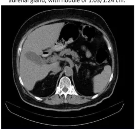
Figure 1.Abdominal CT-showed minimally enlarged right adrenal gland, with nodule 0.88/0.88 cm; enlarged left
adrenal gland, with nodule of 1.03/1.24 cm.
Figure 2.Thoracic CT revealed pulmonary tumor located in Fowler segment of left superior lung lobe.
Treatment
We initiated treatment with Ketoconazole 400 mg, 1 day, and then 600 mg, for 2 days, but with inadequate correction of alkalosis and kypokalemia – pH was 7.54-7.59, BE 5.7-9.8 mmol/l, K 3.16 mmol/l. The third day patient became septic (probably MRSA Staphylococcus) due to central catheter and
interstitial pneumonia – fibrinogen 660 mg/dl, with high liver enzymes – AST 87-160 UI/l, ALT 95-103 UI/L, GGT 348-365 UI/l, total bilirubine 2.44 mg/dl, leucocytes 13,400/mm3, granulocytes 8,500/mm3. Cortisol levels were 26.3-29.2 mcg/dl and Ketoconazole was increased to 1,200 mg/day, also associating Tavanic 500 mg initially, then Tigecycline 100 mg/day. The high values of ALT and AST were due to sepsis and did not increase after doubling Ketoconazole dosage. After 1 day of high dose Ketoconazole, K was 4.7 mmol/l, allowing introduction of Mifepristone 200 mg/day. The seventh day after Mifepristone was introduced, cortisol levels were 18.7 mcg/dl (4.2-38.4), allowing surgery.
Due to denutrition, pulmonary sepsis, lack of localization of tumor – lung/thyroid/ileum, recent syncope, severe brain atrophy with cognitive impairment, we decided to perform left adrenal gland resection.
The adrenal resection was difficult due to diffuse bleeding and lack of tissue elasticity.
Hepatic biopsy showed periportal fibrosis, but no necrosis of hepatocytes, probably due to toxic substances used at work; left adrenal was 7/3/1.5 cm in diameters, with focal hemorrhage. Imunochemistry – Ck7, Ck20, CEA, TF1, ER – negative, MELAN A positive – suggested diffuse hyperplasia of left adrenal gland.
Evolution of patient
One hour afterleft adrenalectomy – cortisol was 18.2 mcg/dl, ACTH 42.3 pg/ml, patient needed inotrop support with Noradrenaline, hydrocortisone 75 mg 1 day, 50 mg in the second day.
36
Figure 3.Cromogranine staining – magnification 20X – proves neuroendocrine tumor.
Figure 4.Ki67 staining – magnification 40X – reveals Ki67 of 3% in pulmonary nodule (well-differentiated tumor).
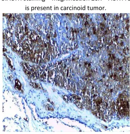
Figure 5.ACTH staining – magnification 20X – ACTH receptor is present in carcinoid tumor.
10 days after adrenal resection cortisol was 26.6 mcg/dl, K 3.9 mmol/l, calcium was normal, Mg was 1.57 mg/dl, allowing second operation – resection of lung tumor – proved to be typical carcinoid with ki-67 3%, ACTH, synaptophysin and cromograninepositive.
Evolution after second surgery: 1 day after carcinoid resection ACTH was 5.95 pg/ml (3-66), cortisol 17.38 mcg/dl –la s e e do e at „C. I. Pa ho I stitute.
12 days after carcinoid resection: ACTH 16.56 pg/ml (7.2-63); Cortisol 10.72 mcg/dl (6.2-19.4).
At 22 days: she lost 8 kg, ACTH 19.1 pg/ml, normal ALT and AST, GGT 236 UI/l, Mg 1.39 mg/dl, Ca 9.96 mg/dl.
At 47 days: she lost 15 kg, ACTH 16.5 pg/ml; cortisol 9 mcg/dl, Mg low even with supplementation.
At 78 days: GGT 259 UI/l (5-36), Mg 1.45 mg/dl, K 3.83 mmol/l, glucose 142 mg/dl, cortisol 20.87 mcg/dl, TSH 2 microUi/ml.
At 3 months: basal cortisol 11.2 mcg/dl (6.2-19.4), basal ACTH 17.46 pg/ml (3-88), cortisol 24 EET – 3.24 mcg/dl, cortisol during suppression test with Dexamethasone 1 mg overnight – 0.48 mcg/dl, PTH 12.32 pg/ml (15-165), low 25OH–D 12.8 ng/ml (30-100), UFC 40.3 mcg/24 h (21-111).
We also performed control thoracic CT – revealed left breast tumor of 0.76/1.21 cm, right adrenal with stationary aspect; portal vein was enlarged 14.5 mm; patient performed FNAB of left thyroid nodule on 20 April 2015, showing benign adenoma.
Mild kypokaliemia and hypomagnesemia, even with oral supplementation, sartan therapy and normal levels of cortisol and ACTH persisted after surgery, probably due to severe deficit of intracellular compartment, even at 3 months after carcinoid resection. Patient does not remember the 2 months prior to surgery, even if cognitive impairment is mild now.
COMMENTS
Vol. CXVIII •New Series • No. 1/2015• Romanian Journal of Military Medicine
37 family. Patient needed more than 30 days of hospital
admittance in two different hospitals and five clinics in order to obtain a good clinical result. The vital risk was high due to sepsis, denutrition, metabolic and ionic imbalance, hepatic lesions, anesthesia, brain atrophy, relative adrenal insufficiency after surgery.
There are no guidelines that state the adequate
cortisol levels to be reached before surgery, nor the duration of Ketoconazole wash-out to prevent adrenal insufficiency.
Recently the patient discovered ductal invasive left breast carcinoma, operated at 6 months after thoracic surgery and will soon start chemotherapy.
References:
1. Paraneoplastic Syndromes: An Approach to Diagnosis and Treatment, Lorraine C. Pelosof MD, PhD and David E. Gerber MD, Mayo Clin Proc. 2010 Sep; 85(9): 838–854. doi: 10. 4065/mcp. 2010. 0099
2. Paraneoplastic Cushi g s s d o e p ese ti g as
psychosis – case report, Cristina Spiroiu, Aurelian Emil Ranetti, Ana-Maria Mihai, Adina Mazilu & Nicolae Diaconu,
Endocrine Abstracts (2007) 14 P501
3. Ketoconazole in the management of paraneoplastic
Cushing's syndrome secondary to ectopic
adrenocorticotropin production. Winquist EW, Laskey J, Crump M, Khamsi F, Shepherd FA, Journal of Clinical Oncology, 1995 Jan; 13(1):157-64.