449 Chondrosarcoma in a patient with multiple osteochondromatosis
Radiol Bras 2006;39(6):449–451 Case Report
CHONDROSARCOMA IN A PATIENT WITH MULTIPLE
OSTEOCHONDROMATOSIS: A CASE REPORT AND REVIEW
OF THE LITERATURE*
Anna Caroline Nobre Gomes1
,Cláudio Régis Sampaio Silveira2
, Roberto Guido Santos Paiva2 , Antônio Gilson Monte Aragão Jr.2
, José Roberto Cavalcante Castro Jr.3
The authors report a case of chondrosarcoma in a 14-year-old female patient with multiple osteochondroma-tosis who has sought medical assistance complaining of a tumor on the left coxa. CT images of the affected limb have demonstrated osteodestructive lesion on the superior third of the left femur. The diagnosis has been made through a review of the biopsy slide from the service of origin of the patient, confirming the finding of a grade I chondrosarcoma in a chondromatous lesion. Disarticulation of the femoral neck was made because of the large extent of the lesion in muscular groups, subcutaneous cellular tissue and skin. The literature shows a wide variation in rates of malignant transformation of exostosis in patients with this dis-ease, and this is a significant complication in patients affected by this disease.
Keywords: Chondrosarcoma; Multiple osteochondromatosis; Computed tomography; Femur.
Condrossarcoma em paciente com osteocondromatose múltipla: relato de caso e revisão da literatura.
Os autores relatam um caso de condrossarcoma em uma paciente de 14 anos portadora de osteocondroma-tose múltipla, que procurou assistência médica com a queixa de tumoração na coxa esquerda. Cortes tomo-gráficos do membro afetado evidenciaram lesão osteodestrutiva no terço superior do fêmur esquerdo. O diagnóstico foi feito através da revisão da lâmina do serviço de origem da paciente, confirmando o achado de condrossarcoma grau I em lesão condromatosa. Foi realizada desarticulação do colo femoral devido ao extenso comprometimento de grupos musculares, de tecido celular subcutâneo e de pele. A literatura mostra ampla variação nas taxas de transformação maligna das exostoses em pacientes portadores desta doença, sendo esta uma importante complicação nestes pacientes.
Unitermos: Condrossarcoma; Osteocondromatose múltipla; Tomografia computadorizada; Fêmur. Abstract
Resumo
* Study developed at Hospital do Câncer do Ceará Service of Radiology, Fortaleza, CE, Brazil.
1. Student in the fourth year at Faculty of Medicine – Univer-sidade Federal do Ceará.
2. MDs, Radiologists at Hospital do Câncer do Ceará, Titular Members of Brazilian College of Radiology and Diagnostic Imag-ing.
3. MD, Orthopedist at Hospital do Câncer do Ceará, Titular Member of Brazilian Society of Orthopedics and Traumatology. Mailing address: Anna Caroline Nobre Gomes. Conjunto Cas-telo Branco, Quadra F, 187, Presidente Kennedy. Fortaleza, CE, Brazil 60357-250. E-mail: acngomes@yahoo.com.br
Received March 3, 2005. Accepted after revision May 25, 2005.
INTRODUCTION
Multiple osteochondromatosis, also called hereditary multiple exostosis or dia-physeal aclasis, is a bony metaphysis ge-netically heterogeneous disorder transmit-ted in an autosomal dominant manner, pre-senting an incomplete penetrance in female individuals(1). Kivioja et al.(2) and Schmale
et al.(1) report a 1/50,000 prevalence of this disorder, although another study has shown a 9/1,000,000 in Europe(3). Osteochondro-mas develop only in bones of endochondral origin as a result of a peripheral dysplasia
of the growth plate(1), causing displacement in areas of the plate. This is the most fre-quent type of benign bone tumor(1) and af-fects primarily long bones(1,4,5), pelvis, scapula(2,4), ankle and knee(1).
The most severe complication presented by patients affected by multiple osteochon-dromatosis is the malignant degeneration of cartilaginous exostosis into chondrosa-rcomas or, more rarely, into other types of sarcomas. A high variation is found in rates of malignant degeneration reported in the literature, with a more recent study report-ing a rate of less than 5%(6).
Peripheral chondrosarcomas are less aggressive (or possibly more accessible to surgical excision) than central tumors(5).An adequate treatment implies a complete re-section of the tumor and of the bone seg-ment involved. The prognosis for patients with dedifferentiated chondrosarcoma is poor, independently from the therapy method employed; most of patients die as a result of distant metastases within one year after the initial diagnosis(7).
In this study, we present a case of a sec-ondary grade I chondrosarcoma in a patient with multiple osteochondromatosis, and also a literature review.
CASE REPORT
A female, 14-year-old patient referred to the orthopedics ambulatory, complaining of a tumor on the left coxa. The patient pre-sented with multiple osteochondromatosis, with a painful and slow growing tumor on the lateral surface of the proximal third of the left coxa for two years. There was no familial history of multiple exostoses.
At physical examination, the patients presented with a tumor affecting the antero-lateral surface of the posterior third of the left coxa. The left coxa presented 56 cm in diameter, and the right coxa, 46 cm.
450
Gomes ACN et al.
Radiol Bras 2006;39(6):449–451 Axial CT imaging was performed in our
service, with 10 mm-thick slices, after io-dine contrast mean administration, evi-dencing an expansile osteodestructive le-sion affecting the proximal diaphysis, per-meated by gross calcifications and with a solid component invading soft tissues, in-volving the anterior muscular compartment at the level of the root of the hip. The femo-ral muscular-nervous bundle was medially dislocated by the lesion, but the vascular permeability was not affected. The lesion measured 13 × 12 × 11.5 cm in orthogo-nal planes. Bone excrescences compatible with osteochondroma were observed on the lateral and medial femoral distal diaphysis, with absence of cortical rupture.
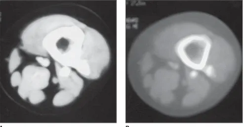
Figure 1. CT image of coxa, bone window (A) and soft tissues (B). Exostoses on the left femur posterior surface.
A B
A B
Figure 2. CT image of coxa, bone window (A) and soft tissues (B). Large expansile, osteodestructive lesion on the left femur proximal diaphysis, permeated by gross calcifications. Chondrosarcoma.
Disarticulation of the femoral neck was made because of an extensive involvement of muscular groups, subcutaneous cellular tissue and skin.
DISCUSSION
In 1786, John Hunter established the differentiation between solitary and mul-tiple osteochondromatosis, therefore the diagnosis of this disease currently is well established(3). In approximately 10% of
cases documented in the series of Schmale
et al.(1) there was no familial history of
multiple exostosis, confirming the informa-tion already existent in the literature(4). So,
the appearance of this disease was
associ-ated with a spontaneous genes mutation(4).
In the study of Schmale et al.(1) an
incom-plete penetrance of this disorder was not detected in female individuals, but rather an absent detection of small exostoses in this group because of the large deposition of fat tissue during puberty. The data ob-tained also support the concept that the gene expression is more severe in men.
The radiological finding determining the diagnosis of diaphyseal aclasis is the direct continuity of the mass with the pri-mary bone medullary cavity and absence of subjacent cortex. This occurs because of the bone metaphysis displacement during its growth through a deficient perichon-drium, and later formation of spongiosa bone inside the mass as the vessels invade the cartilage. The growth of exostoses oc-curs during the childhood, ceasing with the end of the adjacent plate growth, and may cause symptoms as a result of local tissues compression, deformities and alterations in length of bones(2).
451 Chondrosarcoma in a patient with multiple osteochondromatosis
Radiol Bras 2006;39(6):449–451 and thickening > 1 cm of the cartilaginous cap in adults(4,6,8). Due their risk of devel-opment of a malignant tumor, besides the difficulty in detecting alert symptoms in regions like the pelvis(2), it is advised that such patients are submitted to periodical follow-up with biennial x-rays(2), especially those with exostoses in the most frequent sites of sarcomatous degeneration, that is to say, the pelvis(1,2,4,5,8) and shoulder(1,5,8). In case of suspect protuberances, a mag-netic resonance imaging (MRI) should be performed(2). Chondrosarcomas rarely af-fect distal parts of limbs.
Therefore, this complication frequently occurs after puberty and is rare during childhood. Malignization is more frequent in the age range between 20 and 40 years(8), the risk increase being directly proportional to the age of the patient (1).
Chondrosarcomas may be primary or secondary, the latest appearing as a malig-nant transformation of an enchondroma or, rarely, of an osteochondroma cartilaginous cap. Depending on the site, chondrosarco-mas are further subclassified as intramed-ullary or juxtacortical, and, histologically as conventional (hyaline and/or myxoid), clear cell, dedifferentiated and mesenchy-mal variants. These sarcomas, usually, have an indolent natural history, presenting with pain and swelling.
The malignant transformation process occurs with a frequency of approximately, 5%(1,5). However, the literature presents wide variations in this rate. Rates as low as 0,6%(2) and as high as 25%(7) also are de-scribed, but probably are a function of: bias in ascertainment and incomplete detection of affected individuals who did not develop a sarcoma, but presented a familial history of hereditary multiple exostosis(1); a pro-longed follow-up period, which increases the risk of sarcomatous degeneration(1); differences of ages between study series(5); malignancy criterion(5); and grade of
spe-cialization of the services where the cases come from(5).
X-rays, CT, MRI, arteriography and bone scintigraphy provide findings sug-gesting the diagnosis of chondrosarcoma(8). The cartilage nodular growth pattern pro-duces radiographically prominent en-dosteal scalloping. The calcified matrix appears like foci of flaked density, the car-tilaginous matrix presenting with mottled, popcorn, stippled or anular calcification(8). Serial x-rays usually provide some indica-tion of malignant transformaindica-tion(4,5). X-ray as single examination in these cases is rarely conclusive; most frequently the evaluation of all the other evidences should be taken into consideration(5). Sharply de-fined margins of a lesion may become in-distinct(4,8), or the lesion may increase in size(4,5,8), or its mineralization may show a ground grass appearance(4).A relative lu-cency in a previously mineralized region of the cartilaginous cap also implies the pos-sibility of sarcomatous degeneration(4,8). Although the thickness of the demineral-ized cartilaginous cap usually is < 1 cm in benign osteochondromas, generally, in cases of malignant transformation, it is > 2 cm(4). Generally, but not always, the evi-dence of the cartilaginous cap thickness is a reliable sign of malignancy or benig-nity(4). It may be difficult to evaluate this thickness by means of CT, because of the similar radiodensity of perichondrium, bursa and other juxtaposed tissues(4). The more radiotransparent is the tumor, the higher is the probability of a high grade tumor. A slow-growing and low-grade tu-mor causes a reactive cortical thickening, while a more aggressive high-grade neo-plasm destructs the cortex and forms a soft tissue mass. CT is not useful for differen-tiating benign from malignant lesions, al-though a negative CT may rule out the pos-sibility of an exostosis malignant transfor-mation(2). Kivioja et al.(2) , in their study,
have utilized MRI as a technique for screening in the pelvis, proximal femur and upper humerus, but its utilization for early detection of chondrosarcomas and follow-up of patients with multiple exostoses yet is very limited. For Solomon(5), the diagno-sis issue is aggravated by one’s attempt to establish a precise differentiation between a benign lesion and a chondrosarcoma. By definition, the term sarcoma only can be
applied to designate a neoplasm likely to metastize, and this is what is expected to be avoided with an appropriate manage-ment of a tumor before it manifests such feature(5). Therefore, it is important to evaluate the probable behavior of the tumor rather than its precise designation(5).
REFERENCES
1. Schmale GA, Conrad EU 3rd, Raskind WH. The natural history of hereditary multiple exostoses. J Bone Joint Surg Am 1994;76:986–992. 2. Kivioja A, Hervasti H, Kinnunen J, Kaitila I, Wolf
M, Böhling T. Chondrosarcoma in a family with multiple hereditary exostoses. J Bone Joint Surg Br 2000;82:261–266.
3. Taniguchi K. A practical classification system for multiple cartilaginous exostosis in children. J Pediatr Orthop 1995;15:585–591.
4. Ostlere SJ, Gold RH, Mirra JM, Perlman RD. Case report 658: Chondrosarcoma of the proxi-mal phalanx of right fourth finger secondary to multiple hereditary exostoses (MHE). Skeletal Radiol 1991;20:145–148.
5. Solomon L. Chondrosarcoma in hereditary mul-tiple exostosis. South Afr Med J 1974;48:671– 676.
6. Bovée JVMG, Sakkers RJB, Geirnaerdt MJA, Taminiau AHM, Hogendoorn PCW. Intermediate grade osteosarcoma and chondrosarcoma arising in an osteochondroma. A case report of a patient with hereditary multiple exostoses. J Clin Pathol 2002;55:226–229.
7. Kilpatrick SE, Pike EJ, Ward WG, Pope TL. Dedifferentiated chondrosarcoma in patients with multiple osteochondromatosis: report of a case and review of the literature. Skeletal Radiol 1997;26:370–374.