1N e u ro s u rg e ry Department General Hospital of Fortaleza, Fortaleza CE, Brazil;2N e u ro s u rg e ry Service, Federal University of São Paulo, São Paulo SP, Brazil; 3Federal University of Ceará, Fortaleza CE, Brazil.
Received 30 January 2006, received in final form 23 June 2006. Accepted 9 August 2006.
Dr. Jackson A. Gondim - Av. Engenheiro Santana Junior 2977 - 60175-650 Fortaleza CE - Brasil. E-mail: [email protected]
INTRASELLAR PRESSURE AND TUMOR
VOLUME IN PITUITARY TUMOR
Relation study
Jackson A. Gondim
1, Osvaldo I. Tella Jr.
2, Michele Schops
3ABSTRACT -Objective:To determine if there was a relationship between intrassellar pressure (ISP) and p i t u i t a ry tumor volume. Method:Between August 2002 and May 2004, 60 patients aged between 13 and 75 years old (39 males), having a pituitary adenoma were submitted to an endoscope transseptal appro a c h . During the surg e ry and before tumor resection, 2 mm of the sella’s floor were removed and a 1.5 mm dur-al opening made to place a transducer into the pituitary adenoma. The transducer was connected to a p re s s u re monitor. Results:The intrasellar pre s s u re, ranged from 2-51 mmHg and was measured based on the classification of Hard y - Vezina. The most elevated was in the type II macro adenomas with 32.6 mmHg, sharply superior to the value of a normal intracranial pre s s u re . Conclusion:These values showed that the m a c roadenomas confined to the sella, without destruction of the floor and integrity of the diaphragm, type II of Hardy-Vezina, presented a value of ISP much higher than intra-extrasellar macroadenoma’s.
KEY WORDS: pituitary adenoma, intrasellar pressure, endonasal approach.
Relação entre pressão intra-selar e volume de tumor de hipófise
Objetivo:Determinar se existia uma relação entre a pressão intraselar (ISP) e o volume de tumor de hipó-f i s e . Método:E n t re agosto de 2002 e maio de 2004, 60 pacientes com idades variando entre 13 e 75 anos (39 homens), port a d o res de adenoma hipofisários foram operados por via transesfenoidal. Durante o ato c i r ú rgico e antes da resseção do tumor, uma osteotomia de 2 mm foi realizada no assoalho selar e uma a b e rtura de 1,5 mm na duramater para a introdução de um transdutor dentro do tumor. O transdutor foi conectado a um monitor de pressão e esta foi medida por 2 minutos. Resultados:A pressão intra-selar variou entre 251 mmHg e a correlação entre tamanho do tumor e ISP foi baseada na classificação de Hard y -Vezina. A média da ISP mais elevada foi encontrada nos macroadenomas tipo II com 32,6 mmHg, nitida-mente superior a pressão intra selar norm a l . Conclusão:Estes valores mostram que os macro a d e n o m a s confinados a sela sem destruição do assoalho selar e com integridade do diafragma, classificados como tipo II de Hardy-Vezina, apresentam uma ISP muito superior aos outros adenomas.
PALAVRAS-CHAVE: adenoma hipofisário, pressão intra-selar, via transesfenoidal.
T h e re are studies in the literature corre l a t i n g intrasellar pre s s u re(ISP) and adenoma blood flow1, ISP and stalk compression syndrome2, ISP and endo-crine function3 , 4, ISP and headaches3, pituitary vol-ume and headache5, ISP and pituitary tumor apo-p l e x y6, but there are no specific studies corre l a t i n g ISP and tumor volume.
The walls of the sella turcica are a relatively rigid s t ru c t u reand under normal circumstances, may serv e to protect the pituitary gland from trauma and sur-rounding pressure fluctuations. The growth of a tu-mor within the sella, a normally inelastic space, is likely to cause an increase of ISP. It has been
the highest ISP is found in macroadenomas confined to an enlarged sella without disruption of the floor and with integrity of the diaphragm as type II tumor of Hardy Vezina6.
The objective of this study is to determine if there is a relationship between ISP and pituitary tumor vol-ume.
METHOD
Between August 2002 and May 2004 sixty consecutive patients (39 males) with pituitary adenomas were operat-ed by transnasal transsphenoidal endoscopic surg e ry and had their ISP measured during surg e ryfor pituitary adeno-ma at our institution. Their age was between 13 and 75 years old. Among the 60 cases, studied, 42 (70%) were func-tional adenomas (eighteen producing adre n o c o rt i c o t ro p i n h o rmone, fourteen producing gr owth hormone, six pro-ducing prolactine hormone and four cases of plurihorm o n-al adenomas) and eighteen null cells adenomas. All patients w e re ambulatory. No patients were using glucocort i c o i d s or had pituit ary apoplexy, but some patients had adeno-mas with cystic component. The patients with horm o n a l hypopituitarism, the compensation usually begins in the transoperative period. The study was approved by the Institutional Review Board of General Hospital of Fort a l e z a , an informed consent was obtained from each patient.
All patients underwent pituitary computerized tomog-raphy (CT) and magnetic resonance image (MRI) at 1.5 T. The MRI examination included coronal and sagittal T1-weighted spin-eco sequences with a maximum section thick-ness of 3 mm, before and after intravenous administration of a gadolinium-based contrast medium. For the estima-tion of the tumor volume, it was assumed that the
pitu-i t a ry tumors had an ellpitu-ipsopitu-id form5 , 7 - 9. If the tumor was
l a rge and multilobed, the tumor volume was assumed to consist of separat ed ellipses and the sum of each volum e was calculated.
Tumors were subdivided according to radiological
clas-sifications of Hardy and Ve z i n a6. The tumors were
classi-fied as microadenoma grade 0 where they weren´t visible on imaging. Immunohistochemical characteristics of tumor were available in all patients. The tumor volume distribu-tion classificadistribu-tion is showed in the Table.
P re-operative pituitary function evaluation was per-f o rmed in all patients, and included basal serum per-free T4, f ree T3, thyroid stimulation hormone, follicle-stimulating hormone, luteinizing hormone, testosterone (men), corti-sol levels on multiple days, adrenocorticotropic hormone, p rolactin, growth hormone and somatomedin-C and a glu-cose tolerance test with GH.
The transsphenoidal exploration was carried out under
general anesthesia with normotension and normocapnia. The pituitary sella’s floor was exposed through a transnasal
transseptal approach assisted by endoscope1 0. On entering
the sphenoid air sinus a 2-mm diameter window of the bo-ne sella’s floor was remove and a 1.5 mm dural opening was made to allow the transducer placed into a needle in the tumor mass without extravasation of intrasell ar con-tent. Once the transducer placed the needle is taken off and the ISP measured. In some patients with cystic compo-nent a careful attention is made for extravasations of tumor component. If there were tumor component extravasations, the ISP measurement would be stopped and the patient wouldn't count. The fiberoptic transducer is located at the tip and has a 1.3 mm diameter. Sixty seconds later, after a stable pre s s u re obtained, the pre s s u re was re c o rded, the transducer removed and the tumor resection initiated. We used the Coodman Intracranial Pre s s u re Monitoring Kit (Camino Laboratories San Diego, CA) to determinate the I S P. The kit uses a fiberoptic transducer connected to a pre s-sure monitor.
The statistic analysis was done using the test of Levene to see the homogeneity of variances. The F test was done to know if there was a diff e rence between the five intrasel-lar pre s s u re median groups. The individual comparisons between the pairs of the five group of ISP were done using the multiple comparasions test of Bonferroni. Statistic analy-sis using the F test was done with the prolactine medium in each group of pa tients (Cushing, acromegaly and nul l cells adenoma). The ISP was also compared into the four g roup of pathology (acro m e g a l y, Cushing, pro l a c t i n o m a and nulls cells adenoma). Variables with significant pro b-ability values (p< 0.05) were considered a possible signifi-cant. Statistical evaluation w as perf o rmed with commer-cially available statistical software (SPSS version 10.0).
RESULTS
Tumor volume was measured in all adenomas and varied between 0 cm3(in six patient with clinical and laboratorial diagnosis of Cushing´s disease, but with no radiological image) and 134.5 cm3in a non-func-tioning pituitary adenoma with a cystic component. The tumors of this series were solid in 51 patients (85%) and had a cystic component in 9 (15%).
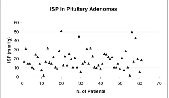
The ISP was measured in all 60 patients and rang-ed from 2-51 mmHg with mean (±sd) of 18.7±10.8 and a median of 16 mmHg (Fig 1). The test of homog e n e-ity of variances Levene Statistic showed significant (1.639 p=0.178) the amostrage of ISP measure m e n t . The median comparison of the five groups of ISP showed all diff e rent (F 17.69 p=0.001). The statistic
Table. Tumor volume distribution in Hardy-Vezina classification.
Hardy-Vezina´s Grade Grade 0 Grade 1 Grade 2 Grade 3 Grade 4
study was done to known the specific pair-wise analy-ses and showed that ISP of group 2 of Hard y - Ve z i n a was diff e rent in the pair comparison of all other g roup (0 an2 p=0.026; 1 and 2 p=0.0001, 2 and 3 p=0.0001; 2 and 4 p=0.0001).
Six patients showed immunohistochemical char-acteristics of prolactin adenoma and were excluded f rom the analysis of correlation between ISP meas-u rements and sermeas-um prolactin. The prolactin sermeas-u m was ranged from 5-57µg/L with mean of 24.3±8 . 2 and a median of 25.1µg/L, The statistic analysis bet-ween ISP and prolactine in the group of patients with a c romegaly (R=0.767 p=0.075), Cushing (R=0.588 p=0.491) and null cels adenoma (R=–0.161 p=0.600) do not show significant statistic variation.
In the analyses of diff e rent immunohistochemi-cal characteristics types of pituitary tumors in this series and the ISP re g i s t e r, indicate the absence of relationship between ISP and tumor type (R=4.543 r=0.022).
DISCUSSION
The measurement of ISP is simple, re p ro d u c i b l e and can be done using standard equipment as the same for intra cranial pre s s u remonitoring. The nor-mal ISP is unknown but is unlikely to exceed the ICP. T h e re are no studies of ISP in patients without intra-sellar tumor in the literature, but some studies1 , 4 showed that when ISP was measured in patients with microadenoma and empty sella the ISP were 13,5±3 mmhg. In our series we measured the ISP in six
pa-tients with Cushing´s disease, and no tumor found in the MRI, but with petrous sinus sample confirm a t i o n the pituitary origin of the disease. The ISP measure d in these six cases was 15 mmHg, 6 mmHg and 2 mmHg, 8 mmHg, 2 mmHg and 8 mmHg re s p e c t i v e-l y, with an average of 6.83±6.5 mmHg, mediam of 7 mmHg. The observations in others studies suggest that normal ISP is of the order of 10 mmHg4.
The perfusion of the anterior pituitary depends on the balance between the portal venous input and the local tissue pre s s u re. The stru c t u re of the long p o rtal vessels is similar to that of peripheral veins1 1 - 1 3 and even a minor elevation in the ISP would reduce blood flow to the pituitary. Normal portal venous pressure is unlike to greatly exceed systemic venous p re s s u re. The perfusion pre s s u reof the anterior pitu-itary must be considerably lower than the arterially supplied tissue and may be seriously compro m i s e d by a relatively small ISP’s rise2. The observation of lar-ge tumors with high ISP and no significant compro-mise in the pituitary function is probably due to a direct extraportal arterial supplies10.
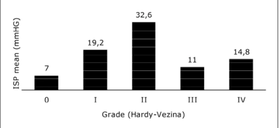
The ISP was measured separately in the patients a c c o rding to Hard y - Ve z i n a ’s classification, and it was verified that the ISP was more elevated in the macro-adenomas type II with medium of 32.6 mmHg, sharply superior to the normal ICP. In the adenomas type 0 the ISP was (7 mmHg), in I (19.2 mmHg), in II (32.6 mmHg), in III (11 mmHg) and in IV (14.8 mmHg) (Fig 2). These values show that the macroadenomas confined into the sella, without destruction of the
floor, and integrity of the diaphragm, presented an ISP much higher than those tumor with both intra and extrasellar extension (Fig 3). It does not seem c o h e rent, but the explanation can be the following: while the tumor is a microadenoma (grade 0 or 1) it will be contained inside the sella, a closed cavity. The g rowth of the tumor inside this cavity will incre a s e ISP progressively. In the moment that the sella is no m o re competent to contain the pituitary and the tu-m o r, there will be a ru p t u reof the content of the sel-la into the cranial cavity and/or the sphenoid sinus. In case of an almost ruptured sella, there will be an ISP in its maximum value. In this series, this value was 51 mmHg. In the series of Arafah3this value re a c h e d 60 mmHg and in the series of Kru s e1it was of 62 mmHg. When the tumor leaves the limits of the sel-la cavity, the universe of the adenoma in terms of ISP is no more the sella, but the cranial cavity. At this
time ISP tends to be equal to ICP, having a decrease in relation to ISP of the intrasellar adenomas.
When there is an important growth of the ade-noma, the ISP can influence the ICP, mainly if there is a blockade of the cerebral blood flow. If the ade-noma does not grow towards the diaphragm, but down, destroying the floor of the sella, (free tumor within the sinus), the ISP will tend to fall, as a con-sequence of equilibration with atmospheric pre s s u re via the sphenoid air sinus4. An important factor involved in the genesis of ISP, is the speed of the tumor growth and the ability of the sella’s walls to modulate this growth. In general, patient with fast g rowing tumor confined to the sella (pituitary a p o p l e x y, metastases tumors), are more susceptible to present higher ISP than those with slow growing tumor.
In patients with hyperprolactinemia and no pro-Fig 2. Shows the distribution of tumor grade and median ISP value according to the
Hardy-Vezina classification.
lactin secretor tumor, there were no corre l a t i o n between the ISP and the level of prolactine.
In patients with hypopituitarism there is also no correlation between this situation and ISP.
In patients with predominantly cystic tumors and with thin walls there are possibilities of error in intro-ducing the transducer into the cystic part of the tu-m o r. The exit of the cystic liquid tu-may give a negative false result.
In conclusion, the pituitary adenomas classified as grade II of Hard y - Vezina are probably the tumor with the highest ISP, higher than the normal ICP.
Acknowledgements -The authors grateffuly thank Cecilia Schops Oliveira for the English version of this paper.
REFERENCES
1. K ruse A, A s t rup J, Gyldensted C, Cold GE. Hyperprolactinaemia in patients with pituitary adenomas: the pituitary stalk compression syn-drome. Br J Neurosurg 1995;9:453-457.
2. Lees PD, Pickard JD. Hyperprolactinemia, intrasellar pituitary tissue pressure, and the pituitary stalk compression syndrome. J Neurosurg 1987;67:192-196.
3. Arafah BM, Prunty D, Ybarra J, Hlavin ML, Selman WR. The dominant role of increased intrasellar pre s s u re in the pathogenesis of hypopitu-itarism, hyperprolactinemia, and headaches in patients with pituitary adenomas. J Clin Endocrinol Metab 2000;85:1789-1793.
4. Lees PD, Fahlbusch R, Zrinro A, Pickard JD. Intrasellar pituitary tis-sue pre s s u re, tumor size and endocrine status, an international com-parison in 107 patients. Br J Neurosurgery 1994;8:313-318.
5. Levy MJ, Jager R, Powell M, Matharu M, Meeran K, Goadsby PJ. Pi-tuitary volume and headache. Arch Neurol 2004;61:721-725. 6. Zayour DH, Selman WR, Arafah BM. Extreme elevation of intrasellar
p re s s u rein patients with pituitary tumor apoplexy: relation to pitu-itary function. J Clin Endocrinol Metab 2004;89:5649-5654.
7. H a rdy J, Vezina JL. Transsphenoidal neuro s u rgery of intracranial neo-plasm. In Tompson RA, Green JR (eds). Advances in Neurology: New York, Raven Press, 1976;15:261-275.
8. Di Chiro G. The width (third dimension) of the sella turcica. Am J Roent-genol Rad Therapy & Nuclear Med 1960;84:26-37.
9. Lundin P, Pedersen F. Volume of pituitary macroadenomas: assessment by MRI. J Comput Assisted Tomogr 1992;16:519-528.
10. Gondim J, Schops M, Tella OI Jr. Transnasal endoscopic surgery of the sellar region: study of the first 100 cases. A rq Neuropsiquiatr 2003; 61:836-841.
11. G o rczyca W, Hardy J. Microadenomas of the human pituitary and their vascularization. Neurosurgery 1988;22:1-6.
12. Daniel PM, Prichard MML. Studies of the hypothalamus and the pitu-itary gland with special re f e rences to the effects of transaction of the pituitary stalk. Acta Endocrinol 1975;80(Suppl 201):S1-S216. 13. Popa G, Fielding U. A portal circulation from the pituitary to the