JSCS–4272 535.375+535.372
Original scientific paper
Synthesis and properties of
5,10,15,20-tetrakis[4-(3,5-dioctyloxy-benzamido)phenyl]porphyrin and its metal complexes
WENHUI LIAN, YUANYUAN SUN, BINBIN WANG, NING SHAN and TONGSHUN SHI*College of Chemistry, Jilin University, Changchun 130023, P. R. China (Received 16 May, revised 3 October 2011)
Abstract: A novel 5,10,15,20-tetrakis[4-(3,5-dioctyloxybenzamido)phenyl]-porphyrin and its transition metal complexes are reported in this paper. Their molecular structures were characterized by elemental analysis as well as IR,
1H-NMR and UV–Vis spectroscopy. Their spectroscopic properties were
studied by Raman and fluorescence spectroscopy, and X-ray photoelectron spectroscopy (XPS). The fluorescence quantum yields were measured at room temperature. The fluorescence intensity of the porphyrin ligand was stronger than the intensity of the complexes. There were large differences in the Raman spectrum of the porphyrin ligand and those of the metal complexes due to changes in the symmetry of porphyrin plane. In the XPS spectra, the replacement of the free-base protons by a metal ion to form the metalloporphyrin not only increases the symmetry of the molecule, but also introduces an electron withdrawing group into the center of the porphyrin ligand, which increases the N1s binding energy.
Keywords:porphyrin; transition metal complex; XPS spectra; Raman spectra; fluorescence spectra.
INTRODUCTION
Recently, investigation of porphyrins has become of increasing interest.1,2
Porphyrins and metalloporphyrins are functional molecules that are used for a va-riety of applications and devices, such as optoelectronic, luminescent and, mole-cular logic devices, supramolemole-cular self-assembly, solar energy harvesting sys-tems, photonic materials, and therapeutics.3–7.These applications are affected by
the diverse electrochemical and photophysical properties of the porphyrins and the ability to fine-tune these properties by the exocyclic substituents on the mac-rocycle and via the choice of chelated metal ion.
Porphyrins have many desirable features, such as high stability, intense ab-sorption of sunlight, a highly conjugated plane and a small gap between the
est occupied molecular orbital (HOMO) and lowest unoccupied molecular orbital (LUMO) energy level. The highly conjugated π-electron skeleton of a porphyrin provides an adequate number of π electrons and π–π* transitions normally give a strong absorption in the UV and visible regions of the spectrum.8
Metallopor-phyrins generally have high thermal stability and show strong electronic transi-tions in the visible and ultraviolet regions. In order to evaluate the applications of metalloporphyrins, it is necessary to understand the electronic structure and pho-tophysical properties. Raman spectroscopy, fluorescence spectra, X-ray photo-electron spectroscopy, etc, have been widely used to study the porphyrin ligand and metalloporphyrins, and the results could provide important information to determine their electronic structure. X-Ray photoelectron spectroscopy (XPS) is a powerful tool for the characterization of both the chemical composition and the electronic environments of each atom in a molecular system.9–11
To date, the synthesis and application of the substituent tetraphenylporphyrin (TPP) bearing special functional groups have caused great interest, but those bearing an amide group as side chains have been little reported. In this research, a novel TPP derivative bearing an amide group and its transition metal complexes were synthesized. The benzene core was replaced by a rigid porphyrin core, which is linked via amide bonds to four triphenylenes. Since the porphyrin di-mension is much larger than that of a benzene ring and the symmetry changes from three-fold to four-fold, an entirely different self-assembly via hydrogen bonding was to be expected. Their structures were characterized by their UV– –Vis, IR and 1H-NMR spectra and the changes in the fluorescence, Raman
spec-tra and XPS behavior of these compounds were investigated.
RESULTS AND DISCUSSION
Physical, analytic and spectral data for 7a–7d
5,10,15,20-Tetrakis[4-(3,5-dioctyloxybenzamido)phenyl]porphyrin (7a). Purple solid. Yield: 62.4 %; Anal. Calcd. for C136H178N8O12: C, 77.12; H, 8.42; N, 5.26
%. Found: C, 77.16; H, 8.48; N, 5.20 %. IR (KBr, cm–1): 3314 (N–H, pyrrole),
2925, 2855 (C–H), 1674 (C=O), 1523 (N–H, amide), 1245 (Ar–O–C), 966 (N–H, pyrrole), 721 (–(CH2)n–, n > 4). 1H-NMR (500MHz, CDCl3, δ / ppm): 8.92 (8H,
s, pyrrole ring), 8.30 (4H, s, –NH–CO–), 8.20–8.26 (8H, d, J = 30 Hz, o–C6H4),
8.08–8.03 (8H, d, J = 25 Hz, m-C6H4), 6.98–7.04 (8H, d, J = 30 Hz, o–C6H3),
6.64–6.69 (4H, d, J = 25 Hz, m–C6H3), 4.10–4.22 (16H, m, –O–CH2–), 1.78–1.98
(16H, m, –O–C–CH2–), 1.49–1.64(16H, m, –CH2–CH3), 1.21–1.40 (64H, m, –O– C–C–(CH2)4–C–CH3), 0.85–0.95 (24H, t, J = 25 Hz, –O–C–C–(C)4–C–CH3),
–2.74 (2H, s, N–H, pyrrole). UV–Vis (CHCl3, λmax / nm): 425 (Soret band), 520,
555, 595, 650 (four Q bands).
8.35; N, 5.02 %. Found: C, 74.99; H, 8.30; N, 5.07 %. IR (KBr, cm–1): 3285 (N–H,
imide), 2924, 2851 (C–H), 1664 (C=O), 1527 (N–H, amide), 1245 (Ar–O–C), 996 (N–Zn), 721 (–(CH2)n–, n > 4). 1H-NMR (500 MHz ,CDCl3, δ / ppm), 8.99
(8H, s, pyrrole ring), 8.12–8.30 (8H, d, J = 40 Hz, ο–C6H4), 8.06 (4H, s,–CON–H), 7.95–7.96 (8H, d, J = 5 Hz, m–C6H4), 7.26–7.27 (8H, d, J = 5 Hz, o–C6H3),
6.96–6.87 (4H, t, J = 22.5 Hz, m-C6H3), 3.92–4.03 (16H, t, J = 7.5 Hz, –O–CH2–), 1.68–1.98 (16H, m, –O–C–CH2–), 1.22–1.52 (80H, m, –C–C–(CH2)5–CH3),
0.861–0.905 (24H, t, J = 11 Hz, –C–C–(C)5–CH3). UV–Vis (CHCl3, λmax / nm):
425 (Soret band), 551, 592 (two Q bands).
Manganese 5,10,15,20-tetrakis[4-(3,5-dioctyloxybenzamido)phenyl]por-phyrin (7c). Dark green solid. Yield: 82.4 %; Anal. Calcd. for C136H176MnN8O12Cl: C, 74.12; H, 7.99; N, 5.04 %. Found: C, 74.07; H, 8.04; N, 5.08 %. IR (KBr, cm–1):
3282 (N–H, imide), 2925, 2854 (C–H), 1677 (C=O), 1519 (N–H, amide), 1244 (Ar–O–C), 1008 (N–Mn), 721 (–(CH2)n–, n > 4). UV–Vis (CHCl3, λmax / nm):
480 (Soret band), 585, 625 (two Q bands).
Cobalt 5,10,15,20-tetrakis[4-(3,5-dioctyloxybenzamide)phenyl]porphyrin (7d). Purple red solid. Yield: 85.7 %; Anal. Calcd. for C136H176CoN8O12: C, 75.21; H,
8.11; N, 5.09 %. Found: C, 75.14; H, 8.16 N, 5.15 %. IR (KBr, cm–1) 3280 (N–H
(imide)), 2925, 2854 (C–H), 1658 (C=O), 1517 (N–H, amide), 1252 (Ar–O–C), 1000 (N–Co), 717 (–(CH2)n–, n > 4). UV–vis (CHCl3, λmax / nm): 415 (Soret
band), 530, 675 (two Q bands). UV–Vis spectra
The electronic absorption spectra of porphyrins result from electronic tran-sitions from the ground state (S0) to the two lowest singlet excited states S1 and
S2. The S0→S1 transition gives rise to weak Q bands in the visible region while
the S0→S2 transition produces a strong Soret band in the near UV region.12,13
The absorption bands of 7a appear at 425, 520, 555, 595 and 650 nm. Compared with the absorption bands of TPP, the absorption bands of 7a are red-shifted.14 Possibly, this is because the substituent group on the phenyl group at the meso-position of the porphyrin ring is an electron-donating group, thereby enabling the electronic density on the phenyl ring to strengthen. Thus, the phenyl ring con-jugates to a certain degree with the porphyrin macrocycle. This kind of conjuga-tion acconjuga-tion causes a reducconjuga-tion of the electron transiconjuga-tion energy of the porphyrin macrocycle, resulting in red shifts of the absorption bands.
and the absorption frequencies shift. When the metal ion coordinates with por-phyrin ligand, the symmetry of the molecule is changed from D2h to D4h, the
cleavage degree of the molecular orbital decreases and the degeneracy increases; hence, the number of Q bands therefore decreases.
Infrared spectra
The IR bands at 3314 and 966 cm–1 of 7a are attributed to N–H stretching
and bending vibrations of the porphyrin ligand core, respectively, but they were absent in the spectra of the complexes, because the hydrogen atom in the N–H bond was replaced by a transition metal ion.15 In addition, a new band in the
spectrum of 7b, 7c and 7d appeared at 996, 1008 and 1000 cm–1, respectively, which is characteristic of metalloporphyrins. The bands at about 3280–3285 cm–1
of the complexes are assigned to N–H stretching vibrations of the amide group on the side chains, but in the spectrum of the porphyrin ligand, it overlapped the N–H stretching vibration of the porphyrin core and could not be distinguished. The bands in the range of 1652–1670 cm–1 are assigned to C=O stretching
vib-ration (amide I). The bands at about 1517–1521 cm–1 are assigned to N–H in-
-plane bending and C–N stretching (imide II). The bands at about 1245–1252cm–1
are assigned to Ar–O–C stretching vibrations.The bands at 717–721 cm–1 are
as-signed to the methylene in-plane rocking vibration of a straight alkyl chain con-taining more than four carbon atoms.
Fluorescence spectra
Excited-state processes in porphyrins are extremely important for their appli-cation in molecular devices. The room temperature fluorescence spectra of the porphyrin ligand and metalloporphyrins in chloroform (2×10⁻⁶ mol L–1) were
re-corded. No fluorescence signal was detected for the metalloporphyrins 7c and 7d
Fig. 1. Excitation spectra of
under the employed experimental conditions. The excitation spectra of 7a and 7b are shown in Fig. 1. In the region of 350–700 nm, the excitation spectra are ap-proximately mirror images of the absorption spectra, indicating that they corres-pond to a similar electron transition process.
The emission spectra of 7a and 7b are shown in Fig. 2 for an excitation wavelength of 420 nm. The emission spectra data of 7a and 7b are given in Table I. Excitation to the S2 (B band) and the S1 (Q band) in porphyrin compounds
re-sults in fluorescence. The fluorescence of the S2 (B band) is attributed to the
transition from the second excited singlet state S2 to the ground state S0, S2→S0,
and it corresponds with the Soret band in the electronic absorption spectra. In addition, fluorescence of the Q band is attributed to the transition from the lowest excited singlet state S1 to the ground state S0. The fluorescence of S2→S0 was
too weak to be observed in this study, owing to light scattering and resorption of strong Soret absorption band. It is well known that metal-free porphyrins usually show two strong emission bands around 650 and 720 nm.16 The fluorescence
bands of 7a were at 653 and 716 nm.Compared with the fluorescent bands at 650 and 713 nm of TPP, the emission peaks of porphyrin ligand were red-shifted by 3 nm.The fluorescence bands of the Zn complex were at 597 and 644 nm, which were red-shifted compared to ZnTPP (592 and 641 nm).These red shifts may be due to the interaction of the arylamido group with the conjugated π-electron sys-tem of the porphyrin macrocycle. The conjugation of the porphyrin macrocycle is affected by the electronic donating groups. The conjugation is enhanced when the alkylamide groups are linked on the phenyl group.
Fig. 2. Fluorescence spectra of 7a and 7b in chloroform.
sample ZnTPP
sample ZnTPP
ZnTPP sample
F A
F A
φ = φ
where Fsample and FZnTPP are the measured fluorescence integral areas of the
sample and the reference ZnTPP, respectively. Asample and AZnTPP are the
absor-bance of the sample and the reference, respectively. φsample and φZnTPP (φZnTPP =
= 0.033)17 are the quantum yields of the sample and the reference ZnTPP at same
excitation wavelength. The quantum yields of the porphyrin ligand are much lower than that of TPP (φTPP = 0.1118).As is known, porphyrins can be considered
as specific donor–acceptor systems, and the decrease in the fluorescence quan-tum yields of the porphyrin ligand in comparison to that of TPP could be the re-sult of intramolecular energy migration or electron transfer from the donor part of the molecule to the acceptor part. The fluorescence quantum yields of 7a and 7b were very small. Thus, the excited state S1 was primarily deactivated by
radia-tionless decay in porphyrins. This indicates fairly certainly that the spin forbid-den process S1→Tn is dominant for the radiationless deactivation of S1 in
por-phyrin compounds. The fluorescence yield of 7b was much smaller than that of 7a because zinc weakened the fluorescence radiation.
TABLE I. Emission spectra data and quantum yields (φf) of 7a and 7b
Compound Q(0–0) / nm Q(0–1) / nm φf
7a 653 716 0.064
7b 597 644 0.032
Resonance Raman spectra
The resonance Raman spectra of the porphyrin ligand and complexes were obtained by excitation at 514.5 nm. The important Raman frequencies and their assignments are listed in Table II. The Raman spectra of the porphyrin ligand and the Mn complex are shown in Fig. 3. The resonance Raman spectra of tetraphe-nylporphyrin derivatives have been studied extensively.19–21 Thus, the
assign-ments of Raman bands of porphyrin ligand and complexes are only discussed briefly here.
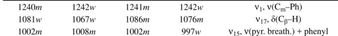
TABLE II. Raman spectra and assignment of the porphyrin ligand and its complexes (vs: very strong, s: strong, m: medium, w: weak)
Compound
Assigned
7a 7b 7c 7d
1553vs 1550s 1573vs 1561vs ν2, ν(CβCβ)
1496m 1494m 1497s 1502m φ5, phenyl
1455s 1454w 1456m 1455w ν3, ν(CαCm)
1360m 1353s 1372m 1367s ν4, ν(Cα–N) / ν(CαCβ)
1240m 1242w 1241m 1242w ν1, ν(Cm–Ph)
1081w 1067w 1086m 1076m ν17, δ(Cβ–H) 1002m 1008m 1002m 997w ν15, ν(pyr. breath.) + phenyl
TABLE II. Continued
Compound
Assigned
7a 7b 7c 7d
963w – – – ν6, ν(pyrrole breath.)
719w 717w 713w 710w π3, phenyl
404w 397w 395 ν(M–N)
335 – 313w 312w ν8, ν(pyr. translation)
Fig. 3. Raman spectra of the porphyrin ligand and the Mn complex.
In the 900–1650 cm–1 high-frequency region of the Raman spectra of
por-phyrin ligand and complexes, Raman bands generally arise from the totally sym-metric vibrational modes, such as CβCβ, CαCβ, CαCm, pyrrole quarter-ring, and
pyrrole half-ring stretching. The wavenumber positions of Raman bands in the high-frequency region are sensitive to the core size, axial ligation and electron density of the central metal ion. In this region, the band at 1553 cm–1 of
porphy-rin ligand was assigned to the CβCβ stretch ν2 mode, which was up-shifted to
1573 cm–1 in the Mn complex and to 1561 cm–1 in Co complex, but it was
down-shifted to 1548 cm–1 in the Zn complex. The ν2 mode was observed with
en-hanced intensity in the complexes. In fact, it is one of the most intense bands in the high-frequency region. The band at 1496 cm–1 of porphyrin ligand was
positions. Raman bands in 1300–1450 cm–1 were due to the out-of-phase coupled
CαCβ/CαN stretching modes. The 1360 and 1330 cm–1 bands of the porphyrin
ligand were assigned to the ν4 and ν20 mode, respectively. The ν4 mode of 7b, 7c and 7d appeared at 1353, 1372 and 1367 cm–1, respectively. The ν20 mode of 7c shifted to 1341 cm–1, while the ν20 mode of 7b and 7d were too weak to be
observed in the present experiments. The 1240 cm–1 band of the porphyrin ligand
and the 1240–1242 cm–1 band of the complexes were attributed to the Cm−ph
stretching ν1 mode. The band at 1081 cm–1 of the porphyrin ligand was assigned
to the vibrations of the pyrrole Cβ–H stretching ν9 mode, which shifted to 1067,
1086 and 1076 cm–1 in 7b, 7c and 7d, respectively. The band at 1002 cm–1 of the
porphyrin ligand was assigned to the vibration of pyrrole breathing and phenyl stretching ν15 mode, which did not shift in the Mn complex, but was up-shifted
to 1008 cm–1 in the Zn complex and downshifted to 997 cm–1 in the Co complex.
The band at 963 cm–1 of the porphyrin ligand was assigned to pyrrole breathing
ν6 mode, but it was absent in the spectra of the metalloporphyrins because the
hydrogen atom in the N−H bonding had been replaced by a metal ion. In the low- -frequency region, the Raman bands of the metalloporphyrin complexes were very different to those of the porphyrin ligand because the structures or vibratio-nal dynamics, especially around the CαCmCβ bond-angles, were altered by the
metal ions. For the porphyrin ligand, the only weak Raman band was observed at 335 cm–1, which was assigned to the ν8 mode. The ν8 mode consists of the in-
-plane translational motion of the pyrrole, which can be described as a uniform breathing of the whole porphine ring accompanied by an in-plane deformation of CαCmCβ in the pyrrole ring.22
X-Ray photoelectron spectra
The XPS spectra of the porphyrin films were obtained. All elements of the porphyrins, including C, N, O and different metals for the metalloporphyrins, were found in the XPS spectra of their films grafted on Si (100). The X-ray pho-toelectron spectra data of the porphyrin ligand and its complexes are given in Table III. The XPS spectra of the N1s region are shown in Fig. 4.
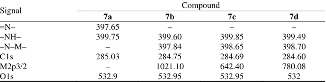
TABLE III. Binding energies (eV) of the porphyrin ligand and complexes obtained in the XPS experiments
Signal Compound
7a 7b 7c 7d
=N– 397.65 – – –
–NH– 399.75 399.60 399.85 399.49
–N–M– – 397.84 398.65 398.70
C1s 285.03 284.75 284.69 284.60
M2p3/2 – 1021.10 642.40 780.08
Free base porphyrins exhibit two distinct N1s signals corresponding to the
imine (–C=N–) nitrogen and the pyrrole (–NH–) nitrogen. The pyrrole nitrogen usually has a higher N1s binding energy than the imine nitrogen.23–26 The N1s
binding energies of these species were ≈400 and ≈397 eV, respectively, so that they should have been readily resolved in the performed experiment. In the por-phyrin ligand 7a, two N signals could be observed, one related to the N atoms of unprotonated porphyrin rings (397.65 eV), and the other to the N atoms of pro-tonated porphyrin-rings and the N atoms of the amide around the porphyrin-ring (399.75 eV). The N1s spectra of the metalloporphyrins also exhibited two signals,
the signal with the lower binding energy can be assigned to the metal-binding nitrogen atoms. The binding energy of the protonated porphyrin-ring nitrogen co-incides with the amide nitrogen, and their band energies were higher than for the unprotonated porphyrin-ring. This is because of the protonation of the imine nit-rogen atoms, which decreases the intensity of the N1s peak at the lower binding
energy.
Fig. 4. N1s region of the porphyrin ligand and its complexes.
The metal ion region of the XPS spectra of the films of metalloporphyrins 7b, 7c, and 7d on the Si (100) surface are shown in Fig. 4. The metalloporphryins exhibit a 2p3/2 signal. The M2p3/2 (M = Zn, Mn or Co) region of 7b, 7c and 7d shows sharp peaks with bonding energy at 1021.10, 642.40 and 780.08 eV, res-pectively, and it is lower than the M2p3/2 binding energy of corresponding metal
EXPERIMENTAL General synthesis procedures
Chemicals and solvents were obtained from various commercial sources and used with-out further purification unless otherwise stated. Pyrrole was freshly distilled before use. DMF was pre-dried over activated 4Å molecular sieve and vacuum distilled from calcium hydride (CaH2) prior to use. Dry CHCl3 and Et3N were obtained by redistillation from CaH2. Acetone was dried with anhydrous magnesium sulfate. Anhydrous potassium carbonate was dried under vacuum at 80 °C for 30 min.
The synthesis procedures are illustrated in Schemes 1 and 2. 5,10,15,20-Tetra-(4-nitro-phenyl)porphyrin was synthesized through the reaction between pyrrole and p -nitrobenzal-dehyde in the presence of lactic acid according to a published procedure.27
5,10,15,20-Tetra(4-aminophenyl)porphyrin (TAPPH2) was prepared using SnCl2/HCl according to the method of Kruper.28
Scheme 1. Synthetic route to 3,5-dioctyloxybenzoyl chloride.
Methyl 3,5-dioctyloxybenzoate(2). Compound 1 (3.36 g, 20.0 mmol), anhydrous
potas-sium carbonate (8.28 g, 60.0 mmol) and a catalytic amount of KI in acetone (150 ml) were refluxed for 0.5 h and then 1-bromooctane (9.6 g, 50 mmol) was added in the solution. The reaction mixture was gently refluxed for 36 h under an N2 atmosphere. The resulting mixture was filtered to remove the impurities. The solution was evaporated to dryness. The solid remaining was recrystallized from methanol to obtain 2 (m.p. 32 °C; yield: 89 %).
3,5-Dioctyloxybenzoic acid(3). It was synthesized according to a method similar to that
described in previous papers.29,30 The product was recrystallized from ethanol (m.p. 53 °C;
yield: 92 %).
3,5-Dioctyloxybenzoyl chloride (4).Compound 3 (3.78 g, 10.0 mmol) was dissolved in
20 ml thionyl chloride. The solution was refluxed for 12 h and then the solvent was evapo-rated using a water vacuum pump.
Synthesis of the ligand 5,10,15,20-tetrakis[4-(3,5-dioctyloxybenzamido)phenyl]porphyrin (7a)
extracted four times with freshly distilled chloroform and then dried over anhydrous magne-sium sulfate. The solvent was removed on a rotary evaporator. The residue was further puri-fied by column chromatography (silica gel); the eluent was chloroform followed by chloro-form/ethanol=50:1 (v:v). The first band was collected and evaporated to dryness to give a purple solid, which was further recrystallized from chloroform/methanol.
Scheme 2. Synthetic route to the porphyrin ligand and its metal complexes.
Synthesis of zinc 5,10,15,20-tetrakis[4-(3,5-dioctyloxybenzamido)phenyl]porphyrin (7b)
Synthesis of manganese 5,10,15,20-tetrakis[4-(3,5-dioctyloxybenzamido)phenyl]porphyrin (7c)
Complex 7c was prepared by reaction of 7a (100 mg) with MnCl2·4H2O (120 mg) in a mixture of freshly distilled CHCl3 (10 ml) and dried DMF (15 ml) at 70 °C under an N2 atmosphere for about 4 h. The progress of the reaction was also monitored by measuring the UV–Visible spectrum of the reaction mixture. After completion of the reaction, the resulting mixture was cooled, extracted several times with CHCl3 and distilled water. Then the solvent was removed to dryness, and the product was chromatographed on a silica gel column using the eluent CHCl3/C2H5OH 10:1 (v:v).
Synthesis of cobalt 5,10,15,20-tetrakis[4-(3,5-dioctyloxybenzamido)phenyl]porphyrin (7d)
Compound 7d was synthesized in a similar manner to that described for 7c. Characterization
The 1H-NMR spectra were acquired on a Varian Unity-500 (MHz) NMR spectrometer
using standard pulse sequences. The spectra were recorded at 298 K in CDCl3, unless other-wise stated. Chemical shifts are reported on the δ scale relative to TMS. Elemental analyses were performed on a Perkin-Elmer 240 C auto elementary analyzer. The infrared spectra were obtained using a Nicolet 5PC-FT-IR spectrometer in the region of 4000–400 cm-1. The
elec-tronic absorption spectra were measured with a Shimadzu UV-3000 spectrometer. Steady-state emission measurements were obtained on a Shimadzu RF-5301PC fluorescence spectrometer with both the excitation and emission slit set at 5 nm. The resonance Raman (RR) spectra were obtained using a Renishaw inVia microscopic instrument. Radiation of 514.5 nm was obtained from an Ar+ laser. The chemical composition of the surface was investigated by
X-ray photoelectron spectroscopy using an ESCALAB Mark II spectrometer. CONCLUSIONS
In this study, a novel meso -tetrakis[4-(3,5-dioctyloxybenzamido)phenyl]por-phyrin and its transition metal complexes were synthesized and characterized. Their properties were clearly influenced by the nature of the central transition metal ions. Compared with the porphyrin ligand, the number of the electronic absorption bands of the complexes decreased and exhibited some shifts. The fluorescence results showed that the transition metal in the central porphyrin ring quenched the fluorescence of porphyrin. From the XPS spectra, the information concerning the character of the chemical bonding in the porphyrin ligand and its complexes was obtained. In addition, the investigation could provide very useful information for further study of these derivatives.
Acknowledgement. This work was supported by the National Natural Science Foundation of the People’s Republic of China.
Ј 5,10,15,20- [4-(3,5-
)-] Њ
WENHUI LIAN, YUANYUAN SUN, BINBIN WANG, NING SHAN TONGSHUN SHI
College of Chemistry, Jilin University, Changchun 130023, P. R. China
5,10,15,20- [4-(3,5- ) ]
IR, 1H-NMR UV–Vis .
, - (XPS).
ђ . ђ
.
ђ ,
. XPS
-
-- , N1s .
( 16. , 3. 2011)
REFERENCES
1. G. A. Bogdanovic, V. Medakovic, M. K. Milcic, S. D. Zaric, Int. J. Mol. Sci. 5 (2004) 174
2. P. I. Premović, I. R. Tonsa, D. M. Đorđević, M. S. Pavlović, J. Serb. Chem. Soc. 65
(2000) 113
3. R. K. Lammi, A. Ambroise, T. Balasubramanian, R. W. Wagner, D. F. Bocian, D. Holten, J. S. Lindsey, J. Am. Chem. Soc.122 (2000) 7579
4. C. M. Drain, X. Chen, Encyclopedia of Nanoscience & Nanotechnology, American Scien-tific Press, New York,2004, pp. 593–616
5. F. Remacle, S. Speiser, R. D. Levine, J. Phys. Chem., B105 (2001) 5589
6. P. Bhyrappa, G. Vaijayanthimala, B. Verghese, Tetrahedron Lett. 43 (2002) 6427
7. C. M. Drain, I. Goldberg, I. Sylvain, A. Falber, Top. Curr. Chem. 245 (2005)55
8. R. H. Jin, Chem. Commun. (2002) 198
9. D. M. Chen, T. J. He, D.-F. Cong, Y.-H. Zhang, F.-C. Liu, J. Phys. Chem., A105 (2001) 3981
10. C. Y. Lin, T. G. Spiro, Inorg. Chem. 35 (1996) 5237
11. A. Rienzo, L. C. Mayor, G. Magnano, C. J. Satterley, E. Ataman, J. Chem. Phys. 132
(2010) 084703
12. M. H. Qi, G. F. Liu, Solid State Sci.6 (2004) 287
13. D. M. Chen, Y. H. Zhang, T. J. He, F. C. Liu, Spectrochim. Acta, A 58 (2002) 2291
14. M. B. Lan, H. L. Zhao, H. H. Yuan, C. R. Jiang, S. H. Zou, Y. Jiang, Dyes Pigm.74
(2007) 357
15. W. Liu, Y. H. Shi, T. S. Shi, Chem. J. Chin. Univ.24 (2003) 200
16. X. Zhang, Y. Li, D. Qi, J. Jiang, X. Yan, Y. Bian, J. Phys. Chem., B114 (2010) 13143
17. S. S. Song, D. M. Li, J. F. Wu, C. F. Zhuang, H, Ding, Eur. J. Inorg. Chem. (2007) 1844 18. B. D. Stasio, C. Frochot, D. Dumas, P. Even, J. Zwier, F. Guillemin,M. L. Viriot, M.
Barberi-Heyob, Eur. J.Med. Chem.40 (2005) 1111
19. G. S. S. Saini, Spectrochim. Acta, A64 (2006) 981
20. F. Paulat, V. K. K. Praneeth, C. Nather, N. Lehnert, Inorg. Chem.45 (2006) 2835
21. I. Halvorsen., E. Steene., A. Ghosh, J. Porphyrins Phthalocyanines5 (2001) 721
22. P. M. Kozlowski, A. A. Jarzecki, P. Pulay, X. Y. Li, M. Z. Zgierski, J. Phys. Chem.100
(1996) 13885
23. J. M. Gottfried, K. Flechtner, A. Kretschmann, T. Lukasczyk, H. P. Steinruck, J. Am. Chem. Soc. 128 (2006) 5644
24. N. Nishimura, M. Ooi, K. Shimazu, H. Fujii, K. Uosaki, J. Electroanal. Chem.473 (1999) 75
25. D. K. Sarkar, X. J. Zhou, A. Tannous, M. Louis, K. T. Leung, Solid State Commun.125
(2003) 365
27. B. K. Zhu, Z. K. Xu, Chin. J. Appl. Chem.16 (1999) 68
28. W. J. Kruper, T. A. Chamberlin, M. Kochanny, J. Org. Chem.54 (1989)
29. T. Ohtake, M. Ogasawara, K. Ito-Akita, N. Nishina, S. Ujiie, H. Ohno,T. Koto, Chem Mater. 12 (2000) 782