Article
J. Braz. Chem. Soc., Vol. 26, No. 12, 2615-2622, 2015. Printed in Brazil - ©2015 Sociedade Brasileira de Química 0103 - 5053 $6.00+0.00
A
*e-mail: [email protected]
Chlorophyllin Derivatives as Photosensitizers: Synthesis and Photodynamic
Properties
Adjaci F. Uchoa,a,b Aaron M. Konopkoa and Maurício S. Baptista*,a
aDepartamento de Bioquímica, Universidade de São Paulo, 05513-970 São Paulo-SP, Brazil bInstituto de Engenharia Biomédica, Universidade Camilo Castelo Branco,
12247-016 São Jose dos Campos-SP, Brazil
Two new photosensitizers (PSs) derived from copper-chlorophyllin were designed to have excitation wavelengths appropriate for the use in photodynamic therapy (PDT) and to have amphiphilic character with positive charge, which favors binding to cell membranes and walls and the intracellular localization in mitochondria. Herein we describe the synthesis and characterization of several properties of these two new PS, i.e., photophysical (absorption, fluorescence and singlet oxygen emission quantum yields, Φf and Φ∆, respectively), physical-chemical (aggregation) and
photobiological (binding, incorporation and cell killing). As expected, the aggregation affected not only the absorption spectra but also lowered considerably the values of Φf and Φ∆, which
could be controlled by the interaction of the PS with aqueous micelles. In vitro studies were performed in cells, mitochondria, and vesicles to determine uptake, membrane binding, cytotoxicity, phototoxicity, and intracellular localization. The positively charged derivatives showed to be considerably more efficient for cell killing than methylene blue.
Keywords: photodynamic therapy, chlorophyll, aggregation, singlet oxygen
Introduction
Copper-chlorophyllin (Cu2+-Chl) is a commercially available chlorin, non-symmetric and meso-substituted with three carboxyl groups at positions 23, 181 and 202. It has several applications, including anti-microbial (against human immunodeficiency virus-HIV and bacteria), antitumoral and food coloring.1,2 It is obtained from chlorophyll by replacing the methyl and phytyl ester groups with alkali and replacing the magnesium with copper.3 The spectroscopic (electronic spectrum and infrared) of Cu2+-Chl is very similar to that of chlorophyll. It has an intense Q band at 630 nm, which is ideal for use in photodynamic therapy (PDT), a technique that has been used for the treatment of cancer and other diseases and that is based on the damage of living tissues by visible light in the presence of a photosensitizer (PS) and molecular oxygen.4,5 Chlorophyll is a raw material in the synthesis of various PSs used in PDT such as chlorin e66 and other synthetic chlorins.7-9 However, Cu2+-Chl does not work as a PS, since the paramagnetic Cu2+ avoids the formation of long-lived excited states.10
The process of photosensitization in PDT leads to the generation of singlet oxygen and of several radicals (R.) and reactive oxygen species (ROS).11,12 These species damage membranes, proteins and deoxyribonucleic acid (DNA), resulting in several mechanisms of cell death.13-15 The PS should be non-toxic in the absence of light and should be selectively up-taken by the target tissues.4,5 Several features are being considered in the development of new PSs: molecular targeting to diseased tissues, optimization of the bioavailability and of the photophysical and photochemical properties, decrease in photobleaching and self-aggregation.7,16,17 The efficiency of the PDT is also driven by the main site of intracellular localization, which is related to the structure of the PS.5,7,17,18
Experimental
All solvents and compounds were purchased from Acros Organics Chemicals and were used as they arrived unless otherwise specified. The trisodium salt of Cu-chlorophyllin: copper trisodium (2S-trans )-[18-carboxy- 20-(carboxymethyl)-13-ethyl-2,3-dihydro-3,7,12,17-tetramethyl-8-vinyl-21-H,23-H-porphine-2-propionato was obtained from Sigma-Aldrich and used without any purification.
Removal of Cu2+ and esterification
500 mg (0.7 mol) of trisodium salt of Cu-chlorophyllin was dissolved in methanol saturated with HCl and the reactional mixture was stirred for 4 hours. The mixture was partitioned three times with a dichloromethane (DCM)/ HCl solvent mixture, in a second step, the organic phase was neutralized with ammonia solution and made a second partition. The organic phase was dried with magnesium sulfate, and the solvent was removed in rotavap, the solid residue was purified by column chromatography using silica gel and eluted with DCM:MeOH 50:1. The collected fractions were analyzed by absorption spectroscopy and the subsequent; 18-methyl formate-20-(methyl acetate)-
13-ethyl-2,3-dihydro-3,7,12,17-tetramethyl-8-vinyl-21-H,23-H-porphine-2-methyl propionate, (trimethylester chlorophyllin, compound 2), was isolated. The process yielded 210 mg (0.3 mol, i.e., 48% yield).
Synthesis of compound 4,18-(N-(3-(dimethylamino)propyl) formamide)-20-(N -(3-(dimethylamino)propyl)acetamide)-13-ethyl-2,3-dihydro-3,7,12,17-tetramethyl-8-vinyl-21-H,23-H -porphine-2-N-(3-(dimethylamino)propyl)propionamide
200 mg (0.3 mmol) of compound 2, was dissolved in 4 mL of CHCl3 and added to 20 mL aqueous HCl 2 mol L-1. After 4 h, the solvents were distilled off under reduced pressure, thus obtaining the compound 3 (122 mg, 0.2 mmol, 66%)19 without further purification. After that, 100 mg (0.17 mmol) of the compound 3 was refluxed under a dry nitrogen atmosphere with an excess of fresh oxalyl chloride (2 mL) for 2 h, followed by the distillation of the remaining oxalyl chloride, thus yielding a green residue as a film.17 Finally, this last obtained compound was stirred with an excess of 3-dimethylamino-1-propylamine (2 mL) for 2 h, under a dry nitrogen atmosphere and at room temperature. The amine excess was distilled off under reduced pressure, and the solid was dissolved in DCM and washed with an aqueous solution of ammonium hydroxide (0.5 mol L-1). The organic phase was separated
and the solvent distilled off to obtain a green solid. The product was purified by column chromatography over basic alumina, (Merck: Brockmann Grade V) using DCM/MeOH = 10:1 (v/v) as eluent, and the obtained precipitate was crystallized with DCM and hexanes to give compound 4 (108 mg, 0.12 mmol) in 76% yield.
Compound 4 was characterized by Fourier transform infrared (FTIR), nuclear magnetic resonance (NMR) and electrospray ionization time-of-flight mass spectrometry (ESI-MS-TOF). IR signature C=O at 1661 cm-1; 1H NMR (500 MHz, CDCl3) d (ppm): −2.3 (s, 2H, H-21 and H-22), 1.56 (t, 3H, CH3-132), 1.62-1.64 (m, 6H, H-26, H-184 and H-205), 2.13 (s, 18H, 6CH
3, H-29, H-187 and H-208), 2.39-2.41 (m, 6H, H-27, H-185 and H-206), 2.99-2.86 (m, 6H, H-25, H-183 and H-204), 3.37, 3.35 and 3.27 (3s, 12H, CH3-31, CH3-71 and CH3-171), 3.58 (t, 2H, J 7.7 Hz, CH2-22), 4.07 (t, 2H, J 7.7 Hz, CH2-21), 4.09 (s, 2H, CH2, 201), 6.46-6.36 (m, 2H, 82), 7.99 (m, 1H, 81), 8.77 (s, H, CH-10), 9.67 (s, 1H, CH-20), 10.09 (s, 1H, CH-15); 13C NMR (125.77 MHz, CDCl
3) d (ppm): 12.2, 14.3, 14.4 and 19.9 (C-71, C-132,C-121 and C-171), 22.9 (C-21), 29.9, 29.7 and 29.7 (C-26, C-184 and C-205), 32.1 (C, CH
2-22), 38.0 (C, CH2-201), 30.0, 30.1, and 31.0 (C-25, C-183 and C-204), 45.5, 45.3, 45.2, 45.2 and 44.9 (C-29, C-187 and C-208), 58.5, 58.3 and 58.1 (C-27, C-185 and C-206), 191.6, 185.0 and 173.3 (3C, C-23, C-181 and C-202); m/z 847.57 calculated for C49H71N10O3+ (MH+); found: 847.60.
Synthesis of compound 5, 8-(3-formamido-N,N,N -trimethylpropan-1-aminium iodide)-20-(3-acetamido-N,N,N -trimethylpropan-1-aminium iodide)-13-ethyl-2,3-dihydro-3,7,12,17-tetramethyl-8-vinyl-21-H,23-H-porphine-2-N,N,N -trimethyl-3-propionamidopropan-1-aminium iodide
50 mg (0.05 mmol) of the compound 4 was dissolved in anhydrous DCM and excess of CH3I was added to give compound 5 with quantitative yield. The reaction medium was maintained with stirring for 12 hours, and the residues were purified by recrystallization in DCM:MeOH. Compound 5 was characterized by FTIR, NMR and ESI-MS-TOF. IR signature observed at 1657 cm-1 (C=O); 1H NMR (500 MHz, DMSO-d
6) d (ppm): −2.38 (s, 2H,
H-21 and H-22), 1.83 (t, 3H , J 7.0 Hz, CH3-132), 2.06-1.78 (m, 6H, H-26, H-184 and H-205), 2.90-2.77 (m, 6H, H-27, H-185 and H-206), 3.09-3.07 (m, 6H, H-25, H-183 and H-204), 3.14, 3.12 and 3.10 (3s, 12H, CH
17.3 (C-31, C-71,132, 121 and 171), 20.0 (C-131), 22.9 (C-21), 19.4, 22.1 and 23.0 (C-26, C-184 and C-205), 35.5 (C-22), 35.2 (C-201), 34.2, 35.5 and 36.2 (C-25, C-183 and C-204), 52.3, 52.3, 52.3, 52.4, 52.5 and 52.5 (9C, C-29, C-187 and C-208), 62.4, 63.4 and 63.5 (C-27, C-185 and C-206), 161.6, 172.5 and 175.0 (C-23, C-181 and C-202); m/z 1272.34 calculated for C52H79N10O3+ (MH+); found: 1272.34.
Spectroscopic studies
Ground-state absorption spectra were recorded using a SPEX UV-2401 PC Scanning Dual Beam Spectrometer by measuring the sample against its background from 350 nm to 700 nm. Extinction coefficients were determined by measuring absorption as a function of concentration in MeOH. In order to evaluate the state of aggregation, equal volumes of aqueous solutions of compounds 4 and 5 were diluted in a volumetric flask in deionized water, and dissolved with surfactant stock solution to give 10 mmol L-1 sodium dodecyl sulfate (SDS), or 10 mmol L-1 certimoniumn bromide (CTAB).
Fluorescence studies were conducted using a SPEX Fluorolog 1934D spectrofluorimeter. Fluorescence quantum yields (Φf) were determined by measuring the area under the curve against a known standard (methylene blue-MB, Φf = 0.03; excitation 532 nm, emission 555-900 nm) with optical densities of solutions under 0.1. Singlet oxygen measurements were performed using a phosphorescence detection method, utilizing a Continuum Surelite III Nd:YAG laser, exciting at 532 nm. The radiation emitted at 1270 nm by the sample was detected by a Hamamatsu R5509 photomultiplier. To obtain singlet oxygen quantum yields (Φ∆), a standard of hematoporphyrin IX (Φ∆ = 0.74) was used in the same solvent (methanol) and excitation as the compounds 4 and 5.17-20
n-Octanol-water partition coefficient studies
Equal volumes of buffer of specifiedpH and n-octanol were shaken vigorously for 20 minutes. The phases were allowed to separate until there was mutual saturation between the solvents. The compound was dissolved in the pH buffer or in n-octanol, whichever was more soluble, to an optical density of 1 at the Soret band, complimented with an equal volume of either buffer or n-octanol, and added to the saturated solvent system. The mixture was agitated vigorously for 2 hours followed by centrifugation at 4000 rpm at room temperature for 10 minutes. The absorbance at the Soret band of both phases was determined and analyzed to determine the n-octanol/water partition coefficient.21
Mitochondrial uptake of photosensitizers
Mitochondria were isolated from the livers of rats as described previously.17 Protein concentration was determined by the biuret method in buffer containing 0.250 mol L-1 saccharrose, 10-2 mol L-1 4-(2-hydroxyethyl)-1-piperazineethanesulfonic acid (HEPES), 1 mmol L-1 ethylene glycol tetraacetic acid (EGTA), 2 × 10⁻³ mol L-1 succinate, 10-6 mol L-1 sodium phosphate, and 10-6 mol L-1 of both rotenone and oligomycin. A suspension of 13 mg mL-1 of mitochondria was mixed with 2 × 10⁻⁶ mol L-1 compounds 4 and 5 each and mixed in phosphate buffer for 10 minutes, after which, the suspension was centrifuged for 10 minutes. The incorporation was determined based on the difference of absorption from a control sample unexposed to the mitochondrial suspension.16-18
Liposomal uptake of photosensitizers
46 mg of phosphatidylglycerol was dissolved in 1 mL of CHCl3 and evaporated by a flow of argon. 2 mL of phosphate buffer was added to the test tube, and the system was shaken vigorously for 3 minutes. The tube was centrifuged for 3 minutes at 14,000 rpm. The supernatant was discarded, and the process was repeated in triplicate. To the precipitate, 1 mL of phosphate buffer was added, and shaken vigorously for 3 minutes. Of this solution, 25 µL was taken and added to 1 mL of a 2 × 10⁻⁵ mol L-1 solution of either compounds 4 and 5. The system was then subjected to mechanical agitation for 30 minutes, followed by rest for 30 minutes. The solution was centrifuged again as above. Concentration incorporated was determined by comparing a control solution of 2 × 10⁻⁵ mol L-1 of the compounds unexposed to the liposomal solution.17,21,22
Cell culture studies
Human adenocarcinoma cell line (HeLa) was grown according to protocol in 75 cm2 plastic culture tissue flasks in Dulbecco’s minimum eagle medium (DMEM) supplemented with 10% fetal bovine serum (FBS) and 1% penicillin/streptomycin (P/S). The flasks were maintained with constant humidity at 37 °C with 5% CO2.17
Cellular uptake in HeLa cells
(HeLa) was grown according to protocol in 75 cm2 plastic culture tissue flasks in DMEM supplemented with 10% fetal bovine serum (FBS) and 1% penicillin/streptomycin (P/S). The flasks were maintained with constant humidity at 37 °C with 5% CO2, either compounds 4 and 5 was added. The cells were incubated with the photosensitizer for 3 hours, the medium was removed, and the difference of absorbance versus unexposed DMEM with compounds 4 and 5 was used to compare uptake by HeLa cells.17
Cellular toxicity studies
HeLa cells were seeded in 1.8 cm diameter Petri dishes and incubated with 2 × 10⁻⁵ mol L-1 compounds 4 and 5, or methylene blue (MB) as above. After 3 hours, the medium with sensitizer was removed and the cells were washed with fresh phosphate-buffered saline (PBS) in triplicate. Cells were then exposed to laser light from a Morgotron laser of 532 nm (for MB, 20 mW) or 650 nm laser (for compounds 4 and 5, 20 mW) in fresh PBS. The laser sheaf was held to the culture dish cover and cells were exposed to the light for 1 minute on, 1 minute off, for a total of 7 minutes of irradiation. The laser dose measured at the surface of the culture dish was 2.7 mJ cm-2 and power of 4 mW for 532 nm laser, and 2.5 mJ cm-2 and power of 3.6 mW for the 650 nm laser. After irradiation, the PBS was removed, and the cells were placed in fresh DMEM with 10% FBS and 1% P/S and maintained overnight. Cellular viability was determined 24 hours later by standard (3-(4,5-dimethylthizol-2-yl)-2,5-diphenyltetrazolium bromide (MTT) assay.
Results and Discussion
Synthesis of compounds 4 and 5
We have synthesized two new porphyrins with an easy synthetic pathway and using reagents that are simple and cost-effective. Compared to other possible sources of lead compounds for this synthesis such as chlorophyll a, Cu2+-Chl (1) is commercially available, stable, and has
low-cost.3,4 The removal of the central copper of Cu2+-Chl is crucial, because the paramagnetic nature of copper avoids the formation of the long-lived triplet states.2 After removing the copper center, the spectral properties of the demetallated product (compound 2) are very similar to those of chlorophyll a, since both compounds have the same chromophoric group (Scheme 1).1,2 The elimination of copper was found to be most effective by the use saturated HCl in methanol, and yielded a shift in the Q4 band from 630 nm to 650 nm in the demetallated, compound 2. Although minor changes in the Soret band remained, the major changes in the Q4 band that resulted from the removal of Cu2+, could be accurately attributed to metal complex (Figure 1).
We were interested in producing an efficient and low-cost synthesis. Reagents and conditions were optimized from several experimental conditions and only the best conditions are described. As such, we chose reagents that do not produce harmful by-products allowing simple purification schemes and that would also be simple to replicate on a larger scale. To produce the
N N N
N
O-Na+ O
O O
O-Na+ O-Na+
Cu MeOH/HCl N NH N HN O O O O O O N NH N HN NH O O O NH NH N
H2N
1)
2)
N
N
N
CH3I
N NH N HN NH O O O NH NH
N+I
-N+I
--I+N Cl
O
Cl O
1 2 4 5
8 5 10 15 20 13 181 185 201 206 1 21 27 2 H2O/HCl
N NH N HN OH O O O OH OH 3
Scheme 1. Scheme of the synthetic route used to prepare the new derivatives of copper-chlorophyllin.
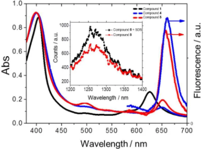
Figure 1. Absorption (left) and fluorescence (right) spectra in methanol
of compounds 1 (only absorption is shown), 4 and 5. For the fluorescence emission, λexc = 500 nm; λemi = 570-720 nm. Insert show singlet oxygen emission of compounds 4 and 5. λexc = 532 nm, 10 pulses second-1,
chlorophyllin acid chloride (compound 3, Scheme 1), we chose oxalyl chloride.3 The bis-amine substituent reagent (N,N-dimethylpropane-1,3-diamine) was chosen to facilitate the nucleophilic attach in the acid chloride and to allow the product with exposed amine side group.4
Spectroscopic properties
The UV-Vis and fluorescence spectra for both (compounds 4 and 5) in methanol are typical for chlorophyll derivatives, with a strong Soret band at 400 nm and four minor “Q” bands, with Q4 at 650 nm (Figure 1) being the band of major interest for PDT.2 Both compounds exhibit hyperchromic displacement of 4 nm for Soret band in comparison to Cu2+-Chl. Compound 4 showed a Soret band at 400 nm, Q4 at 651 nm and emission at 657 nm. Compound 5 showed a Soret band at 400 nm, Q4 at 650 nm and emission at 653 nm (Figure 1, Table 1).
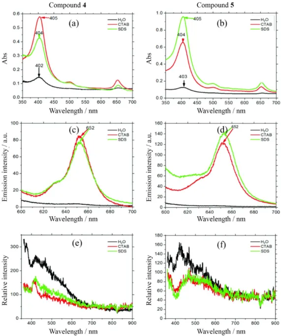
Chlorophyll and porphyrin derivatives have a large tendency to aggregate, distorting the absorption spectra and decreasing severely the photophysical properties relevant to PDT and micelles are known to control aggregation.17 We analyzed the effects of surfactants on the absorption spectra of both compounds 4 and 5. There is a 7- and 5-fold increase in absorbance at the Soret band of compound 5 in SDS and CTAB, respectively, compared with the absorbance in water (SDS and CTAB are anionic and cationic surfactants, respectively) (Figure 2a). The increase in absorbance was higher for compound 5 in SDS and for compound 4 in CTAB. There was also a substantial increase in fluorescence and decrease resonance light scattering in the presence of micelles indicating the lower photophysical parameters in water due to aggregation and the improvement of these properties in the presence of micelles. The fact that compound 5 interacts more efficiently with SDS micelles and compound 4 with CTAB micelles (absorbance data) indicates that there are two main forces driving this interaction, i.e., electrostatic and hydrophobic.
The fluorescence quantum yields (Φ
f) were determined using methylene blue (MB) as a standard and were about half of that of MB for both compounds 4 and compound 5 (Φf = 0.013 and 0.014, respectively). Singlet oxygen quantum yields (Φ∆) were determined by phosphorescence detection at 1270 nm against a standard of hematoporphyrin IX. Φ∆ values were 0.57 and 0.56 for compounds 4 and 5, respectively, in methanol. Compound 5 in 2 × 10⁻⁵ mol L-1 SDS in D2O exhibited a 37% increase in singlet oxygen emission as compared to 2 × 10⁻⁵ mol L-1 of compound 5 in D2O. This confirms that the monomer state of the sensitizer is the only state that is an effective PDT agent.
Water to n-octanol partition and viability studies
Water to n-octanol partition coefficients, which were expressed as logPO/W, were used to estimate the photosensitizer’s affinity for cell membranes.5 Figure 4a show logPO/W values for compounds 4 and 5 across the relevant pH range. Compound 5 has negative logPO/W values in the whole pH range, being always more hydrophilic than compound 4, which is explained by its cationic structure. logPO/W becomes more negative with the decrease in pH due to protonation of the chlorophyllin ring nitrogens. Compound 4 exhibits hydrophilicity at low pHs, a condition that allows protonation of both the ring and the side-group nitrogens, nearly equal affinity for the organic or aqueous phase from pH 3 to 8, a range in which only the side-group nitrogens are protonated, and is lipophilic at pH > 8, for loosing protonation of the side-group nitrogens.
Photosensitizer incorporation in mitochondria, liposomes, and HeLa cells were determined and are shown in Figure 4b. For compound 4, incorporation was 58%, 28%, and 10% for mitochondria, liposomes, and HeLa cells, respectively. For compound 5, incorporation was 61%, 48%, and 13% for mitochondria, liposomes, and HeLa cells, respectively. Therefore, both compounds interact well with cells and membranes, and compound 5 interacts a bit more favorably with liposomes.
Table 1. Spectroscopic properties of compounds 1, 2, 4, 5
Spectroscopic and photophysical parameters
Compound Absorption Emission
Soret (λ) / nm εa / (mol L-1 cm-1) Q4b (λ) / nm εa / (mol L-1 cm-1) Φ∆c Φfd
1 404 48293 626 7192 − −
2 400 20121 650 3612 − −
4 400 20825 651 3797 0.57 0.014
5 400 19736 650 3584 0.56 0.013
aε: molar absorption coefficient; bQ4: band Q4; cΦ
Figure 2. (a,b) Absorption; (c,d) fluorescence emission; (e,f) resonance light scattering spectra of compounds 4 and 5 in water, CTAB, and SDS solutions, shown in black, red, and green lines, respectively. Fluorescence emission (λexc = 532 nm; λemi = 600-700 nm); for resonance light scattering λexc = λemi.
The photodynamic efficiency of compounds 4 and 5 were tested in HeLa cell culture and compared with the photodynamic efficiency of MB. Cells cultured and incubated at the same PS concentration (2 × 10⁻⁵ mol L-1), were irradiated at 650 nm. HeLa cell viability was determined to be 11.2% for MB, 4.8% for 7.7% for 4 and 5 in relation to a control group (no light), indicating a similar overall phototoxicity for all three compounds with a bit of better efficiency for compounds 4 and 5.
To determine the intrinsic photototoxicity of these compounds we compared the amount of absorbed photons in each case. It was not possible to directly measure the absorption of PS in cells, but we were able to estimate the
absorption factor (fraction of incident photons that are absorbed, equation 1) using ε (molar absorption coefficient) of each compound and the percentage of incorporation of the PS in cells at 2 × 10⁻⁵ mol L-1 of PS concentration, which was kept constant. At 650 nm, ε values are 68,000 mol L-1 cm-1 for MB,23 3797 and 3584 mol L-1 cm-1 for compounds 4 and 5, respectively.
α = 1 − 10-Abs (1)
obtains the values of absorption, and by using equation 1, absorption factors are calculated which were 0.9 for cells treated with MB and 0.09 for cells treated with the compounds 4 or 5 (Figure 4b). In other words, cells treated with compounds 4 and 5 absorb 10 times less photons than MB at 650 nm. Therefore, if one calculates the efficiency of cell killing divided by the number of absorbed photons (Figure 4c), one realizes that the new compounds induce cell death much more efficient per
absorbed photon. The reason for this difference may be due to several factors, including intracellular sub-location and avoidance of bleaching reactions. It should be noted that MB can be reduced in the cellular environment,24 having photodynamic activity decreased, which does not happen for the chlorophyllins 4 and 5.
Conclusions
New chlorophyllin non-symmetric and positively-charged photosensitizers were obtained by using inexpensive copper-chlorophyllin as raw material and a simple chemical route. These compounds
Figure 3. (a) Octanol/water partition coefficient of chlorophyllins (compounds 4 and 5) as a function of pH; (b) incorporation of compounds 4 and 5 in HeLa cells (106 cells mL-1 in DMEM), in liposomes (1.5 mg mL-1 in phosphate buffer) and in mitochondria suspensions (13 mg L-1 in phosphate buffer).
[PS] = 2 × 10⁻⁵ mol L-1.
Figure 4. (a) Viability in percentage of the control; (b) absorption factor; (c) intrinsic phototoxicity of methylene blue and of chlorophyllin compounds 4
and 5 in HeLa cells. [PS] = 2 × 10⁻⁵ mol L-1.
absorb reasonable well in the far-red spectral region (λ > 650 nm), are efficient singlet oxygen generators Φ∆ ca. 0.6 and have a strong tendency to interact with membranes. The overall photodynamic efficiency was similar to that of MB. However, the photodynamic efficiency per absorbed photons was considerably higher than that of MB, suggesting that compounds 4 and 5 have optimized properties for use in PDT.
Acknowledgments
FAPESP (grant No. 12/50680-5 and 13/07937-8) CNPq and NAP-Phototech are acknowledged for providing funds for this research. The funders had no role in study design, data collection and analysis, decision to publish, or preparation of the manuscript.
References
1. Neurath, A. R.; Strick, N.; Haberfield, P.; Jiang, S.; Antiviral Chem. Chemother.1992, 31, 55.
3. Woodward, R. B.; Ayer, W. A.; Beaton, J. M.; Bickelhaupt, F.; Bonnet, R.; Buchschacher, P.; Closs, G. L.; Dutler, H.; Hannah, J.; Hauck, F. P.; Ito, S.; Langemann, A.; Goff, E. L.; Leimgruber, W.; Lwowski, W.; Sauer, J.; Valenta, Z.; Volz, H.;
Tetrahedron 1990, 46, 7599.
4. Wainwright, M.; J. Antimicrob. Chemother.1998, 42, 13. 5. Allison, R. R.; Sibata, C. H.; Photodiagn. Photodyn. Ther. 2010,
7, 61.
6. Katoa, H.; Furukawaa, K.; Satob, M.; Okunakaa, T.; Kusunokic, Y.; Kawaharad, M.; Fukuokae, M.; Miyazawaf, T.; Yanag, T.; Matsuih, K.; Shiraishii, T.; Horinouchij, H.; Lung Cancer2003, 42, 103.
7. Uchoa, A. F.; de Oliveira, K. T.; Baptista, M. S.; Bortoluzzi, A. J.; Iamamoto, Y.; Serra, O. A.; J. Org. Chem.2011, 76, 8824. 8. Pangka, V. S.; Morgan, A. R.; Dolphin, D.; J. Org. Chem.1986,
51, 1094.
9. de Oliveira, K. T.; Momo, P. B.; de Assis, F. F.; Ferreira; M. A. B.; Brocks, T. J.; Curr. Org. Synth.2014, 11, 42.
10. Becker, R. S.; Allison, J. B.; J. Phys. Chem.1963, 67, 2662. 11. Baptista, M. S.; Indig, G. L.; J. Phys. Chem. B 1998, 102, 4678.
12. Indig, G. L.; Anderson, G. S.; Nichols, M. G.; Bartlett, J. A.; Mellon, W. S.; Sieber, F.; J. Pharm. Sci. 2000, 89, 88. 13. Dolmans, D. E.; Fukumura, D.; Jain, R. K.; Nat. Rev. Cancer
2003, 3, 380.
14. Hacham, H.; Freeman, S. E.; Gange, R. W.; Maytum, D. J.; Sutherland, J. C.; Sutherland, B. M.; Photochem. Photobiol.
1990, 52, 893.
15. Ravanat, J. L.; Martinez, G. R.; Medeiros, M. H.; di Mascio, P.; Cadet, J.; Arch. Biochem. Biophys. 2004, 423, 23.
16. He, Y.-Y.; Liu, H.-Y.; An, J.-Y.; Han, R.; Jiang, L.-J.; Dyes Pigm.
1999, 44, 63.
17. Uchoa, A. F.; Oliveira, C. S.; Baptista, M. S.; J. Porphyrins Phthalocyanines 2010, 14, 832.
18. Oliveira, C. S.; Turchiello, R.; Kowaltowski, A. J.; Indig, G. L.; Baptista, M. S.; Free Radical Biol. Med.2011, 51, 824.
19. Salamão, G. H. A.; Uchoa, A. F.; Baptista, M. S.; Tardivo, J. P.; Christofolini, D. M.; Correa, J.; Lasers Surg. Med. 2015, 47, 421.
20. Uchoa, A. F.; Knox, P. P.; Turchielle, R.; Seifullina, N. K.; Baptista, M. S.; Eur. Biophys. J. 2008, 37, 843.
21. Pavani, C.; Uchoa, A. F.; Oliveira, C. S.; Iamamoto, Y.; Baptista, M. S.; Photochem. Photobiol. Sci. 2009, 8, 233.
22. Küpper, H.; Spiller, M.; Küpper, F. C.; Anal. Biochem.2000,
286, 247.
23. Junqueira, H. C.; Severino, D.; Dias, L. G.; Gugliotti, M.; Baptista, M. S.; Phys. Chem. Chem. Phys.2002, 4, 2320.
24. Gabrieli, D.; Belisle, E.; Severino, D.; Kowaltowski, A. J.; Baptista, M. S.; Photochem. Photobiol.2004, 79, 227.
Submitted: July 3, 2015
Published online: October 30, 2015