J of Evolution of Med and Dent Sci/ eISSN- 2278-4802, pISSN- 2278-4748/ Vol. 3/ Issue 19/May 12, 2014 Page 5135
PERLS’ PRUSSIAN BLUE POSITIVITY IN EXFOLIATED BUCCAL CELLS OF
THALESSEMIA MAJOR PATIENTS AND ITS CORRELATION WITH SERUM
FERRITIN
Sonam Gupta1, V. K. Trichal2, Reeni Malik3, R. K. Nigam4, Rajni Choudhary5, Atul Shrivastava6
HOW TO CITE THIS ARTICLE:
Sonam Gupta, V. K. Trichal, Reeni Malik, R. K. Nigam, Rajni Choudhary, Atul Shrivastava. Perls’ Prussian Blue Positivity in Exfoliated Buccal Cells of Thalassemia Major Patients and its Correlation with Serum Ferritin. Journal of Evolution of Medical and Dental Sciences 2014; Vol. 3, Issue 19, May 12; Page: 5135-5140,
DOI: 10.14260/jemds/2014/2565
ABSTRACT: BACKGROUND: Iron absorption is carefully regulated to maintain equilibrium between the absorption and the body loss of iron. Thalassemia is a genetic disorder of hemoglobin synthesis requiring regular blood transfusion leading to iron overload in the body which leads to morbidity and mortality in these patients. AIMS AND OBJECTIVES: To demonstrate iron overload at an early stage
by exfoliative cytology using Perls’ Prussian blue method and to compare with that of serum ferritin
levels of those patients. MATERIALS AND METHODS: Smears were obtained from buccal mucosa of 60 thalassemia major patients who were undergoing repeated blood transfusion and 30 healthy subjects as control. Scrapings were obtained from the buccal mucosa and stained with Perls’ Prussian blue stain and were examined under light microscope. RESULT: Perls’ positivity was observed in 61.6% of thalassemic patients with a moderately positive correlation to serum ferritin levels.
CONCLUSION: Exfoliative cytology is a simple painless and a non-invasive technique therefore it can be used as a diagnostic tool in demonstration of iron overload in thalassemic patients.
KEYWORDS: Iron overload, thalassemia, exfoliative cytology, Perls’ Prussian blue reaction.
INTRODUCTION: Minerals are inorganic elements that are essential for life. Among them iron is an essential nutrient which are required by human cell. Approximately 4.5 g of iron can be found in an average adult man most of which is contained in the hemoglobin molecule and in other haem containing proteins.1 Under physiologic conditions the concentration of iron in the human body is carefully distributed between functional, transport and storage components.2
In 1925 Thomas Cooley and Pearl Lee described a form of severe anemia associated with splenomegaly and characteristic bony changes. As all earlier cases were reported in children of the Mediterranean origin, the disease was later named thalassemia, from the Greek word for sea Thalessa.3
In Thalassemia major the production of beta globin chains is severely impaired because both beta globin genes are mutated which results in a severe or total suppression of beta chain synthesis. Beta thalassemia major is a life threatening condition characterized by severe anemia, hepatosplenomegaly, growth retardation endocrine dysfunction and skeletal changes due to hypertrophy and expansion of the hematopoietic marrow.4
J of Evolution of Med and Dent Sci/ eISSN- 2278-4802, pISSN- 2278-4748/ Vol. 3/ Issue 19/May 12, 2014 Page 5136 In patients who are not receiving transfusions, abnormally regulated iron absorption results in increasing in body iron burden ranging from 2-5 g/yr. Regular transfusion may double this rate of iron accumulation.
After approximately 1 yr. iron begins to be deposited in parenchymal tissue where it may cause substantial toxicity as compared with that within reticuloendothelial cells.
Storage iron occurs in two forms- ferritin and hemosiderin. In states of iron overload hemosiderin increases to a greater degree than ferritin and becomes the dominant form.6 Ferritin although may be present in the cells cannot be visualized under light microscope. Hemosiderin because of its large size can be visualized under light microscope as blue coloured granules in the cytoplasm of the cells.7
)n our study exfoliated cells from the buccal mucosa of β thalassemia major patients comprising the study undergoing transfusions and a control group 0f 30 normal individuals who had no confirmed acute and chronic liver damage, malignancy and megaloblastic anemia were considered
and the smears obtained were stained with Perls’ Prussian blue stain.
AIMS AND OBJECTIVES:
1. To demonstrate iron overload in an early stage in the exfoliated buccal cells of beta thalassemia major patients.
2. To correlate Perls’ Prussian blue staining with serum ferritin levels.
MATERIALS AND METHODS: The study was conducted after approval from institutional ethical committee. The study group comprised of 60 hemoglobin electrophoresis confirmed beta thalassemia major patients ranging in the age group of 1- 16 yrs. receiving regular blood transfusion. The control group comprised of 30 both clinically & hematologically healthy individuals in the age range of 1-16 yrs. who had no confirmed acute and chronic liver damage
,
malignancy and megaloblastic anemia.For the preparation of the smears clean, fresh and dry glass slides were used. Scrapings were obtained from the subject’s mouth by using a wet wooden spatula, by using a gentle scraping motion with little pressure over buccal mucosa of both the study and control groups. The scraps were smeared into the centre of the glass slides & immediately fixed in 90% alcohol for one hour, then
stained with Perls’ Prussian blue stain which consist of Potassium ferrocyanide and (Cl which reacts with the ferritin in the cells to form blue coloured granules.8
J of Evolution of Med and Dent Sci/ eISSN- 2278-4802, pISSN- 2278-4748/ Vol. 3/ Issue 19/May 12, 2014 Page 5137
RESULT:
Figure 1: Smear at higher magnification showing clumps of blue colored granules (Perls' Prussian blue stain, x 400).
Figure 2: Smear at higher magnification showing dispersed blue colored granules Perls’ Prussian blue stain, x400)
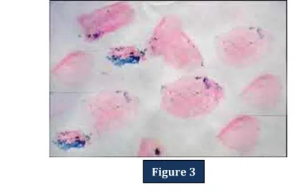
Figure 3: Smear at higher magnification showing clumps of blue colored granules (Perls' Prussian blue stain, x 400)
Figure 1
Figure 2
J of Evolution of Med and Dent Sci/ eISSN- 2278-4802, pISSN- 2278-4748/ Vol. 3/ Issue 19/May 12, 2014 Page 5138
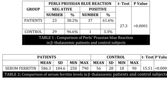
GROUP
PERLS PRUSSIAN BLUE REACTION t- Test P Value
NEG ATIVE POSITIVE
27.3 <0.0001
NUMBER % NUMBER %
PATIENTS 23 38.2% 37 61.6%
CONTROL 29 96.6% 1 3.3%
TABLE 1 : Comparison of Perls’ Prussian blue Reaction
in -thalassemic patients and control subjects
PATIENTS CONTROL t- Test P Value
MEAN SD MIN MAX MEAN SD MIN MAX
15.51 <0.0001
SERUM FERRITIN 586.3 184.6 250 790 56 28 18 90
TABLE 2: Comparison of serum ferritin levels in -thalassemic
patients and control subjects
Serum ferritin Perls’ Prussian blue staining positivity
Correlation coefficient 0.76
P Value <0.0001 Sig
N 60
TABLE 3:Correlation of Perls’ Prussian staining positivity and
serum ferritin levels in - Thalassemic patients
Serum ferritin Number of Blood Transfusions
Correlation coefficient 0.68
P value <0.0001 sig
N 60
TABLE4
:
Correlation between number of blood transfusionsand serum ferritin levels in - Thalassemic patients
DISCUSSION: Perls’ Prussian blue reaction is considered to be the first classical histochemical
reaction carried out, and is widely applied in the field of haematology.9 We have applied this technique to exfoliated buccal cells.
In our study, exfoliated cells from the buccal mucosa of 37 of the 60 thalassemic patients (61.6%) group revealed positivity for Perls’ Prussian blue reaction. Chi –square test was applied and the P value was <0.0001 and the test was considered significant. And one of the control subjects
showed false Perls’ Prussian blue positivity.
Our results were similar to those observed by Nandprasad et al (2010) who recorded 65 %
Perls’ positivity although these were lower than Atul A Bhat et al who recorded . % Perls’
J of Evolution of Med and Dent Sci/ eISSN- 2278-4802, pISSN- 2278-4748/ Vol. 3/ Issue 19/May 12, 2014 Page 5139
SL. No. Study year Positive cases % positivity
1) Gururaj and Sundaram et al 2004 10/10 100%
2) Nandprasad et al2 2010 65/100 65%
3) Atul A Bhat et al10 2013 43/60 71.7%
4) Harika Chittamsetty et al11 2013 29/40 72.5%
5) Present study 2014 37/60 61.6%
Tables 5: Comparative study of similar studies
The serum ferritin was higher in our study groups as compared to control group.
Our results showed a moderately positive correlation between Perls’ Prussian blue positivity
and serum ferritin which indicates that the positive staining in exfoliated buccal cells may give a clue of increased serum ferritin levels and thus indicating iron overload in the body tissues.
CONCLUSION: Measurement of iron concentration in a liver biopsy specimen is a reference method for assessing the body iron stores. The discomfort and risk associated with the liver biopsy procedure as well as cost factor associated with MRI as compelled us to search for a simple and a cheaper technique.
Oral exfoliative cytology is a simple and non-invasive technique which can be used for screening and diagnostic tool for assessing iron status in thalassemia patients undergoing repeated blood transfusions.
Further advancement in this field is advocated to establish this technique as a screening tool & also to assess complications associated with iron overload.
I would like to extend special acknowledgement to our laboratory technician Mr. K.P. Verma and department staff Mr. BK. Shrivastava for their help and support in this project.
REFERENCES:
1. I.S Young, Woodside. Antioxidants in health & disease. Journal of Clinical pathology 2001; 54: 176-186.
2. Nandprasad S, Sharada P, Vidya M, Karkera B, Hemanth M, Prakash N. Oral exfoliative cytology in beta thalassemia patients undergoing repeated blood transfusions. Internet J Pathol. 2009; 10: 7.
3. Marcel E Conrad, Jay Numbrelt. Iron absorption & transport - an update. Am J Hematol. 2000; 64: 287-288.
4. Abu Alhaija ES, Al-Wahadni AM, Al-Omari MA. Uvulo-glosso-pharyngeal dimensions in subjects with beta-thalassaemia major. Eur J Orthod 2002 Dec; 24(6): 699-703.
5. Al-Wahadni A, Qudeimat MA, Al-Omari M. Dental arch morphological and dimensional characteristics in Jordanian children and young adults with beta-thalassaemia major. Int J Paediatr Dent. 2005; 15: 98–104. [PubMed]
6. Firkin F, Chesterman C, Pennington D, Rush B, editors. 5th ed. Noida, India: Blackwell Publishing; 1989. De Gruchy's clinical hematology in medical practice.
J of Evolution of Med and Dent Sci/ eISSN- 2278-4802, pISSN- 2278-4748/ Vol. 3/ Issue 19/May 12, 2014 Page 5140 8. Churukian CJ. Pigments and minerals. In: Bancroft JD, Gamble M, editors. Theory and practice of
histological techniques. 6th ed. China: Churchill Livingstone Elsevier Ltd; 2008. pp. 233–6. 9. Gururaj N, Sundharam S. Demonstration of iron in the exfoliated cells of the oral mucosa. J Oral
Maxillofac Pathol. 2003; 1: 37–9.
10.Atul A Bhat, Rajkumar N. Parwani & Sangeeta P. Wanjari. Demonstration of Iron in exfoliated buccal cells of thalassemia major Patients. J Cytol.2013 Jul-Sep; 30(3): 169-173.
11.Harika Chittamsetty, Munishekar, Syed. Afroz Ahmed, Churu Suri Sridevi Palla et al. A Non- invasive Technique which demonstrate iron in the buccal mucosa of sickle cell anaemia & thalessemia patients who undergo repeated blood transfusion. Journal of Clinical and Diagnostic Research.2013 June, Vol-7(6): 1219-1222.
AUTHORS:
1. Sonam Gupta 2. V. K. Trichal 3. Reeni Malik 4. R. K. Nigam 5. Rajni Choudhary 6. Atul Shrivastava
PARTICULARS OF CONTRIBUTORS:
3. D.C.P, Student, Department of Pathology, Gandhi Medical College, Bhopal.
4. Associate Professor, Department of Pathology, Gandhi Medical College, Bhopal.
5. Professor and HOD, Department of Pathology, Gandhi Medical College, Bhopal.
6. Professor, Department of Pathology, Gandhi Medical College, Bhopal.
1. Assistant Professor, Department of Pathology, Gandhi Medical College, Bhopal.
2. Senior Resident Officer, Department of Population Based Cancer Registry, ICMR, Gandhi Medical College, Bhopal, M. P.
NAME ADDRESS EMAIL ID OF THE CORRESPONDING AUTHOR:
Dr. Sonam Gupta, #G-2,
Meenakshi Regency Idgah Hills, Bhopal.
E-mail: drgsonam@gmail.com